
Abstract
Gender determination is an important part of fingerprint identification research, with many applications in several scientific domains. This skill extends beyond forensic science to include other disciplines such as human biology and anthropology, altogether aiding in studying variable yet unique identifiable qualities among populations and civilisations. Thereby, this present study aims to seeks to contribute to the broader field of fingerprint analysis and its application in gender determination.Fingerprints from 100 university students (50 males and 50 females) aged 18–25 years were analysed. Fingerprints were obtained using an inkpad, which were then tallied for epidermal ridges drawn inside the bisected lines drawn and calibrated on the upper right end of the fingerprint on a translucent film and the ridge density and characteristics were calculated digitally using Adobe Photoshop CS5.Findings indicate that females tend to have slightly higher minimum and maximum ridge densities compared to males in both hands. However, the mean ridge densities for males and females are relatively similar, with females showing a marginally higher mean ridge density in both the right and left hands. The frequency of ulnar loop was found in higher in females as compared to males, with minute characteristics that were of ridge breaks, although of little or insignificant difference between both genders.The data highlights minor but consistent differences in fingerprint ridge density between genders, supporting the potential for using ridge density as a biometric marker for gender determination. To the best of our knowledge, this is the first study to quantitatively assess fingerprint ridge density as well as ridge characteristics specifically in the middle finger among the young adult population in Eastern Uttar Pradesh, thereby providing region-specific forensic biometric data for gender differentiation. Further studies with increased sample size and utilising comparison between more than one finger of the same hand for both the density as well as the characteristics of the ridges would be of immense help in providing consistent and robust results.
Introduction
Dermatoglyphics is the least invasive and most economical method of determining a person’s gender by analysing their fingerprints. 1 A ‘fingerprint’ is an impression of the fleshy distal region of the finger’s epidermal ridges. Dermatoglyphics is the study of fingerprints. 2 It was said that Cummins and Mildo coined the word ‘dermatoglyphics’. It is the study of how the palms, soles, and fingers’ dermal ridges are arranged. Fingerprint patterns, in contrast to most other physical traits, develop between weeks 6 and 13 of pregnancy. This makes them even more special because they were formed before any environmental factors could have an impact. 3 Gender determination is an important part of fingerprint identification research, with many applications in several scientific domains. This skill extends beyond forensic science to include other disciplines such as human biology and anthropology, altogether aiding in studying variable yet unique identifiable qualities among populations and civilisations. 4 Scientifically, the ability to determine gender from fingerprints is based on physiological and genetic differences between males and females, manifesting as dermatoglyphic patterns. In forensic science, gender determination from fingerprints can narrow down the pool of suspects, making it a useful tool in criminal investigations. 5 Features that are extensively utilised and gain relative importance include ridge counts, pattern type and pattern intensity index. However, while being features of significant importance because of their direct relevance in personal identity, other aspects, such as minutiae and epidermal ridge width (ridge density), have not traditionally been examined extensively in comparison to the feature described above. Forensic professionals use distinctive patterns and ridge counts for identification that usually differ between males and females. For example, research has revealed that females have finer ridge patterns, whereas males frequently have coarser ridge patterns. Understanding gender-specific variations in fingerprints can aid in the diagnosis and investigation of forensic medicine, thereby indicating its role to be added as an integral part in identification purposes and to solve cases.6, 7 Dermatoglyphic analysis can also shed light on the occurrence of chromosomal anomalies such as Turner syndrome or Klinefelter syndrome, in which fingerprint patterns can be used as an additional diagnostic technique. Fingerprint-based gender determination is useful in anthropological sciences as well, since it provides a non-invasive tool for studying genetic and evolutionary features across groups and civilisations. It enables researchers to trace lineage, better understand migratory patterns and investigate the distribution of genetic features across demographic groups. For example, changes in fingerprint patterns between ethnic groups can be investigated better to understand evolutionary adaptations and environmental influences on human populations. These distinctions serve as a valid biomarker for gender reveal, aiding in criminal investigations, medical diagnostics and anthropological studies, demonstrating the broad applications of fingerprint identification science.8, 9 The study of these characteristics not only aids in criminal investigations but also contributes to our understanding of human biology and population genetics. 10
By examining the specific features and patterns unique to the middle finger, this research aims to comprehensively understand how these characteristics can be utilised effectively in various scientific and practical applications. Through detailed analysis and comparison, this study seeks to contribute to the broader field of fingerprint analysis and its application in gender determination.
Although several international and pan-Indian studies, particularly from Southern as well as Eastern parts of India, have explored fingerprint ridge density for gender determination, there is a lack of focused data from the Eastern Uttar Pradesh region—an area with distinct ethnic, genetic, and environmental influences. Moreover, prior research rarely isolates ridge density analysis to the middle finger, despite its forensic relevance due to frequent recovery in crime scene prints. This study addresses these gaps by providing a localised, digit-specific forensic profile for gender estimation.
Material and Methods
This study was conducted in King George’s Medical University in Lucknow, Uttar Pradesh. Ethical permission for this study was obtained from the Institutional Ethics Committee (Ref. No.2352/Ethics/2024). Written informed consent for the same was taken from all participating students.
Study Population and Sampling
Fingerprints from 100 undergraduate university students (50 males and 50 females) aged 18–25 years were analysed. Non-resident Indians and subjects from Central, Western and Eastern India were excluded from the study. Participants were recruited using simple random sampling from the undergraduate student pool. Individuals with scars, deformities or dermatological conditions on the fingers were excluded.
Sample Size Calculation
Using a two-sided α = 0.05 and 80% power, based on the hypothesis that the observed mean differences between the male and female population would be negligible, the sample size formula for comparing two independent means was calculated as,
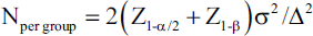
Substituting the value, the sample size obtained was (≈4,600 per group for the right hand; ≈6,100 per group for the left hand) because the observed effect is extremely small. Detecting a moderate effect (d = 0.50; ∇ ≈ 1.38 ridges/mm²) would require ≈63 per group (total ≈126). Since it was practically constraining to obtain such a large sample in a single tertiary care centre for hypothesis testing, the present study recruited a total sample of 50 males and 50 females, which was underpowered to detect such minute differences.
Method
Before collecting the fingerprints, individuals were asked to wash and clean their hands. Fingerprints were obtained using an inkpad. Fingerprint samples (male and female) were tallied for epidermal ridges inside perpendicular lines drawn and the area within these on the upper right end on a translucent film, as shown in Figure 1. The ridge density value that was used for analysis refers to the number of ridges within the described area. These lines were put in the upper left corner of the centre core region to capture a right-hand fingerprint. For fingerprints from the left hand, the lines measuring 5 mm in length were positioned at the upper right corner in relation to the centre core area. Figure 1 shows how to place lines on a right-handed fingerprint sample. This sampling approach isolates ridges within a given area, simplifying ridge counting. Unlike previous studies that use full fingerprint analysis or varying finger types, our methodology specifically targets the middle finger, considered the most frequently recovered finger in forensic samples. Ridge characteristics were recorded based on the criteria by Michael Kuckenas,
11
which are as follows:
Type 1 Whorl pattern: These are formed by any random ridges that encircle a core with two or more tri-radii. Type 2 Loop pattern: This pattern has numerous ridges that enter the finger tip from one side and leave from the same side. Type 3 Arch pattern: These are usually composed of ridges that cross from one side of the fingertip to the other. It has no tri-radii.
Showing the Dermatoglyphic Pattern, of Which the Ridge Characteristics Were Noted.
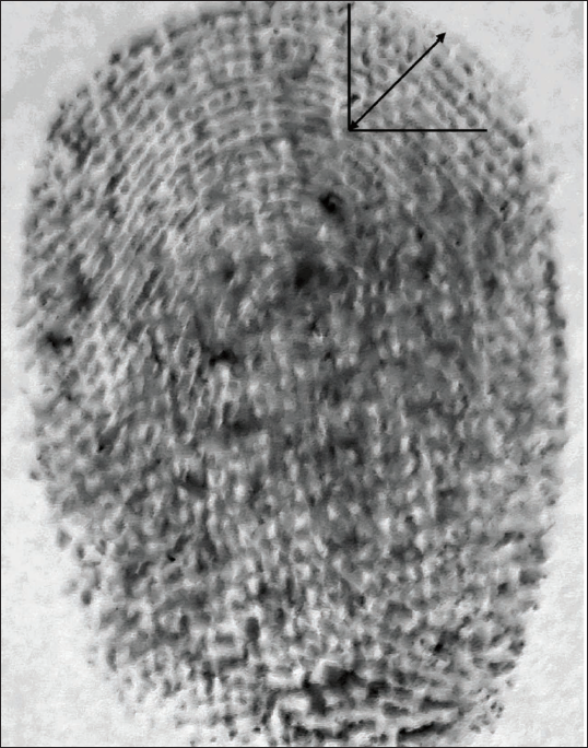
Although we collected middle-finger prints from both hands, our primary analysis focused on the right-sided ridge characteristics because (a) latent fingerprints recovered at crime scenes in India are more often from the dominant/right hand, reflecting the predominance of right-handedness in the population; (b) using a single, consistently defined area on one side minimises intra-individual variability and improves reproducibility of measurements; and (c) published reference data on ridge density for Indian populations most frequently report right-hand metrics, enabling more direct comparisons with earlier studies. With regard to the placement of the designated area, the upper corner of the core region was chosen rather than the central zone because pilot testing showed fewer smudges, clearer ridge definition and less overlap with creases at this location, facilitating more reliable and reproducible ridge counts. Central zones often showed greater ridge convergence and distortion, which could lead to counting errors. Therefore, the upper-corner placement offers a standardised, high-quality area for analysis while still sampling the core region of the print.
The counting was done digitally using Adobe Photoshop CS5 after scanning the original prints on the paper, and the measurements were then entered separately in Microsoft Excel. Additionally, lakes and bifurcations were recorded as two ridges, while dots were regarded as one ridge. By setting pixels between 4,000 and 3,000, the image size was standardised to create a life-size image of a fingerprint. This was accomplished with the aid of the ruler tool from analysis. With the aid of an arbitrary picture rotation tool, the fingerprint’s centre was maintained at a 90-degree angle. Two straight lines were drawn, which bisect each other; this bisecting point was set at the right upper centre of the fingerprint as per the calibration done on paper. SPSS version 20.0 software was then used for statistical analysis, after which the results were obtained, and a conclusion was drawn.
Statistical Analysis
Minimum, maximum, mean and standard deviation of ridge density (ridges/mm²) were computed separately for each hand and gender. Independent-samples t-tests compared mean ridge densities between males and females. p < .05 was considered statistically significant.
Results
For males, the ridge density in the right hand ranged from a minimum of 7 ridges/mm² to a maximum of 18 ridges/mm², with a mean ridge density of 13.00 ridges/mm² and a standard deviation of 2.852. The left hand showed similar results, with a ridge density range of 7 to 18 ridges/mm², a mean of 13.26 ridges/mm², and a standard deviation of 2.891. The results are significant, indicating the fact these differences exist among the genders in the population (Table 1).
Shows Description of Ridge Density in the Middle Finger of 100 Individual’s Both Males and Females.
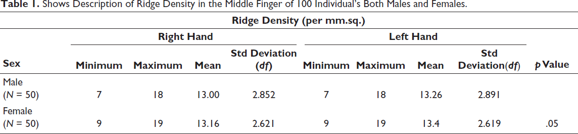
For females, the ridge density in the right hand varied from a minimum of 9 ridges/mm² to a maximum of 19 ridges/mm², with an average ridge density of 13.16 ridges/mm² and a standard deviation of 2.621. The left hand exhibited a ridge density range from 9 to 19 ridges/mm², a mean of 13.40 ridges/mm², and a standard deviation of 2.619. The results obtained are of significance (Table 1).
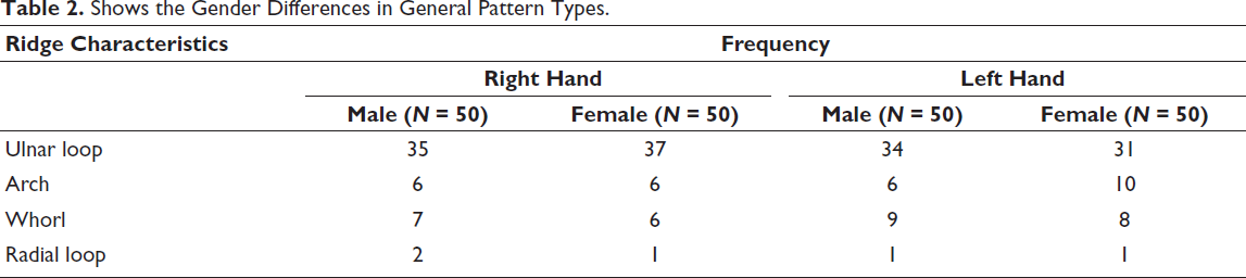
The calculation of ridge characteristics pointed out to the fact that the ulnar loop was found in the maximum of the participants, with a frequency of 74% in females and 70% in males in the right hand and 68% in males and 62% in females in the left hand (Table 2). In decreasing order of frequency of the appearance were arch (12% in both gender in right hand 12% in males, 20% in females in left hand), whorl (14% in males, 12% in females in right hand, 15% in males, 16% in females in left hand) and radial loop (4% in males, 2% in females in right hand, 2% in both genders in left hand). The p value was found to be significant for the same (>.05), suggesting the significant difference in appearance in the ulnar loop in both the hand as well as gender difference for the same, which was found to be appearing at a higher frequency, suggesting its potential significance to be used in identification.
Shows the Gender Differences in General Pattern Types.
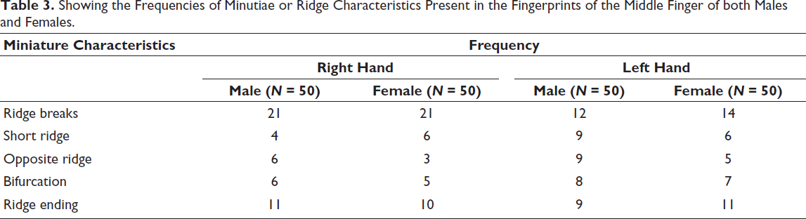
Furthermore, in miniature characteristics, the maximum characteristic recorded was of ridge breaks, with a frequency of 42% in both females as well as males in the right hand and 24% in males, 28% in females in the left hand (Table 3). In decreasing order of frequency of the appearance were short ridge (8% in males, 12% in females in right hand, 18% in males, 12% in females in left hand), opposite ridge (12% in males, 6% in females in right hand, 18% in males, 1% in females in left hand), bifurcation (12% in males, 1% in females in right hand, 16% in males, 14% in females in left hand) and ridge ending (22% in males, 20% in females in right hand, 18% in males, 22% in females in left hand). The p value was found to be significant for the same (>.05), indicating that the ridge breaks can serve a purpose for forensic analysis for both genders.
Showing the Frequencies of Minutiae or Ridge Characteristics Present in the Fingerprints of the Middle Finger of both Males and Females.
Overall, the data highlights minor but consistent differences in fingerprint ridge density between genders, supporting the potential for using ridge density as a biometric marker for gender determination.
This consistent result obtained with both left and right hand ridge count in females may be attributed to more uniform epidermal ridge development, a pattern that may carry implications in forensic sex estimation, especially in cases involving partial or single-hand fingerprints.
Discussion
Fingerprint ridge density is the number of ridges inside a certain area of the fingerprint, which is commonly quantified in ridges per unit of distance. Numerous studies have found detectable gender differences in fingerprint ridge density, which can be used to determine gender with high accuracy.
Summary of Principal Findings
In this preliminary study of 100 young adults (50 males, 50 females) from Eastern Uttar Pradesh, mean ridge densities were marginally higher in females than in males for both right and left middle fingers. However, the absolute mean differences were very small (≈0.14–0.16 ridges/mm²) and effect sizes (Cohen’s d) were ≈0.05, indicating negligible practical separation between sexes with the present measurement procedure and sample. However, a study performed by Nithin et al. 3 in the South Indian population have utilised all the fingers and have classified them separately using Henry’s classification system, and have found that in females, 55.28% of ulnar loop pattern was observed, 26.84% of the whorl pattern, and in males, 49.32% of ulnar loop pattern, with 30.64% of the whorl pattern in the ring finger. Thereby, the findings of this study contrast with the use of the middle finger for concluding results. However, due to regional differences, the same result and hypothesis testing could not be applied for our study, and hence, comparison with other fingers requires further validation and comparison for obtaining robust results. Although the ridge characteristic pattern was consistence with that found in our study, the frequency percentages vary.
In contrast, a study done by Acree et al. (1999) 7 did a thorough study on fingerprint ridge density and discovered that females had a higher ridge count per unit area than males across all fingers, including the middle finger. This study, with a large sample size, provided strong evidence for the existence of gender variations in ridge density. The differences observed in our sample were small (Cohen’s d ≈ 0.05), hence not statistically robust with our limited sample size. Significant bimanual asymmetry for patterns in the second and third interdigital areas of palms was seen in a study by Qutub et al. in 1988 12 that involved 100 Black Americans, 100 of whom were male and 100 of whom were female. Excessive digital arch patterns in females and greater mean finger ridge counts in males were among the notable sex differences. Therefore, interpalm ridge characteristics can also be considered for the analysis to derive the result. Furthermore, the increased ridge density found in the female population as compared to males was consistent with the findings by Thakar et al. 13 The study utilised both the index as well as middle fingers; however, our study only utilised the middle finger, forming it as a drawback of the present study. The frequency of miniature characteristics pointed out in the study was that ridge breaks were the most frequently encountered, with little difference between the genders. In contrast, the above study showed a significant increase appearance of ridge endings, bridge, and enclosures.
The developmental biology of fingerprints provides the physiological reason for this variation. Genetic and hormonal variables play a role in the creation of ridges in the dermal layers during foetal development. The two main sex hormones, testosterone and oestrogen, are essential to this process. The more common hormone in females, oestrogen, encourages the development of finer, more closely spaced ridges. On the other hand, coarser and less tightly packed ridges are linked to testosterone, which is more common in men.
These conclusions have been supported by research done on a variety of populations. For instance, statistically significant variations were seen between male and female fingerprint ridge densities in an Indian sample investigated by Nayak et al. (2010), with females consistently exhibiting higher densities. Gungadin’s (2007) study in a Mauritian population also revealed that females had higher ridge density, which further supports the application of this measure to determine gender across various ethnic groups.
Forensic science can benefit from these gender variations in ridge density. Given that partial fingerprints are frequently retrieved from crime scenes, forensic professionals can infer the gender of the fingerprint leaver based on the ridge density. When the fingerprint is the main piece of evidence or when other biometric data are lacking, this can be especially helpful.
Knowing the gender variations in fingerprint ridge density has benefits for forensic applications as well as for more general anthropological and biological studies. It makes it possible to investigate the genetic and evolutionary influences on fingerprint patterns and offers insights into how natural selection and adaptation have moulded these traits in various human populations.
This study contributes novel forensic evidence from a demographically underrepresented region. Ridge density data from Eastern Uttar Pradesh have not previously been reported in the context of forensic gender estimation. The focus on the middle finger also provides a practical advantage in forensic casework, where partial prints often include this digit.
Limitations of the study and future implications:
The study was conducted in a single institute in Eastern Uttar Pradesh; the findings cannot be generalised to the entire population. The sample size taken was underpowered; a larger sample size targeting at least 700 participants would suffice to support the results, warranting future study for the same. Since only the middle finger was used for the analysis, the observed differences could also lead to misclassification. Comparison of two or more finger ridges of both hands is warranted for future studies so that robust results and concrete findings can be developed through future studies. Since both the middle fingers have been used by different hands, comparison of the fingers of the same hand as per utilised in previous studies can be utilised to generate robust results. Minimal differences seen between the genders may lead to observer bias; therefore, a large sample size is recommended for future studies in order to know which characteristics would be definitive for determining the gender.
Regional Contribution
Despite limitations, the present work provides preliminary ridge-density data for middle-finger prints in young adults from Eastern Uttar Pradesh—a region with distinct ethnic and environmental influences—filling a gap in the Indian dermatoglyphic literature.
Forensic Implications
Although ridge density alone may not be sufficient for definitive sex classification, it can serve as a supportive parameter in combination with other features.
Conclusion
In summary, the data firmly indicate that there are gender variations in fingerprint ridge density, with females showing higher ridge densities than males. This trait is helpful for forensic gender determination, but it also offers important data for anthropological and human biology research. The reliability of ridge density as a biometric marker for gender identification is highlighted by the findings’ consistency across different research and groups. The present study offers novel insight into fingerprint-based gender differentiation using ridge density in a specific Northern Indian population. These findings can serve as a valuable reference in forensic identification cases within this region and may aid in developing a national fingerprint density database tailored for sex estimation. Further studies are warranted to overcome the aforementioned limitations.
Footnotes
Data Availability
All the data are available with the manuscript.
Declaration of Conflicting Interests
The authors declared no potential conflicts of interest with respect to the research, authorship and/or publication of this article.
Ethical Approval
The ethical approval was provided by the Institutional Ethical Committee (ETHICAL NO: 2352/Ethics/2024). Informed consent was taken from participants according to the Declaration of Helsinki, who agreed to participate in the study.
Funding
The authors received no financial support for the research, authorship and/or publication of this article.
Informed Consent
Informed consent was taken from the participants for publication.