
Abstract
This research work deals with the development of a polymeric super-hydrophobic surface involving nano silica–titania core–shell particles. This core–shell structure enhanced the properties of two different materials in a single nanoparticle in an outstanding manner; polymeric coatings containing core silica and shell titania have improved the mechanical behavior and hydrophobicity of coating surfaces, respectively. This nano core–shell was synthesized through two different methodologies which were prepared at high and low processing temperature separately, that is, called sol–gel and peptization synthesis. Further surface properties of the prepared nanoparticles were investigated individually in solvent-based emulsions and water-based emulsions. Nanocoating formulations were developed on mild steel substrate for analysis on the mechanical behavior of the coating and contact angle measurement. In the coating formulation, nano core–shell concentrations ranged from 1% (wt) to 6% (wt), and used nanoparticles were functionalized with methyl trimethoxy silane for better surface properties. Based on the results of the experiment, core–shell nanocoatings have been found mechanically robust and superhydrophobic (∼145.1° ± 2°) coating.
Keywords
Introduction
In the past few years, super-hydrophobic surfaces have gained substantial attention for their various real applications, like self-cleaning, water/oil separation, corrosion resistance, anti-fouling, anti-bacterial, possess unique wettability properties, etc. (Figure 1), the extended life expectancy of the coatings and substrates.1–6 Research analysis on these surfaces has specified that two main elements determine the surface wettability; surface roughness which generates from special nano/microstructures and surface chemistry.7–12 To date, super-hydrophobic coatings have been developed by various methods considering sol–gel process, lithographic processes, CVD, dip-coating, etc. using several nanoparticles such as iron, magnesium, nickel, and zinc 13–17 but most of the methods suffered from many constraints such as time-consuming, being complicated, requiring special equipment as well as the raw materials, and being expensive with high-priced equipment. In these approaches, limitations of substrate materials restrict the large-scale applications.18–23 So considering widespread applications of super-hydrophobic surface, inexpensive, facile methods are urgently needed. Although, the weak mechanical behavior of the nano/microstructures on the coated surface bound the applications of super-hydrophobic coatings.24–26 To overcome these limitations, we have developed nanocomposites through peptization synthesis that improved excellently the surface hydrophobicity and mechanical properties of the coating surface which is rarely explored for marine coating applications.27–30 Most likely this is the first time introduced by our research group to explore such multifunctional silica–titania nano core–shell for nanopaint application by utilizing mechanical properties of the core silica and hydrophobicity of the shell titania in a single nanocomposite.31–33 Developed core–shell materials are non-toxic itself and completely environment-friendly in nature. (a) Schematic for self-cleaning phenomena on super-hydrophobic coating surface: (b) Applications of super-hydrophobic nanocoatings.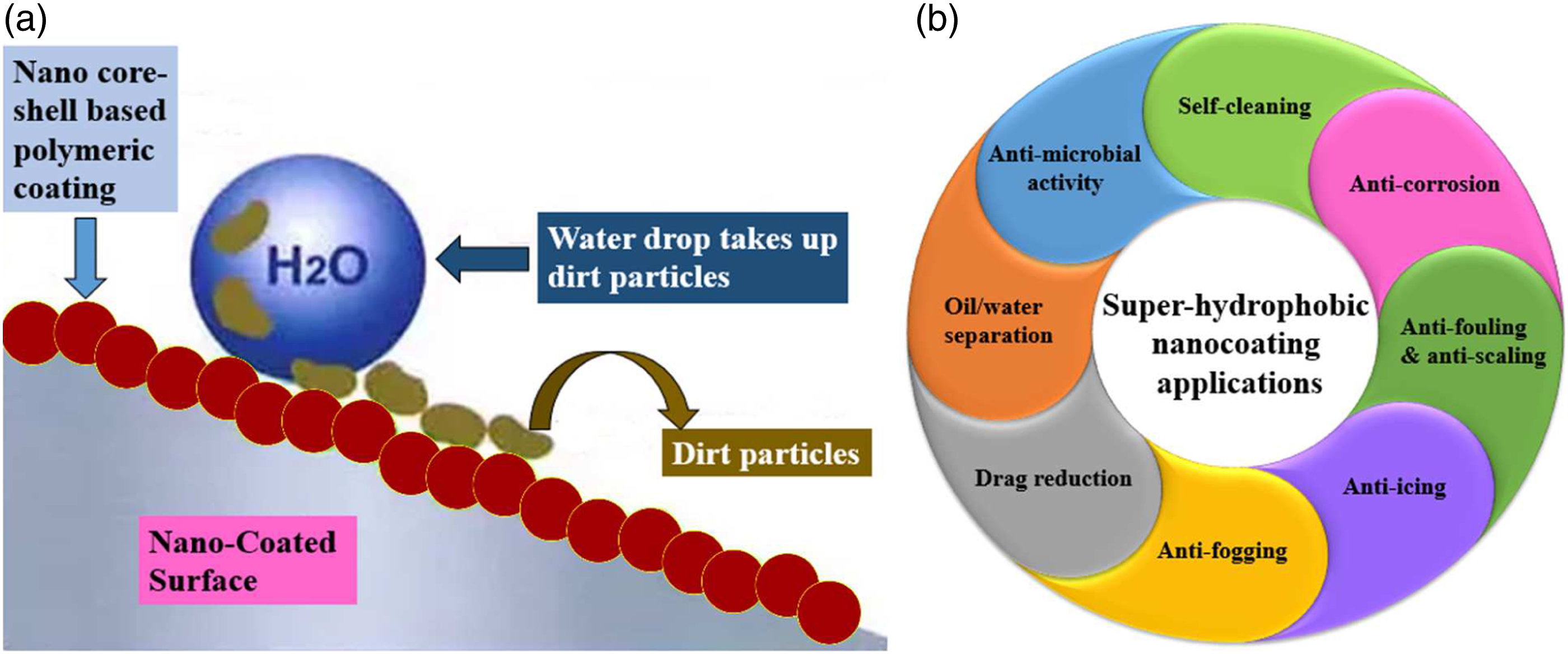
In the present study, a high-performance super-hydrophobic nanocoating has been developed with SiO2–TiO2 nano core–shell particles along with a solvent-based binder system. Comparative studies were continued on contact angle measurement with a water-based emulsion system as well. Further mechanical properties of the developed coating system have been investigated. Surface modification of this core–shell nanoparticle was carried out to improve the hydrophobicity as well as mechanical properties of the coating surface. These super-hydrophobic nanoparticle coatings promise a great application potential in the future because they could be extensively expanded and functionalized on various substrates.
Experimental
Materials
Titanium tetra-isopropoxide (TTIP) and tetraethyl ortho-silicate (TEOS) were purchased from Sigma Aldrich Chemicals Pvt. Limited (India). Ethanol was bought from Merck (India) and ammonium hydroxide from Qualikems Fine Chemical Pvt. Ltd. (India). Acetone, nitric acid, and isopropyl-alcohol (IPA) were purchased from Fischer scientific (India). Poly-acrylic and polyurethane were purchased from Dalton chemicals Pvt. Limited (India) for the development of coating formulations with all prepared nanoparticles. Methyl trimethoxy-silane (MTMS) was purchased from Sigma Aldrich Chemicals Pvt. Limited (India) for surface modifications.
Methodology
Synthesis of nano silica–titania (SiO2–TiO2) core–shell particles
In this study, SiO2–TiO2 nano core–shell particles were synthesized through two different processes for better comparative study, that is, sol–gel and peptization (Figure 2). Synthesis of SiO2–TiO2 core–shell nanoparticles.
Sol–Gel Synthesis
Silica nanoparticle was firstly prepared for the development of nano silica–titania core–shell structure. During synthesis, tetraethyl ortho-silicate (TEOS) was used as a precursor in presence of ethanol, H2O, and ammonium hydroxide. This ratio of ethanol and H2O was maintained at 1:3 in TEOS. A Further sample was heated overnight at 100°C and calcinated at a temperature of 650°C for 2 h. Detailed synthesis procedures and characterization of silica nanoparticles are already reported in our previously published articles.34,35 So in this study, we mainly focused on the development of nano core–shell–based super-hydrophobic coating with improved mechanical properties.
After the synthesis of silica nanoparticles, a further experiment was carried out for the preparation of core–shell nanoparticles. These silica–titania core–shell nanoparticles were synthesized by adding titanium tetra-isopropoxide (TTIP) in the prepared silica nanoparticles. This TTIP was mixed in isopropyl alcohol and water mixture (1:1 ratio) in the presence of NH4OH. A white precipitate was formed which is separated by centrifugation at 8000 rpm. The centrifuged sample was heated at 100°C overnight and calcinated at 500°C temperature for 4 h.
Peptization Synthesis
In the peptization method, the procedure of silica synthesis was the same as described in section Sol–Gel Synthesis. Low processing temperature is the major achievement of this synthesis technique for nano core–shell development. In this method, TTIP was suspended in water only and further steps continued in the presence of prepared silica and one electrolyte such as; nitric acid or sulfuric acid, or hydrochloric acid for better dispersion. In this experiment, we tested with all three electrolytes mentioned above but found the best result only with nitric acid electrolyte in the synthesis of the core–shell nanoparticle.
Maximum synthesis temperature was used at 70°C for this core–shell preparation.39 This low processing synthesis methodology is also known as the Kim method.
Nano core–shell functionalization
Better dispersion of the prepared nanoparticles and uniform coating surface was achieved by functionalization (modification) of the nanoparticles. Functionalization of nano core–shell was performed by methyl trimethoxy-silane (MTMS). In this experiment, 1 gm of core–shell nanoparticle was dispersed in 20 mL of ethanol followed by 0.2 mL MTMS under continuous stirring conditions for 4 h. Further, 1 mL of NH4OH was added to this solution dropwise. To remove the ethanol, the sample was heated at 70°C (20–30 min) and obtained the functionalized nano core–shell particles.
Materials characterization
Nano core–shell prepared through both of the synthesis approaches, that is, sol–gel and peptization have been characterized by various characterization techniques such as XRD, UV–Vis spectroscopy, SEM analysis, and FTIR study. In this study, XRD analysis of prepared nanoparticles was carried out through Bruker D8 focus X-Ray diffractometer, Zeiss SEM for surface morphology, and UV–Vis spectrophotometer Shimadzu (UV-1800) for UV spectrum. Further analyses were carried out on surface wettability through contact angle measurement and mechanical properties of the developed surfaces. Goniometric measurements (DSA25E Kruss Germany) were used to measure the static water droplet contact angle of these coated substrates. Mechanical properties were analyzed through a universal material tester (CETR-UMT 3) and Erichsen scratch tester (anti-scratch property).
Results and discussion
Surface morphology of nano core–shell particles
Investigation on the surface morphology of the prepared nano core–shell particles has been carried out through SEM study as mentioned in Figure 3. Nano core–shell formation through both of the synthesis processes has shown in Figure 3(a) and (b), that is, sol–gel and peptization. Individual nano-silica has shown spherical and smooth structure, whereas TiO2 /nano core–shell have shown faceted structure when it was prepared through the sol–gel method. TiO2 and nano core–shell particles have shown spherical shape, when it was produced by peptization synthesis. This result supports the formation of the core–shell structure. Element identification of prepared core–shell nanoparticles was done by energy dispersive spectroscopy as appearing in Figure 3(c). SEM images of nano SiO2–TiO2 core–shell particles developed through: (a) sol–gel synthesis (b) peptization process (c) elemental analysis EDX (peptization synthesis). XRD analysis of nano core–shell particles synthesized by: (d) sol–gel process (e) peptization process. UV–visible spectrum of samples in suspension form for nano core–shell particles synthesized by: (f) sol–gel (g) peptization.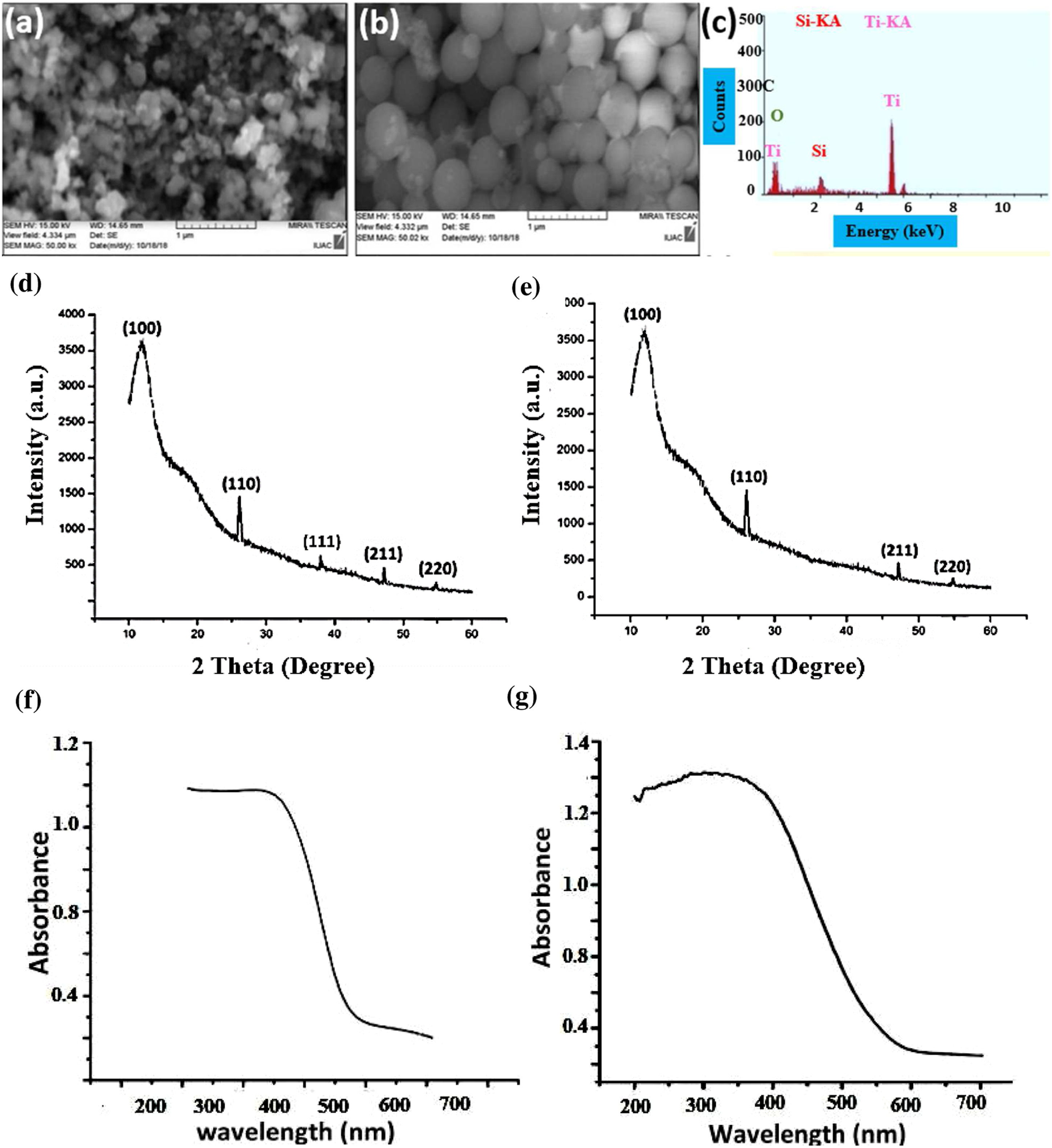
XRD analysis of nano core–shell particles
The crystallinity of the prepared nanoparticles was analyzed by an X-Ray diffractometer (Figure 3(d) and (e)). Diffraction peaks were monitored and recorded at room temperature for all the nanoparticles in the range of 20°–80° on a 2Ɵ scale by applying CuKα (1.5406 Å) radiation. The broad peak of nano SiO2 has its amorphous nature with 2θ at 15° (hkl=100) and sharp peaks of nano TiO2 have shown its crystalline nature and peaks were achieved at 2θ=25°, 37°, 46°, and 55° (hkl=110, 111, 211, 220). The planes observed in TiO2 confirm its anatase nature. One broad peak has been recorded with 2θ at 15° (hkl=100) in the XRD pattern of nano core–shell particles, that specified the existence of core silica material and other sharp peaks confirms the presence of nano TiO2 at 2θ=25°, 46°, and 55°(hkl=110, 111, 211, 220) in both type of core–shell structure only changes in the diffraction intensities has been observed for TiO2 diffraction pattern in core–shell structure, these intensities are low in respect to pure nano titania. This XRD pattern indicates and supports the thin layer formation of titania on core material SiO2 and carry the evidence for the development of nano core–shell particle.
UV–Vis analysis of nano core–shell particles
For UV–Vis analysis, samples were suspended in DI water followed by 5 min sonication through Shimadzu UV–Visible spectrophotometer. Absorbance versus wavelength curve has been recorded for all the suspended samples. This measurement was performed in the wavelength range of 200–700 nm. One maximum absorbance intensity spectrum showed at a particular wavelength (λmax) for individual nanoparticles which justified the formation of the particular nanoparticle. This analysis has shown absorption peak (λmax) at 350 nm for nano titania and nano core–shell particles that were synthesized by sol–gel method; 340 nm for both nano TiO2 and nano SiO2–TiO2 core–shell particles that synthesized by peptization method. The value of λmax was observed 290 nm for individual nano-silica. This result supports the confirmation for the TiO2 coating development on core material SiO2 and consequently the development of core–shell structure (Figure 3(f) and (g)).
FTIR analysis of the nano core–shell particles
FTIR study was performed for all the synthesized nanoparticles through Shimadzu 8400 spectrophotometer. The FTIR measurement was performed in transmission mode in the range of 4000–400 cm−1 for both types of the core–shell nanoparticles, individual FTIR spectra were recorded for SiO2 and TiO2 for a better understanding of nano core–shell structure. In sol–gel synthesis (core–shell s-g) the FTIR peak of nano SiO2, the band was around 1070 cm−1corresponds to the asymmetric stretching vibration of Si-O-Si bond, whereas 3300 cm−1 for H-O-H stretching and 1640 cm−1 bands have appeared for bending of absorbed water. Another peak was observed near 910 cm−1 corresponds to Si-OH bond. In the case of SiO2–TiO2 spectra recording, along with the peaks of SiO2 spectra, a band has shown at around 950 cm−1 corresponding to Si-O-Ti bond which confirms SiO2–TiO2 nano core–shell formation (Figure 4a). FTIR analysis of nano core–shell particles developed before and after functionalization through: (a) sol–gel (b) peptization synthesis.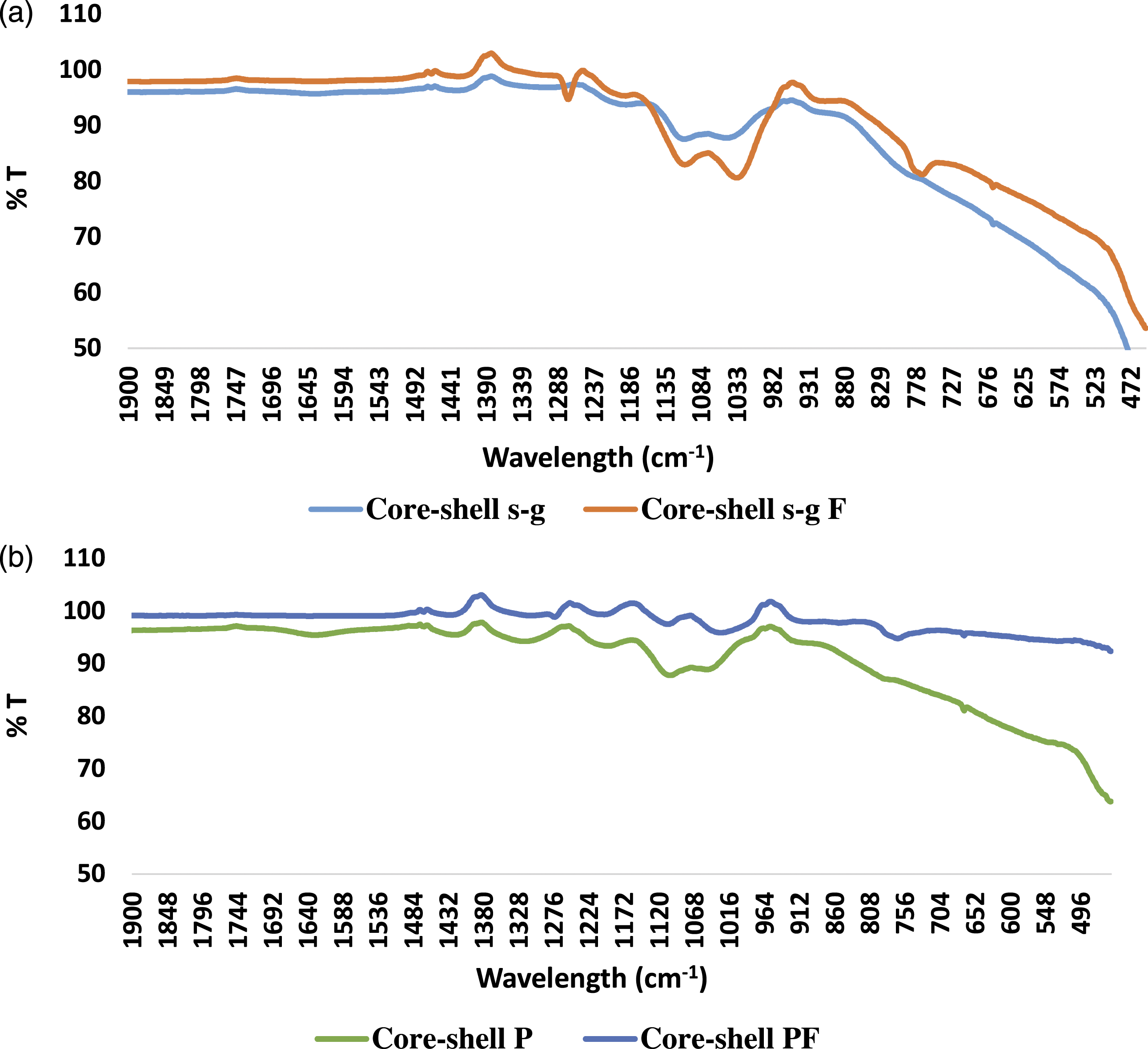
In Figure 4b, FTIR spectra of core–shell prepared through peptization method (core–shell P) clearly shows three bands in which the first band is the broadest, and appeared at 3500 cm−1 and it is representing the stretching vibration of the hydroxyl group (O-H) of the TiO2 nanoparticles. Another second band is detected around 1630 cm−1 which refers to the bending modes of water Ti-OH, and last, there was a prominent peak observed at 1383 cm−1 corresponding to Ti-O modes.
FTIR analysis of functionalized nano core–shell particles
In the previous section, FTIR analysis was discussed for all the prepared nanoparticles. But for improvement in hydrophobicity of the surface and to carry out anti-scratch testing, a smooth surface is really needed. So we functionalized the core–shell nanoparticles using methyl trimethoxy silane for the betterment of the surface and examined by FTIR. Figure 4 shows the FTIR spectrum of nano SiO2–TiO2 core–shell particles synthesized by both the methods before and after functionalization. The detailed description of the bond formation in FTIR analysis is already described in previous section FTIR analysis of the nano core–shell particles.
After functionalization, in the case of core–shell prepared through sol–gel (core–shell s-g F), two new peaks were obtained: i) C-H bond formation which comes at around 1275 cm-1and ii) Si-C is appearing at around 770 cm-1. Intensities of other peaks are increased like at 1055 cm-1 for antisymmetric Si-O-Si and at 1124 cm-1 for further Si-O bond formation from methyl tri-methoxy silane (Figure 4a).
In nano core–shell particles prepared through peptization synthesis, after functionalization (core–shell PF), a new additional peak was observed at 770 cm-1 for Si-C bond formation on the surface of nano core–shell particles. For other peaks like C-H and other organic or inorganic groups peak positions are almost the same, only intensities of the peak were varied (Figure 4b).
Coating development and surface analysis
Coating development was carried out for all the prepared nanoparticles with solvent-based as well as water-based binder systems, that is, polyurethane (PU) and poly-acrylic (PA). Nanoparticle concentration was varied from 1 to 6 % (wt) in the coating formulations which were developed on a mild steel substrate (2x2 cm2 area) by paint-brush and the coating sample was dried at 100°C for 15 minutes. The coating layer thickness measured around 40 μm. The surface wettability of the coated samples was explored through water contact angle measurements.
In Figure 5, from SEM images, we can see the changes in the surface appearance due to proper dispersion of nanoparticles after the functionalization. A well adhesive and uniform surface coating developed after functionalization of the core–shell nanoparticles on the mild steel substrate. (a) Core–shell (peptization)–based coating before functionalization (b) Core–shell (peptization)–based coating after functionalization (c) EDX spectrum of nano core–shell–based coating.
Analysis on surface wettability of the coatings
Contact angle measurement of poly-acrylic coating system with nano SiO2–TiO2 core–shell particles developed by sol–gel and peptization synthesis.

Contact angle (CA) measurement of polyurethane (PU) coating system with nano SiO2–TiO2 core–shell particles developed by sol–gel and peptization synthesis.

These coatings were formulated by varying the concentration of nanoparticles from 1 to 6 wt%. The poly-acrylic coating system did not provide a hydrophobic surface and showed a contact angle only up to 88.5°. Individual silica coating at 4wt% has shown 36.3° contact angle with poly-acrylic and 70.9° with polyurethane. Hydrophobicity improved only after TiO2 shell formation on core silica and improved contact angle up to 133.3° with the polyurethane coating system. In the peptization process, the maximum temperature used was 70°C for core–shell preparation. The reaction temperature had a strong effect on the coating property of the core–shell particles.
7
So TiO2 prepared through this process had some organic matter due to processing temperature which was maintained at 70°C and that caused improvement in hydrophobicity of the coating surface which was developed with nano core–shell particles synthesized through peptization process at 4wt% in PU coating system (Figure 6). (a) Contact angle analysis of poly-acrylic (PA) and polyurethane (PA) coating system at different concentrations of prepared core–shell nanoparticles through sol–gel (S–G) and peptization (P) synthesis on the mild steel substrate. Contact angle measurement of nano-coating formulated with nano core–shell particles through peptization synthesis: (b) before functionalization (c) after functionalization.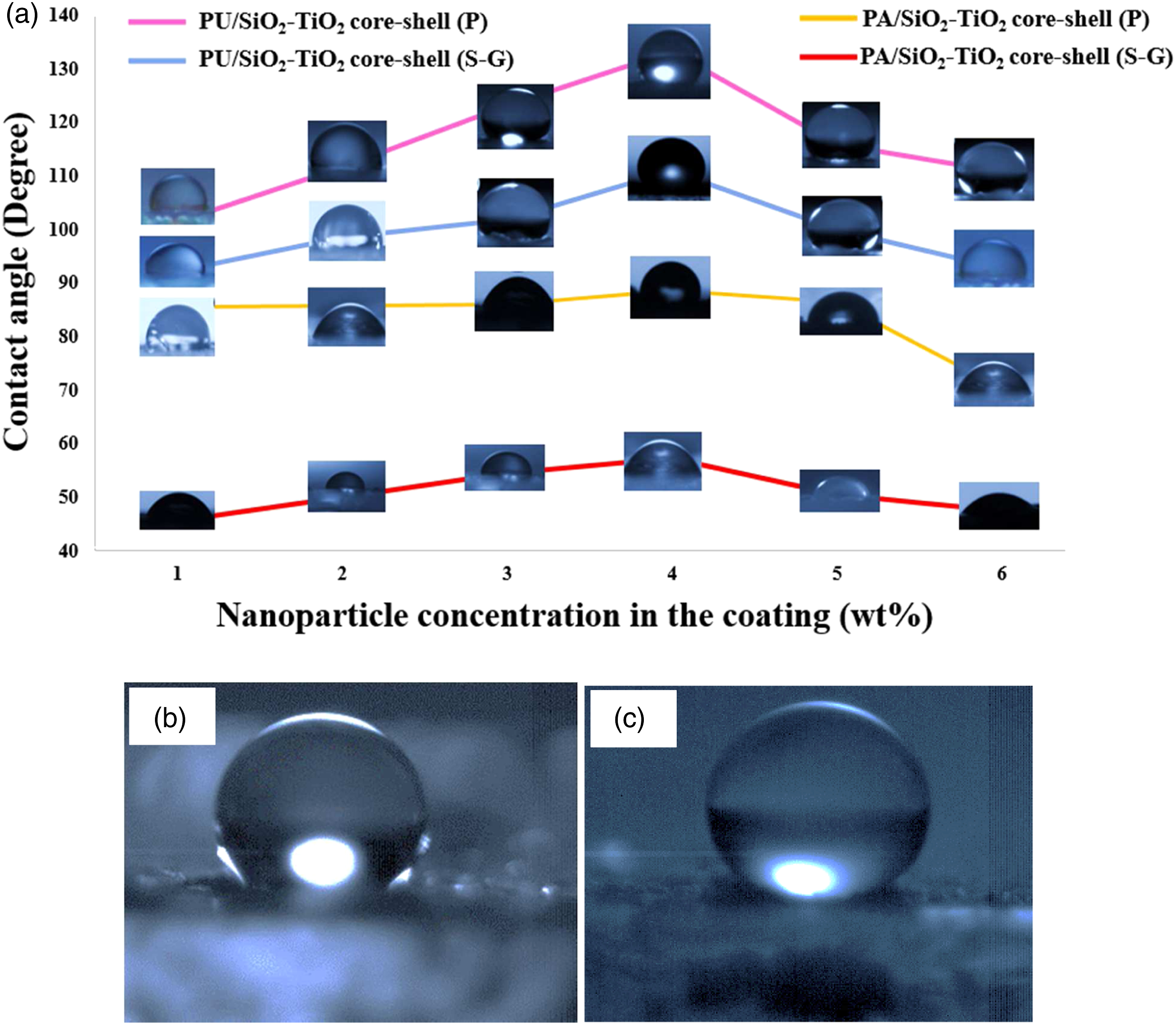
Improvement in hydrophobicity with functionalized nano core–shell particles
For the super-hydrophobicity assessment of the developed coating, further contact angle measurement was carried out with functionalized nanoparticles in PU and PA-based coating systems. Analysis of contact angle measurement data after functionalization of nano core–shell, we found an improvement in hydrophobicity. Here we reported only the best results after functionalization, that is, nano core–shell (4wt%) which was prepared through peptization synthesis because it has shown the highest value of contact angle before modifications also. This functionalized nanoparticle enhanced coating properties and improved the hydrophobic surface to super-hydrophobic. Contact angle value improved from 133.3° to 145.1° as shown in Figure 6(b) and (c) for PU/core–shell (4wt%)–based nanocoating.
Mechanical behavior of the developed nanocoating
Super-hydrophobic coatings with good resistance to mechanical loads are significant in various industrial applications to obtain long-term performance but mechanical robustness is the major challenge for super-hydrophobic coatings and was thus considered first. In this study, UMT CETR Unit-3 was used to perform the anti-scratch testing of coated mild steel samples at various loads from 3N to 9N. UMT CETR Unit-3 is a high-density cast iron vibration dampened frame. In this frame for lateral positioning system, position resolution is maintained at 1 micron and vertical positioning system at 2 microns. Encoder resolution was sustained at 0.25 microns for the lateral positioning system and 0.5 microns for the vertical positioning system. Applied scratch-length was 9 mm on the coated substrate at various loads (3N–9 N). Investigation on scratched samples was carried out by SEM images at a magnification of 150X (Figure 7). SEM images at random loads on the mild steel substrate at 3N, 5N, 7N and 9N: (a), (b), (c), (d) Scratched PU coating. (e), (f), (g), (h) Scratched PU coating containing nano SiO2 before functionalization. (i), (j), (k), (l) Scratched PU coating with nano core–shell particles before functionalization. (m), (n), (o), (p) Scratched PU coating with nano silica after functionalization. (q), (r), (s), (t) Scratched PU coating with nano core–shell particles after functionalization.
For further analysis on higher load than 9N anti-scratch testing was executed by Erichsen scratch tester up to 20N load for coating developed with non-functionalized and functionalized nanoparticles. Through SEM images, it can be seen that only PU coating could not resist the minimum applied load of 3N but SiO2/PU coating allowed to pass load up to 5N and crack started from 7N. PU coating developed with SiO2–TiO2 nano core–shell particle (4wt%) allowed to pass load up to 3N only before functionalization. After functionalization of nano core–shell, PU coating with SiO2 and nano core–shell particles at 4wt% concentration passed up to 20N load easily.
Some additional mechanical properties were also studied of these coatings like adhesion test which was performed through cross-hatch tester, and performed at 1×1 m cross that allowed to pass 100/100. Further, measurement of impact resistance data was also satisfactory, in which 500 g weight was applied from the height of 50 cm on the core–shell (peptization)–based coated sample. In this analysis, PU-based coating with 4wt% of functionalized nano core–shell has shown the best result. Flexibility analysis of these coating films was also measured, which was satisfactory and passed 12 mm cylindrical mandrel.
As we know that, humidity could have a remarkable effect on the performance or activity of the materials and coatings. The product’s lifespan can be extended by adequately protecting against humidity. Investigation on the anti-corrosion performance of these coatings was also successfully tested by high humidity testing. This test is worldwide universal to all types of stainless materials. In this testing procedure, we maintained the required atmosphere in a sealed chamber, and the temperature was maintained at 100°F. In this chamber, the humidity level was maintained near 97%. The sample was placed for 24 h in the chamber and we found that nano core–shell (peptization at 4wt%)–based PU coated sample passed this test, exhibit no type of signs of rust or staining at the coating surface.
Conclusion
A detailed study on the super-hydrophobic surfaces is presented in which the effects of surface wettability and the types of particles are considered at various concentrations. To summarize this research work, polyurethane (PU) and poly-acrylic (PA) –based nanocoating were successfully formulated with SiO2–TiO2 nano core–shell synthesized through two techniques; sol–gel and peptization methodology for contact angle measurement and mechanical testing. Super-hydrophobic nanocoating with good mechanical robustness and substrate adhesion has been achieved by using this core–shell nanoparticle from the peptization process. The contact angle of the coating was improved up to 145.1° with functionalized core–shell nanoparticles in PU-based coating system which was achieved at 4% (wt) nanoparticle concentration. PU-based coating formulations with nano core–shell particles developed through the peptization process were utilized for further study on the mechanical behavior of the coating because this preparation method gave the best results. This PU/nano core–shell–based coating has shown anti-scratch results up to 20 N load with good adhesion, flexibility, impact resistance as well as good anti-corrosion performance. So concluding all the data, mechanical properties, as well as super-hydrophobic surface was achieved through this single SiO2–TiO2 nano core–shell which can be useful for marine paint applications. When these coating would be applied to the underwater hull of ships, discourage or repel the water attachment to the surface that would help to improve the boat life. This coating would also improve the water flow passing the hull of a fishing vessel and high-performance racing yacht because of the smoothness of the coatings as well these multifunctional coatings may offer many advantages toward protecting various surfaces.
Footnotes
Acknowledgments
We would like to acknowledge the financial support from Science and Engineering Research Board (SERB), DST Project No: ECR/2016/000020. We also thank Dr. A. Bhattacharya from Amity Institute of Nanotechnology for her support in FTIR analysis. We would like to acknowledge IUAC Delhi for the SEM facility, IUAC Indore for XPS facility, and BPIL, New Delhi, for providing some of the mechanical testing facility.
Declaration of conflicting interests
The author(s) declared no potential conflicts of interest with respect to the research, authorship, and/or publication of this article.
Funding
The author(s) received no financial support for the research, authorship, and/or publication of this article.