
Abstract
Surface sealants are reported to ensure surface smoothness and improve the surface quality of composite restorations. These sealants should also reduce the bacterial adhesion on composite surfaces however, there is not much information regarding their performance on bulk-fill composite materials. The aim of this study was to evaluate the effect of surface sealant application on surface roughness and bacterial adhesion of various restorative materials. Disc-shaped samples were prepared from a compomer, a conventional composite and three bulk-fill composites. Specimens of each group were divided into two groups (n = 9): with/without surface sealant (Biscover LV, [BLV]). Surface roughness values were examined by profilometry and two samples of each group were examined for bacterial adhesion on a confocal laser scanning microscope (CLSM). Bacterial counts were calculated by both broth cultivation and microscopic images. Results were analyzed with one-way ANOVA and Bonferroni/Dunn tests. Following the BLV application, there was a decrease in the surface roughness values of all groups however, only Tetric N-Ceram Bulk and Beautifil-Bulk groups showed significantly smoother surfaces (p < 0.001). There were no significant differences among material groups without BLV application. Evaluating bacterial adhesion after BLV application, conventional composite had the lowest values among all followed by the compomer group. Beautifil-Bulk had significantly the highest bacterial adhesion (p < 0.05), followed by Tetric N-Ceram Bulk group. Without BLV application, there was no significant difference among bacterial adhesion values of groups (p > 0.05). CLSM images showed cell viability in groups. Bulk-fill composites showed higher bacterial adhesion than conventional composite and compomer materials. The surface sealant was found to be highly effective in lowering bacterial adhesion, but not so superior in smoothing the surfaces of restorative materials. So, surface sealants could be used on the restorations of patients with high caries risk.
Introduction
Various types of tooth-colored restorative materials with different physical characteristics are available to practitioners. Today, great improvements have been made in the chemical formulae of the compositions, resulting in alterations in the structures of the materials, many of their properties have not been widely evaluated in which tendency to bacterial adhesion is one such property.
Bacterial adhesion is the first determinant of colonization, 1 which could lead to secondary caries, periodontal inflammation, and even pulpitis. 2 Therefore, it is crucial to evaluate the surface properties, polishing abilities and the tendency for bacterial adhesion of novel materials. For instance, bulk-fill composites were recently launched to be used in one increment of 4 mm, which reduces the procedure time. 3 These types of composites have more translucent matrices, different photoinitiators, and smaller fillers. 4 Many studies have evaluated the mechanical, 5 physical, 6 and biological properties of bulk-fill composites, 3 but only one study 7 has evaluated bacterial adhesion.
The main factor in the pathogenesis of dental caries is Streptococcus mutans (S. mutans), as it is the pioneer organism in biofilm formation, leading to the subsequent colonization of the surface by other species. 8 In terms of preventing possible secondary caries, some studies have investigated the tendency of bacterial adhesion of various resin composites.9,10 However, there is still no consensus in the literature regarding the influence of the surface properties on bacterial adhesion 11 which may be caused by both physical factors and chemical bonds occurring between restorative materials and bacterial adhesion forces. Surface roughness, hydrophobicity, wettability, and surface free energy (SFE) constitute most of the physical attractions 12 and van der Waals and electrostatic forces 13 are the attractive chemical bonds.
On the other hand, various glaze materials or “surface sealants” are low-viscosity resins that alter the surface characteristics of restorative materials. These surface sealants are polymerized onto composite surfaces in order to penetrate into microstructural defects and smoothen the surfaces. 13 These materials are known to maintain ideal surface properties, ensure adequate marginal sealing, and improve the surface quality of the composite restoration. 14 Aside from these advantageous effects of sealants on resin restorations, sealed surfaces were reported to reduce bacterial adhesion as well. 13 The possible mechanism for surface sealants regarding bacterial adhesion is to lower the SFE of restorations and alter the hydrophobicity levels of surfaces. S. mutans attaches to rougher surfaces with high SFE and also tends to hydrophobic surfaces, in which these characteristics of the restoration surfaces are directly related to the monomer content of materials. Moreover, restorative materials may release or recharge fluoride ions. 15 The ionic exchange from the materials’ surfaces may therefore result in irregularities and greater bacterial colonization, which may be reduced by surface sealants. In terms of bacterial adhesion, the effects of surface sealants on restorative materials are highly dependent on the content of the materials and there is inadequate information about the relation between novel types of composite resin, bulk-fill materials and their properties on bacterial adhesion.
The present in vitro study involves various types of restorative resin materials with different physicochemical intrinsic properties. In many studies, the influence of materials against bacterial adhesion is well-defined by surface roughness values.16,17 Thus, the purpose of this in vitro study was to compare S. mutans adhesion to, and surface roughness values of, various tooth-colored restorative materials coated with a surface sealant (Biscover LV, [BLV]). Bacterial counts were performed on both cultivation findings and microscopic calculation by a confocal laser scanning microscope (CLSM). The following null hypotheses were tested: (1) after the application of surface sealant, there would not be significant differences in the surface roughness values of materials; (2) in terms of bacterial adhesion, there would not be significant differences among the tested restorative materials; and (3) the surface sealant applications would not reduce initial adhesion of S. mutans to composite resin materials.
Materials and methods
Materials
Three bulk-fill resin composites, a conventional composite, and a compomer were used as substrates, which were prepared with/without an unfilled surface sealant. Manufacturers, lot numbers, and the main compositions of the materials are shown in Table 1.
Composition of the materials used in the present study.

Specimen preparation
The disc-shaped specimens (5 mm in diameter and 2 mm thick) of the test materials were prepared using a metallic plate with calibrated holes. The materials were placed into the holes and confined between two opposing transparent matrix strips (SS White, Philadelphia, PA, USA). A glass microscope slide (1 mm in thickness) was then placed on the mold, with constant pressure applied to extrude the excess material. Thereafter, specimens were cured for 20 s on the top sides—with Elipar DeepCure (3 M ESPE, St. Paul MN, USA), operating in standard mode and emitting no less than 600 mW/cm2, as measured with a light meter (Hilux Curing Light Meter, Benlioglu Dental, Ankara, Turkey). The light meter was placed on the curing unit before initiating polymerization. In addition, the light-curing unit’s guide was placed perpendicular to the specimen surface, at a distance of 1 mm. Specimens were then carefully removed from the plate and kept in distilled water in an incubator, at a temperature of 37°C. This was done to reduce the number of uncured monomers. After 24 h, specimens were removed from the incubator and carefully dried. The top surfaces of all specimens were serially polished with the series of Sof-Lex discs (3M ESPE, St. Paul, MN, USA), for 30 s using a slow-speed micromotor at 15.000 rpm and under dry conditions. Each disc was renewed after being used five times.
Afterward, the non-polished surfaces of the specimens were numbered using a water-resistant pen. They were randomly divided into two subgroups, depending on the post-treatment procedures (n = 9). Specimens numbered from 1 to 9 were assigned to the “treatment group” and were manipulated with the surface sealant according to the manufacturer’s instructions. The materials’ surfaces were first acid-etched (Uni-Etch, Bisco, Schaumburg, IL, USA) for 15 s. The acid was then rinsed off with running water, and the surfaces were air-dried. One thin coat of BisCover LV (BLV) was applied for 15 s with a micro-brush, using a light brushing motion. Thereafter, the specimens’ top surfaces were gently air-thinned for 3 s, with 15 s being allowed for evaporation.
Finally, the specimens were light-cured for 20 s with the LED unit that was used at the beginning of the experiment. All of the specimens were stored in distilled water at 37 ± 1°C for another 24 h, to ensure complete polymerization of the sealing resins.
Surface roughness measurement
Specimens were relocated in the metallic plate, for immobilization for the measurement. The surface roughness test was performed with a profilometer (Surtonic 25, Taylor Hobson, Leicester, England). The cut-off value for surface roughness was arranged to 0.25 mm, and the evaluation length for each measurement was set to 1.5 mm. The stylus moved with a crosshead speed of 0.25 mm/s. The tester was calibrated against a standard before the experimental period and during each new measuring session. Three successive measurements from separate locations were recorded for all specimens in each group, and roughness (Ra) values were averaged.
Assessment of S. mutans adhesion
Before initiating bacterial experiments, specimens were placed in an ultrasonic cleaner (Sonica, Soltec, Milan, Italy) filled with 10% ethanol. This was done for 3 min to disinfect the surfaces. After air-drying the surfaces, specimens were kept in the incubator until the bacterial experiments commenced.
Freeze-dried strains of S. mutans (ATCC 25175) that are located on the commercial strip were inoculated in blood agar (Merck, 110886) with the spread plate culture technique. The strains were then incubated at 37°C for 24 h, in aerobic conditions. Following the incubation period, bacteria were collected with a sterile swab and were then suspended in Trypton Soya Broth (Oxoid, CM0129) containing 20% glycerol. This bacterial suspension was frozen at −80°C, as a stock bacterial solution. For the purposes of the experiment, 10 µl of the frozen bacterial sample was inoculated in brain heart infusion (BHI) broth (Oxoid, CM1135) and incubated at 37°C for 24 h, in aerobic conditions. Thereafter, 50 µl of overnight growth bacterial suspensions were spread on BHI agar (Oxoid, CM1136) and incubated at 37°C for 24 h, in aerobic conditions. The bacterial sample was then transferred in BHI broth and was adjusted to give a turbidity equivalent to that of a 0.5 McFarland turbidity standard. Disc-shaped materials were placed into a flat-bottom 96-well plate (Tech Labs Medical, Istanbul, Turkey), with one specimen per well, and 100 µl of the bacterial suspension was added into each well. After incubation at 37°C for 24 h, the test materials were washed three times with 200 µl of sterile saline to remove non-adhering cells. Adhered cells were collected using swabs and were transferred into Falcon tubes filled with 5 ml of saline. The tubes were vortexed for 60 s to detach bacteria from swab surfaces. The detached cells from the two specimens’ surfaces were separated for microscopic analyses.
For conventional culture analyses, a serial dilution was carried out with sterile saline solution (1- to 100-fold dilution). For bacterial count determination, 100 µl of samples from each dilution was spread on the BHI agar plate (Tech Labs Medical, Istanbul, Turkey) in triplicate. They were incubated at 37°C for 24 h, in aerobic conditions. The bacterial count found in 1 mm2 of material surfaces was calculated according to the specimens’ surface area and dilution factor.
Confocal laser scanning microscope 3M ESPE imaging (CLSM)
Two specimens of each subgroup were randomly selected for microscopic evaluation with CLSM (SP8 Lightning confocal microscope, Leica Microsystems, Wetzlar, Germany), immediately following microbiological procedures. Detached cells in 100 µl broth were stained using a Cell-Check Viability/Cytotoxicity bacterial kit (ABP Biosciences, Rockville, MD, USA). As per instructions, 1.5 µl of NucView Green and 1.5 µl of propidium iodide were added to a tube, as was 7 µl of saline solution.
A swab was used to obtain 10 µl of bacterial broth, which was mixed with 1 µl of dye solution in a tube. The tube was then incubated at room temperature in the dark, for 15 min. The mixture was spread over a glass plate, and the images of cells were observed under 5× or 10× magnification.
Three images were taken from each specimen, and live bacteria were counted. The bacterial count found in 1 mm2 of surfaces was calculated according to the formula for microscopic measuring given below, and the results were averaged. 18

(N = number of microorganisms per milliliter S = filtration area n = average number of microorganisms in the studied area C = microscopic area V = filtered sample volume D = dilution rate of the sample)
Statistical analysis
The statistical analysis was performed using SPSS Statistics 24.0 (SPSS, Chicago, IL, USA), at a significance level of 0.05. The results were primarily analyzed using the Kolmogorov-Smirnov test to determine the existence of a normal distribution. Due to the normally-distributed data, the differences observed within each of the materials were analyzed using the Student’s t-test. Further statistical analyses were performed using a one-way ANOVA and Bonferroni/Dunn test, to cross-compare the test materials.
Results
The means and standard deviations of each group’s surface roughness values are summarized in Table 2.
Mean surface roughness values (Ra, μm) with standard deviations (SD) and significance among groups regarding BLV application.*
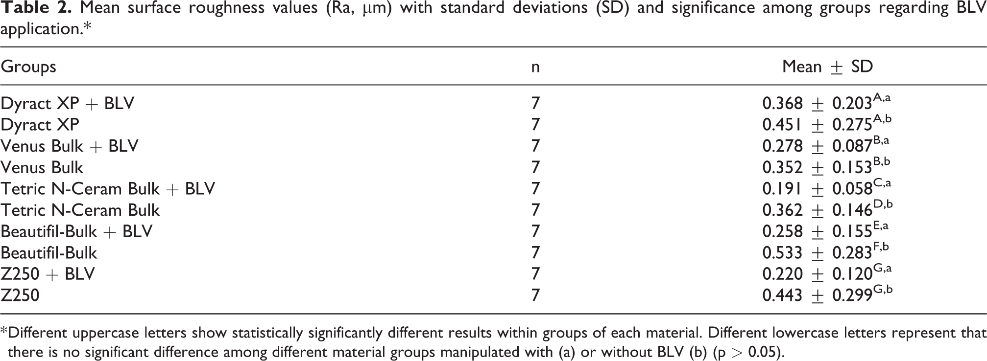
* Different uppercase letters show statistically significantly different results within groups of each material. Different lowercase letters represent that there is no significant difference among different material groups manipulated with (a) or without BLV (b) (p > 0.05).
Table 2 shows that after BLV application, all of the groups had superior surface roughness values; however, the use of surface sealant had significantly lowered the roughness values (p < 0.001) only in condensable bulk-fill composites (Tetric N-Ceram Bulk and Beautifil-Bulk). In addition, no significant differences were observed among groups without BLV application and groups with BLV application except for condensable bulk-fill composites, respectively (p = 0.482, p = 0.078).
Tetric N-Ceram Bulk + BLV showed the lowest roughness, followed by Z250 + BLV, and Beautifil-Bulk + BLV. Dyract XP had the roughest surface among BLV groups and Beautifil-Bulk showed the roughest surfaces among groups without sealant application. The mean bacterial count values in 1 mm2 of broth cultivation results are summarized in Table 3.
Mean values of the bacterial count with standard deviations and significance among groups.*

* Different uppercase letters show statistically significantly different results within groups regarding BLV application of each material. Different lowercase letters statistically significant results among groups with BLV application. The same lowercase “d” letter shows that there is no significant difference among groups without BLV application.
In Table 3, it is clear that, except for the Beautifil-Bulk group, every material with BLV application resulted in lower bacterial adhesion. The Z250 + BLV group had the lowest and the Beautifil-Bulk + BLV group had the highest bacterial adhesion. No statistically significant differences were observed among groups without BLV application (p > 0.05).
However, there were significant differences in groups with BLV applications. For instance, Beautifil-Bulk + BLV group had significantly the highest and Z250 + BLV group had significantly the lowest bacterial adhesion than among groups (p < 0.05). Dyract XP + BLV group showed the second-lowest bacterial adhesion, followed by the BLV groups of Venus Bulk and Tetric N-Ceram Bulk. Moreover, BLV application to Tetric N-Ceram bulk-fill composite did not significantly affect its bacterial adhesion (p = 0.763).
CLSM evaluation
The number of live bacteria on CLSM images of all groups was calculated using the formula (provided above). The numerical results are shown in Figure 1.

Bacterial counts of specimens randomly selected for CLSM imaging calculated with microscopic measuring.
According to the formula, the Beautifil-Bulk group had the highest bacterial adhesion, regardless of the BLV application. In BLV applied groups, the Tetric N-Ceram Bulk group had the lowest bacterial count, followed by the Z250 and Dyract XP groups. Groups without BLV application, Z250, Dyract XP and Tetric N-Ceram showed similar results. Bacterial adhesion on the specimens of all groups was also represented in CLSM images, obtained with the Cell-Check Viability/Cytotoxicity Kit (Figure 2).

Representative CLSM images of S. Mutans adhesion on BLV applied (BLV+) and non-applied (BLV−) specimens. Green dye shows live bacteria and red dye shows dead bacteria. (a) Dyract XP specimen, (b) Venus Bulk Fill, (c) Tetric N-Ceram Bulk Fill, (d) Beautiful-Bulk Fill, (e) Filtek Z250.
In CSLM analysis (Figure 2), biofilm structures were observed on all material surfaces. It is obvious that images with BLV application represented lower bacterial intensity. In accordance with the results of Figure 1, the Beautifil-Bulk group had the most intense biofilm of bacteria, regardless of the BLV application. In all the images, living cells were observed at a higher prevalence on specimen surfaces. However, the dead S. mutans bacteria on the surface of Venus Bulk, Tetric N-Ceram Bulk and Z250 materials were higher than in other groups. In BLV applied groups, Dyract XP, Z250 and Tetric N-Ceram Bulk had similar bacterial intensity. In groups without BLV application, Dyract XP and Z250 groups, and Tetric N-Ceram XP and Venus Bulk groups showed similar tendencies for S. mutans.
Discussion
Bulk-fill composites are commonly used by clinicians, as they are easy to use, have time-saving applications, and reduce polymerization shrinkage. 7 However, the bacterial adhesion properties of bulk-fill composites have not been widely evaluated. Yet, a few studies have been published with various methods of in vitro design.7,19,20 In the present study, surface sealant was applied to various bulk-fill composites to investigate the effect on surface roughness and bacterial adhesion. According to the literature, surface-coating materials, such as sealants and glaze materials, are beneficial not only for covering irregularities and inhibiting marginal microleakage but also for lowering bacterial adhesion. 13
Many of the studies evaluating bacterial adhesion6,12,21 compared tested materials with a high fluoride content; therefore, a compomer material, which has both strontium fluoride and fluoride glass, was included in the present study. To perform quantitative bacterial adhesion measurements, bacterial adhesion was counted, both on cultivation and on CLSM images. However, both techniques have limitations or disadvantages. A series of experimental procedures are involved in cultivation counting which could cause false quantitative results due to the structure of adherent live bacteria. On the other hand, CLSM imaging is a simple method of quantitative and qualitative measurement, without removal from the substrate. Furthermore, dead and live bacteria can be evaluated separately, depending on their staining. However, with CLSM, it is difficult to eliminate the differences between adherent and loose bacteria on the surface. 22 In order to remove the loosened bacteria, specimens were washed three times before preparation for bacterial counts. Although bacterial counts were gained in both methods, only the cultivation results were statistically analyzed. The main reason was that two similar results on the same parameter could be confusing when comparing materials. Moreover, as seen in Figures 1 and 2, the two methods of bacteria counting were not 100% identical. This outcome is in accordance with another study in which bacterial biofilm was evaluated; the study showed that the classical culture method and microscopic results could differ. 23
Regarding surface roughness characteristics, the microstructure of the surface is as important as the intrinsic physicochemical properties of the materials. 24 For the purposes of the present research, the surface roughness values of the tested materials were examined using a contact profilometer, which is used in various other studies.9,25,26 According to results, Tetric N-Ceram Bulk and Beautifil-Bulk groups showed significantly lower roughness values after the BLV application. Thus, the first hypothesis stating that there would be no significant difference among surface roughness values of materials after surface sealant application is partially rejected. It is also clear that the BLV application improved almost all surfaces, bringing them to the acceptable threshold level of 0.2 µm. 27 However, there are conflicting results regarding the effects of sealant materials on the surface roughness values. Although some studies showed that sealants reduce the roughness scores of composites,28,29 no improvements on the surfaces were found in a recent study. 30
On the other hand, Dos Santos’s study 14 explained that sealants with fillers could affect the final roughness values of resin materials. Depending on that, a low-viscosity, filler-free sealant (BLV) was used in the present study. However, this did not lead to a significant change in the roughness values of all materials. It is impossible to obtain a uniform sealant thickness in the composite materials, 31 and differences in the thickness could lead to extra layers on the top surface, which could affect surface roughness values. It is reported 32 that surface sealants could form a layer that is 0–70 µm thick; therefore, sealant thicknesses could be responsible for the inconsistent results.
Obtained results from surface roughness values of groups, with or without BLV application, could be attributed to the intrinsic physicochemical properties of materials as well. Finishing and polishing procedures were performed by one dentist, who used flexible Sof-Lex discs (as they are still accepted as the gold standard). Therefore, iatrogenic factors were eliminated regarding surface properties. Additionally, material-related properties, such as filler size, loading, and shape, could directly determine the surface roughness values of materials.17,33 When evaluating the tested composites, it was determined that the bulk-fill groups had nearly the same filler ratio, of between 65% and 75% of the weight and similar filler sizes, varying from 0.01 to 3.5 µm. These similar filler characteristics could be the reason for parallel roughness scores among groups.
Moreover, the compomer material, Dyract XP, formed higher roughness values as expected. Due to the lower ratio of fillers (47% wt.) and soluble strontium fluoride salts, which cannot be homogenously involved in the polymer matrix, Dyract XP showed inferior results in roughness studies.12,34 However, because of the lack of immersion media, specimens of Dyract XP may demonstrate similar results among other groups. In addition, flowable materials were shown to have 20–25% fewer filler loadings 15 and the only flowable composite of the study, Venus Bulk, showed one of the lowest roughness scores compared to Z250 which is a micro-hybrid composite with higher polishability. Even though there is no significant difference regarding the surface roughness scores of Z250 group without BLV application, results could still be attributed to the similarity between the micron-sized particles and filler loading of other resin-based materials.
According to results, both conventional composite resin and compomer material had significantly lower bacterial adhesion than bulk-fill composites. Therefore, the second hypothesis, which states that there would not be significant differences regarding bacterial adhesion between tested restorative materials, is partially rejected. There were no significant differences among groups without BLV application. However, the lowest bacterial adhesion was detected in Z250, and the highest bacterial adhesion was detected in Beautifil-Bulk material. Yu et al. 35 declared that in the early stages of attachment (2 and 4 h), a rougher surface tended to have a higher attraction to bacteria than a smoother surface. Bacteria prefer to adhere to rough surfaces, as they receive better protection against displacing forces and therefore have enough time to change into an irreversible plaque formation, from a reversible one. It is also plausible that S. mutans, similar to many streptococci, prefer to adhere to surfaces with high SFE. 6 It is therefore evident that S. mutans would prefer to accumulate on rougher dental surfaces than on polished ones.7,9 This result pertains to all tested materials except for the Beautifil-Bulk group, which showed average roughness values but the highest bacterial adhesion in both cultivation and CLSM counts. Beautifil-Bulk is a giomer bulk-fill composite with a high silicate glass ratio and ionic (S-PRG) fillers. Studies36,37 have reported that regardless of surface roughness, the bacterial adherence in resin materials with S-PRG filler was found to be lower than those without S-PRG filler. This was attributed to the release of six ions from the filler (i.e., fluoride, sodium, borate, aluminum, silicate, and strontium), which may contribute to the antimicrobial activity of restorative material against various oral bacteria. However, a novel study 38 outlined some limitations in terms of releasing S-PRG filler ions. It was stated that these ions could only be released if the pH of the related media turns acidic, and they could inhibit the metabolism of S. mutans, if they were in the active growth phase. The inferior results of Beautifil-Bulk may have been caused by the in vitro design of the present study in which acidic media and all elements for biofilm formation were therefore not imitated. Another study has reported that a sealant with S-PRG filler has lower antibacterial potential against S. mutans and higher biofilm formation than other fluoride-releasing agents. 39 Therefore, conflicting results were found against S-PRG filler, in the literature so, inferior values of Beautifil-Bulk may be attributed to the organic part of the composite material, other active-passive physical forces formed between bacteria and material surfaces, or the lack of an acidic element in the experimental environment.
Among tested restorative materials, lower bacterial adhesion was observed with the application of BLV, which is an anti-adherent, low-viscosity, and ethanol-based sealant. Even though the effect of BLV against Candida albicans over dentures has been proven in many studies,10,40 researches against the S. mutans biofilm on composite materials involving BLV are very rare. Furthermore, it appears that this is the first study to investigate the effect of BLV on bacterial adhesion of bulk-fill composites. In the present study, the bacterial count was significantly reduced among groups after the BLV application, except for Tetric N-Ceram Bulk-Fill group. Thus, the last hypothesis stated as surface sealant applications would not reduce initial adhesion of S. mutans to composite resin, is partially rejected. BLV had an acrylate monomer (dipentaerythritol pentaacrylate) and ethanol so that, the structure gains flexibility and moisture-resistance. 41 Moreover, a cross-linking material (PENTA-dipentaerythritol pentaacrylate monophosphate), which is frequently used in adhesive bonding systems, 42 is from the same phosphoric ester family of the acrylate monomer of BLV. It is therefore expected that the BLV material will be in accordance with monomer structures of tested resin composites and may form a barrier for foreign structures such as S. mutans. However, SFE of the specimens may be responsible for the inferior results of the Tetric N-Ceram Bulk-Fill group which is one of the main factors affecting bacterial adhesion. 1 If the SFE is low in these group of specimens, the wettability of the sealant will be low, yet BLV would not function ideally. Another parameter affecting S. mutans biofilm formation is the “hydrophilicity” or “hydrophobicity” of the restorative materials. 1 These terms are related to SFE, so that if a hydrophobic surface becomes rougher, both of its hydrophobicity and SFE values increases as well. Within the parameters of hydrophobicity, it is not only the materials’ surfaces but also the opponent bacteria’s cell membranes that are important for the purposes of ensuring interactive forces regarding adhesion. In a number of studies, it was found that S. mutans has moderate to high cell hydrophobicity.1,43 Hydrophobic surfaces and hydrophobic mutans cells form stronger and closer adhesive forces. 44 BLV application alters the water-contact angle of the composite surfaces 5 so that, after the sealant application, the hydrophilicity or hydrophobicity of the surfaces also increases. In addition, it was remarkable that the Venus Bulk + BLV group showed significantly similar bacterial adhesion to the Z250 group. Apart from having common compounds (such as UDMA monomer or ytterbium fluoride fillers) that are similar in structure to Tetric N-Ceram Bulk and Z250, Venus Bulk’s superior results could be relevant to BLV’s wettability or the fact that it could be a more compatible material with BLV application as a flowable bulk-fill.
The purpose of the present study was not to investigate the materials’ surface properties; therefore, related physical parameters were not evaluated. However, with BLV application, tested material surfaces may be more hydrophilic, thus resulting in less S. mutans adhesion to materials. The existence of hydroxide ions (OH−) or ester bonding in the structure of monomers could affect the materials’ hydrophobicity properties. Although BisGMA and TEGDMA are hydrophilic monomers, the UDMA monomer is known to be hydrophobic. The ethoxylated version of BisGMA (BisEMA) and a novel monomer, BisMPEPP, showed lower water sorption than BisGMA. 45 Therefore, the lower bacterial counts of Z250 and Dyract XP could be attributed to the hydrophilic structure of Z250 and the high fluoride and UDMA content of Dytract XP. On the other hand, the highest bacterial proliferation was detected in the Beautifil-Bulk group, as can be seen in CLSM images. This could be explained by its higher hydrophobic UDMA and BisMPEPP monomers. According to monomer-based hydrophobicity results, the present study is in accordance with the studies by Buergers et al. 11 and Kim and Kwon. 13
The aim of the current study was to compare the materials’ properties regarding bacterial adhesion and to evaluate them with CLSM images. However, there were a number of limitations in the study. Firstly, the in vitro model that was used investigated the adhesion of S. mutans at a neutral pH and under conditions without any fundamental oral elements, like saliva. The reason for this is that, depending on the origin and type of collection of saliva, its protein composition varies greatly. This causes difficulties in standardization. However, saliva would only alter the study’s quantitative results if there were no changes in the comparison of the materials. Secondly, a biofilm formation was not included; therefore, S. mutans adhesion was only tested after 24 h. Thirdly, the environment was not acidic enough for the bioactive material with S-PRG filler to release its ions, which could have been an advantage for the material. Therefore, further in vitro studies are needed to mimic the oral environment for all the compounds included, which would also be beneficial to evaluate the mechanisms of bacterial adhesion reduction using sealant application.
Conclusion
Within the limitations of the present study, the test conditions were adopted to meet the study’s aim of evaluating the material-based differences in a medium optimized for bacterial viability. However, bacterial counting of microbiological cultures did not completely conform with CLSM counts. As bacterial adhesion to restorative materials contains many physicochemical sources and depends greatly on material properties, the related surface sealant on lowering bacterial counts is highly effective, but not superior when it comes to smoothing the surfaces. Therefore, considering the complexity of adhesion, studies including multiple surface and adhesion parameters should be designed, with various methods of cultivation.
Footnotes
Acknowledgments
Authors would like to thank PhD students of Khusan Khodzhaev, MSc and Burcu Salman, MSc from Aziz Sancar Institute of Experimental Medicine Department of Genetics for their valuable contributions on CLSM imaging.
Declaration of conflicting interests
The author(s) declared no potential conflicts of interest with respect to the research, authorship, and/or publication of this article.
Ethical approval
This article does not contain any studies with human participants or animals performed by any of the authors.
Funding
The author(s) disclosed receipt of the following financial support for the research, authorship, and/or publication of this article: The present study was supported by Scientific Research Projects Coordination Unit of Istanbul University (Grant Number: 30073).