
Abstract
Xiao Y, Qin T, Sun L, et al. Resveratrol Ameliorates the Malignant Progression of Pancreatic Cancer by Inhibiting Hypoxia-induced Pancreatic Stellate Cell Activation. Cell Transplantation 2020, Vol. 29 1–14. DOI: 10.1177/0963689720929987
In this article, two representative pictures in adjacent groups were mistakenly mixed and labeled. In the corrected version, Figure 2A has been updated to reflect the image from the MiaPaca-2 Hypoxia+si-HIF1α group. Figure 3A has been updated to display from the Panc-1 CM+RSV group. These changes do not affect the outcome of the article. The updated figures and legends are as follows:
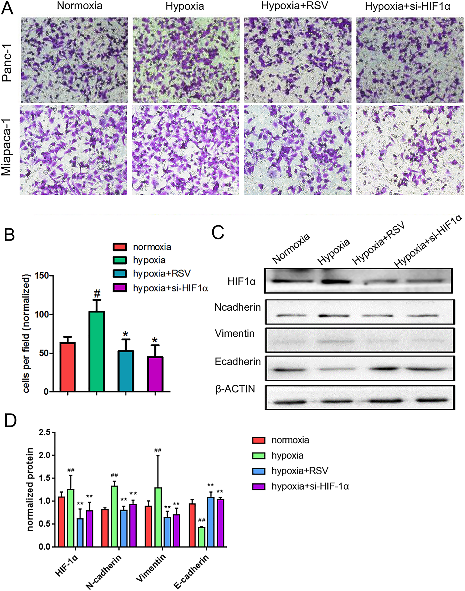
Resveratrol suppresses the pancreatic cancer cell epithelial–mesenchymal transition and invasion induced by hypoxia. (A, B) Panc-1 or Mia Paca-2 cells were incubated under normoxic conditions for 24 h or under hypoxic condition for 24 h in the presence of 50 μM resveratrol or si-HIF-1α. The cells were then seeded into a Matrigel-coated invasion chamber under normal or hypoxic conditions for 24 h. The invaded cells were quantified by counting the number of cells in 10 random fields at 200× magnification. (C) Subconfluent pancreatic cancer cells (Panc-1) were exposed to normoxia for 24 h or hypoxia for 24 h in the presence of 50 μM resveratrol or si-HIF-1α. HIF-1α, Ncadherin, vimentin, and E-cadherin expression levels in Panc-1 cells were analyzed by western blotting. #P<0.05; ##P <0.01 comparing the hypoxia group with the control group. *P <0.05; **P<0.01 comparing the hypoxiaþRSV or hypoxiaþsi-HIF-1a group with the hypoxia group. All data represent at least three independent experiments. HIF-1α: hypoxia-inducible factor 1; RSV: resveratrol.
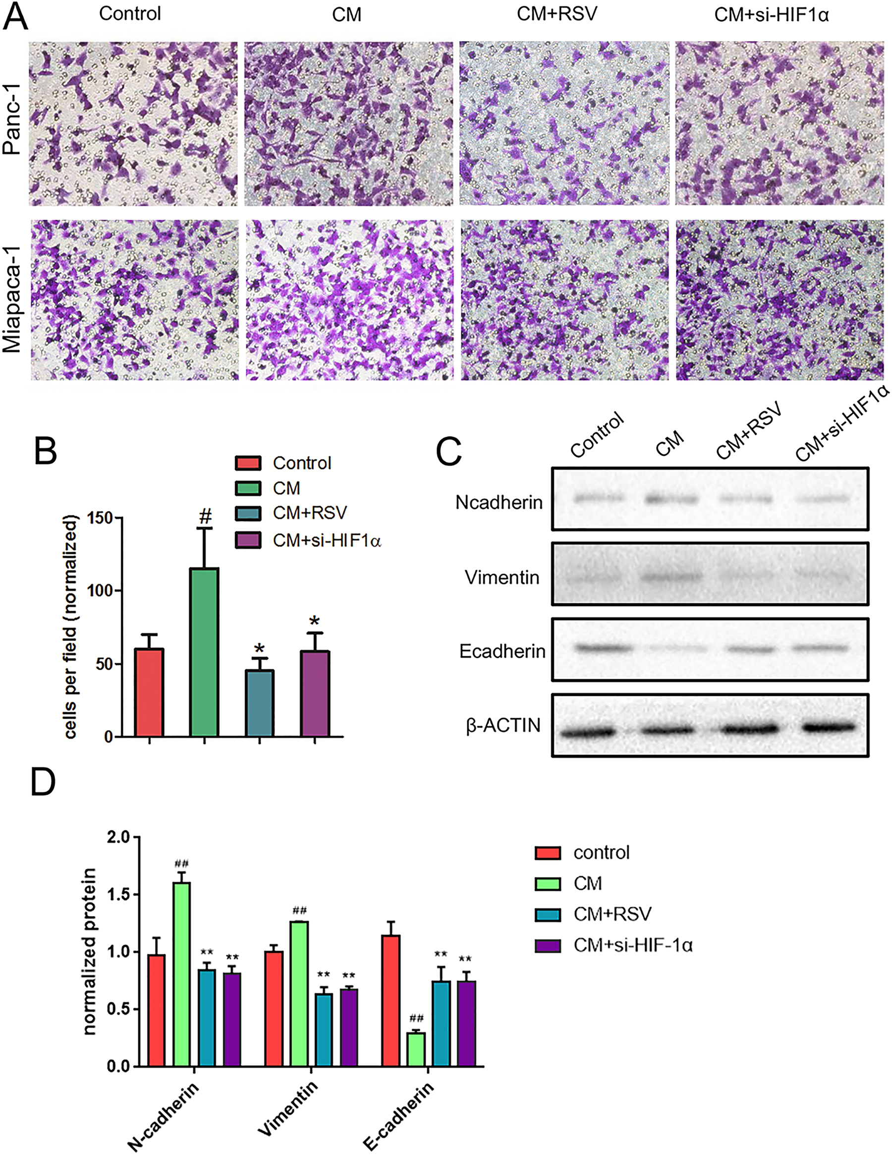
Resveratrol suppresses the pancreatic cancer cell invasion and epithelial–mesenchymal transition induced by CM from PSCs. CM were from PSCs activated by hypoxia. (A, B) Panc-1 or Mia Paca-2 cells were cultured under normoxic conditions with CM from PSCs in the presence or absence of 50 μM resveratrol or si-HIF-1α for 24 h. The cells were seeded into a Matrigel-coated invasion chamber under normoxic conditions for 24 h. The invaded cells were quantified by counting the number of cells at a 200× magnification in 10 random fields. (C) Subconfluent Panc-1 cells were treated under normoxic conditions with CM from PSCs treated with or without 50 μM resveratrol or si-HIF-1α for 24 h. Then, the cells were lysed, and the N-cadherin, vimentin, and E-cadherin expression levels in the Panc-1 cells were analyzed by western blotting. #P <0.05; ##P <0.01 comparing the CM group with the control group. *P<0.05; **P <0.01 comparing the CM+RSV or CM+si-HIF-1α group with the CM group. All data represent at least three independent experiments. CM: conditioned medium; HIF-1α: hypoxia-inducible factor 1; PSC: pancreatic stellate cell; RSV: resveratrol.