
Abstract
Epidemiological evidence of an association between exposure to chemical carcinogens and an increased risk for development of glioblastoma (GBM) is limited to weak statistical associations in cohorts of firefighters, farmers, residents exposed to air pollution, and soldiers exposed to toxic chemicals (e.g., military burn pits, oil-well fire smoke). A history of ionizing radiation therapy to the head or neck is associated with an increased risk of GBM. Ionizing radiation induces point mutations, frameshift mutations, double-strand breaks, and chromosomal insertions or deletions. Mutational profiles associated with chemical exposures overlap with the broad mutational patterns seen with ionizing radiation. Data on 16 agents (15 chemicals and radio frequency radiation) that induced tumors in the rodent brain were extracted from 602 Technical Reports on 2-years cancer bioassays found in the National Toxicology Program database. Ten of the 15 chemical agents that induce brain tumors are alkylating agents. Three of the 15 chemical agents have idiosyncratic structures and might be alkylating agents. Only two of the 15 chemical agents are definitively not alkylating agents. The rat model is thought to be of possible relevance to humans suggesting that exposure to alkylating chemicals should be considered in epidemiology studies on GBM and other brain tumors.
Keywords
Introduction
Glioblastoma (GBM) is the most common primary brain cancer in adults with about 12,000 cases diagnosed in the United States each year 1 encompassing 16% of all primary brain and central nervous system neoplasms. 2 All GBMs are WHO grade IV brain tumors leading to a median length of survival following diagnosis of only 15–18 months and a 5-years survival rate around 10%. 1
The clearest relationship of an environmental exposure to GBM is radiation. Between 1910 and 1959, an estimated 200,000 children worldwide were exposed to X-rays 3 from employing the Adamson-Kienbock procedure for Tinea capitas, i.e., the fungal scalp infection called ringworm. 4 Long-term follow up studies revealed that these scalp X-ray treatments were associated with an increased risk most frequently for meningiomas but also for gliomas.1,5 Radiation is a powerful mutagen associated with many cancers, but it is difficult to assign a particular mutagenic mechanism to the induction of radiation associated GBMs, as X-ray irradiation induces several different types of DNA damage including chromosomal loss, chromosomal deletions, chromosomal nondisjunction, and localized mutations affecting small sections of the chromosome.6,7
In addition to ionizing radiation, at least three genetic syndromes are associated with an increased risk for development of GBM – Li-Fraumeni syndrome,8–12 Neurofibromatosis 1,13–19 and Turcot syndrome.20–23 Ionizing radiation, and these three genetic syndromes, are associated with other brain tumors in addition to GBM, and each of these risk factors displays different mutation patterns thought to influence the initiation or progression of the associated brain tumor type. The lack of specificity regarding brain tumor type, and of putatively causative mutations, implies that different molecular pathways can lead to a GBM or other brain tumor types.
Uncertainty of induction of glioblastoma by chemical exposures
Exposure to environmental chemicals has not been proven to be associated with an increased risk for development of GBM in humans. 24 This ambiguity is not surprising given the challenges faced by an epidemiological approach. In 2020, there were 1,603,844 new cancer cases in the US, and 602,347 cancer deaths. 25 The 12,000 yearly GBM cases and deaths represent only 0.75% of total US cancer cases, and 2% of total cancer deaths. The relative rarity of GBM renders studying its causation via a prospective design much more difficult than employing a less statistically robust retrospective design. 26 A prospective design requires the logistically complex and expensive enrollment of a very large number of research subjects and an extended follow-up period to facilitate detection of a significant number of GBM cases.
Retrospective epidemiology studies and meta-analyses report weak but statistically significant associations between putative exposure to chemicals and brain tumors including GBM. Several retrospective studies have examined working as a firefighter and increased risk for brain tumors.27–36 Similarly, retrospective epidemiology studies have also reported an increased risk of brain cancer in farmers.37–42 In contrast, other studies on farmers have reported no increased risk for central nervous system (CNS) tumors.43–46
Employing both retrospective and prospective study designs, a significant effort has been expended toward studying the possible relationship between exposure to air pollution and the development of brain tumors. Based on 12,928 brain tumor cases and 22,961 controls, a Danish studied reported total tumors of the brain were associated with combined organic carbon and emitted black carbon odds ratio (OR) 1.053 [95% Confidence Interval (CI), 1.005-1.103, per interquartile range of exposure]. The strongest associations were reported for malignant tumors with an OR per interquartile range of exposure for combined organic carbon and emitted black carbon of 1.063, (95% CI 1.007-1.123). 47 Based on 210 cases of malignant brain cancer and 555 cases of meningioma in Los Angeles County, brain cancer risk in men increased with exposure to benzene [hazard ratio (HR) 3.52, 95% CI 1.55-7.55] and PM10 (HR 1.80, 95% CI 1.00-3.23). 48 A prospective Danish study enrolled 25,143 cancer-free nurses and followed them for 15.7 years during which 121 brain cancers developed. In nurses over age 44, a weak positive association between air pollution exposure and brain tumors was seen, with a stronger association seen in obese subjects. Associations for benign tumors and meningeal tumors were stronger than for other brain cancers. 49
A large European prospective study enrolled 282,194 subjects from five Swedish cohorts, one Norwegian cohort, one Danish cohort, two Dutch cohorts, one Austrian cohort, and two Italian cohorts (cities of Varese and Turin). 50 By international standards, the areas selected for study do not experience significant levels of air pollution. More heavily polluted areas might have provided a better test of the possible effects of air pollution. During 12 years of follow-up, 466 malignant brain tumors were diagnosed. Data on nonmalignant brain tumors was available for a subgroup of 106,786 subjects that presented with 176 benign brain tumors and 190 malignant brain tumors. A nonsignificant positive association was seen for malignant brain tumors and exposure to PM2.5 (HR 1.67, 95% CI 0.89-3.14 per 10-5/m3). For other air pollutants, only weakly positive or null associations were observed.
A Taiwanese study examined the association between brain cancer and residential exposure to petrochemical air pollution. 51 Areas with the highest levels of petrochemical air pollution reported a significantly higher risk for brain cancer as compared with areas with the lowest petrochemical air pollution levels (OR 1.65, 95% CI 1.00-2.73). An analysis of 1284 brain cancer cases from the Cancer Prevention Study-II sponsored by the American Cancer Society reported no increase in brain cancer related to air pollution exposure. 52 A prospective Danish study reported a positive association between exposure to auto exhaust emissions and brain cancer [2.28 IRR (incidence rate ratio), 95% CI 1.25-4.19, per 100 μg/m3 NO(x)]. 53 A pooled analysis of 623 cases of malignant CNS tumors from six European cohorts reported an HR of 1.07 (95% CI 0.95-1.21) per 10 μg/m³ NO2, 1.17 (0.96-1.41) per 5 μg/m³ PM2.5, 1.10 (0.97-1.25) per 0.5 10−5m−1 black carbon (BC), and 0.99 (0.84-1.17) per 10 μg/m³ O3. 54 A study conducted in Montreal and Toronto evaluated the association between ultrafine particles (UFPs) and brain tumors. For 1400 brain tumors presenting during the follow-up period, each 10,000/cm increase in UFPs was positively associated with brain tumor incidence (HR 1.112, 95% CI 1.042 -1.188). 55 Overall, the data are mixed with some studies reporting small but statistically significant results, and other studies reporting no relationship.
GBM and the military environment
In 2015, American President Joseph R. Biden’s son Delaware Army National Guard Major Beau Biden died at age 46 from glioblastoma. Major Biden’s deployment to Iraq for almost 1 year from 2008-2009 raised public concerns regarding potential health effects of chemical exposures from military burn pit emissions. 56 Intense interest in the possible adverse health effects of deployment associated exposures culminated in August 2022 with the US Congress’ passage of the PACT Act (The Sargeant First Class Heath Robinson Honoring our Promise to Address Comprehensive Toxics Act of 2022). 57 The PACT Act presumes that GBM is a service-connected disability for veterans who served on or after September 11, 2001, in any of these locations: Afghanistan, Djibouti, Egypt, Jordan, Lebanon, Syria, Uzbekistan, and Yemen. Also covered are veterans who served on or after August 2, 1990, in any of these locations: Bahrain, Iraq, Kuwait, Oman, Qatar, Saudi Arabia, Somalia, and the United Arab Emirates (UAE). Veterans who served in the airspace above these locations are also covered by the PACT Act. 58
Unrelated to burn pit exposure, military personnel from different countries have been studied for brain cancer risk. In an Italian case-control study on 161 histologically confirmed glioma and meningioma patients, and 483 control subjects, a statistically significant association was seen between diagnosed brain tumors and military occupations (p = .013). 59 Using data from the US Department of Defense’s Automated Central Tumor Registry (ACTUR) and the National Cancer Institute’s Surveillance, Epidemiology, and End Results 9 (SEER-9) registries, the age and sex-adjusted incidence rate for malignant neuroepithelial brain cancer was significantly lower in active-duty military personnel than in the US general population [IRR 0.62, 95% CI 0.56-0.68]. 60 A case-control study analyzed data from ACTUR and the Military Health System Data Repository and reported a significant association between psychiatric illnesses and brain cancer (OR = 2.63, 95% CI 2.18-3.16) and other cancers (OR = 1.80, 95% CI 1.49-2.19), compared to non-cancer controls. 61 A small case-control study on 40 Brazilian navy personnel presenting with a CNS tumor reported a higher risk among health personnel (OR = 2.13, 95% CI 1.07-4.97) than in other occupational categories. 62 In 6159 Norwegian peacekeepers (5884 men and 275 women) who served in Kosovo during 1999–2016, there was no increase in cancer rates including brain cancer. 63
Interleukin 15 (IL-15) has been investigated for its therapeutic potential to induce and maintain T cell responses, and to reduce inflammation. 64 Interleukin-16 (IL-16) acts as a chemoattractant for CD4+ cells, as a modulator of T-cell activation, and plays a key role in asthma. 65 Based on 457 case-control sets in US military personnel, higher levels of IL-15 and IL-16 were independently associated with lower glioma risks (Ptrend = 0.002 and Ptrend = 0.001). 66 This result is consistent with the observation of an inverse association between atopy and glioma.67–70
GBM and military burn pits
Epidemiology studies conducted by the Veterans Administration (VA) have provided suggestive evidence that chemical exposure could be associated with development of GBM. In Gulf War veterans potentially exposed during the demolition of nerve agents during March 1991 at Khamisiyah, Iraq, the RR of brain cancer deaths was 1.94 (95% CI 1.12-3.34). Two-day exposure with an RR of 3.26 (95% CI 1.33-7.96) carried more risk than 1-day exposure with an RR of 1.72 (95% CI 0.95-3.10). 71 At 21 years of follow-up, U.S. Army veterans possibly exposed to nerve agents at Khamisiyah had similar rates of brain cancer mortality compared to those not possibly exposed. Notably, veterans possibly exposed had a higher risk of brain cancer in the period immediately following the Gulf War suggesting the possibility that a susceptible subgroup might have developed brain cancer related to exposure and then expired, with the rest of the cohort unaffected moving forward. 72
Despite the large number of veterans that can be studied in the VA epidemiology studies, the lack of specific exposure data for the GBM cases limits the ability to infer causal associations. In the following study, we evaluate the biological plausibility of the induction of GBM via chemical exposure by correlating the mode of action of the known environmental risk factors for GBM with results from the extensive National Toxicology Program (NTP) Database. 73 The NTP Database contains the results from 602 Technical Reports (as of October 2023) on chemicals evaluated in 2-years cancer bioassays primarily employing rats and mice. 74
Brain tumors in rodents
There are histological differences and some similarities between spontaneous brain tumors in rats, mice, and humans. Krinke et al. (2000)
75
have compared the characteristics of spontaneous brain tumors in rodents with those arising in humans. CNS lesions in rodents are usually classified as low grade or high grade, rather than as benign or malignant. Compared with human CNS tumors, rodent CNS tumors are less well differentiated. Medulloblastoma and pineoblastoma occur in both rodents and humans. In contrast, rodents do not develop brain tumors analogous to human gliomas, ependymomas, and meningiomas. Spontaneous CNS tumors occur significantly more often in rats (>1% of total tumors) than in mice (>0.001% of total tumors). A common type among rat CNS tumors is the presence of cells with an eosinophilic granular cytoplasm usually associated with the meninges. These granular tumor types are frequently found on the cerebellar surface. Rat astrocytomas tend to be diffuse, multifocal, and invasive to perivascular spaces and meninges. They are frequently located in the brain stem and basal ganglia. Rat oligodendroglial tumors are well delineated and frequently grow into the walls of brain ventricles. At the periphery of the oligodendroglial tumors, atypical vascular endothelial proliferation occurs. In rats, pineal tumors or medulloblastomas share similar features with analogous human tumors. In contrast with the granulomatous meningiomas seen in rats, mouse meningeal tumors are mostly devoid of granular cells. Astrocytomas are similar in rats and mice. Spontaneous oligodendrogliomas are rare in mice
In contrast with the absence of spontaneous rodent tumors analogous to human gliomas, the C6, 9L, and F89 rat tumor glioma models were developed by repeated injections of nitrosourea to adult rats. 76 C6 is the most widely used rat glioma model and was developed by repeatedly administering N-methyl-N′-nitrosourea (MNU) to outbred Wistar rats over 8 months.77,78 Many small alkylating agents are potent carcinogens and mutagens including dimethyl nitrosamine, diethyl nitrosamine, MNU, N-ethyl-N′-nitrosourea, and 1,2-dimethyl hydrazine. 79 Some small alkylating agents (like nitroamides) do not require metabolic activation but directly react with DNA. However, N-nitrosamines require metabolic activation in the liver to express their carcinogenicity. Each of these alkylating agents forms a major DNA adduct by direct alkylation of the N7 of deoxyguanosine via monomolecular (SN1) or bimolecular (SN2) nucleophilic substitution reactions. Small alkylating agents can bind to the DNA phosphate backbone and can also bind to DNA bases producing low levels of 1-alkyl adenine, 3-alkyl adenine, 7-alkyl adenine, O3-alkyl guanine, O6-alkyl guanine, O3-alkyl thymine, O2−alkyl thymine, O4-alkyl thymine, O3-alkyl cytosine, or O2-alkyl cytosine. Alkylating agents like methyl-methane sulfonate (MMS) and dimethyl sulfate operating via an SN2 reaction mechanism form N7 deoxyguanosine adducts predominately. Small alkylating agents like N-methyl-N′-nitro-N-nitrosoguanidine (MNNG) and N-nitroso-N′-methylurea (MNU) operating via an SN1 reaction mechanism produce relatively more adducts at deoxyguanosine O6. 79 These observations support the relationship between exposure to alkylating agents and formation of aggressive brain tumors in multiple species.
Methods and results
Chemicals inducing brain tumors in rodents exposed for 2-year in NTP cancer bioassays
Chemicals tested by the national toxicology program (NTP) reporting brain tumors in rodent.
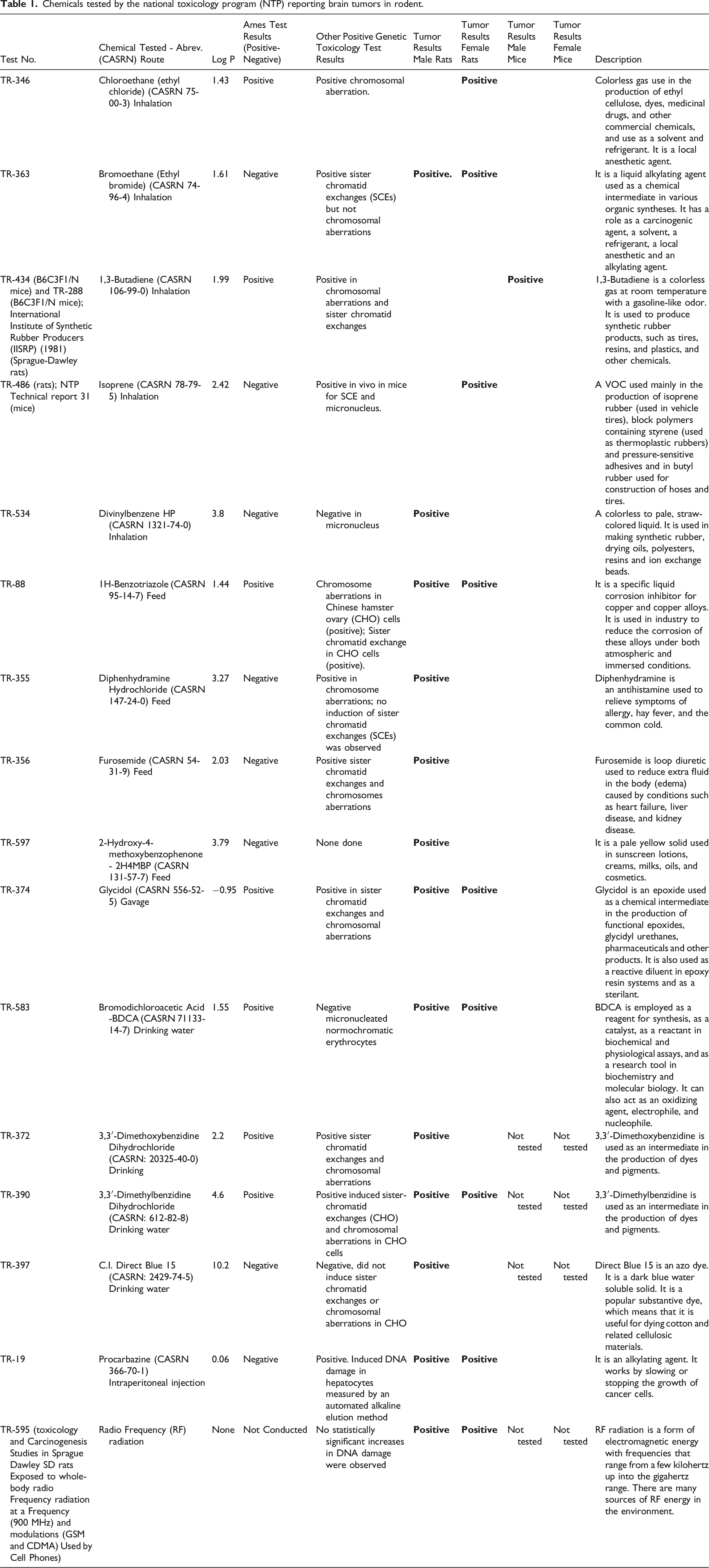
Out of the 602 TRs reviewed, 16 agents (15 chemicals and radio frequency radiation) induced brain tumors in either rats or mice, or both. One agent was tested twice (first in rats and then again in mice) resulting in 16 agents being described in 17 TRs. For each of the 16 agents, Table 1 lists the TR number, chemical name and Chemical Abstract Service Registry Number (CASRN), route of administration, log p value, mutagenicity (Ames results), other genotoxicity data, and tumor results [male rats (MR), female rats (FR), male mice (MM), and female mice (FM))], and a description of the agent tested.
Consistent with the much lower spontaneous brain tumor formation rate in mice as compared with rats, 75 13 agents induced brain tumors in male rats, nine agents induced brain tumors in female rats, but only one agent induced brain tumors in male mice, and no agents induced brain tumors in female mice. Six agents produced tumors in both male and female rats: ethyl bromide, 1H-benzotriazole, 3,3′-dimethylbenzidine dihydrochloride, glycidol, procarbazine, and radio frequency (RF) radiation. Each of these agents is either an alkylating or a possible alkylating agent. No agent produced brain tumors in both rats and mice of either sex.
Determination of the chemical composition of military burn pit smoke
The composition of military burn pit smoke was estimated from an internet search. The search was conducted from September 10, 2023, through September 27, 2023. The search terms employed included the following: military burn pits, effluent from burn pits, military bases with burn pits, Afghanistan burn pits, Iraqi burn pits, materials burned in military burn pits, chemicals from burn pits, metals from burn pits, polyaromatic hydrocarbons from burn pits, volatile organic compounds in military burn pits, halogenated dioxins and furans in burn pits, PM2.5 from military burn pits, PM10 from military burn pits, and alkylating agents in military burn pits. No quotes, special fields, truncations, etc. were used in the searches. No filters of any kind were used in the searches. The only restrictions were that the burn pits were on military bases in Afghanistan or Iraq during the period 2001-2021.
Much of the information regarding chemical composition, presence of metals, and complex mixtures associated with the effluent from military burn pits, came from a detailed report titled “Screening Health Risk Assessment Burn Pit Exposures, Balad Air Base, Iraq and Addendum Report” issued by Lieutenant Colonel Jay A. Vietas (United States Air Force) and his team, published in 2008 at the Aberdeen Proving Ground, Maryland. 81 Additionally, Vietas et al. (2008) 81 provided detailed information on the waste materials being burned in the pits. The report provided data on 77 volatile organic compounds (VOCs), 10 metals, 17 polycyclic aromatic hydrocarbons (PAHs), 17 halogenated dioxins and furans, and particulate matter (PM2.5 and PM10) using analytical methods verified in the literature. Sampling was conducted over a 24-hr period. Forty-one validated samples were collected for the determination of volatile organic compounds (VOCs). Sixty validated samples were collected for PM and metals determinations. Thirty-two validated samples were collected for polycyclic aromatic hydrocarbons (PAHs) determinations. Thirty validated samples were collected for halogenated dioxins and furans determinations. Data on 121 chemicals and metals, as well as two complex mixtures (PM2.5 and PM10) are reported for the effluent from military burn pits. 81 Information from internet searches corroborated the data from Vietas et al. (2008). 81
Chemical agents tested by NTP that induced brain tumors.

aAt frequency tested.
The limited overlap between the 15 chemicals tested by NTP that induced rodent brain tumors, and the chemicals measured in actual and simulated burn pit smoke, is to be expected as NTP selects candidate chemicals for evaluation in the expensive 2-years cancer bioassay protocol based on potential exposure to workers or the general population. The last column in Table 1, i.e., under Description, illustrates that the 15 chemicals tested by NTP that induced rodent brain tumors are commercial products.
Discussion
Chemicals found in burn pit smoke.

The chemical composition of tobacco smoke serves as an exemplar of the enormous number of different chemicals produced by pyrolyzing or burning biological materials across a wide temperature range. To-date, nearly 5700 chemicals have been identified in tobacco smoke. 82 Oxidation, reduction, addition, condensation, hydrogenation, pyrolysis, pyrosynthesis, decarboxylation, and dehydration are but a few of the many chemical reactions known to be involved in tobacco pyrolysis and incomplete combustion of tobacco. 83 Tobacco smoke is generated during a puff interval and during a smolder interval. The temperature that tobacco reaches during a puff is between 800 and 1000 C. The temperature that tobacco experiences during smolder is 500–650 C. Additionally, there are ageing effects and artifact formation that can occur during the sampling and testing of cigarette smoke. 84 These reactions are capable of producing thousands of reaction products. The comparison of tobacco smoke to burn pit effluent is reasonable as the temperatures experienced by both are similar and the majority (over 50–60% of the mass) 85 undergoing pyrolysis and incomplete combustion is plant material (i.e., tobacco, paper, cardboard, wood, etc.). 86 The major limiting factor in the discovery and identification of additional chemical components in tobacco smoke was always the development of new and ever-improved analytical technologies. 83
Previously, we analyzed all the rodent tumor types reported in the NTP database.80,87–91 Both the limited number of chemical agents that induce rodent brain tumors (16 brain tumor-inducing agents from 602 TRs), and the preponderance of alkylating agents among the brain tumor-inducing agents are notable findings. As compared with any other organ system, the blood-brain barrier (BBB) protects the brain from exposure to chemicals administered outside the BBB. From a study on CNS active drugs, Hansch and Leo (1979) 92 found that blood-brain barrier penetration is optimal when the logarithm of the octanol-water partition coefficient (log P) values is 1.5–2.7, with a mean value of 2.1. These data suggest that chemicals possessing log p values of about 0.5–3.0 can readily cross the blood brain barrier and enter the brain. The 15 NTP-tested chemicals that induce brain tumors in rodents generally fall within this approximate range with the exceptions of Procarbazine being water soluble with a log p = .06, and C.I. Direct Blue 15 being extremely hydrophobic with a log p = 10.2.
Compared with the large number of chemicals that induce tumors in rats and mice following metabolic activation by liver enzymes, alkylating agents are direct acting carcinogens. 79 Given the relatively low metabolic activation potential in the brain, 93 the direct carcinogenic action of alkylating agents might be a factor in their ability to induce brain tumors. Exposure from military burn pits would be expected to result primarily via the inhalation route, although chemicals and particles can deposit on the skin, and sometimes be ingested. The results from the NTP database suggest that alkylating agents with modest octanol-water partition coefficients can be considered as possible inducers of GBM. The epidemiology studies conducted by the VA considered GBM cases for which only general estimates of exposure were available.71,72 Due to the concerns that led to the passage of the PACT Act, 57 future deployed veterans are unlikely to experience exposure to military burn pits. Future epidemiology studies on cohorts exposed to smoke constituents, e.g., forest fireman, should consider the potential risk from inhalation of alkylating agents.
Following a lag period post-treatment, several chemotherapy alkylating agents including: mechlorethamine, chlorambucil, cyclophosphamide, melphalan, lomustine, carmustine, and busulfan have been shown to induce secondary tumors. 94 The oral alkylating agent Temozolomide (TMZ) is the main chemotherapy agent administered following surgical resection of GBM and astrocytomas.95,96 TMZ can induce a hypermutator phenotype, causing post-treatment recurrent GBM to accumulate (initiate) new potential driver mutations, increasing GBM’s overall mutational burden, with concomitant further treatment resistance. 23 The mutagenic potential of TMZ suggests that if the median GBM survival time of only 15–18 months 97 was extended significantly (from months to years) that induction of primary brain tumors secondary to treatment could become clinically significant. Development of efficacious treatments for post-surgical GBM that act via non-mutagenic mechanisms could potentially address at least some of the profound clinical challenges presented by recurrent GBM.
Conclusions and relevance
Identification of chemicals that could potentially increase the risk of GBM or other CNS tumors could assist clinicians and epidemiologists in future studies attempting to determine if environmental exposure to chemicals increases the risk of developing brain tumors.
Footnotes
Author contributions
JWA initiated the overall project addressing the possible relationship between military burn pit exposures and GBM and organized numerous conference calls with different ad hoc expert subgroups. SKS provided the clinical neurosurgery perspective that led to conduction of the current review and its sister study analyzing the National Toxicology Program (NTP) database for biological plausibility of chemical induction of GBM. CJS and TAP conceived the structure of the current review and the sister study analysis. CC and CV provided expertise on oncogenic mutational profiles in causation pathways. CJS and TAP performed the literature search and collated the data. All authors have read and agreed to the published version of the manuscript.
Declaration of conflicting interests
The author(s) declared no potential conflicts of interest with respect to the research, authorship, and/or publication of this article.
Funding
The author(s) received no financial support for the research, authorship, and/or publication of this article.