
Abstract
Clinical relevance:
MicroRNAs (miRNAs) have been reported to be involved in the progression of various diseases. Studying the regulatory mechanisms of miRNAs can help clinical treatment.
Background:
Diabetic retinopathy (DR) is one of the complications of diabetes. The objective of this study was to elucidate the underlying molecular mechanisms by which miR-200c-3p regulates the pyroptosis of DR cell.
Methods:
Human retinal microvascular endothelial cells (HRMECs) and high glucose (HG) cultures established DR cell model in vitro. RT-qPCR is used to detect the expression level of miRNAs. CCK-8 assays and flow cytometry are used to detect apoptosis of HRMECs cell. Western blotting is used to detect cleaved caspase-3, cleaved caspase-1, and N-GSDMD proteins levels in HRMECs. The ELISA assay is used to detect the expression of IL-1β and IL-18. Predict and validate potential binding sites between miR-200c-3p and SLC30A7 by dual luciferase reporter gene analysis.
Results:
The results showed that HG caused damage to HRMECs through the pyroptosis pathway rather than the apoptosis pathway. MiR-200c-3p is highly expressed in HG induced-HRMECs, and knockdown of miR-200c-3p mitigates HG-induced HRMECs pyroptosis. MiR-200c-3p negatively targets SLC30A7 in HRMECs, and miR-200c-3p regulates pyroptosis of HG-induced HRMECs by targeting SLC30A7.
Conclusion:
The results suggest that miR-200c-3p might be a promising interference target for DR prevention and treatment. The results of current study may provide new insights into development of therapeutic strategies for DR.
Introduction
Diabetic retinopathy (DR) is a highly specific microvascular complication of diabetic and is one of the leading causes of blindness in adults. 1 Inflammation is one of the key factors in DR progression, as multiple high levels of inflammatory cytokines such as IL-1, IL-6, IL-18 and IL-1β have been observed in serum and eye samples from patients with diabetic and DR. 2 Pyroptosis is a novel form of inflammation that regulates cell death, unlike apoptosis and necrosis, previous studies have shown that caspase-1 and N-GSDMD were are markers of pyroptosis, and their activation or increase indicates the occurrence of pyroptosis.3,4 GSDMD is an effector of pyroptosis that can be cleaved by caspase-1, thereby releasing an N-terminal pored domain for insertion into the plasma membrane. 5 However, the underlying mechanisms remain unclear. Therefore, further elucidating the molecular mechanisms of DR pyroptosis will help provide novel therapeutic strategies and effective prognostic indicators for DR.
There is growing evidence that miRNAs are involved in DR-associated microvascular formation play a beneficial role in the pathogenesis of DR, and regulating their levels can reverse dyslipidemia and alleviate DR progression.6,7 Thus, miRNAs can act as biomarkers of DR. 8 On the other hand, miRNAs can obviously mediate cellular pyroptosis through different molecular mechanisms. For example, miR-21 promotes NLRP3 inflammasome activation to mediate pyroptosis and endotoxic shock. 9 MiR-223-3p inhibits rTp17-induced inflammatory activation and pyroptosis by targeting NLRP3. 10 In addition, miRNA-1 promotes cardiomyocyte pyroptosis and the release of inflammatory factors by downregulating the expression level of PIK3R1 through the FoxO3a pathway, 11 and miRNA-214 promotes the cervical cancer pyroptosis and inhibits proliferation by regulating the expression of NLRP3. 12
MiR-200c-3p is a member of the miR-200 family and is involved in the regulation of various disease progressions by modulating post-transcriptional translation. Studies by Ding et al. have showed that miR-200c-3p inhibits malignant behavior in nephroblastoma cells by negatively targeting FRS2. 13 Chen et al. showed that miR-200c-3p, which targets SOX2, reduces the resistance of breast cancer cells to Palitassel. 14 In addition, studies have shown that miR-200c-3p can promote the proliferation of ischemia reperfusion injury by inhibiting the NF-κB pathway, and the NF-κB pathway plays a role in the inflammatory response. 15 MiR-200c-3p is involved in accumulation of inflammatory cytokines. 16 These studies suggest that miR-200c-3p is an inflammatory-associated miRNA. However, the effect of miR-200c-3p on causing DR pyroptosis is unclear. Therefore, further researche will help identify and assess the therapeutic potential of miR-200c-3p in DR treatment.
Zinc transporter 7 in the SLC30 family (SLC30A7, ZnT7) is a widely expressed zinc transporter involving the isolation of zinc into Golgi apparatus and vascular compartments. 17 SLC30A7 plays a vital role in diabetes-related diseases. For example, SLC30A7 is involved in the regulation of diabetic cardiomyopathy progression by influencing muco-mitochondrial coupling in hyperglycemic cardiomyocytes. 18 Craig et al. study shows that SLC30A7 relieves diabetes-related diseases by controlling insulin secretion. 19 While the various features of SLC30A7 have been elucidated, whether it is involved in regulating DR progression is even unknown. The objective of this study was to explore the effects of SLC30A7 on DR and its molecular mechanisms.
In summary, the objectives of this study were to elucidate the regulatory role of miR-200c-3p in DR pyroptosis and the potential mechanism by which SLC30A7 regulates DR progression. We were expected to demonstrate that miR-200c-3p regulates pyroptosis in DR by targeting SLC30A7. This study confirms for the first the role of miR-200c-3p in DR and provides new insights into the development of DR therapeutic strategies.
Methods
Cell culture and treatment
Human retinal microvascular endothelial cells were purchased from Procell Life Science & Technology Co., Ltd (Wuhan, China). Endothelial cell culture medium contains 10% fetal bovine serum (FBS), 100 μg/mL streptomycin, and 100 U/mL penicillin (Invitgen, Carlad, CA) to maintain cell growth. All cells are cultured in a wet incubators containing 5% CO2 at 37°C. Cells are treated with 5 mM glucose as a normal glucose control (Control) and 25 mM glucose as high-glucose (HG). 20
Cell transfection
The miR-200c-3p mimic, mimic-NC, miR-200c-3p inhibitor, inhibitor-NC, si-NC and si-SLC30A7 were designed and acquired from GenePharma (Shanghai, China). For cell transfection, HG-induced HRMECs were randomly divided into 3 groups: (1) NC group transfection NC inhibitor and si-NC vector (2 sg/well), miR-200c-3p inhibitor; (2) si-NC group transfection miR-200c-3p inhibitor and si-NC vectors, miR-200c-3p inhibitor; (3) si-SLC30A7 group transfected with miR-200c-3p inhibitor and si-SLC30A7 vector. Plasmids were transfected using Lipofectamine™ 3000 Transfection Reagent (Takara, Kusatsu, Japan). Oligonucleotide sequences are listed below. After 48 h of transfection, HRMECs were indicated for subsequent experiments. 21
miR-200c-3p mimics:
5′-UAAUACUGCCGGGUAAUGAUGGA-3’;
miR-mimics-NC:
5′-CAGUACUUUUGUGUAGUACAA-3’;
miR-200c-3p inhibitor:
5′-AUUAUGACGGCCCAUUACUACCU-3’;
miR-inhibitor-NC:
5′-CGAACGUGUCACGUTT-3’.
si-NC:
5′-CAACAAGATGAAGAGCACCAA-3’;
si-SLC30A7:
5′-GCCAUAGUCACGAAGCCAATT-3’.
RT-qPCR
Primer information.
CCK-8 assay
Cell Counting Kit-8 (CCK-8. Beyotime, Nanjing, China) for the determination of HRMECs cell viability. Briefly, cells are seeded into 96-well plates (3650, Corning, N.Y., U.S.) and incubated with 25 mM HG for 48 h. Subsequently, 10 μL of CCK-8 solution is added to the cell wells, incubated at 37°C for 2 h, and finally the absorbance reflecting the cell viability at 450 nm is detected with a fluorescent microplate. 23
Flow cytometry (FACs)
A FACs is performed to assess apoptosis of HRMECs. Briefly, HRMECs are inoculated into 96-well plates (3650; Corning, N.Y., U.S.) and incubated with 25 mM HG for 48 h. Add heterocyanate fluorin (FITC) and propylene iodide (PI) to HRMECs (5 μL/well) and incubate at 37°C for 2 h. Flow cytometry counts the number of apoptotic cells. Apoptosis is defined as FITC (+) and PI (+). 24
Western blot assay
Isolate the HRMEC total protein transfected with cell lysis buffer (Beyotime, Nanjing). Western blotting is performed according to the method described earlier. 21 All antibodies used in this study were from Abcam (1:1000), including Cleaved-Caspase-1, Cleaved-Caspase-3 and N-GSDMD. Take β-actin as the internal reference. Using ImageJ Software Inc. Quantify the optical density of protein strips. 25
ELISA assay
Human retinal microvascular endothelial cell transfected with the corresponding plasmid was lysed with a 1 mL protein extraction reagent (Beyotime, Nanjing, China) and IL-1β and IL-18 levels were detected with corresponding ELISA kit produced by Roche basel, Switzerland. 26
Bioinformatics and luciferase reporter assay
First, Genecards (https://www.genecards.org) was used to screen out miRNAs involved in DR regulation. In all the data sifted by Genecards, we selected the results by setting category to ‘RNA Gene’ and correlation was greater than 1.0. Subsequently, ENCORI, miRWalk, and miRDB were used to predict the miR-200c-3p target gene. The wild-type SCL30A7 3′UTR and mutant SCL30A7 3′UTR were developed and synthesized by For Science (Shanghai, China). In addition, 2×10⁵ HRMECs were added to a 12-well plate, incubated with 25 mM HG for 48 h, SLC30A7 3′-UTR vector wild-type or MUT plasmid and miR-200c-3p mimic or mimic NC (the plasmid used in this study was purchased from Sangon Biotech) was co-transfected with HG-induced HRMECs using Lipofectamine® 2000 (Invitgen, Waltham, MA, Invitgen). Finally, luciferase reporter assay was performed using the Luciferase Reporter Assay Kit (Biovision, Wuhan, China) with the Renilla functioned as the internal control. 27
Statistical analysis
The mean ± standard deviation (SD) represents data from three independent experiments. GraphPad Prism 5.0 Software (GraphPad Software, Inc.) is used for statistical analysis of all data. T-test or one-way analysis of variance (ANOVA), followed by Tukey posttests, are used for comparison between two and multiple groups, respectively. When p < 0.05, the difference was statistically significant. 28
Results
High glucose induced damage to HRMECs via pyroptosis pathway rather than apoptosis pathway
To investigate the underlying pathogenesis of DR, HRMECs are traeted with high glucose (HG, 25 mM) to induce damage models. The results of the study showed that HG-induced inhibition of HRMECs viability and accelerated apoptosis of HRMECs without affecting the levels of Cleaved-caspase-3 proteins (Figure 1(a)–(c)) showed that HG induced damage to HRMECs, but not through the apoptosis pathway. Further analysis showed that HG induced levels of inflammatory cytokines in supernatant of HRMECs, including IL-1β and IL-18 compared with controls, and accelerated levels of pyroptosis-related proteins in the HRMECs, including Cleaved-caspase-1 and N-GSDMD (Figure 2(a) and (b)), indicating that HG causes damage to HRMECs through the pyroptosis pathway. Taken together, these findings suggest that HG causes damage to HRMECs through the pyroptosis pathway rather than the apoptosis pathway. High glucose induced damage to HRMECs, but not via apoptotic pathway. HRMECs were randomly divided into 2 groups, control group, cells were treated with the same volume of PBS, HG group, cells were treated with high glucose (25 mM) for 48 h. (a) CCK-8 assay was performed to assess HRMECs viability. (b) FACs was performed to detect HRMECs apoptosis. (c) Western blot was performed to measure the level of Cleaved-caspase-3 protein in HRMECs. Data represent the mean of three independent experiments. Error bars represent SD.
**
p < 0.01. High glucose induced damage to HRMECs via pyroptosis pathway. HRMECs were randomly divided into 2 group, control group, cells were treated with same volume of PBS, HG group, cells were treated high glucose (25 mM) for 48 h. (a) ELISA was performed to detect the levels of IL-1β and IL-18 in supernatant of HRMECs. (b) Western blot was performed to measure the levels of pyroptosis-related proteins, including Cleaved-caspase-1 and N-GSDMD. Data represent the mean of three independent experiments. Error bars represent SD.
*
p < 0.05,
***
p < 0.001.

MiR-200c-3p knockdown alleviated HG induced-HRMECs pyroptosis
The findings suggest that miRNAs have an important effect on gene expression by modulating transcription and translation, and may be associated with the regulation of a variety of diseases progression.
29
In order to explore whether miR-200c-3p is involved in the regulation of the DR process, primarily, Genecard (https://www.genecards.org) is mainly used to screen miRNAs involved in DR regulation. Bioinformatics analysis shows that multiple genes are involved in DR progression, including miR-200b-3p, miR-29c-3p, miR-429, miR-29a-3p and miR-200c-3p. Subsequently, RT-qPCR analysis indicated that miR-200c-3p was the miRNA with the largest differential expression in HG-induced HRMECs, while other miRNA levels remained almost unchanged (Figure 3). Further functional studies have shown that elevated HG-induced levels of IL-1β and IL-18 in HRMECs are antagonized by miR-200c-3p knockdown (Figure 4(a)). Consistently, Western blot analysis showed that miR-200c-3p knocked down HG-induced upregulation levels of Cleaved-caspase-1and N-GSDMD in antagonistic HRMECs (Figure 4(b)). Taken together, these results suggest that miR-200c-3p knockdown mitigates HG-induced pyroptosis of HRMECs. MiR-200c-3p was highly expressed in HG induced-HRMECs. HRMECs were randomly divided into 2 groups, control group, cells were treated with the same volume of PBS, HG group, cells were treated high glucose (25 mM) for 48 h. RT-qPCR was performed to assess the levels of DR-related miRNAs, including miR-200b-3p, miR-29c-3p, miR-429, miR-29a-3p and miR-200c-3p. Data represent the mean of three independent experiments. Error bars represent SD. *p < 0.05,
**
p < 0.01,
***
p < 0.001. MiR-200c-3p knockdown alleviated HG induced HRMECs pyroptosis. HRMECs were randomly divided into3 group, NC group, cells were transfected with NC inhibit (2 μg per well); NC+HG group, cells were transfected with NC inhibit and treated high glucose (25 mM) for 48 h; miR-200c-3p inhibit + HG group, cells were transfected with miR-200c-3p inhibit and treated high glucose (25 mM) for 48 h. (A) ELISA was performed to assess the levels of IL-1β and IL-18 in supernatant of HRMECs. (B) Western blot was performed to measure the levels of pyroptosis-related proteins, including Cleaved-caspase-1 and N-GSDMD. Data represent the mean of three independent experiments. Error bars represent SD.
***
p < 0.001 vs NC-inhibit group,
##
p < 0.01,
###
p < 0.001 vs NC-inhibit + HG group.

MiR-200c-3p negatively targeted SLC30A7
It has been noted that miR-200c-3p is involved in the regulation of disease progression by modulating the transcription of target gene.
30
To determine downstream regulators of miR-200c-3p in HRMEC, predict possible target genes for miR-200c-3p using ENCORI, miRWalk, and miRDB, and perform Funrich functional annotation on 126 cross-target genes. The findings indicate that the binding site of miR-200c-3p is observed in the 3′-UTR of SLC30A7 (Figure 5(a) and (b)). Subsequently, detection of the dual luciferase reporter gene showed that co-transfection of miR-200c-3p mimic inhibited luciferase activity in the wild-type SLC30A7 reporter gene, but co-transfection with miR-200c-3p mimic did not induce luciferase activity in the mutant SLC30A7 reporter gene (Figure 5(c)). In addition, Western blot analysis showed that the miR-200c-3p overexpression inhibited SLC30A7 levels in HG-induced HRMECs compared to control (Figure 5(d)). Combined with reading, the results showed that miR-200c-3p was negatively targeted to SLC30A7 in HG-induced HRMECs. MiR-200c-3p negatively targeted SLC30A7. (a) ENCORI, miRWalk and miRDB were adopted to predict the putative target gene of miR-200c-3p. (b) The binding site of miR-200c-3p was observed in 3′- UTR of SLC30A7. (c) the dual luciferase reporter assay was performed to confirm the direct binding relationship between miR-200c-3p and SLC30A7. (d), Western blot was performed to detect the level of SLC30A7. Data represent the mean of three independent experiments. Error bars represent SD.
***
p < 0.001.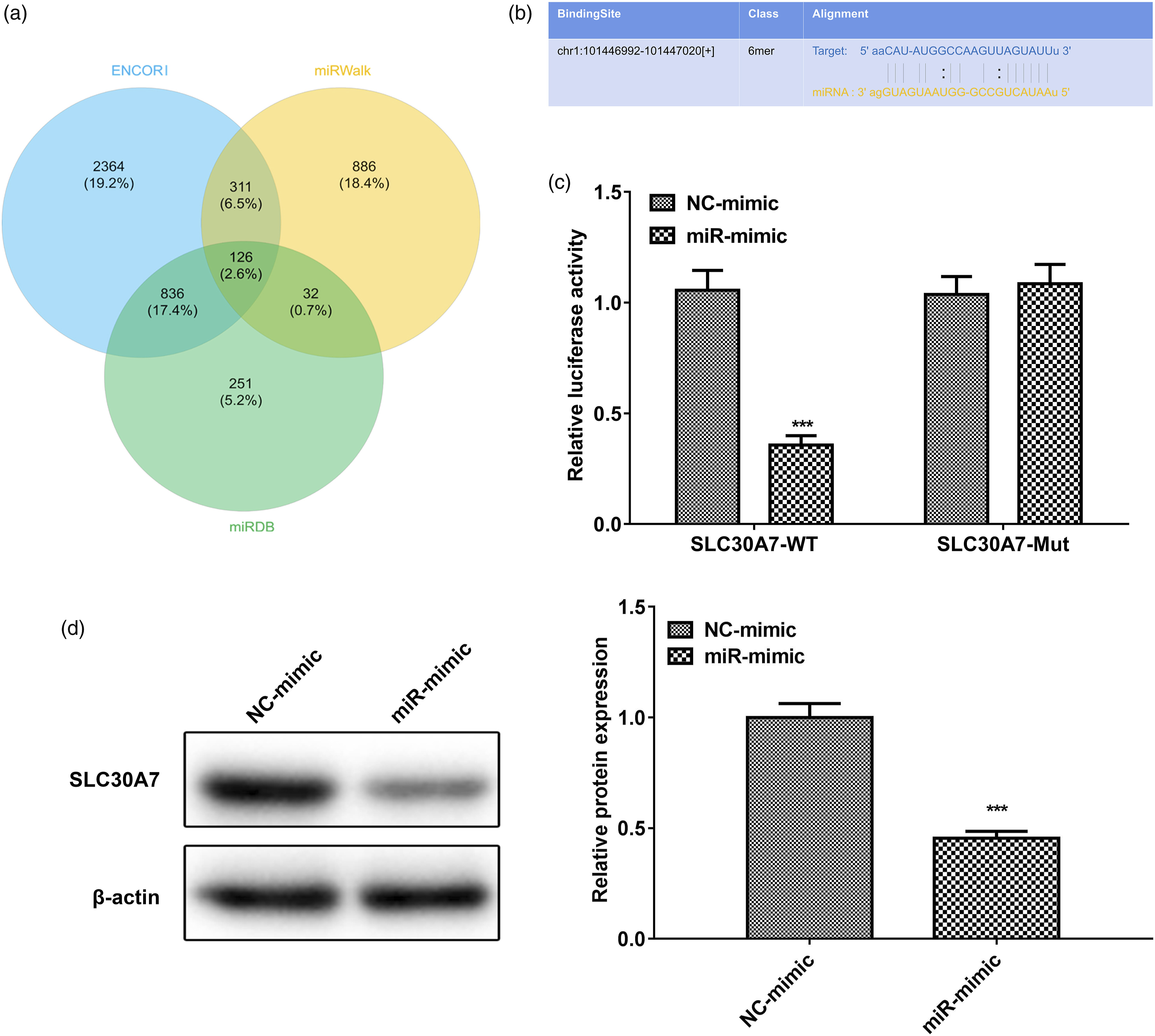
MiR-200c-3p regulated HG-induced pyroptosis via targeting SLC30A7
To further investigate whether miR-200c-3p modulates DR progression by targeting SLC30A7, miR-200c-3p inhibitor or si-SLC30A7 vector were transfected into HG-induced HRMECs to inhibit miR-200c-3p or SLC30A7. ELISA analysis showed that miR-200c-3p inhibitor downgraded the IL-1β and IL-18 levels required for HG in HRMECs, while SLC30A7 knockdown mitigated the inhibition (Figure 6(a)). Consistently, Western blot analysis showed that HG-induced Cleaved-caspase-1 and N-GSDMD levels decreased due to miR-200c-3p inhibitor, while SLC30A7 knockdown reversed the downward trend (Figure 6(b)). Taken together, these results suggest miR-200c-3p modulating HG-induced pyroptosis by targeting SLC30A7. MiR-200c-3p regulated HG-induced pyroptosis via targeting SLC30A7. miR-200c-3p inhibit or Si-SLC30A7 vector was transfected into HG-induced HRMECs for miR-200c-3p or SLC30A7 inhibition. NC inhibit served as the negative control for miR-200c-3p inhibit, Si-NC served as the negative control for Si-SLC30A7. (a) ELISA was performed to assess the levels of IL-1β and IL-18 in supernatant of HG-induced HRMECs. (b) Western blot was performed to measure the levels of pyroptosis-related proteins in HG-induced HRMECs, including Cleaved-caspase-1 and N-GSDMD. Data represent the mean of three independent experiments. Error bars represent SD.
***
p < 0.001 vs NC-inhibit+Si-NC group,
##
p < 0.01,
###
p < 0.001 vs miR-inhibit +Si-NC group.
Discussion
Diabetic retinopathy is the leading cause of new-onset blindness and visual impairment in adults aged 20–65 years and is accompanied by inflammation, with excessive inflammation accelerating DR progression. 21 Pyroptosis is a highly inflammatory programmed cell death characterized by cell rupture, nuclear formation, and high concentrations of nuclear serous fluid. 31 Caspase-1 is activated downstream of the inflammasome, and the lysis of caspase-1 induces cellular pyroptosis by cleaving members of the gasdermin family, leading to nucleosis, membrane rupture, and release of inflammatory cytokines IL-1β and IL-18. 32 So far, this inflammation-mediated process has been observed in diabetic nephropathy and DR.33,34 The results showed that the levels of IL-1β, IL-18, Cleaved-caspase-1 and N-GSDMD in HG-treated HRMECs were significantly elevated, suggesting that pyroptosis was involved in the pathogenesis of DR.
Emerging evidence suggests that miRNAs can serve as effective biomarkers or therapeutic targets for a variety of diseases, 21 and various miRNAs have been shown to play a key regulatory role in the progression of DR. Studied by Ye et al. have shown that miR-1273g-3p is involved in DR progression by modulating the autophagy-lysosomal pathway, demonstrating miR-1273g-3p as a new target for DR therapy. 35 Gu et al. showed that miR-590-3p targeted NLRP1 to inhibit pyroptosis in DR rats. 21 In this study, we considered the role of miR-200c-3p in HG-induced DR models. First, bioinformatics results of miRNAs downloaded from Genecards related to DR development were analyzed. Then, in vitro, RT-qPCR analysis showed miR-200c-3p as the miRNA with the largest differential expression of any DR-associated candidate miRNA. In addition, we found that miR-200c-3p was highly expressed in HG-induced HRMECs, suggesting that miR-200c-3p may exacerbate DR progression. Further functional studies have shown that inhibition of miR-200c-3p downregulats levels of IL-1β, IL-18, Cleaved-caspase-1 and N-GSDMD in HG-induced HRMECs. Among them, N-GSDMD is a fragment produced by caspase-1 cutting GSDMD. These results suggest that inhibiting miR-200c-3p mitigates pyroptosis in DR.
Studies have shown that miRNAs have an important effect on gene expression by regulating transcriptional translation and may be involved in regulating the progression of a variety of diseases, including DR. 25 Yan et al. suggest that miRNA-451a regulates the mitochondrial function of proliferative DR by targeting transcription factor 2. 36 The results of this study show that miR-200c-3p negatively targets SLC30A7, which encodes the ubiquitous zinc-expressing transporter and involves the delivery of cytoplasmic zinc to the Golgi apparatus and vascular compartments containing ZnT7. 37 SLC30A7 plays an important role in the progression of diabetes-related diseases by regulating insulin levels. 38 The functional studies of this study further demonstrated that inhibition of miR-200c-3p downregulated levels of IL-1β, IL-18, Cleaved-caspase-1 and N-GSDMD in HG-induced HRMECs, while SLC30A7 knockdown mitigated inhibition, indicating that miR-200c-3p modulated HG-induced pyroptosis by targeting SLC30A7 in HG-induced HRMECs. Therefore, SLC30A7 plays a key role in the pyroptosis of DR. However, this study still has some limitations. For example, the mechanism by which miR-200c-3p regulates pyroptosis in vivo remains to be determined. In future work, we will continue to explore the function and regulatory mechanism of miR-200c-3p in vivo to make its clinical relevance more accurate and effective.
Conclusion
In summary, the results of this study suggest for the first time that HG-induced HRMECs produce higher level of miR-200c-3p. Down-regulating miR-200c-3p mitigates pyroptosis by targeting SLC30A7 in HG-induced HRMECs. These findings suggest that miR-200c-3p may regulate pyroptosis by targeting SLC30A7 in DR. This study is the first to demonstrate the expression and function of miR-200c-3p in DR and provide new insights into the development of DR therapeutic strategies.
Footnotes
Authors’ contributions
Weina Li conceived and designed the project. Sheng Yang and Guangsheng Chen performed the experiments and acquired the data. Weina Li and Shiping He analyzed the data. Sheng Yang and Shiping He wrote the manuscript. Weina Li revised the manuscript.
Declaration of conflicting interests
The author(s) declared no potential conflicts of interest with respect to the research, authorship, and/or publication of this article.
Funding
The author(s) received no financial support for the research, authorship, and/or publication of this article.
Availability of data and materials
The datasets used and/or analysed during the current study are available from the corresponding author on reasonable request.