
Abstract
Introduction:
In the study, it was aimed to investigate the possible protective effects of curcumin, a potent antioxidant, against the toxic effect of nonylphenol on bone development.
Methods:
Thirty pregnant female Wistar albino rats were used. The rats were randomly divided into the following five groups; the control group, corn oil group (150 µl/kg/day), nonylphenol group (50 µl/kg/day), curcumin group (100 mg/kg/day) and curcumin + nonylphenol group (100 mg/kg/day + 50 µl/kg/day). The doses were given by gavage from the 5th day to the 20th day of gestation. The fetuses were removed out on the 20th day of pregnancy by cesarean at the end of the study. After the sacrifice of the animals, double skeletal staining in front extremity (clavicula, scapula, humerus, radius, ulna) and hind extremity (femur, tibia, fibula), additionally histological and immunohistochemical examinations in femur bone were performed.
Results:
The nonylphenol group offspring have the lowest weights of fetuses and placenta, head-to-hip lengths, biparietal and occipitofrontal length, and also, bone length percentage and percentage of the ossification area in all measurements of the front extremity and hind extremity Interestingly, the groups treated with curcumin showed close to the control group in terms of double skeletal staining, histological, and immunohistochemical examinations.
Conclusions:
Our findings demonstrated an association between bone development and exposure to nonylphenol. The findings suggest that curcumin treatments may be effective in accelerating bone formation.
Introduction
Alkylphenolic compounds are organic chemicals generally produced for the manufacture of alkyphenol ethoxylates, which are mainly used as non-ionic surfactants, but also in a wide range of applications. Exposure to these chemicals can occur occupationally during their production or with exposure to domestic and industrial detergents, specialty paints, pesticides, cosmetics and hair dyes, among others, 1 but also as a consequence of non-occupational exposures, such as diet and water intake, use of personal care and household cleaning products. These chemicals imitate the hormone system in the body and disrupt body development and cell metabolism. 2 Nonylphenol, octylphenol and their ethoxylates are the most commonly used alkyphenol compounds.1,3 It is one of the most widely used synthetic xenoestrogens in detergents, plastic products, paints and the most important environmental degradation factor.4,5 Due to environmental pollution, living things exposed to these substances also have reproductive problems along with health problems. 5 Nonylphenol has been identified in urine, blood, and tissues that people are constantly exposed to, as it has a wide range of uses in the industry. 6 Although, nonylphenol is a substance that acts as estrogen by binding to estrogen receptors, the effects on calcium are different from estrogen. Because estrogen acts in increasing plasma calcium level and also in calcitonin level. Nonylphenol causes both to decrease, plasma calcium and calcitonin levels. 7 In addition, it has been reported that estrogen affects bone metabolism and osteoblastic differentiation. 8
Curcumin is an antioxidant belonging to the Zingiberaceae family, obtained from the rhizomes of the Curcuma longa plant with yellow flowers. Curcumin is the active ingredient that gives the yellow color for the curry spice. The roots of this plant are dried and used in Asian medicine for thousands of years. 9 Curcumin, a polyphenol and a major biologic component in the extract of the root of Curcuma longa, exhibits biologic properties because of its anti-inflammatory, antioxidant, anti-tumor and antimicrobial activities.10,11 In recent studies, have suggested that curcumin exerted a protective effect on bone disorders and inflammatory diseases, such as osteolysis, rheumatoid arthritis and osteoporosis. 12
Alkylphenols are endocrine disruptors that show estrogen-like effects in various species. However, only a few information are available about the action of these chemicals on bone development. Therefore, this study may be the first reported research in this area. In this study, the determination of negative structural changes caused by nonylphenol on bone development and is aimed to determine the possible protective effects of curcumin, which is known to have antioxidant properties against these changes, using double-staining and immunohistochemical methods.
Materials and methods
Animal protocol
The Ethical Committee of Animal Experiments of Erciyes University was applied for the study and approved by decision 16/137 dated 16 November 2016. All the procedures performed at each stage of the study and were carried out in accordance with the rules specified in the ethics committee directive. Consistent with the ethical rules of animal use and experimental design, the minimum number of rats and fetuses were used. Thirty adult female Wistar rats weighing between 200 and 250 g were used for this study. Female rats were mated with male rats to get pregnant. Two female rats and one male rat were placed in the cages (polypropylene steel wire mesh cages) for mating from 5 pm to 8 am. The female rats with a sperm-positive vaginal smear were accepted to be on day 0.5 of pregnancy. The pregnant rats were housed in individual cages and maintained in a room with a controlled temperature of 21 ± 3°C and a normal photoperiod (12-h light/12-h dark cycle). They were fed ad libitum with a fattening meal (granule pellet containing 21% raw protein). The water needs of the rats were provided by tap water, which was changed daily and controlled.
Experimental groups
In the present study, rats considered to be 0.5 days pregnant were randomly divided into five groups, with six pregnant rats in each group. This study focused on fetal bone development, and pregnant rats were injected with nonylphenol between the 5th and 20th days of their pregnancy. Control group: the group that was given standard food and water. Corn oil group: 50 µl /kg/day corn oil was administered to gavage between the 5th and 20th days of pregnancy once a day. Nonylphenol group: 50 µl /kg/day dissolved in 100 µl corn oil and prepared daily with 50 µl dose of nonylphenol (total 150 µl) was given gavage between the 5th and 20th days of pregnancy once a day. Curcumin group: 100 mg/kg/day dose of curcumin was given gavage, prepared fresh daily, and dissolved in 150 µl corn oil (total 150 µl) between the 5th and 20th days of pregnancy once a day. Curcumin + nonylphenol group: Half an hour after the application of curcumin at 100 mg/kg/day and 50 µl dose of nonylphenol, prepared fresh again (total 150 µl). All doses were given by gavage and the experiment continued between the 5th and 20th days of pregnancy once a day.
The number of pregnant rats in each group was determined by taking measurements of at least 18 fetuses and taking into consideration the losses that may occur during the dissection, double skeletal, histological, and immunohistochemistry stages. Experimental studies in all groups of the study began at the same time. The solution was prepared daily by dissolving 50 µl nonylphenol in 100 µl corn oil. Pregnant rats in the control group were given only standard food and water were given to rats in this group until the 20th day of their pregnancy. The animals were regularly weighed until the end of the experiment.
Maternal and fetal evaluation
Maternal body weights were measured on gestation days 0, 4, 8, 12, 16, and 20. The rats were anesthetized with ketamine (75 mg/kg) and xylazine (10 mg/kg), for cesarean operation on day 20. The abdominal area of the anesthetized rats was cleaned with a 70% alcohol solution, and the area was opened with a transverse incision. The uterus was dissected together with the fetuses inside. Weights of fetuses separated from the placenta were weighed and recorded by measuring the head-to-hip lengths, biparietal and occipitofrontal lengths with digital calipers. In each group, 18 of the fetuses were used for the double skeletal staining method, Masson trichrome (MT), and immunohistochemical staining (IHC) methods. Fetuses used for double skeletal staining were separately measured on both extremities (18 right front and hind extremity, 18 left front and hind extremity). The femur bone of the hind extremity of each fetus was used for MT, and IHC methods.
Double skeletal staining methods in fetuses
In order to stain the skeletal system of the fetuses by the double skeletal staining method, all fetuses were hold in 70% ethyl alcohol for 4–7 days, to facilitate dehydration. Following this procedure, they were hold in pure acetone for 1–3 days to remove the oil. Then, their skins were peeled, and their internal organs and eyes were removed. All offsprings were subjected to a skeletal examination with the double-staining technique.13,14 The stained fetuses were then placed in 20%, 50%, and 80% glycerin and finally 100% pure glycerin. Images of the front and hind extremity bones were taken for morphometric measurements of fetuses with bone and cartilaginous structures identified by double skeletal staining. Images of the bones placed on millimeter paper were recorded via a stereomicroscope on which a Nikon E5700 camera was mounted. The obtained images were transfered to a computer and measurements were made of the bone and cartilage areas by the ImageJ software program. Calibration was adjusted between the original image and the photo image so that the measurement process worked correctly. First, the photo image was opened with the ImageJ program, to be measured. The original 1 mm on the millimeter paper was measured by a straight line in ImageJ program. Then, proper calibration was performed using the ImageJ program’s Analyze-Set Scala-Known distance menu. The total bone length, the length of the ossification region, the total bone surface area and the surface area of the ossification region of the bones subjected to double skeletal staining were measured with the ImageJ program. For the ratio of the surface area of the ossification region of the bones to whole the total bone surface area, the (Ossification area/Total bone area) × 100 formula was used.
Histological analysis
After the pregnant rats were decapitated on the 20th day of their pregnancy and their offspring were removed by cesarean section, excised offspring femur tissues were stored in 10% formaldehyde for fixation. The femur tissues were kept in fixation solution for 72 h and then washed with running tap water. They were passed through graded alcohol series and cleared using xylol. Then they were embedded into the paraffin and blocked. Sections of 5 µm were taken from the femur tissues and placed onto polylysine-coated laminas. Using a standard histologic follow-up method, the paraffin of the laminas were removed with xylol, and the laminas were passed through graded alcohol series (100%, 96%, 80%, 70%, 50%) and washed with water. In order to determine the general histologic structure, the sections were stained using MT, passed first through increasing alcohol series and then through xylol, and covered with a lamella using Entellan. Olympus BX51 light microscope (Olympus BX51, Tokyo, Japan) equipped with DP 70 digital camera was used for histological evaluation of sections stained with MT. All preparations were examined under this microscope and images representing the groups were obtained.
Immunohistochemical analysis
The avidin-biotin-peroxidase technique was used for the immunohistochemical studies to analyze AP (Anti-alkaline phosphatase) and TRAP (tartrate-resistant acid phosphatase) expression. In this study, the immunoreactivity intensity was analyzed in the femur of fetuses in all groups. Cross-sections prepared from tissue blocks that were 5 µm thick were incubated at 60°C. Tissues were rinsed with sequential alcohol solutions for deparaffinization, and distilled water was used to remove the alcohol from dehydrated tissues. For the next steps, a staining kit (Thermo Fisher Scientific, USA) was used. The surfaces of the tissues in the preparations were incubated overnight by dripping TRAP polyclonal primary antibody from Abcam (Cambridge, Massachusetts, USA) and AP primary antibody from Abcam (Cambridge, Massachusetts, USA). After the washing process, the sections were incubated with the biotin-secondary antibody for 15 min, and then the washing process was repeated. The sections that had been exposed to streptavidin peroxidase for 15 min were washed, they were exposed to the substrate for 5 min to make immunoreactivity apparent. After that, they were washed with deionized H2O for 5 min. Lastly, chromagen DAB containing diaminobenzidine substrate was added to the medium, and the immune reaction was allowed for approximately 5–10 min. Mayer’s hematoxylin was used as the background stain. The prepared sections were examined using an Olympus BX51 microscope (Tokyo, Japan). Photographs were taken from IHC applied preparations using the same microscope. Random 10 fields were observed from the preparations belonging to each group. These photos were transferred to the computer. Expression measurements of TRAP and AP were done on photographs by ImageJ program.
Statistical analysis
The suitability of the data to normal distribution was evaluated by the histogram, q–q graphs, Shapiro–Wilk test. The suitability Variance homogeneity was tested by the Levene test. One-way analysis of variance and Kruskal Wallis tests were used for comparisons between more than two groups. Dunn-Bonferroni test was applied for multiple comparisons. The data were evaluated with R 3.2.2. (www.r-project.org) software. The significance level of the data was accepted as p < 0.05.
Results
Weight findings of pregnant rats
In the study, the weights of the mother rats belonging to all experimental groups were weighed and recorded on days 0.5, 4, 8, 12, 16, and 20 during their pregnancy, and the data were compared between all groups according to the gestational days. Statistical analysis results are shown in Table 1.
Effect of nonylphenol on maternal body weights.

Data are expressed as mean ± standard deviation. The same letters in the same column indicate similarity between groups and different letters indicate difference between groups.
Findings of growth parameters
In the study, individual weights of offspring taken by cesarean section on the 20th day of their gestation from mother rats belonging to all experimental groups were weighed and recorded. Accordingly, the lowest mean fetal weight belonged to the nonylphenol group and there was a statistically significant decrease compared to all other groups including the curcumin + nonylphenol group (p < 0.001). When the weights of the placentas belonging to fetuses were compared, the lowest placental weight was also observed in the nonylphenol group. Placental weights of fetuses belonging to the nonylphenol group were significantly lower than the control, curcumin, and curcumin + nonylphenol groups (p < 0.001), but were not significant with the corn oil group (p > 0.05). Statistical analysis results are shown in Table 2.
Comparison of the fetus and placental weight data and Head-to-hip length, Bipariatel, and Frontal - Occipital length data parameters.
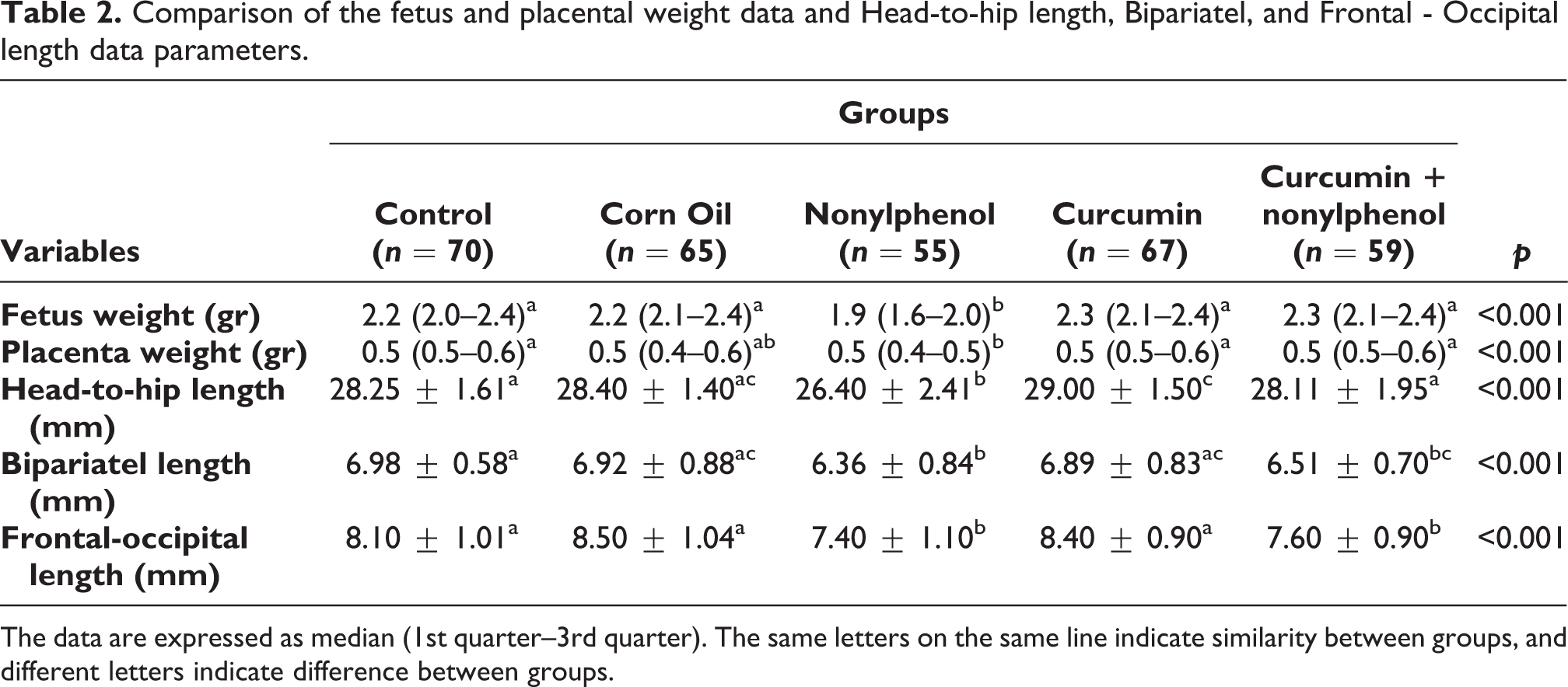
The data are expressed as median (1st quarter–3rd quarter). The same letters on the same line indicate similarity between groups, and different letters indicate difference between groups.
The head-to-hip lengths, biparietal and occipitofrontal lengths of fetuses obtained by cesarean in mother rats belonging to the experimental groups were measured and recorded with a digital caliper (Figure 1). When the data obtained were evaluated statistically, the fetuses with the lowest height and frontal-occipital length values were obtained from mother rats belonging to the nonylphenol group (p < 0.001). When the biparietal length measurement results were examined, while there was a significant decrease in the nonylphenol group compared to the control, corn oil, and curcumin groups (p < 0.001), the biparietal length results in the curcumin + nonylphenol group were higher than the nonylphenol group, but the data was not statistically significant (p > 0.05). Statistical analysis results are shown in Table 2.

(A) Head-to-hip lengths measurement of fetuses, (B) occipito-frontal length measurement of fetuses, (C) biparietal length measurement of fetuses.
Findings of double skeletal staining method
In the study, the front and hind extremity bones of fetuses belonging to all experimental groups were examined in both the right and left extremities one by one. Measurements were made as to the total length of the bone, the percentage of the length of the region showing ossification, the total area of the bone, and the area of the region showing ossification. The total ossification percentage of the bones was calculated using the data in area measurements. Right and left bilateral examinations of 18 fetuses in total were performed for each bone, and thus 36 bones from each group were included in the examination.
Accordingly, it was observed in fetuses belonging to the nonylphenol group that the length and area percentages of the ossification regions of the right and left front extremity belonging to all experimental groups were the lowest. This decrease in the percentage of decreased bone length and area observed in the right and left front extremity of the fetuses belonging to the nonylphenol group was statistically significant when compared with all other experimental groups (p < 0.001) (Figure 2). This situation showed the damage caused by nonylphenol in the fetus’s skeletal system due to the late occurrence of cartilage destruction. However, in the curcumin + nonylphenol group, increased bone total length and the length of the ossification region were observed compared to the nonylphenol group. Statistical analysis results of front extremity measurements are shown in Table 3.

Images of the front extremity bones. The area stained with red color (Alizarin Red-S) on the images shows the ossification area and the area stained with blue color (Alcian Blue) indicates the cartilage area. U; Ulna, R; Radius.
Comparison of the ossification area of the forearm and bone length percentage ratio.
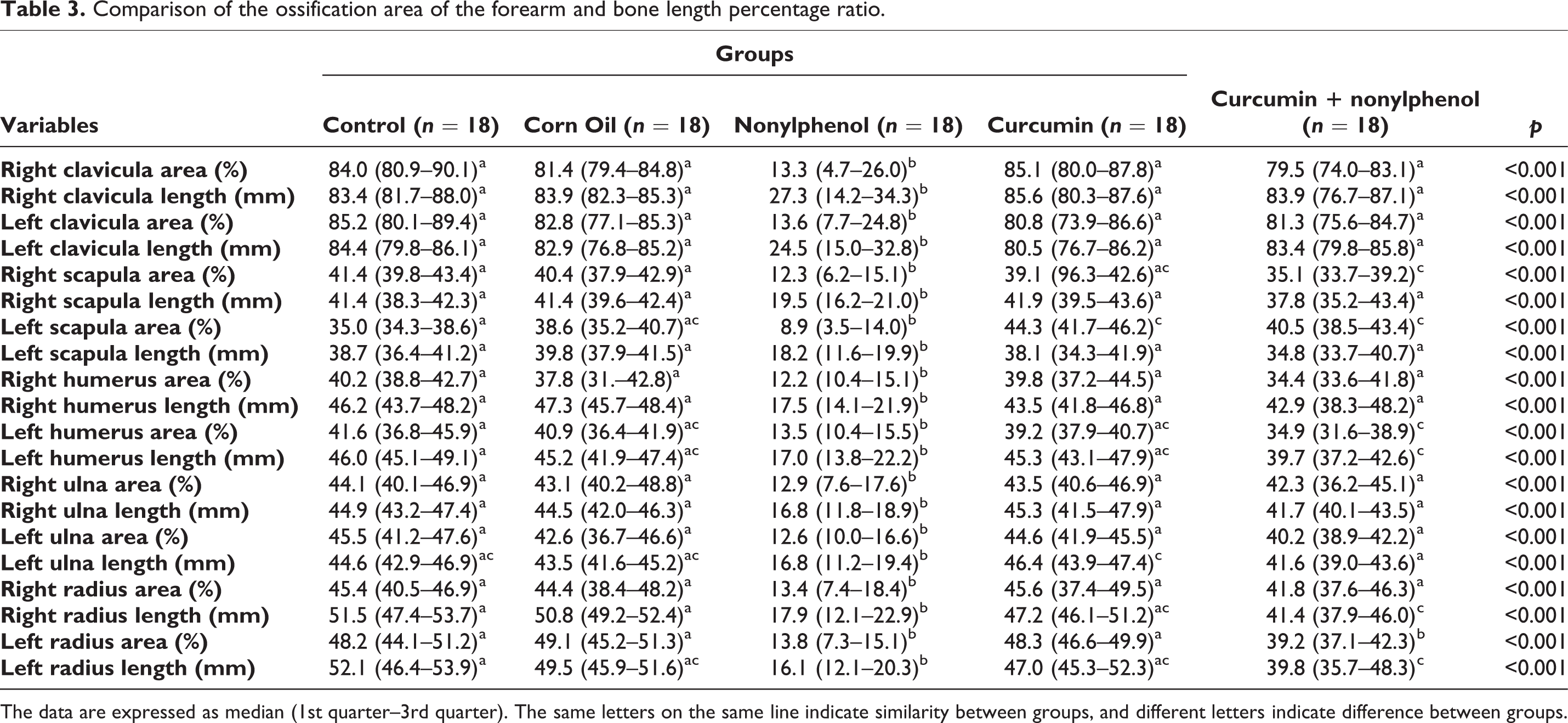
The data are expressed as median (1st quarter–3rd quarter). The same letters on the same line indicate similarity between groups, and different letters indicate difference between groups.
In the study, the experimental group treated with nonylphenol, the total length of the right and left hind extremity bone and the percentage of the area showing ossification showed a statistically significant decrease compared to all other groups (p < 0.001). There was a significant increase in ossification in the curcumin + nonylphenol group compared to the nonylphenol group (p < 0.001). In the control group, there was no significant difference between the corn oil and only curcumin applied groups in terms of the ossification area percentage of the right and left hind extremity bones (p > 0.05). The results of our study are showing that curcumin has a protective effect on the right and left hind extremity bones on ossification caused by nonylphenol (Figure 3). All statistical analysis results of the right and left hind extremity bones are shown in Table 4.

Images of the hind extremity bones. The area stained with red color (Alizarin Red-S) on the images shows the ossification area and the area stained with blue color (Alcian Blue) indicates the cartilage area. F; Fibula, T; Tibia..
Comparison of lower extremity ossification area and bone length percentage ratio.

The data are expressed as median (1st quarter–3rd quarter). The same letters on the same line indicate similarity between groups, and different letters indicate difference between groups.
Histological findings
In the light microscopic examination of the femur section belonging to the control group, the ossification region was clearly observed in all femur sections with newly formed bone spicules (Figure 4). In the light microscopic examination of the femoral sections of the groups in which corn oil and only curcumin was applied, it was observed that the endochondral ossification regions had the same morphological features. When the femoral sections of the nonylphenol applied group were examined, it was noticed that the ossification areas were less than the other groups. In particular, the number of chondrocytes in the hypertrophy zone was significantly reduced. According to microscopic findings, it was clear that nonylphenol delayed ossification. In femur sections of the group treated with curcumin + nonylphenol, it was observed that the ossification regions compared to the nonylphenol group had characteristics almost similar to the control group (Figure 4).
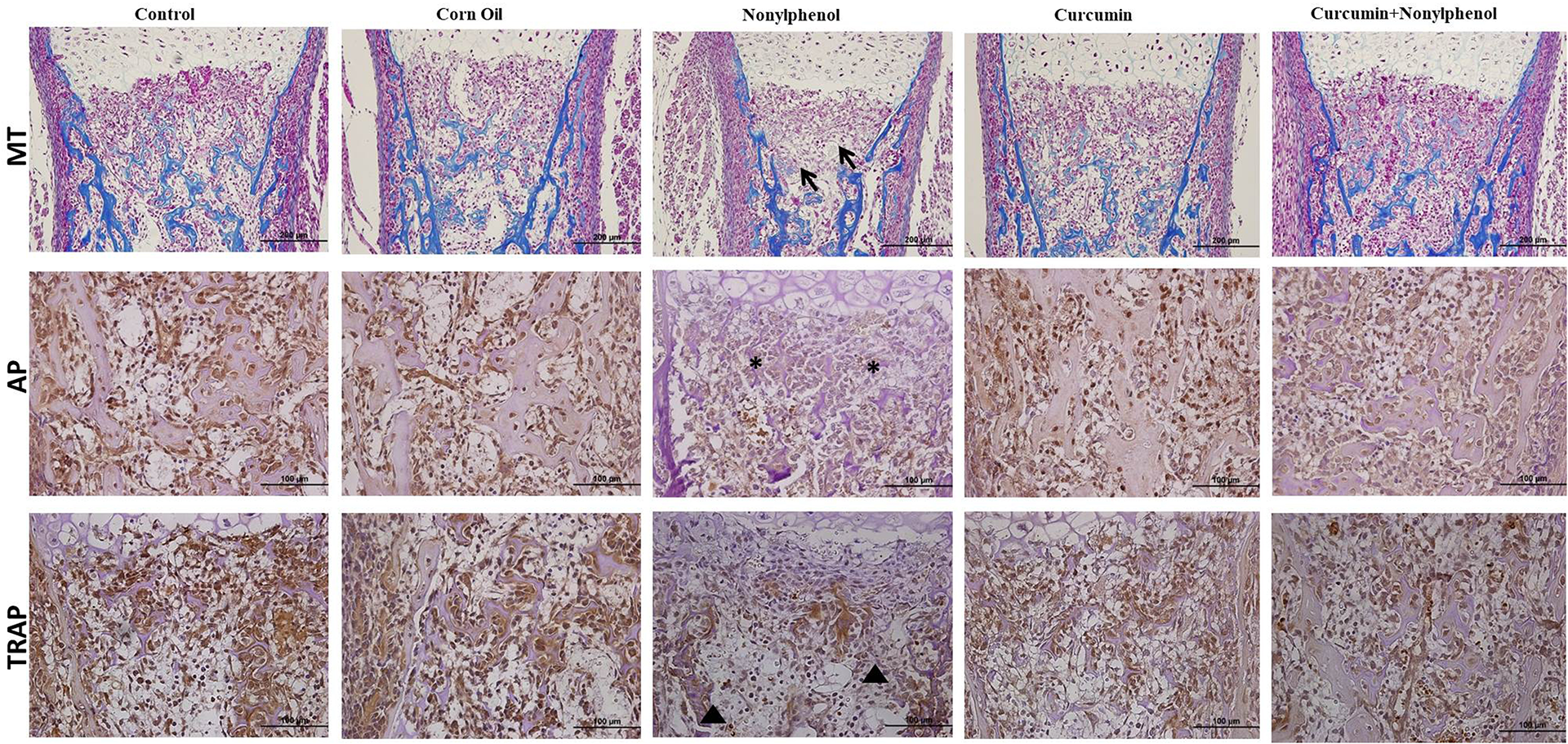
Effects of curcumin on histological changes in the bone development caused by nonylphenol. MT, ×200. Immunohistochemical staining of AP and TRAP in rat femur from all experimental groups. Arrow; areas of reduced ossification. Star; decreased AP immunoreactivity intensity. Arrow head; decreased TRAP immunoreactivity intensity. AP: alkaline phosphatase. TRAP: tartrate resistant acid phosphatase. IHC, ×200.
Immunohistochemical findings
In femur sections of the nonylphenol applied group, the AP immunoreactivity intensity was significantly decreased compared to all other groups (p < 0.001). However, AP immunoreactivity density in femur sections of the group treated with curcumin + nonylphenol was significantly increased compared to the nonylphenol group (p < 0.001). In the curcumin + nonylphenol group, AP immunoreactivity intensity was almost the same, and there was no statistically significant difference among the control, corn oil, and curcumin groups (p > 0.05) (Figure 4) (Table 5). The results of the study show that nonylphenol exposed during pregnancy has a destructive effect on osteoblast cells responsible for bone formation and negatively affects ossification.
Immunohistochemistry results of AP and TRAP of all experimental groups are given in developing-bone tissue samples.

Data are expressed as mean ± standard deviation. The same letters on the same line indicate similarity between groups, and different letters indicate difference between groups.
When the immunoreactivity densities of the femur sections in which TRAP immunohistochemistry was applied in the femur sections belonging to the control group were examined, only the nonylphenol applied group had a significantly higher immunoreactivity density compared to the other experimental groups (p < 0.001). Although the density of TRAP immunoreactivity in the femur sections of the group treated with curcumin + nonylphenol was higher than the nonylphenol group, the data were not statistically significant (p > 0.05) (Figure 4, Table 5).
Discussion
The fetal changes induced by the administration of nonylphenol to pregnant dams in this study were characterized by lower birth weights, shorter head-to-hip lengths, delayed bone development, and decreased ossification rates. These results were based on both double skeletal staining, MT and IHC methods.
During pregnancy, mothers can be exposed to a variety of chemicals. 15 One of these is nonylphenol, common environmental contaminant, a chemical that has adverse effects on the endocrine system. Nowadays, most of the chemicals that are used in our daily life cause the most damage to the environment. Some of these chemicals have severe toxic effects on humans and the environment. Plasticizers are among such chemicals. In this regard, nonylphenol, a persistent organic pollutant, is the most commonly used organic plasticizer. 16 Nonylphenol is in industrial production and daily life, such as detergents, polyvinyl chloride pipes, and in the food processing and packaging industry. Therefore, human beings are inevitably exposed to nonylphenol through the food chain, air, water, and various industrial products which are routinely used.17,18 Nonylphenol can cause adverse effects on the endocrine, immune, central nervous, and reproductive systems of wildlife and humans. 19 Thus, environmental nonylphenol exposure should be a concern to public health and give rise to more attention. In this study, the effects of nonylphenol on bone development were examined by the double skeletal staining, MT and IHC methods.
Natural plant products have been used for various purposes throughout human history. Curcumin (1,7-bis(4-hydroxy-3-methoxyphenyl)-1,6-hepta-diene-3,5-dione) is a phenolic natural product derived from the yellow-pigmented fraction of turmeric. A great deal of attention on curcumin was due to that it exhibited a wide range of biological activities, including anti-inflammation, antimicrobial and antioxidant properties. Numerous articles have discussed the molecular basis of curcumin’s potential antioxidant activities during the last decade.20,21 Curcumin prevented bone loss in ovariectomized mature rodent model of postmenopausal osteoporosis 22 and experimental periodontitis bone loss model. 23 Recently, it was reported that curcumin treatment attenuated hind-limb suspension-induced bone loss in rats. 24 Previous studies showed that curcumin could be used to reduce bone loss, which could be achieved by inhibiting the proliferation and differentiation of osteoclasts while promoting their apoptosis. 25 In addition, curcumin could promote the osteogenic differentiation of mesenchymal stem cells (MSCs) and regulate bone formation. 11 Although, the reports suggest that curcumin may have a positive effect in bone remodeling, 26 very few studies revealed that curcumin might have the ability to regulate bone remodeling. Considering the toxic effect of nonylphenol and the anti-inflammatory properties of curcumin, we designed the present proof of principle study to assess the biological effect of curcumin on structural changes caused by nonylphenol on bone development.
Han et al. reported that long-term administration of nonylphenol in high doses can decrease the bodyweight of the rats compared to the control. 27 Couderc et al. also reported that pregnant female rats receiving nonylphenol showed a significant weight loss in comparison to the control group. This weight loss occurred during the first few days. 28 Although, in another study, no changes in body weight of the rats under treatment by nonylphenol were reported for a long duration. 29 In this study, the bodyweight of the experimental animals naturally increased with progressing pregnancy. According to the present study, this weight gain can be observed in the control group. However, considerable weight loss in the nonylphenol group compares to the control group at the end of the experiment may confirm the toxic effects of administration of nonylphenol. Nagao et al. reported decreased viability of offspring in a 50 mg/kg nonylphenol exposure group. 30 We found a significant alteration in the body weight of pups among the other groups and the nonylphenol group. The body weight of pups from the nonylphenol group which indicated there was systemic toxicity in offspring rats was reduced in relation to other groups including the curcumin + nonylphenol group. Similarly, placental weights of pups belonging to the nonylphenol group were lower than the curcumin + nonylphenol and other groups. Furthermore, it showed that lowest height of the head-to-hip lengths, biparietal and occipitofrontal lengths in pups belonging to the nonylphenol group according to all other groups. All growth parameters of the offspring belonging to the curcumin + nonylphenol group were better than the nonylphenol group.
Skeletal tissues of rat fetuses in developmental toxicity studies are frequently evaluated following the double skeletal staining method. To assist in the classification of cartilage and ossified bone in fetal evaluations (to discriminate cartilage from ossified bone and delayed ossification), a double-staining technique utilizing Alizarin Red S and Alcian blue can be used. It is an important method used in teratological investigations in the embryonic and postnatal periods.14,31 This study demonstrated the usefulness of the double-staining technique for evaluating the skeletal changes induced by nonylphenol. It has been well known that certain periods during development, such as the prenatal period and early postnatal life, are critically sensitive windows of exposure to some chemicals. 32 Although the placenta may provide protective defenses against harmful exposures, many lipophilic chemicals, such as nonylphenol, can enter into umbilical cord blood via the placental barrier. 33 Previous studies have reported that endocrine disruptors like Bisphenol A exhibit adverse effects at “very low doses”. 14 However, only a few studies have reported the effects of nonylphenol at low doses.
There is increasing interest in the discovery of natural compounds that could favorably affect the skeletal system. Studies have confirmed that curcumin can promote the osteogenic differentiation of MSCs. 34 Another study revealed that it could also effectively restore bone histomorphological parameters inhibited as a result of estrogen deficiency. 35 Curcumin greatly contributes to the treatment of osteoporosis as it was shown that curcumin could inhibit osteoclastogenesis. 36 In the study, the front and hind extremity bones of fetuses belonging to all experimental groups were examined in both, the right and left extremities. After the double-staining method was applied, the measurements were carried out on the clavicula, scapula, humerus, radius, ulna in the front extremity, and on the femur, tibia, fibula in the hind extremity. It was observed in pups belonging to the nonylphenol group that the length and area percentages of the ossification regions of the right and left front extremity belonging to all experimental groups were the lowest, while the bone total length and the length of the ossification region was increased in the curcumin + nonylphenol group. Moreover, there was a significant increase in the total length of the right and left hind extremity bone and the percentage of the area showing ossification in the curcumin + nonylphenol group compared to the nonylphenol group while no significant difference was observed in other groups.
Endochondral ossification of the long bones depends on the proliferation, differentiation, and interaction of various cell types. 37 Bone modeling is maintained by a balance between bone resorption by osteoclasts and bone formation by osteoblasts. Osteoblasts play a central role in bone formation by synthesizing multiple bone matrix proteins, differentiating into osteocytes, and regulating osteoclast maturation by soluble factors and cognate interaction. 38 The osteoblasts and osteocytes become active, promote osteogenesis, and build bone in bone development. Osteoclasts, on the other hand, resorb bone and helps in bone remodeling. 39 Bone formation is regulated by multiple signaling pathways, and it is necessary to conduct further investigations to identify whether curcumin could affect osteogenic differentiation. In our study, the endochondral ossification has only been analyzed at 21 days after birth, because the knee joint epiphysis exhibit, at this age, all the various zones of endochondral ossification on which the analysis is focused.
Two key enzymes involved in the processes of bone matrix deposition and resorption are AP and TRAP. AP is one of the primary enzymes secreted by osteoblasts during bone deposition, and by chondroblasts during cartilage deposition. 40 At the same time, AP is a useful biomarker for overall bone-building activity. 41 TRAP, on the other hand, is secreted by osteoclasts. The osteoclasts secrete TRAP and TRAP is secreted into the acidic space between the osteoclast and bone matrix and aids in bone degradation and resorption. In addition, there is a balance between osteoblasts and osteoclasts in the normal bone cycle. 42 AP and TRAP, therefore, occupy different extracellular sites associated with the bone matrix and function within different pH environments.43,44 When the sections of femurs taken from fetuses belonging to all experimental groups were stained with MT and the ossification regions were examined under the light microscope, It was found that the number of chondrocytes in the hypertrophy zone was significantly reduced in the femur section of nonylphenol group. IHC was used to examine the direct effect of nonylphenol on fetal bone metabolism in the present study. In the present study, the concentration of AP and TRAP expression was determined in the femur bone tissues of pups. Accordingly, in femur sections of the nonylphenol applied group, the AP and TRAP immunoreactivity intensity was significantly decreased compared to all other groups. However, AP and TRAP immunoreactivity density in femur sections of the group treated with curcumin + nonylphenol was significantly increased compared to the nonylphenol group. This result demonstrated that nonylphenol could affect bone remodeling during skeletal growth.
Conclusion
Endocrine disruptors, which are alkylphenolic compounds and their degradation products, which pose a threat to both natural life and human health, are widely used in countries. In relation to that, this study also documented the teratogenic effect of nonylphenol on the skeletal development of rat fetuses. The weighing significance of each group and the healing effect of curcumin on the 20th day were demonstrated separately by both, the double skeleton staining technique and IHC method. In conclusion, in our study, the percentage of ossification length in the anterior and posterior extremity bones of pregnant rats exposed to nonylphenol and the percentage of the area showing ossification decreased, the shortening of the head-to-hip lengths, biparietal and occipitofrontal length, the decrease in the weight of the fetus and placenta. Additionally, it has been shown that curcumin can have a protective role against this toxic effect of nonylphenol and significantly increase ossification in all fetuses belonging to the nonylphenol group. The results obtained from the double skeleton staining, MT and IHC methods used in the study supported each other. As a result of the study, it was concluded that nonylphenol negatively affected bone metabolism and curcumin is an effective regulator for toxic effects by delaying bone formation-destruction phases. There are several reports concerning the effects of curcumin on the differentiation, activity and death of osteoclasts and osteoblasts.
Footnotes
Author contributions
Conceived and designed the experiments: AY and TE. Performed the experiments: PAS, AC, OC, and EA. Analyzed the data: PAS, AY, TE, and MN. Wrote the paper: PAS and AY.
Data availability
All relevant data are within the manuscript, and all data are fully available without restriction.
Declaration of conflicting interests
The author(s) declared no potential conflicts of interest with respect to the research, authorship, and/or publication of this article.
Funding
The author(s) disclosed receipt of the following financial support for the research, authorship, and/or publication of this article: This work was supported by Erciyes University Scientific Research Projects Coordination Unit (grant number: TYL-7098).