
Abstract
The present study aims to evaluate the potential genotoxic and associated factors among coal miners, divided by degree of exposure. Blood and buccal smears were collected from 158 workers, who actively participate in different activities in coal mining, and 48 individuals living in the same city but do not have participation in coal mining activities (control group). The workers were divided into three different groups, according to the level of contact with coal extraction. A questionnaire intended to identify factors associated with DNA damage was performed in participants. The results regarding oral mucosa micronucleus test showed a significant difference (p < 0.001) of the worker groups 1 and 2 in relation to the control group, where the group 1 has a higher degree of exposure to coal than group 2. For the lymphocyte micronucleus test and comet assay, there was no significant difference between the exposed groups and control group. There is an association between the outcome and the fact of living in the municipality of the mining company and the exposure to radiation in the last 12 months. Besides, the multivariate analysis showed an association of the tail moment with radiation exposure in the last 12 months. Thus, the findings of this study reveal genotoxicity in oral mucosa cells of workers exposed to coal and that workers with higher degree of contact with coal have a more pronounced response.
Introduction
Coal is an organic fuel used on a large scale for electric power generation. It contains abundant organic and inorganic substances. Among the organic molecules are aliphatic hydrocarbons, polycyclic aromatic hydrocarbons (PAHs), alcohols, carboxylic acids, aldehydes, ketones, and aromatic nitro compounds. 1 At the same time, it is the fuel that most pollutes the environment, from its extraction process to its combustion, having significant socioenvironmental effects on soil, air, water resources, and their biota. 2 Its extraction from the soil causes environmental damage, including atmospheric changes, due to the emission of gases and dust, which are closely linked to human health. 3,4 During extraction, coal particles and dust migrate along air currents, exacerbating pollution in mining areas. 5 In addition, coal, when exposed to high temperatures, releases ash that contain several substances with genotoxic potential, including organic compounds and heavy metals. 6 A constant problem associated with coal mining is the occupational exposure of miners to several genotoxic factors, from both direct and indirect contact with coal.
Located in the state of Rio Grande do Sul, Brazil, is one of the largest coal reserves in the world. Coal extraction in this region takes place through open-pit mines, and the coal itself is used locally to generate electricity in a thermal energy complex. 7 All the processes of extraction and transport are mechanized, with miners working only to control these machines. The vehicles themselves are sealed, so that the miners do not come in contact with dust. However, the extraction results in coal waste polluting the atmosphere and the eventual exposure of workers to a mixture of dangerous particles 8 that can cause adverse effects.
The vast majority of exposure to mineral coal occurs through inhalation of its compounds, making the local population more susceptible to a range of pulmonary diseases, such as pneumoconiosis and bronchitis. 9 When inhaled, coal particles come into contact with various tissues, including nonkeratinized epithelium. Because these tissues are central to gas exchange in physiological processes, they become vulnerable to these toxic substances. 10
Occupational illnesses in coal miners can be influenced by multiple factors, such as age, heredity, education, psychosocial and socioenvironmental factors, 11 and exposure to a mixture of chemical substances; the effects of which on human health are uncertain. 12
Therefore, numerous studies have focused on improving or maintaining workers’ health in the face of occupational stress. The exposure of miners to toxic agents is so high that in many countries, coal mining is considered the most dangerous occupation. 13 Coal miners are occupationally exposed to coal dust containing inorganic compounds, mineral particles, and PAHs and to ionizing radiation. These factors can promote inflammation and oxidative stress, leading to innumerable types of DNA damage, formation of adducts, and quantitative chromosomal abnormalities. 14 Oxidative stress is one of the main causes of DNA damage, being closely related to the action of multiple substances. Among this range of substances is possible to highlight some compounds present in coal and pesticides. In this context, recent researches have reported increased micronuclei and the induction of genotoxicity in human lymphocytes at different concentrations of carbofurans, a widely used typo of pesticide. 15,16 Also, studies using peripheral lymphocytes and buccal mucosa from miners have shown increases in reactive oxygen species that induce cellular damage. 10,17,18
Recent studies in China have demonstrated a strong association between the duration and mode of miners’ contact with coal in the workplace and the severity of subsequent DNA damage 19 and have shown the intrinsic relationship between occupational and lifestyle factors and, finally, how this relationship may influence genetic damage and occupational health. 20 In addition, in a study carried out in Russia, for the comparison of genotoxic damage (using a micronucleus test) in multiple coal mining activities, miners were classified into groups according to the type of activity performed and their workplace in the mine. The article pointed out that administrative staff, such as mine managers, responsible for part of the mine and people subordinate to them, were the only ones to present significant differences in their micronucleus samples in comparison with the control group. 21 Thus, the present study aimed to evaluate the potential genotoxic and associated factors for coal miners, while considering different exposure groups.
Methods
Individuals and sampling
The participants in this study were workers from an opencast coal mine in Candiota city (31°34′44′′ S, 53°42′45′′ W), southern Brazil. A total of 158 workers, 157 men and 1 woman, who actively participated in different coal mining activities, were selected. The nonexposed control group consisted of 48 individuals (all males) who concurrently lived in Candiota city. The control group included people without occupational exposure to genotoxic agents, who were nonusers of drugs, had no recent X-ray exposure, and had no history of cancer. Both study populations (“workers” and the control group) lived in the same region; therefore, they may be considered to have broadly similar lifestyles.
The “workers” group was divided into groups according to the activities performed in the mine: (i) workers 1 (n = 86) comprised surface miners and scale operators, responsible for the extraction and weighing of coal; (ii) workers 2 (n = 56) included coal beneficiation operators and dragline operators; and (iii) workers 3 (n = 16) comprised employees performing administrative functions, laboratory workers, heads of departments, and general services.
A questionnaire was applied by researches to collect information, including personal data (age, height, weight, and ethnicity), health status, cancer history, other chronic diseases, lifestyle, diet, smoking habits, medication intake, alcohol consumption, occupation, time of service (only for the exposed group), exposure to radiation in the previous 12 months, and other socioeconomic factors. The questionnaire followed the one used by Singh et al. 22 All data were typed doubly in Epi Info software (Version 6, Center for Disease Control and Prevetion, EUA) and the mistakes were corrected. Subsequently, consistency and coherence assessments were made. In the end, all resulting data were organized and recorded in databases.
Blood and buccal sample collection
After informed consent was obtained from each individual, blood and buccal samples were collected. Peripheral blood samples (5 mL) were collected by venipuncture using vacutainers with heparin and Ethylenediamine tetraacetic acid (EDTA) for the comet assay and micronucleus test. Buccal samples for buccal micronucleus test were obtained by rubbing the inside of the subjects’ cheeks with a cytobrush. The brushes were kept in falcon tubes containing 10 mL of saline solution. All samples were coded and kept in the dark during transportation to the laboratory and were processed as quickly as possible after their arrival.
Comet assay
The comet assay was carried out according to the original methodology of Pinto et al., 23 with the modifications indicated by Singh et al. 22 Blood samples (15 µL) were mixed with low-melting point agarose at 37°C. This mixture was placed into a slide previously coated with normal melting point agarose processed at 60°C. The slides were placed in a lysis solution, at 4°C in dark, for a minimum of 1 h and a maximum of 24 h. Afterward, the slides were placed in an alkaline buffer solution to unwind the DNA and perform electrophoresis. Finally, slides were stained with SYBR Safe (Thermo Fisher Scientific Waltham, Massachusetts, EUA) and examined at 40× magnification under a fluorescence microscope. Exposure of the samples to direct light was avoided throughout the process. For each individual, 100 randomly selected nucleoids were analyzed. Image analysis was conducted using a software package (ImageJ, Version 1.50i, National Institute of Health, Bethesda, EUA), and the factors considered to quantify DNA damage were the mean values for the parameters such as tail length, percentage of tail DNA, and tail moment.
Micronucleus test
The micronucleus test was performed on peripheral lymphocytes and oral mucosal cells. Cells and micronuclei were evaluated according to the criteria suggested by Holland et al. 24
The lymphocyte micronucleus test used a cytokinesis blocker. Peripheral blood samples (0.4 mL) were added to a flask containing 10.44 g RPMI medium (4 mL), fetal bovine serum (1 mL), fitohemaglutinina (0.2 mL), and L-glutamine (0.1 mL) and were incubated for 72 h at 37°C. After 44 h of culture, 0.2 mL of cytochalasin B was added to the medium. At the end of the 72-h process, hypotonic shock was induced with sodium citrate, and three centrifugations were then performed at 1000 r/min. Subsequently, the supernatant was removed and 4 mL of Carnoy fixative was added. Finally, the slides were Giemsa-stained (Merck, Darmstadt, Germany and/or its affiliates). For the evaluation of micronuclei, 1000 binucleated cells were counted per individual. 25 An optical microscope lens coupled with a camera was used at 40× magnification to perform the count.
For micronucleus testing of the oral mucosa, samples were centrifuged for 10 min at 1000 r/min, the supernatant was removed, and the samples homogenized. Approximately 1 mL of the sample was collected with a Pasteur pipette and smeared on slides. After drying, these were fixed for 10 min with methanol and then Leishman-stained. The number of micronuclei in 1000 cells per subject was evaluated using an optical microscope with 40× magnification and image analysis was conducted using a software package (ImageJ).
Statistical analysis
The number of micronuclei per 1000 oral mucosal cells or lymphocytes and comet assay results (tail length, percentage of DNA, and tail moment) were expressed as means ± standard deviation (SD), medians, and interquartile ranges (IQRs). The normality of the variables was evaluated by Kolmogorov–Smirnov test. Nonparametric Kruskal–Wallis test, followed by Dunn’s post hoc test, was used to evaluate the different parameters measured by the micronuclei test and comet assay.
Bivariate and multivariate Poisson regression analyses were used to evaluate the factors associated with the outcomes (tail moment and number of micronuclei). The analysis using tail moment as an outcome categorized individuals as healthy or unhealthy based on the mean tail moment in the study population, using the criterion adopted by Singh et al. 22 In the second analysis, the frequency of small chromosomal fragments, called micronuclei, in the cytoplasm was taken as an indicator of mutagenic damage. In this study, individuals with a micronucleus frequency of less than 1.5 micronuclei/1000 cells were considered to be healthy, as per the study by Holland et al. 24
The independent variables that constituted the theoretical model of risk and protection determination were primary level (socioeconomic and demographic conditions): age, color, work situation, marital status, and per capita income; secondary level (living and working conditions): duration of residence in the municipality, city of residence, type of housing, source of water supply, sewage, and occupational exposure; tertiary level (lifestyle and dietary habits): smoking, drinking, drug consumption, intake of carbohydrates, cereals, dairy products, red meat, white meat, “chimarrão,” fruits and vegetables, and radiation exposure; and quaternary level (health conditions): reported health problems, drug use, levels of glutamic oxaloacetic transaminase, glutamic pyruvic transaminase, gamma globulin, urea, creatinine, lymphocytes, leukocytes, hematocrit, and hemoglobin.
Ethical aspects
The research was conducted under the auspices of the Research Ethics Committee in Health (CEPAS) at the Universidade Federal do Rio Grande (FURG), and it was awarded approval number 036/2013 (CEPAS/FURG). Informed consent was obtained from all participants after the aims, methodology, benefits, and risks of the study were explained to them. All the information that could be used to identify individual participants is being kept at the “Laboratório de Ensaios Farmacológicos e Toxicológicos” of the FURG in the city of Rio Grande, located in the state of Rio Grande do Sul, Brazil.
Results
The characteristics of the study population, including lifestyle, exposure to coal, individual and family health problems, and sociodemographic indicators, are presented in Table 1. From these data, it can be seen that 50.6% of the participants reported experiencing illness in the preceding 12 months, with problems relating to blood pressure, bronchitis, rhinitis, and sinusitis being the most frequently reported. When asked about the incidence of cancer in the family, 31.6% reported that someone in their family already had some type of cancer: pulmonary (32%), prostatic (24%) gastric (10%), and intestinal (10%) cancers were the most common.
Prevalence of sociodemographic characteristics of the coal mine workers.

aMW means minimum wage, which in Brazil is approximately R$937.00.
bThe most frequent were problems with blood pressure, bronchitis, rhinitis, and sinusitis.
To evaluate current exposure factors, the function performed by the individual within the company was taken into account. The cumulative exposure factor was calculated by evaluating the work performed before the current job. Thus, among the 158 workers interviewed, 96.2% performed functions that exposed them to coal (Table 1). Of these, 84.2% were currently exposed, with 19% having both current and historic occupational exposure.
Table 2 shows the mean, SD, median, and IQR data of the three groups of workers; the control group for the micronucleus tests on the mucosa and lymphocytes, and the lymphocyte comet assay. It should be noted that lymphocyte micronucleus tests were not performed in the control group.
Mean, SD, median, and interquartile range of the evaluation of genotoxicity in all groups studied.
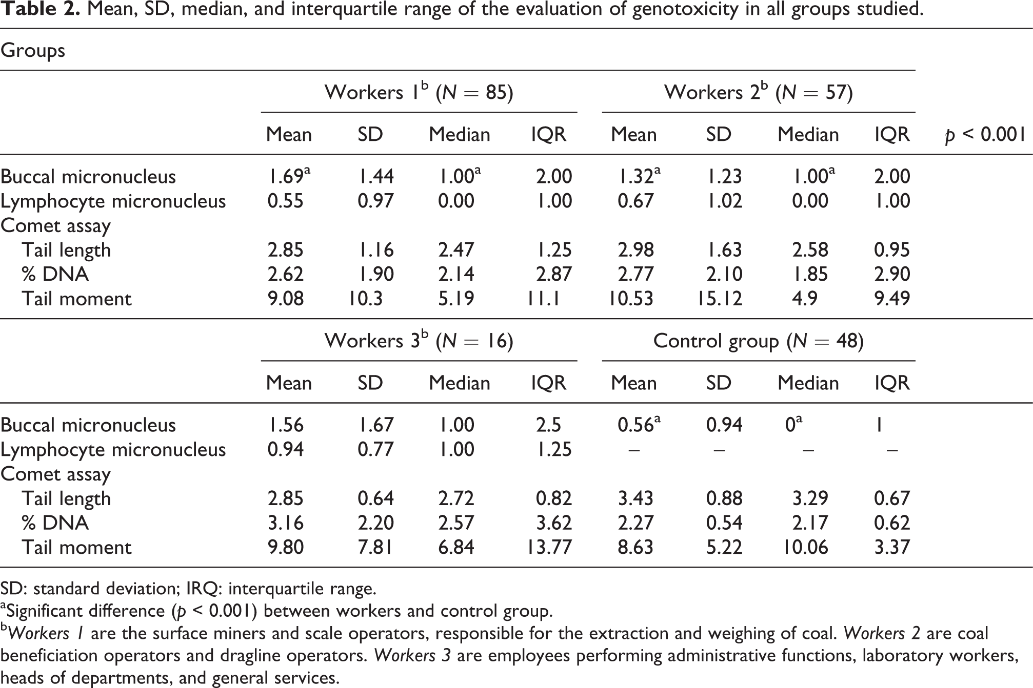
SD: standard deviation; IRQ: interquartile range.
aSignificant difference (p < 0.001) between workers and control group.
b Workers 1 are the surface miners and scale operators, responsible for the extraction and weighing of coal. Workers 2 are coal beneficiation operators and dragline operators. Workers 3 are employees performing administrative functions, laboratory workers, heads of departments, and general services.
The results of the oral mucosa micronucleus test showed a significant difference (p < 0.001) between the control group and worker groups 1 and 2. However, the lymphocyte micronucleus test showed no significant difference between the exposed groups. Of the results of the comet assay, only the tail moment (product of the average tail length, in micrometers, and percentage of DNA in the tail, of 100 nucleoids per individual) was considered for statistical analysis. Comparisons of the tail moment between the groups did not show significant differences. Though, the tail length showed a significant difference of the worker groups 1 and 2 in relation to the control group. Figure 1 presents one slide containing no damage and one with high DNA damage while Figure 2 shows oral mucosa cells stained for visualization of micronuclei.

Illustration of oral mucosa cell stained for the micronucleus test.

Illustration of no damaged cells (a, image collected from a slide of the control population) and damaged cells (b, Image collected from a slide of the workers 1 population) in the comet assay assessment.
Table 3 presents the results of bivariate (brute) and multivariate (adjusted) analyses of factors associated with DNA damage and chromosomal abnormalities that were of statistical significance. Other factors, such as current exposure, alcohol consumption, ethnicity, and income, had no correlation with outcome variables.
Poisson regression analysis of the risk and protection factors associated with outcomea,b.

aSignificance level of 5%.
bTable exhibits only associations that showed statistical significance. PR: Prevalence Ratio; CI: Confidence Interval..
Significant results from the brute analysis showed an association between outcome and both living in the municipality of the mining company and exposure to radiation in the last 12 months. In contrast, multivariate (adjusted) analysis showed an association of the tail moment only with radiation exposure in the last 12 months (Table 3).
From the lymphocyte micronucleus test, the most significant factor in the brute analysis was the duration of residence in the municipality. It is worth mentioning that in this case, association was observed between the groups that lived between 120 and 240 months and over 241 months in the municipality, because a lack of association of these groups with the reference group (living between 0 and 119 months in the municipality) could be verified. The brute analysis of the risk factors evaluated by the oral mucosa cell in the micronucleus test showed none to be significantly associated with the outcome. In contrast, the adjusted analysis showed the presence of a partner to have a significant association with the outcome.
Discussion
Coal is a heterogeneous substance that combines many organic and inorganic compounds, especially hydrocarbons (which can generate PAHs) and minerals, 26 and their mining involves the release of numerous pollutants into the environment. 27 PAHs and inorganic elements, like trace metals and silica, can induce DNA breaks, influence the mechanism of DNA repair, and modulate the gene expression of enzymes. 28 –30 Furthermore, when the combustion of coal is inefficient, there is emission of nanoparticles, and these particles have a large contact surface area where different substances, such as metals, end up binding. As a result of this process, their entry into the cell becomes facilitated. 31,32 This may cause early stages of carcinogenicity 33 due to induction of chromosome breakage and consequent formation of micronuclei, as reported by Dwivedi et al. 34 Moreover, different elements, resulting from coal burning, are able to produce free radicals in biological systems due to redox cycling reactions. 35 Reactive oxygen species generated in these reactions results in damage to the DNA strand through molecules such as DNA glycosylases and apurinic and apyrimidinic endonucleases, thus directly affecting base excision repair, which results in an increase of DNA breaks. 36,37 Therefore, combustion of coal contributes to multiple environmental risks and adverse health effects, which may be directly related to mutagenic and/or genotoxic effects.
The comet assay is a biomarker used in environmental and occupational studies of humans to detect DNA damage induced by genotoxic agents that can be repaired, such as intercalation actions and other injuries caused by alkylating compounds and oxidants. 2,38,39 The micronucleus, when present, is a biomarker for the detection of clastogens (induce ruptures or breaks of chromosomes) and/or aneugens (cause an abnormal number of chromosomes in daughter cells) in the cytoplasm of interphase cells that have undergone division during or after exposure to different agents. It is a characteristic that is highly correlated with the risk of cancer development. 24 –40
Although coal mining activities may be directly related to DNA damage in biological systems, as described in the literature, 6 –10 the results obtained in the current study reveal that we must consider not only the environmental exposure factor but also socioeconomic factors and lifestyle.
The variables that were shown to be of significance were the dwell time and recent exposure to radiation. Although exposure to radiation is commonly cited as a risk factor for the development of DNA damage, 41 this parameter seems to be related to healthy habits of regular medical follow-up. The absence of a partner was shown to be a risk factor, and dwell time in the municipality was shown to be a determining factor for this variable. However, the absence of a partner may also be seen as a lack of an intermediary to ensure the well-being of the exposed individual.
When related to genetic alterations, a dwelling time in the municipality of over 20 years proved to be a protective factor. This result may be associated with physiological or xenobiotic transformation adaptations, which play an important role in the mutagenic activity of environmental contaminants, and this may be linked to different genetic polymorphisms and individual susceptibility to the adverse effects of exposure to coal as described by Espitia-Pérez et al. 42 These relationships may allow a longer period of exposure to become a protective factor against the mutagenic factors in question.
The joint use of biomarkers of exposure and effect, such as the comet assay in blood cells and the micronucleus test in oral mucosa cells, has already been proposed as a strategy for genetic biomonitoring of human populations exposed to environmental contaminants. 43 In the present study, another evaluation of effect, the lymphocyte micronucleus test, was incorporated to complement these tests. This particular method has already been used in another study for the biomonitoring of miners of an open-pit coal mine. 10 It has also been shown to be an effective strategy for monitoring the genetic health of occupationally exposed individuals.
Although the mean values found are in accordance with the literature data, 24 the micronucleus test on oral mucosa revealed a significant difference between the groups considered to be most exposed to coal (worker groups 1 and 2) and the control group. These data are similar to those found by Rohr et al., 2 who carried out a study, also in the municipality of Candiota, with workers exposed to mineral coal, verifying an increase in the number of micronuclei in the oral mucosa of the exposed population when compared with the control. Nevertheless, this previous study did not consider different activities related to the extraction of coal when evaluating the exposure scenario. It is worth mentioning that the worker group 3 did not present a significant difference, perhaps because of the small number of individuals in the group. However, if a larger number of individuals presented similar values, a tendency toward a significant difference between this group and the control group would be suspected. However, the lymphocyte micronucleus test and comet assay did not reveal significant differences between the exposed and control groups.
Holland et al. stated, after careful review, that it was not yet clear to science whether the frequency of micronuclei in oral cells could be associated with the frequency of micronuclei in other cells, such as lymphocytes. 24 However, for anatomo-physiological reasons, it is presumed that the cells of the buccal mucosa are directly exposed to different contaminants, such as those present in daily foods and in the inhaled air. This may be due to the absence of an efficient barrier in this nonkeratinized epithelial tissue. Thus, the micronucleus test on oral mucosa presents more sensitive, though less specific, results.
Conclusion
The findings of this study reveal that the micronucleus in oral mucosal cells of workers exposed to coal is dependent on the degree of contact with coal, although the mean values found were in accordance with the literature data. In addition, it was found that factors such as housing conditions and medical monitoring should be always considered in a genotoxicological evaluation. Furthermore, measures to promote health should be encouraged so that there are improvements in the lifestyles of mine workers.
Footnotes
Acknowledgement
The authors would like to thank the Companhia Riograndense de Mineração for the opportunity to work alongside the company.
Declaration of Conflicting Interests
The author(s) declared no potential conflicts of interest with respect to the research, authorship, and/or publication of this article.
Funding
The author(s) disclosed receipt of the following financial support for the research, authorship, and/or publication of this article: This work was financially supported by Companhia Riograndense de Mineração.