
Abstract
Previous investigations demonstrated that high fluoride (F) exposure may adversely affect the neurodevelopment and learning and memory ability. However, whether maternal F exposure during gestation and lactation can influence the learning, memory ability, and glutamate receptor expressions of offspring has not yet been elucidated. Hence, in the present study, maternal mice were exposed to F (25, 50, or 100 mg/L sodium fluoride (NaF) in drinking water) during gestation and lactation. Results showed that exposure to 100 mg/L NaF significantly enhanced the number of total arm entries and working memory errors of offspring in the radial arm maze test compared to the control group. However, no difference was observed in open-field behaviors. For the subtypes of glutamate receptors in hippocampus, expression of GluR2 mRNA was significantly reduced by 25, 50, and 100 mg/L NaF. Besides, F exposure also suppressed the expression of NR2A, NR2B, and mGluR2 mRNA levels in a dose-dependent manner, where NR2A was significantly suppressed by 50 mg/L NaF and NR2B and mGluR2 by 100 mg/L NaF. However, no significant changes were observed in GluR1 and mGluR5 mRNA expression levels. Collectively, these findings suggested that F can pass through the cord blood and breast milk and may have deleterious impact on learning and memory of the mouse pups, which was mediated by reduced mRNA expression of glutamate receptor subunits.
Introduction
Apart from dental and skeletal fluorosis, excessive fluoride (F) ingestion results in a broad array of symptoms and pathological changes. 1 In recent years, increasing epidemiological investigations have demonstrated the negative relationship between the high F exposure and the low children’s intelligence. 2 –5 It is worth to note that children living in high F areas had lower IQ scores than those in low F areas. 3 Furthermore, animal experiments also confirmed the neurotoxicity of F. To our knowledge, diverse animal behavior methods like Y-maze test, 6 open field test, 7 transfer latency, 8 and step-down test 9 have been used in this field to support that F can induce behavior alteration and learning and memory impairment.
Long-term potentiation (LTP), the widely accepted electrophysiological model for the cellular mechanism underlying the spatial learning and memory formation, is triggered by the special receptors on the excitatory synapses. 10 In mammalian brains, most excitatory synapses are glutamatergic, and they use glutamate (an amino acid neurotransmitter) as the chemical messenger. 11 Jiang et al. and our previous studies reported that the glutamate concentration in hippocampus was reduced in rats exposed to F. 12,13 Glutamate takes up over 30% of the total excitatory neurotransmitter in brain. Interestingly, because the glutamate in the blood serum cannot penetrate the blood–brain barrier, glutamates of brain are synthesized only in brain. 13
Glutamate released from presynaptic nerve terminals binds specifically to glutamate receptors. 14 Up to now, the subtypes of glutamate receptors are categorized as ionotropic receptors including kainate receptors, α-amino-3-hydroxy-5-methyl-4-isoxazolepropionic acid receptors (AMPARs, GluR1-4), N-methyl-D-aspartate receptors (NR1-3), and metabotropic receptors (mGluRs, mGluR1-8). 15 Earlier investigation revealed that the exposure to F decreased mGluR5 expression in rat hippocampus and cortex. 12 Additionally, our previous study showed that F altered the gene and protein levels of NR1 in rat hippocampus. 16
Importantly, F can cross the placenta barrier and diffuse into cord blood, with the ranges from 60% to 91% of maternal F. 17,18 High F in breast milk suggested its accessibility for infants. 19,20 However, the maternal F exposure during gestation and lactation can influence the learning and memory ability and the glutamate receptor expressions of mice offspring are poorly understood. In view of this, in this study, we exposed the maternal mice to various concentrations of F during gestation and lactation, followed by behavioral tests like open field test and radial-arm maze, as well as the mRNA expressions of GluR1, GluR2, NR2A, NR2B, mGluR2, and mGluR5 in hippocampus of mouse pups.
Materials and methods
Experimental animals
Forty-eight adult mice of Kunming strain with half males and half females, weighing 20–25 g each, were obtained from Experimental Animal Center of Shanxi Medical University. The mice were placed in plastic cages and acclimated to the laboratory condition of 12-light/12-h dark cycle and 22–25°C for 1 week before the experiment. All animals received their food and water freely. After a week of acclimatization, one male and two female mice were housed together in one cage in the evening, and sperm plug in the female mice vagina was examined in the next day morning. The day when sperm plug presence was noticed was designated as day 0 of gestation. Each pregnant mouse was then separated and kept individually in a cage and randomly divided into four groups with different sodium fluoride (NaF) concentrations of 0, 25, 50, and 100 mg/L. After delivery, the females were continued with the same treatment until the end of the lactation. The offspring at the postnatal day 21 were allowed to attend the future tests. The experimental design was approved by the Ethics Committee of Shanxi Agricultural University (Taigu, China).
Open field test
As shown in Figure 1, the open field apparatus was a 72 × 72 × 72 cm3 black plastic box which contained an outer field and a center square field (36 × 36 cm2) at the bottom. The test was allowed to record the activities in a quiet test room (walking distance, residence time, and average speed) in the outer field and the center field. The mice were placed individually in the center of the chamber for 3 min before the test. Then, the movements were continuously monitored by the digital video camera fixed on the top of the box for 5 min; 70% ethanol was applied to clean the testing arenas after each trial.

The photo (a) and schematic diagram (b) of open field apparatus which contained an outer field and a center square field (36 × 36 cm2) at the bottom.
Radial arm maze test
The device was composed of eight 50 × 10 × 20 cm3 extending radial arms, with a platform (25 cm in diameter) in the cross center, presented in Figure 2(a). The maze was 50 cm above the floor. The test was performed as previously described by Wang et al. 21 Briefly, in the training phase, each mouse was trained to collect food pellets placed at the ends of the four randomly selected testing arms, while no food was presented in the remaining four arms. The training was conducted for a couple of days. In the testing phase, mice were first placed on the cross-center platform for 15 s and then freely explored in the maze for 5 min. The camera above the maze monitored the activity of the mice. The test indicators included (1) the number of total arm entries and (2) the number of working memory errors, entering a food arm that had been entered previously. The test was conducted twice daily for seven consecutive days.

The photo (a) and schematic diagrams (b and c) of radial arm maze. An illustrative example of a control (b) and a 100-mg/L NaF (c) treatment mouse’s travel pathway showed the mouse in the fluoride group entered into more arms. NAF: sodium fluoride.
RNA extraction and quantitative real-time polymerase chain reaction
Total RNA was extracted from the hippocampus of offspring by using Trizol reagent (Invitrogen, California, USA) according to the manufacturer’s instructions. Specific primers (Invitrogen, Shanghai, China) for GluR1, GluR2, NR2A, NR2B, mGluR2, mGluR5, and β-actin (an internal control) were designed with Primer 3.0 plus and the sequences are presented in Table 1. Quantitative real-time polymerase chain reaction (QRT-PCR) amplification was carried out with two-step SYBR® QRT-PCR kit (Takara, Dalian, China) on the Mx3000P™ QPCR system (Stratagene, San Diego, California, USA) for 40 cycles with denaturation at 95°C for 5 s, annealing at 61°C for 15 s, and extension at 72°C for 6 s. The relative mRNA abundance of each target gene was calculated using comparative 2−ΔΔCt method.
Primer sequences and product sizes for QRT-PCR.

SEM: standard error of the mean; QTR: quantitative real time; PCR: polymerase chain reaction.
Statistical analysis
The raw data of spontaneous behavior and learning and memory were analyzed by SMART version 3.0 tracking system (Panlab, Spain). The data in this study are shown as mean ± standard error of the mean. The difference among groups was compared by one-way analysis of variance with Dunnett as the posttest (GraphPad Software Inc., San Diego, California, USA). A probability of p < 0.05 was considered as being statistically significant.
Results
Open field test
Results showed that F exposure during embryonic to suckling periods had no significant effects on the spontaneous behavior of offspring mouse. No statistical difference was observed in walking distance, residence time, or average speed in both outer and center field in three experimental groups compared to the control group. For the overall activity, there was no difference in the total distance and average speed among the four groups.
Radial arm maze
The data of radial arm maze in Tables 2 and 3 show the effect of F on the learning and memory ability of offspring. Compared to control group, 100 mg/L NaF significantly enhanced the number of total arm entries from day 2 to 7. In 50 mg/L NaF group, a significant increase was observed on day 5 and 6, while 25 mg/L NaF have no effects on the number of total arm entries. With respect to the number of working memory errors, it was significantly increased in 100 mg/L NaF group from day 2 to 6 and in 50 mg/L NaF group on day 3, 5, and 6. No statistical difference was observed in 25 mg/L NaF group during the whole test period.
Effect of maternal fluoride exposure during gestation and lactation on the number of total arm entries of mouse pups in radial arm maze test (mean ± SEM, n = 6).

NAF: sodium fluoride; SEM: standard error of the mean.
a p < 0.05: a statistical difference when compared to the control group.
b p < 0.01: a statistical difference when compared to the control group.
Effect of maternal fluoride exposure during gestation and lactation on the number of working memory errors of mouse pups in radial arm maze test (mean ± SEM, n = 6).

NAF: sodium fluoride; SEM: standard error of the mean.
a p < 0.05: a statistical difference when compared to the control group.
b p < 0.01: a statistical difference when compared to the control group.
mRNA expressions of glutamate receptors
The mRNA expression levels of NR2A, NR2B, GluR1, GluR2, mGluR2, and mGluR5 in hippocampus are shown in Figure 3. For the subtypes of glutamate receptors, compared to controls, expression of GluR2 mRNA was significantly reduced by 25, 50, and 100 mg/L NaF. Besides, F exposure also suppressed the expression of NR2A, NR2B, and mGluR2 mRNA levels in a dose-dependent manner, where NR2A was significantly suppressed by 50mg/L and NR2B and mGluR2 by 100mg/L. However, no significant changes were observed in GluR1and mGluR5 mRNA expression levels.
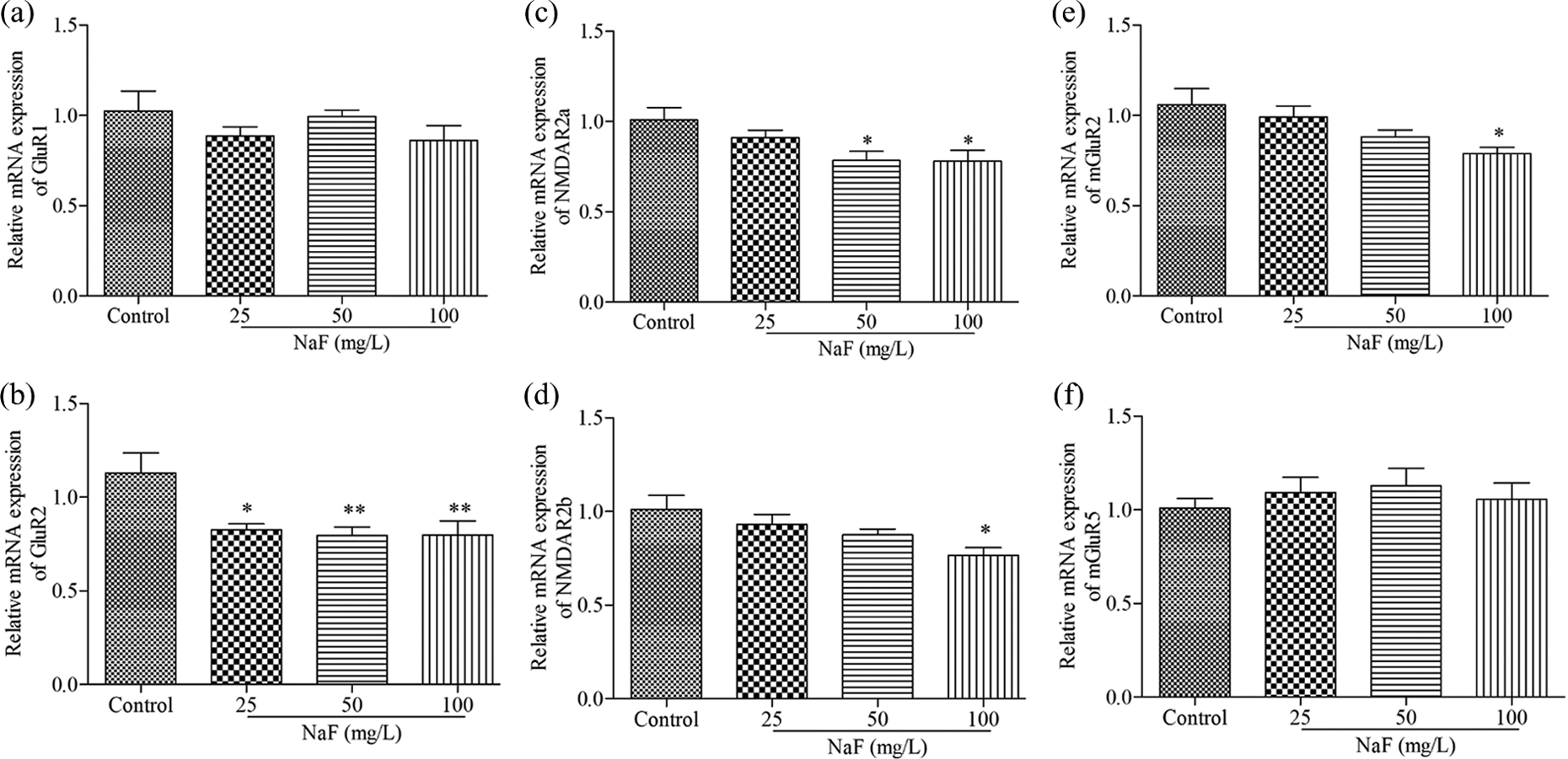
Effect of maternal fluoride exposure during gestation and lactation on the mRNA expressions of GluR1, GluR2, NR2A, NR2B, mGluR2, and mGluR5 in hippocampus of mouse pups. Bars represent mean ± SEM (n = 6). *p < 0.05 or **p < 0.01: a statistical difference when compared to the control group. SEM: standard error of the mean.
Discussion
F accumulation in different brain regions 22,23 and impaired learning and memory 3,24 have been consistently reported in previous investigations. However, the F source of subjects in those studies was from the diet and/or water. In the present study, the F exposure of offspring was through cord blood and breast milk. Results of open field test and radial-arm maze test revealed that F ingestion through this way during developmental period impaired the spatial learning and memory ability of offspring.
Hippocampal neurons are considered to be responsible for encoding space. 25 Results of pharmacological and physiological experiments have been established the link between hippocampal LTP and spatial learning. 26 In addition, glutamate receptors participate in the formation of spatial learning and memory. 26 Therefore, the expressions of different subtypes of glutamate receptors in hippocampus were analyzed in this present study.
AMPARs, which have been demonstrated to underlie the formation of learning and memory, are assembled from four subunits designated glutamate receptor GluR1, 2, 3 and 4. 27 During the first postnatal week, the expression of the GluR1 is higher, but in the later developmental period, GluR2 gets dramatically increased. 28 In adult hippocampus, most AMPARs are comprised of GluR1/GluR2 heteromers with small portion of GluR2/GluR3. 29 Hence, GluR2 is a critical subunit in determining the function of AMPAR in mammals. 28 The different subunits endue AMPAR channel with specific physiological properties. 27 GluR2-containing AMPARs serve for the flow of monovalent cations, while those lacking the GluR2 subunit are permeable to divalent cations, such as calcium. 30 In the current study, the expression level of GluR2 mRNA was significantly inhibited in hippocampus of offspring from mice treated with 25, 50, or 100 mg/L NaF. In contrast, GluR1 mRNA expression in experimental groups had no significant changes. Therefore, we suggest that F impaired learning and memory ability mainly through the reduced GluR2-containing AMPARs.
Resembling AMPARs, NMDA receptor-dependent LTP in the hippocampus is considered to be the crucial neural substrate for spatial learning and memory. 26 Here, the results showed that mRNA expression of NR2B was significantly suppressed in 50mg/L NaF group, while no changes were observed in NR2A subunit. The current mediated by heteromers containing NR2B is about three to four times slower than by those composed of NR2A. 31 Furthermore, the overexpressed NR2B enhanced synaptic plasticity in hippocampus and spatial learning. 32 Taken together, the decreased levels of NR2B in our experiment revealed that NR2B was also involved in the molecular mechanisms underlying fluoride-induced learning and memory impairment.
However, with respect to the subunits of metabotropic glutamate receptors, no changes were observed in the mGluR5 mRNA expression levels, indicating that the learning and memory impairment caused by F was independent to mGluR5-containing receptors. Notably, mGluR2 mRNA expression level in 100 mg/L NaF group was significantly inhibited. Previous studies found that mGluR2 activation modulated a specific olfactory memory formation which helped the females to be successful in the mating test. 33 In this study, the learning and memory formation relies on whether mice could find the food in certain arm of the maze. Hence, whether the low mGluR2 expression induced by F is related to impaired olfactory sensation requires further investigation.
In conclusion, F exposure during embryonic to suckling stages impaired the learning and memory ability of the mouse pups, by inhibiting the mRNA expression of glutamate receptor subunits including GluR2, NR2B, and mGluR2, among which GluR2 was the more sensitive molecule in hippocampus.
Footnotes
Declaration of Conflicting Interests
The author(s) declared no potential conflicts of interest with respect to the research, authorship, and/or publication of this article.
Funding
The author(s) disclosed receipt of the following financial support for the research, authorship, and/or publication of this article: This research was supported by National Natural Science Foundation of China (grant no. 31201965), Program for the Top Young Innovative Talents of Shanxi Agricultural University (grant no. TYIT201408), and Program for the Outstanding Innovative Teams of Higher Learning Institutions of Shanxi.