
Abstract
This investigation was undertaken to test the effect of nilotinib against
Background
Fulminant hepatic failure (FHF) is clinically defined as rapid deterioration of liver function due to massive hepatocyte death in the absence of preexisting liver disease. This syndrome is often associated with coagulopathy, jaundice, multisystem organ failure, and altered mental status. The prognosis of FHF is somewhat poor, and until now, there is no effective treatment for the disease other than liver transplantation. 1
Lipopolysaccharide (LPS) is the main constituent of the cell wall of the gram-negative bacteria that can contribute fundamentally to liver failure.
2
The combination of LPS and
Nilotinib is a highly potent tyrosine kinase inhibitor that can be administered orally. It is used for BCR–ABL-positive chronic myelogenous leukemia treatment in patients showing resistance or intolerance to imatinib. 9 It strongly inhibits the tyrosine kinase activity of ABL/BCR–ABL, as well as that of the discoidin domain receptor, stem cell factor, platelet-derived growth factor (PDGFR), and colony stimulating factor-1 receptor. 10,11 Recent studies have shown that nilotinib has potent antifibrotic 12 and anti-inflammatory activities in different models of inflammatory disorders. 13,14 This has been linked to its ability to specifically inhibit PDGFR tyrosine kinase, TNF-α, and transforming growth factor-β1. 12,14 The current study aimed to test the potential protective effect of nilotinib on acute liver injury induced by GalN/LPS injection in mice and explore possible molecular mechanisms underlying that effect by evaluating its effects on different oxidative stress markers and inflammatory cytokines.
Materials and methods
Experimental design
To induce acute liver injury, male Swiss albino mice (20–22 g) were selected and intraperitoneally injected with GalN (700 mg/kg, Sigma-Aldrich, St. Louis, Missouri, USA) and LPS (10 µg/kg, Escherichia coli serotype O111: B4, Sigma-Aldrich). 6 To test the effect of nilotinib against GalN/LPS-induced acute liver injury, nilotinib (Novartis Pharma AG, Basle, Switzerland) was administered for 3 days prior to challenge with GalN/LPS.
Mice were divided into four groups randomly (n = 8 for each). The first one was control where mice received only normal saline, while the second served as GalN/LPS-untreated control. The other two groups were GalN/LPS + nilotinib-treated where mice were pretreated orally with nilotinib at two different doses (25 and 50 mg/kg) 15 for 3 days.
All mice were kept in an air-conditioned room maintained at 25 ± 2°C with alternatively 12-h light and dark cycles. Animals were permitted a standard laboratory diet for rodent and tap water. The study protocol was conducted according to the ethical principles and guidelines of the handling, use, and care of experimental animals adopted by the Research Ethical Committee of the “Faculty of Pharmacy, Mansoura University, Egypt,” which were in accordance with the principles of Laboratory Animal Care (NIH 1985).
Eight hours after administration of GalN/LPS, mice were anesthetized and blood samples were collected. Serum samples were obtained after blood centrifugation for 15 min at 4000 g at 4°C and kept at −80°C until further analysis. Mice were then killed and liver tissues were dissected and washed with ice-cold saline. Small pieces of liver tissue (0.5 g) were homogenized using 1.15% potassium chloride solution.
The homogenates were centrifuged for 15 min at 900 g, and the supernatant was collected and centrifuged at 9000 g. The supernatant was kept at −80°C until analyzed. An extra sample of liver was excised and fixed in 10% neutral buffered formalin solution for histopathologic analysis.
To observe the effect of nilotinib against GalN/LPS-induced lethality, other groups of mice were arranged, treated as above-mentioned, and the mortality was observed within 24 h. 16
Assessment of survival rate
The number of dead animals and the total number of tested animals at given time were used to calculate the percentage of the survival rate. 17
Assessment of hepatotoxicity markers
Serum levels of alanine aminotransferase and aspartate aminotransferase were measured using kits (bioMérieux, Marcy-l’Etoile, France) based on the manufacturer’s protocols. Alkaline phosphatase (ALP) and lactate dehydrogenase (LDH) were measured using available kits (Human Gesllschaft fur Biochemica und Diagnostica, Germany) according to the manufacturer’s instructions.
Assessment of histopathological lesions
Liver paraffin blocks were sectioned (4–5 μm) and stained with hematoxylin and eosin. The liver specimen was examined under a microscope without the knowledge of the group and in a random order. The histopathological grade of the hepatocyte necrosis and lobular inflammation in the hepatic tissue was described and scored using five semi-quantitative grades as follows: grade 0 (no change), grade 1 (very mild), grade 2 (mild), grade 3 (moderate), and grade 4 (severe). 18
Assessment of oxidative stress markers
Hepatic content of malondialdehyde (MDA), glutathione (GSH), and superoxide dismutase (SOD) activity was measured using detection kits (Bio-Diagnostic, Egypt) according to the manufacturer’s protocols. Briefly, MDA hepatic content was determined by measuring the level of thiobarbituric acid reactive substances spectrophotometrically at 534 nm. Detection of SOD activity in liver homogenates relies on the ability of SOD to inhibit the reduction of nitro-blue tetrazolium dye. GSH content in the liver is proportional to the reduced yellow chromogen measured at 405 nm, that is produced by the reduction of Ellman’s reagent (5,5′ dithiobis-2-nitrobenzoic acid) with sulfhydryl (–SH) group of GSH.
TLR-4 assessment
TLR-4 level was estimated in liver tissue using the rat TLR-4 ELISA kit (MyBiosource Inc., San Diego, California, USA) based on the manufacturer’s instructions.
NF-κB assessment
Immunohistochemical (IHC) staining was done on 4-µm slices of paraffin-embedded tissue miniarray (TmA) sections using primary antibodies against rabbit polyclonal anti-NF-κB p65 antibody (Thermo Fisher Scientific, Fremont, CA, USA) according to previously described methods. 19
Cytokine assessment
Hepatic TNF-α and interleukin-1β (IL-1β) levels were determined using Quantikine mouse kits (R&D Systems, Minneapolis, Minnesota, USA) according to the manufacturer’s protocols.
Statistical analysis
The values are presented as the mean ± SEM for eight mice per group. Statistical analysis was done using one-way analysis of variance followed by Tukey–Kramer multiple comparison test. Histopathological grading was analyzed by the nonparametric Kruskal–Wallis test followed by Dunn’s test. A p value of<0.05 was considered significant.
Results
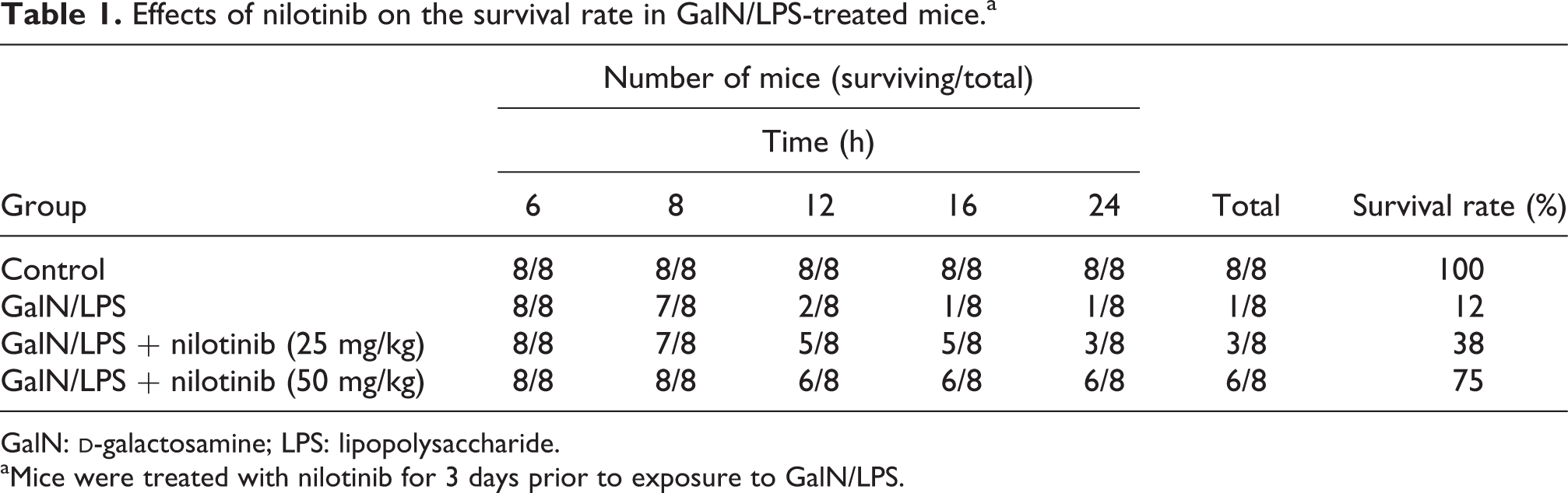
Effects of nilotinib on the survival rate
As shown in Table 1, the survival rate at 24 h in the GalN/LPS control group was 12%. Nilotinib pretreatment increased the survival rates to 38–75% in GalN/LPS-treated mice.
Effects of nilotinib on the survival rate in GalN/LPS-treated mice.a
GalN:
aMice were treated with nilotinib for 3 days prior to exposure to GalN/LPS.
Effects of nilotinib on the serum transaminases and ALP and LDH activities
Mice of GalN/LPS group showed a significant rise in serum transaminases and ALP and LDH levels compared to the control group. Nilotinib pretreatment resulted in a significant decrease in liver transaminases and ALP and LDH levels compared to GalN/LPS group (Table 2).
Effects of nilotinib on biochemical parameters of liver function tests in GalN/LPS-treated mice.a,b,c

GalN:
aMice were treated with nilotinib for 3 days prior to exposure to GalN/LPS.
bSerum was obtained 8 h after GalN/LPS injection.
cData are expressed as mean ± SEM, n = 8.
d p < 0.05 as compared to control group.
e p < 0.001 as compared to control group.
f p < 0.0 as compared to GalN/LPS group (one-way ANOVA).
g p < 0.01 as compared to GalN/LPS group (one-way ANOVA).
h p < 0.001 as compared to GalN/LPS group (one-way ANOVA).
Effects of nilotinib on histopathologic examination of liver
As shown in Table 3 and Figure 1, mice of the control group exhibited normal liver architecture, while mice of the GalN/LPS group showed a significant increase (p < 0.001) in the severity of necrosis and inflammation compared to the control group. Histological examination showed the presence of marked degree of hepatocellular centrilobular focal necrosis, apoptosis, and inflammation in the Gal/LPS-treated mice (Figure 1(b)), while the control livers showed normal no signs of liver damage (Figure 1 (a)). Pretreatment with nilotinib resulted in a significant reduction in the histopathological lesions compared to GalN/LPS group in a dose-dependent manner (Figure 1(c) and (d)).
Effects of nilotinib on the histopathological grade values of the hepatocyte necrosis and lobular inflammation in GalN/LPS-injured liver.a,b
GalN:
aMice were treated with nilotinib for 3 days prior to exposure to GalN/LPS.
bData are expressed as mean ± SEM, n = 8.
c p < 0.05 as compared to control group.
d p < 0.01 as compared to control group.
e p < 0.001 as compared to control group.
f p < 0.01 as compared to GalN/LPS group (Kruskal–Wallis test).
g p < 0.001 as compared to GalN/LPS group (Kruskal–Wallis test).

Effects of nilotinib on GalN/LPS-induced hepatic damage in mice. Mice were treated with nilotinib for 3 days prior to exposure to GalN/LPS. Liver (n = 8) was processed for histological changes at 8 h after GalN/LPS challenge. (a) Control group showing normal liver histology. (b) GalN/LPS group showing severe necrosis and inflammation, indicated by black arrows. (c) GalN/LPS + nilotinib (25 mg/kg)-treated group showing moderate degree of centrilobular focal necrosis and inflammation. (d) GalN/LPS + nilotinib (50 mg/kg) showing mild degree of necrosis and inflammation. H&E, magnification 100×. GalN:
Effects of nilotinib on hepatic oxidative stress
GalN/LPS significantly elevated (p < 0.001) the MDA level in the hepatic tissues with contaminant decrease in the GSH content and SOD activity (p < 0.001) compared to the control group. Nilotinib pretreatment effectively increased the levels of GSH and SOD and lowered the level of MDA compared to GalN/LPS group (Figure 2).

Effects of nilotinib on GalN/LPS-induced changes in hepatic MDA, SOD, and GSH in mice. Mice were treated with nilotinib for 3 days prior to exposure to GalN/LPS. Liver homogenates were prepared for measuring (a) MDA, (b) SOD, and (c) GSH. Data are expressed as mean ± SEM, n = 8. *p < 0.05 and *** p < 0.001 compared to control group; #
p < 0.05, ##
p < 0.01, and ###
p < 0.001 compared to GalN/LPS group (one-way ANOVA). GalN:
Effects of nilotinib on hepatic TLR-4
The level of TLR-4 was significantly increased after administration of GalN/LPS. This increase was not affected by nilotinib pretreatment (Figure 3).
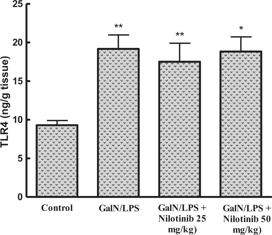
Effects of nilotinib on GalN/LPS-induced increase of hepatic TLR-4 level in mice. Mice were treated with nilotinib for 3 days prior to exposure to GalN/LPS. Data are expressed as mean ± SEM, n = 8. *p < 0.05 and **p < 0.01 compared to control group (one-way ANOVA). GalN:
Effects of nilotinib on hepatic NF-κB
IHC analysis indicated that NF-κB was significantly elevated in GalN/LPS group compared to the control group. Administration of nilotinib significantly reduced the immunostaining of NF-κB compared to GalN/LPS group in a dose-dependent manner (Figure 4).

Effects of nilotinib on GalN/LPS-induced increase of hepatic NF-κB expression in mice. (a) Control group showing minimal brown staining. (b) GalN/LPS group showing severe NF-κB expression. (c) GalN/LPS + nilotinib (25 mg/kg)-treated group showing moderate degree of NF-κB expression. (d) GalN/LPS + nilotinib (50 mg/kg) showing minimal degree of NF-κB expression, magnification 400×. GalN:
Effects of nilotinib on hepatic TNF-α and IL-1β
GalN/LPS administration caused a marked increase in hepatic levels of TNF-α and IL-1β as compared to the control group. Nilotinib pretreatment significantly attenuated the production of both cytokines as compared to the GalN/LPS group (Figure 5).

Effects of nilotinib on GalN/LPS-induced increase of hepatic TNF-α and IL-1β levels in mice. Mice were treated with nilotinib for 3 days prior to exposure to GalN/LPS. Liver homogenates were prepared for measuring (a) TNF-α and (b) IL-1β. Data are expressed as mean ± SEM, n = 8. *p < 0.05 and ***p < 0.001 compared to control group; ##
p < 0.01 and ###
p < 0.001 compared to GalN/LPS group (one-way ANOVA). GalN:
Discussion
GalN/LPS-induced FHF is an experimental model of acute liver injury that is widely used for testing the efficacy of hepatoprotective agents. A combination of LPS and GalN selectively produces fulminant hepatitis within few hours and rapid death within few days. The toxic effects of LPS, which is a potent inflammagen, are markedly potentiated by GalN that reduces the intracellular pool of uracil nucleotides in hepatocytes and thus inhibiting the synthesis of RNA and proteins. 20 Nilotinib has been reported previously to possess potent hepatoprotective effects against liver injury and fibrosis. 21,22 However, its effect on GalN/LPS-induced FHF has not been defined yet. The results of the present study demonstrated, for the first time, that nilotinib has hepatoprotective effects against GalN/LPS-induced FHF, which may be related to nilotinib ability to counteract NF-κB activation and cytokine generation.
As presented in this study, GalN/LPS increased the mortality rate, which reached 88% at 24 h. At 8 h after GalN/LPS injection, marked FHF was observed as indicated by the significant increase in serum transaminases and ALP and LDH activities. This was supported by histopathological examination of liver, which revealed extensive focal necrosis, apoptosis, and sever portal inflammation. These results are in accordance with previously documented hepatotoxic effects of GalN/LPS following in vivo administration to experimental animals. 8,23 On the other hand, the hepatoprotective effects of nilotinib were clearly evident through its ability to decrease the lethality and serum markers of hepatic injury. Histopathological findings further validated the beneficial effect of nilotinib as there was a marked reduction in the hepatocellular necrosis, inflammation, and apoptosis.
The mechanisms underlying the hepatoprotective effect of nilotinib against GalN/LPS-induced FHF were further examined via estimation of oxidative stress, NF-κB, TLR-4, and inflammatory cytokines in the hepatic tissue.
Oxidative stress has a pivotal role in the pathogenesis of acute liver failure. GalN/LPS exposure causes accumulation of neutrophils and Kupffer cells in liver sinusoids resulting in overproduction of reactive oxygen species (ROS), which induce cell injury directly through GSH depletion and subsequent lipid peroxidation. 24 –26 SOD acts as the first-line defense against ROS by converting superoxide to hydrogen peroxide, which will then be further detoxified by several enzymes, such as catalase and glutathione peroxidase. Consistent with the previous studies, the current study showed that GalN/LPS administration increased oxidative stress in the liver, as it is evident from the significant increase in hepatic MDA content and the significant reduction in GSH content and SOD activity. Pretreatment with nilotinib resulted in decreased MDA content and increased GSH and SOD levels in the liver. These results are in accordance with the previous ones that reported the ability of nilotinib to ameliorate the oxidative burden during different inflammatory damages in other organs. 14,27
Recently, the role of TLR inflammatory signaling has been widely implicated in the LPS-induced FHF. GalN/LPS increased the protein expression of TLR-4. 28 Furthermore, the inflammatory response and the liver injury in TLR-4 deficient mice were significantly attenuated following the induction of FHF. This has been attributed to decreased hepatic c-Jun and NF-κB expression and thus decreased TNF-α level. Many agents have been reported to possess potent hepatoprotective effects against LPS-induced liver injury due to inhibition of TLR signaling. 28 –32 Results of this study revealed the significant increase of TLR-4 upon GalN/LPS administration which was not affected by nilotinib pretreatment. Thus, it is reasonable to postulate that the hepatoprotective effect of nilotinib is not mediated through suppression of TLR-4.
Previous reports have evoked the role of NF-κB in the regulation of the expression of multiple genes involved in the inflammatory response, which controls the pathology of acute liver injury and inflammation. 33 NF-κB activation and translocation into the nucleus leads to the activation of inflammatory cytokine genes expression with subsequent release of proinflammatory mediators, such as TNF-α, cyclo-oxygenase 2, and nitric oxide. 19 Results of the present study revealed the increase in NF-κB expression in GalN/LPS mice, which was ameliorated in nilotinib-pretreated animals. Thus, it is acceptable to presume that the protective effects of nilotinib may be partly due to alleviating inflammatory response in GalN/LPS-induced acute liver damage via NF-κB signal pathway.
TNF-α and IL-1β are reported as the key cytokines involved in cell damage during sepsis and play a role in the pathogenesis of GalN/LPS-induced liver failure. 5 GalN/LPS increases mRNA and protein level of TNF-α in hepatic tissue as well as the circulating TNF-α level in serum. TNF-α induces hepatocytes apoptosis in the early stage of liver of GalN-sensitized mice. Furthermore, TNF-α induces neutrophil transmigration occurring in the late stage of liver injury. Unfiltered neutrophils can initiate respiratory burst and neutrophil degranulation leading to extensive hepatocytes necrosis. 3,34 Also, the significant elevation of IL-1β release and expression after GalN/LPS exposure has been reported. 35 Many agents, such as melatonin and mellitin, were shown to suppress the lethal liver injury via regulating TNF-α activity in GalN/LPS-treated mice. 17,23
Results presented here indicate that the levels of TNF-α and IL-1β were significantly increased upon GalN/LPS administration. Nilotinib pretreatment caused a significant attenuation of these inflammatory cytokines. Previous reports have shown the potent anti-inflammatory activity of nilotinib and its ability to decrease inflammatory cytokines. 14,22,36 –38 Some of these reports have attributed nilotinib anti-inflammatory activity to its ability to attenuate p38 MAPK and c-Jun N-terminal kinase activation. 22 Others have focused on nilotinib ability to suppress PDGFR. 12 However, our study linked the hepatoprotective effects of nilotinib with its ability to attenuate TNF-α and IL-1β production via NF-κB signaling pathway.
Taken all together, the current study provides evidence that nilotinib possesses potent anti-inflammatory activity that can protect against GalN/LPS-induced FHF. This protective effect may be mediated through decrease of oxidative burden and suppression of cytokine production through NF-κB signal pathway but not through TLR4 signaling. These results may provide evidence of the beneficial use of nilotinib in acute liver failure.
Footnotes
Author contributions
DS El-Agamy and AA Shaaban contribute equally to all aspects of the study. AM Shebl carried out the histopathological examination and was responsible for manuscript editing.
Declaration of Conflicting Interests
The author(s) declared no potential conflicts of interest with respect to the research, authorship, and/or publication of this article.
Funding
The author(s) received no financial support for the research, authorship, and/or publication of this article.