
Abstract
Organochlorines (OCs) are common environmental pollutants that have been linked to cancer. This work aims to assess the role of OCs as a risk factor for breast cancer and to evaluate the cellular changes induced by exposure to such environmental contaminants. The study included 70 cancer patients subjected to thorough history taking and routine investigations. Samples from tumor and normal adjacent tissue were taken to measure OCs’ levels and to perform molecular analysis (some oncogenic and apoptotic markers) by flow cytometry. There were significantly higher concentrations of methoxychlor, dichloro-diphenyl-trichloroethane (DDT), hexa-chlorobenzene (HCB), and chlordane in tumor tissue samples compared to the surrounding normal tissue. There was a positive statistically significant correlation between G2m and dichloro-diphenyl-dichloroethane, DDT, and methoxychlor. There was also a negative correlation between propidium iodide (PI) and heptachlor as well as between PI, B-cell lymphoma 2, and methoxychlor. Annexin showed a negative correlation with HCB and methoxychlor. In conclusion, the higher level of organochlorine pesticides in the tissue specimens of breast cancer and the resultant molecular dysfunction highlight a possible association. Further research is warranted to elucidate the other possible mechanisms involved in the process of carcinogenesis.
Introduction
Pesticides are chemicals widely used throughout the world. Organochlorine (OCs) are highly toxic due to their persistence, stability, and long half-lives (30–47 years). They can bioaccumulate and biomagnify in food chains. They also travel over long distances and can be detected even in areas where they have never been used. 1,2 They were prohibited from use throughout the world for more than 20 years ago. However, many of the banned pesticides are still sold or manufactured for export to developing countries such as Egypt. 3
Some Egyptian studies documented the presence of OCs in vegetables and fruits, water, milk, and its products despite that the Egyptian Ministry of Agriculture banned them in the 1980s
4
Organochlorines are known to be stored in adipose tissues as they are lipophilic, resulting in extremely high concentrations in humans posing a public health hazard. 1 There is speculation that they may increase cancer risk of hematopoietic system, prostate, breast, pancreas, and liver. 6
Breast cancer, in particular, is a major public health problem as one out of nine women will develop the disease in her lifetime and it is responsible for about 22% of the total cancer mortality. 7 The increasing incidence of breast cancer could only be partially explained by improvements in genetic studies and screening programs. 8
It is supposed that environmental factors may play a crucial role in the pathogenesis of the disease. Long-term exposure to environmental pollutants especially those having the estrogenic effect might make a significant contribution to the process of carcinogenesis particularly hormone-related tumors such as breast cancer. This issue is controversial and is still being researched intensively.
6
The aim of this work was to assess the potential role of organochlorines as a risk factor for breast cancer and to evaluate the cellular changes induced by exposure to such environmental contaminants.
Patients and methods
Study design
The present work is a comparative cross-sectional study that was conducted on breast cancer patients attending the Oncology Center, Mansoura University, Dakahlia Governorate, Egypt, in the period between April 2013 and July 2014.
Inclusion criteria
Patients who agreed to participate in the study were non-smokers. They did not receive either hormonal therapy, chemo-or radiotherapy before the surgery.
Exclusion criteria
Patients with positive family history of breast cancer or had previous surgery and came for recurrence or metastasis were excluded from the study.
Ethical consideration
A written informed consent was obtained from all studied patients participating in the study besides approval from the Ethical Committee of Mansoura University-Faculty of Medicine regarding all the procedures conducted in the present work.
Methods
All patients underwent thorough history taking including sociodemographic data, full detailed patient medical history (present, past, and family history), residence, and marital status.
Preoperative investigations
Preoperative histopathological examination of breast mass by fine needle aspiration cytology (FNAC) or true cut biopsy. Then, postoperative histopathological examination of breast mass, lymph nodes, estrogen receptors, progesterone receptors, and human epidermal receptor 2.
Radiological examination by sono-mammography.
Surgery and collection of tissue samples (10 g each)
Modified radical mastectomy or wide local excision with at least 1 cm safety margin was done. During the surgical procedure, test samples were taken from the fresh malignant breast tissues. Control samples were also obtained from adjacent histologically healthy tissue (6 cm away from the tumor). All samples were divided into two halves – one for OCs detection and the other specimens were transported to the flow cytometry laboratory in Mansoura Children Hospital for molecular investigations.
Extraction and detection of organochlorines
All samples were stored at −20 °C until analysis. Then, samples were extracted and analyzed by gas chromatography for detection of organochlorine pesticides and estimation of their concentrations in breast tissue samples. Analytical procedure: – All solvents and reagents were pesticide analytical grade free of interfering residues. Reference chemical standards (13 organochlorines) were purchased from Ehrenstorfer (Ausberg, Germany). The purities of the standard pesticides were 97.4–99%. – Pesticides analyzed in this study were hexachlorocyclohexane (HCH): α-HCH, β-HCH, hexa-chlorobenzene (HCB), the cyclodienes including aldrin, endrin, heptachlor, heptachlor epoxide, γ-isomer of HCH (γ-HCH, lindane), and γ-chlordane. Also, dichloro-diphenyl-trichloroethane (DDT) including DDT isomer (p,p′-DDT: 1,1,1-trichloro-2,2-bis-p-chlorophenyl-ethane), DDT metabolites: p,p′-p,p′-DDD (1,1-dichloro-2,2-bis-p-diphenyl-dichloroethane) and DDE (1,1-dichloro-2,2-bis-diphenyl-dichloroethylene), and methoxychlor. – The tissue samples were extracted according to Petreas et al.
10
One-fifth of the extract was taken to determine the lipid content gravimetrically. The samples were cleaned up by florisil column chromatography to separate pesticide residues by gas chromatography (Agilent Technologies, Inc., 6890 series) equipped with 63 Nickel Electron Capture Detector (63Ni) and auto-sampler injection. – Gas chromatographic conditions were as follows: silica capillary column PAS-5 (30 m × 0.32 mm inner diameter × 0.25 μm film thickness), injector temperature 280 °C; detector 300 °C; column 160 °C, initial time 2 min hold 10 °C min−1 to 280 °C for 10 min, the carrier gas was nitrogen. Peak areas were used as the basis for quantification. Residue levels are expressed relative to extracted lipid (ng/g lipid). – The limit of detection was 0.5 ng/g of lipid for α, β, and γ-HCH, 0.25 ng/g lipid for HCB, aldrin, heptachlor, heptachlor epoxide, γ-chlordane, and p,p′-DDE while it was 0.99 ng/g lipid for endrin, p,p′ -DDD, p,p′ -DDT, and methoxychlor.
Molecular investigations by flow cytometry
Tissue samples were washed three times with normal saline solution and the surrounded fats were trimmed carefully.
Flow cytometer (Becton Dickinson, Sunnyvale, California, USA) was used for cell-cycle analysis. Preparation of single cell suspension and evaluation of apoptosis and DNA cell-cycle parameters were done.
11
Determination of Bcl2 was done. 15
Cell death was evaluated by determination of propidium iodide (PI) “oncotic marker” and annexin V (apoptotic marker) using flow cytometry: dot plots were generated and divided into four quadrants (UL: upper left; UR: upper right; LL: lower left; LR: lower right). The LL quadrant shows cells negative for both annexin V and PI (living non-apoptotic cells). The UL quadrant shows dead cells (positive for PI, but negative to annexin V), whereas the UR quadrant shows late apoptosis: cells positive for both annexin V and PI. The LR quadrant shows living, early apoptotic cells positive for annexin V, but negative for PI. 16
Statistical analysis
Data entry and statistical analyses were performed using SPSS (statistical package of social sciences) version 20.0 (SPSS Inc., Chicago, Illinois, USA). Distribution of data was first tested by one sample K-S test. Parametric data was expressed in mean and standard deviation. Non-parametric data was expressed in median, minimum, and maximum. Independent t-test was used to compare means for continuous parametric variables, while Mann–Whitney U test was done for comparison of non-parametric continuous variables between two groups. For comparison of qualitative data among groups, χ 2 square test was performed. Spearman’s correlation coefficient was used to illustrate the strength of relationship between non-parametric variables. p value < 0.05 was considered as statistically significant.
Results
Seventy female patients fulfilled the inclusion criteria and were finally enrolled in the study. Their age ranged from 29 to 58 years (mean ± SD was 54.8 ± 12.4). The main characteristics of the studied patients as regards age, residence, occupation, marital status, and the hormonal analysis were illustrated in Table 1.
Sociodemographic data and hormonal analysis of the studied breast cancer cases (n = 70).

PR: progesterone receptors; ER: estrogen receptors; HER2: human epidermal receptors 2; n: number.
Table 2 shows the concentrations of organochlorine pesticides in addition to the odds ratio (OR) of their levels in the tested tissue samples. There were higher concentrations of methoxychlor, DDT, HCB, and chlordane in tumor tissue samples compared to the surrounding normal tissue in the studied cases and the differences were statistically significant (p < 0.05).
Levels of organochlorine pesticides in tissue samples (n = 140).
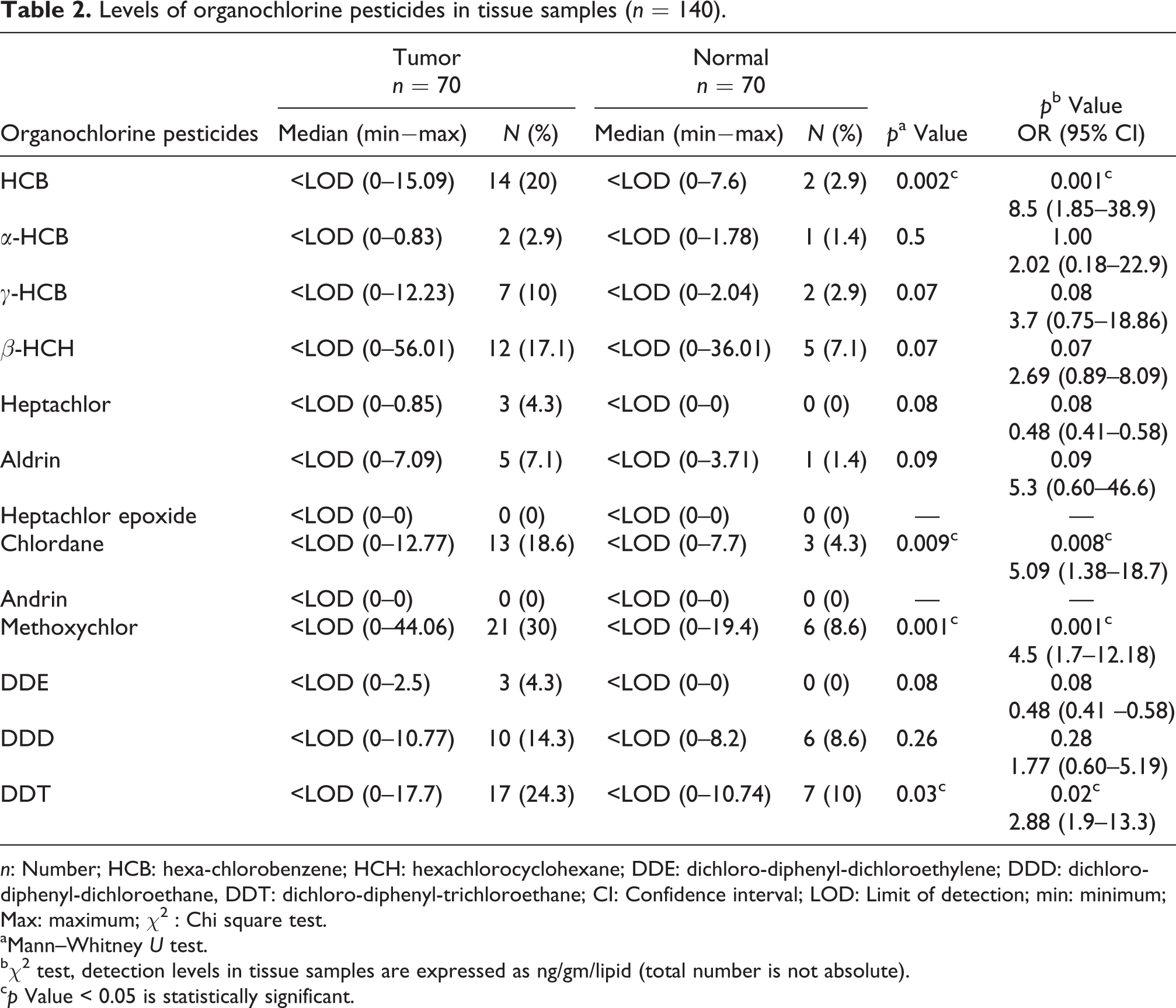
n: Number; HCB: hexa-chlorobenzene; HCH: hexachlorocyclohexane; DDE: dichloro-diphenyl-dichloroethylene; DDD: dichloro-diphenyl-dichloroethane, DDT: dichloro-diphenyl-trichloroethane; CI: Confidence interval; LOD: Limit of detection; min: minimum; Max: maximum; χ2 : Chi square test.
aMann–Whitney U test.
b χ 2 test, detection levels in tissue samples are expressed as ng/gm/lipid (total number is not absolute).
c p Value < 0.05 is statistically significant.
Data of molecular analysis of tissue samples are shown in Table 3. There were higher levels of PI, bcl2, CD95, apoptosis, STm, G2m, annexin (necrosis, early and late apoptosis) in tumor tissue compared to normal samples which were statistically significant (p < 0.05). On the other hand, PIM2, G01, and annexin (viable) were highly detected in normal tissue compared to tumor tissue samples (p < 0.05).
Molecular analysis in malignant versus normal breast tissue samples (n = 140).

N: Number; SD: standard deviation.
aMann-Whitney U test or independent t-test.
b p Value < 0.05 is statistically significant.
Table 4 illustrates the correlation between different molecular analytical data and OC pesticides detected in tumor tissue samples. There was a weak positive statistically significant correlation between G2m and DDD, DDT and methoxychlor (p ≤ 0.05). Also, there was a negative correlation between PIM1 and heptachlor. Annexin necrotic showed a negative correlation with HCB. Methoxychlor showed a negative correlation as regards annexin (early apoptosis), PIM1, and Bcl-2.
Correlation between molecular analytical data and Organochlorine pesticides levels in tumor tissue samples (N = 70).a

N: number; HCB: hexa-chlorobenzene; HCH: hexachlorocyclohexane; DDE: dichloro-diphenyl-dichloroethylene; DDD: dichloro-diphenyl-dichloroethane; DDT: dichloro-diphenyl-trichloroethane.
aHeptachlor epoxide and endrin were excluded because its median levels were nil in tumor tissue.
b p Value < 0.05 is statistically significant.
Discussion
Organochlorines (OCs) are common environmental pollutants that have been linked to various health hazards including cancer. Studies concerning the relation between breast cancer and OCs are too few to provide a conclusion. Some reports did not confirm a possible correlation,
17
while others were positive.
18
Inconclusive results necessitate further investigation. Moreover, mechanisms that explain a potential association still in need to be elucidated.
17
To the best of our knowledge, this work is one of the few studies that assesses the correlation between levels of different OCs and some molecular markers detected in tissue specimens of human breast cancer.
The present findings showed that HCB, chlordane, methoxychlor, and DDT are significantly higher in malignant tissue samples than in the surrounding normal tissue specimens with higher risk of breast cancer in relation to these compounds (OR: 8.5, 5.09, 4.5, 2.88). Endrin and heptachlor epoxide are not detected in any of the tested samples. It is also found that 78.6% of the patients are housewives and 85.7% are residents of rural areas.
The concentration of the detected tissue OCs is lipid adjusted in order to give a better estimation of the burden of exposure as those compounds are lipophilic. 20 Serum OC concentrations are not used in the present work as they have some limitations in comparison to adipose breast tissue. Crinnion 21 reported that serum OC concentrations are only good estimators of recent exposure. They could not indicate long-term exposure, which is affected by current exposure levels and lipid mobilization.
Nearly similar, Salerno et al. 22 investigated the association between farming (a proxy for pesticide exposure) and cancer in the Vercelli suburban area (Italy). They reported that farmers showed higher odds for all cancers (OR = 1.459, p < 0.001) including breast cancer.
In line with the current work, it was found that β-hexachlorocyclohexane (β-HCH), HCB, heptachlor, and p,p′-DDE are positively associated with breast cancer risk. Further statistical adjustment for the selected covariates indicated that only β-HCH, heptachlor, and p,p′-DDE remained statistically significant. 23 In addition, Cohn et al. 24 indicate an association between breast cancer and DDT exposure as evidenced in the present study.
It was also reported that sera of breast cancer patients have a combination of aldrin, DDE, and DDD and the risk of breast cancer was moderately associated with the later. The authors suggested that organochlorine pesticide mixtures could play a possible role in breast cancer. 25
In contrast, other studies did not report an evidence to support a causal association between breast cancer and OCs.
26
Noteworthy, the well-known correlation between prolonged exposure to estrogens and breast cancer suggests that environmental pollutants, namely, xeno-estrogens such as organochlorines, may play a critical role in the cellular and molecular changes occurring during breast carcinogenesis. 28
In the present work, there are higher levels of PIM1, bcl2, CD95, apoptosis, STm, G2m, annexin necrosis, and annexin (early and late apoptosis) in tumor tissue compared to normal tissue (p < 0.05). There is a significant correlation between annexin (necrotic) and HCB as well as between G2m and methoxychlor, DDD, and DDT and between annexin (early apoptosis) and methoxychlor. While a weak negative correlation is noticed regarding heptachlor, methoxychlor, and PIM1 and between Bcl-2 and methoxychlor.
Apoptosis is a type of cell death that plays a role in various pathological conditions as well as in regulation of tumor progression. Induction of apoptosis is actively used by the immune system to eliminate abnormal cells and to prevent the tumor development. Higher incidence of cancers is related to disruption of apoptotic mechanisms. 29
Many molecular markers play a crucial role in carcinogenesis. For example, Bcl-2 is involved in regulation of apoptosis. It may have a role in breast cancer by raising the apoptosis threshold. 30 In addition, CD95 is a type I cell surface glycoprotein that transduces apoptosis in sensitive cells. Increased CD95 has been reported in various cancers. 31 On the other hand, annexins are involved in cell growth and proliferation and alteration of their expression is associated with cancer. 32
AlegrÍa-Torres et al. 33 documented that DDT and its metabolites are able to induce apoptosis in many cell lines such as human peripheral blood mononuclear cells. Heptachlor was also reported to inhibit apoptosis and thus triggering proliferation of rat hepatocytes. 34 A chronic apoptotic stimulus can lead to a high rate of tissue regeneration with elevation of the risk of mitotic errors that may predispose to cancer development 35 which could explain the findings in the current work.
Additionally, it was reported that HCB causes an imbalance between cell death and cell proliferation, resulting in an increased number of apoptotic cells. Consequently, HCB was suggested to be a tumor co-carcinogen in rat mammary gland. 36 It was claimed that HCB-induced apoptosis is mediated through increased CD95 and caspase-8. 37
Moreover, it was observed that DDT constituents (i.e. o,p′-DDT, o,p′-DDE, p,p′-DDT, and p,p′-DDE) induced cell proliferation. 38 In addition, there is an evidence that these chemicals might also cause cancer via other mechanisms, including the induction of enzymes that produce genotoxic intermediates and DNA damage, 39 and disruption of the epigenomic landscape in cancers. 9
Chatterjee et al. 40 also reported that pesticides induced a significantly higher level of apoptotic receptors expression (annexin V and CD95) in addition to an increased percentage of cells in the G0/G1 phase compared to the control group in the bone marrow progenitor cells. However, there was a significant reduction of the cell fraction in the S/G2/M phase, indicating an enhanced level of stem/progenitor cellular apoptosis and impaired cell proliferation following pesticide exposure. This suggests a higher rate of premature cell senescence and restricted cell cycle pattern. The inhibition of cell replication and cell cycle arrest at the G0/G1 phase also signifies the toxic effect of pesticides. 41
The present findings could partly explain the molecular mechanisms induced by organochlorine pesticides to be involved in the pathogenesis of breast cancer. It was reported that these pesticides even at very low levels can cause cancer through their tumor-promoting properties, immunosuppressive effects, endocrine-disrupting impacts, and potential estrogenic activity leading to disturbance of the normal estrogen–progesterone balance 42 –44 and hence these environmental toxic pollutants could contribute to the risk of breast cancer.
Additionally, it was shown that exposure to OCs may modulate cell-cycle control and other molecular parameters in human mammary cells. 28,45 In other words, certain molecular changes have to occur in epithelial cells to convert normal cells into tumor cells. Thus, OCs may promote oncogenesis and cancer development through enhanced cell proliferation, decreased cell-cycle arrest, and decreased apoptosis 46 as evidenced in the current work.
Limitations of our study do exist, mainly that there are no adipose tissue samples are available from healthy controls as it seems unethical to take a tissue biopsy from healthy individuals.
In conclusion, the higher levels of organochlorine pesticides in the tissue specimens of breast cancer and the resultant molecular dysfunction highlight a possible association. Future cancer studies should analyze the interaction between those environmental contaminants with endogenous hormones and with cellular dysfunction. No safe limits of exposure to OCs have yet been determined. Hence, continued exposure to these persistent pollutants should be considered especially in developing countries where alarming rates of BC incidence exist.
Footnotes
Declaration of Conflicting Interests
The author(s) declared no potential conflicts of interest with respect to the research, authorship, and/or publication of this article.
Funding
The author(s) disclosed receipt of the following financial support for the research, authorship, and/or publication of this article: This is a research project funded by Mansoura University Institution.