
Abstract
The effects of drugs of abuse on oral mucosa are only partly understood. The aims of the present study were to: (1) evaluate the frequency of nuclear changes in normal-appearing oral mucosa of alcoholics and crack cocaine users and (2) assess their association with cell proliferation rate. Oral smears were obtained from the border of the tongue and floor of the mouth of 26 crack cocaine users (24 males and 2 females), 29 alcoholics (17 males and 12 females), and 35 controls (17 males and 18 females). Histological slides were submitted to Feulgen staining to assess the frequency of micronuclei (MN), binucleated cells (BN), broken eggs (BE), and karyorrhexis (KR). A significant increase in the frequency of MN was observed in cells exfoliated from the tongue of crack cocaine users (p = 0.01), and alcoholics showed a higher frequency of KR in cells obtained from the floor of the mouth (p = 0.01). Our findings suggest that the use of crack cocaine induces clastogenic effects, whereas alcoholism is associated with higher degrees of keratinization in the floor of the mouth.
Introduction
Oral cytopathology is based on the microscopic evaluation of cells collected by scraping the oral mucosa. This technique is noninvasive, painless, simple, and inexpensive. Currently, it has been used as a strategy to detect early changes in the oral mucosa of subjects exposed to noxious substances as a result of their habits or occupational activities. Some studies have shown changes in epithelial maturation, 1,2 in cell proliferation rates, 3,4 and also in the frequency of micronuclei (MN) 5 –7 in cells exfoliated from the oral mucosa of smokers and smokers and drinkers.
MN are extranuclear bodies composed of chromosomes or chromosomal fragments, which are separated from the daughter nuclei during mitosis. The presence of MN indicates DNA breaks, that is, clastogenicity or aneuploidy due to disturbances in the mitotic spindle caused by exposure to genotoxic agents. 8 Tolbert et al. 9 proposed the inclusion of other nuclear changes, for example, binucleated cells (BN), nuclear buds (also called broken eggs (BE)), and karyorrhexis (KR), to improve the evaluation of the effects of occupational or behavioral exposure to carcinogens. MN quantification appears to be a useful biomarker, as its frequency gradually increases in potentially malignant lesions, showing a direct relationship with tumor progression. 10
The primary aim of the current study was to evaluate the frequency of nuclear changes in cells exfoliated from the oral mucosa of crack cocaine users and alcoholics based on the hypothesis that individuals exposed to these substances are more susceptible to presenting abnormalities. The secondary aim was to investigate whether nuclear changes could be associated to cell proliferation rate that could indicate early stages in tumor progression.
Methods
Characteristics of the sample
This cross-sectional study assessed 90 subjects separated as follows: (1) crack cocaine users: 26 adult crack users (24 males and 2 females) attending a drug addiction treatment group at the International Federation of Red Cross in Porto Alegre, Brazil; (2) alcoholics: 29 adult alcohol abusers (17 males and 12 females), nonusers of crack, attending an alcohol dependence program also at the Red Cross in Porto Alegre, Brazil; and (3) controls: 35 adults (17 males and 18 females), nonusers of crack, nonsmokers, who consumed less than one dose of alcohol weekly (14 g) and received treatment at the School of Dentistry of the Federal University of Rio Grande do Sul, Porto Alegre, Brazil.
Patients with a history of malignant oral tumors or showing oral lesions were excluded from the study. Sample size was determined based on previous oral cytopathology studies involving smokers and drinkers, which have usually included about 25 subjects per group. 3,6,11,12
The present study was conducted in accordance with ethical guidelines set forth in the Declaration of Helsinki. The study protocol was approved by the local ethics committee. All patients signed an informed consent form prior to their inclusion in the study.
Questionnaire and oral examination
Information on age, skin color, monthly income, educational level, use of removable dentures, and alcohol and tobacco consumption were collected by an interview. Total lifetime exposure to cigarette smoke (pack-years) was calculated by multiplying the number of cigarettes smoked per day by the number of years of habit, divided by 20 (1 pack).
Daily alcohol consumption was calculated by multiplying the number of drinks consumed weekly by the average alcohol content of a glass of beer, wine, or cachaça (a typical Brazilian spirit distilled from sugarcane), divided by 7 days. The volume of alcohol was estimated to be 10 mL in a glass of beer, 12 mL in a glass of wine, and 10 mL in a dose of cachaça. Alcohol by volume was converted to alcohol by weight using a 0.8 conversion factor. 13 Because age and exposure to tobacco and alcohol are factors known to modify cell proliferation, these variables were compared only across study groups.
The consumption of other drugs of abuse was assessed using the Alcohol, Smoking and Substance Involvement Screening Test by the World Health Organization. 14
Finally, oral health conditions were recorded in a simplified manner 11 as poor (presence of residual roots, loss of various teeth, and advanced periodontal disease), regular (presence of caries and tartar but loss of few teeth), good (many restorations, no caries, or tartar), and very good (absence of caries, restorations, or tartar).
Cell collection
Before the collection of oral cells, patients rinsed their mouth with tap water for 1 minute. 6 Then smears were collected from the most prevalent oral cancer sites, 15 that is, lateral border of the tongue and floor of the mouth, using cytobrushes. Each sample was spread onto two cytologic slides and stored in plastic vials with 100% ethanol for fixation.
Cytological preparation and sample analyzes
Evaluation of nuclear changes
Two slides (one of tongue border and one of floor of the mouth) from each patient was Feulgen stained to assess the frequency of MN, BN, BE, and KR, according to the criteria proposed by Tolbert et al. 9 A total of 1000 cells/slide were evaluated.
MN were defined as oval or round extra nuclear bodies present in the cytoplasm with similar staining intensity as the main nucleus and having a diameter between 1/3 and 1/16 of the main nucleus. Presence of MN was considered indicative of genotoxicity. 16 BN were characterized by the presence of two main nuclei with similar morphology. This disorder is supposed to be associated with aneuploidy. 17 BE are accessory nuclei connected to the main nucleus by a chromatin filament. The clinical relevance of BE is still controversial, but they seem to precede MN. 18 BE result from the expulsion of amplified DNA from the main nucleus. 19,20 Finally, KR was defined as cells presenting nuclear disintegration involving loss of integrity of the nucleus, which are considered indicative of cell death/apoptosis. 17 Representational images of each nuclear change are presented in Figure 1.
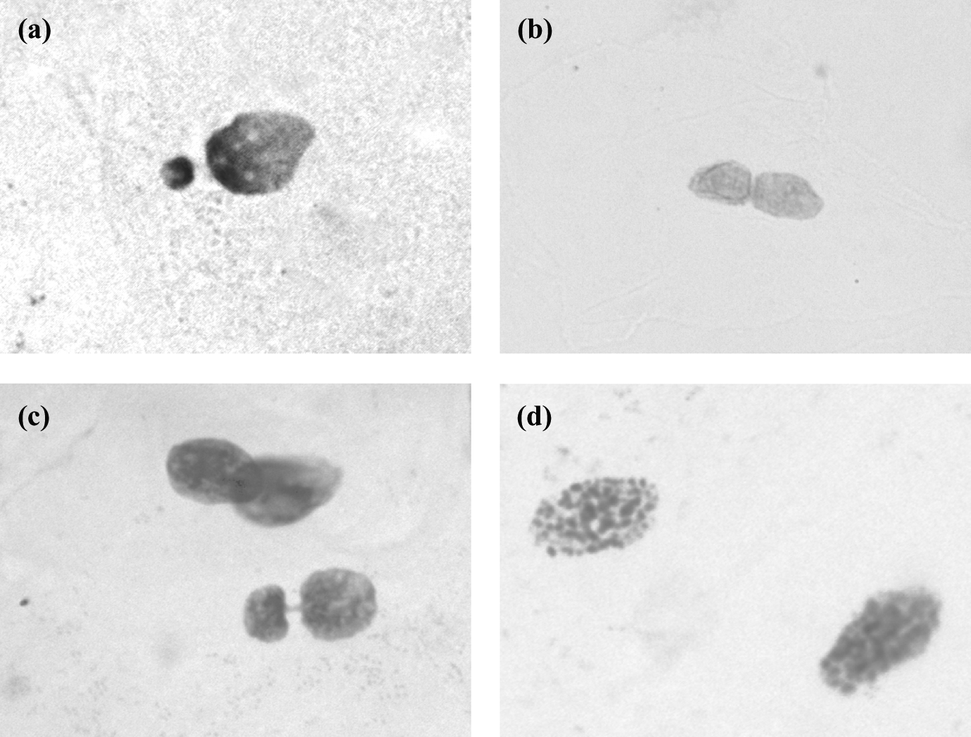
Representational images of the nuclear changes: MN (a), BN (b), BE (c), and KR (d). Feulgen stain, original magnification ×1000. MN: micronucleus; BN: binucleated cell; BE: broken egg; KR: karyorrhexis.
From each slide, 1000 cells were examined using 1000× magnification. The criteria established by Tolbert et al. 9 were used to determine nuclear changes. Only well-spread, nonoverlapping cells were included in the analysis. The examiner was blinded to group allocation.
Evaluation of cell proliferation rate
Data from cell proliferation rate were retrieved from our previous study 21 to assess the potential association with the occurrence of nuclear changes. Briefly, the second slide from each patient was silver stained for nucleolar organizer region (AgNOR) quantification as a parameter of cell proliferation rate according to the technique proposed by Ploton et al. 22 Images were obtained from each slide using an Olympus® digital camera (model Qcolor 5, Coolet, RTV, Japan) coupled to an Olympus® binocular light microscope (model CX41RF) and a Dell computer (Dimension 5150). Images were captured using the Qcapture® software version 2.81 (Quantitative Imaging Corporation, Inc., 2005, Surrey, BC, Canada) at 1000× magnification with oil immersion. The first 50 well-spread, nonoverlapping nucleated cells were analyzed (Figure 2), according to the protocol proposed by Paiva et al. 3 Number of AgNORs per nucleus was visually counted on the recorded images 3,4 and the mean number of AgNORs per nucleus (mAgNOR) was calculated.

Representational images showing cells with three AgNORs/nucleus and five AgNORs/nucleus. Silver stain, original magnification ×1000. AgNOR: silver staining for nucleolar organizer region.
Statistical analysis
To compare the occurrence of nuclear changes among the groups, data distribution were analyzed using the Shapiro–Wilk test. Parametric tests (Student’s t-test or analysis of variance/Tukey) were used to analyze data following a Gaussian distribution. Non-normally distributed data were analyzed using the Kruskal–Wallis test, followed by Dunn’s multiple comparison test. Significance was set at 5%. To define the cutoff for level of occurrence of BN, BE, and KR as low and high, median was used. Since MN data follow the Poisson distribution with the majority of subject presenting no or 1 MN, the cutoff for MN was defined as low (no MN) and high (≥1). To assess whether nuclear changes could, or could not, be associated to increase of cell proliferation rate, mAgNOR from individuals with low and high level of each nuclear changes was compared.
Results
Sample characteristics, including demographic data, oral health conditions, and use of licit and illicit substances are summarized in Tables 1 and 2. Mean age was not significantly different across groups. Males predominated in the crack cocaine user group. The same group showed a lower percentage of individuals with good/very good oral health conditions (Table 1).
Characteristics of the sample showing demographic data and oral health conditions.

a p = 0.10: based on analysis of variance results.
Characteristics of alcoholics and crack cocaine users according to exposure to tobacco, alcohol, and other drugs.

ASSIST: Alcohol, Smoking and Substance Involvement Screening Test.
aStudent’s t-test.
bAdapted from the ASSIST by the World Health Organization. It was considered the use of substances in the last 3 months. One alcoholic (3.45%) reported use of amphetamine-type stimulants. The use of inhalants, hallucinogens, and opioids was not reported by any patient.
Duration of smoking habit was on average 1.5-fold longer and lifetime exposure to tobacco 1.9-fold higher in alcoholics than in crack cocaine users. Crack cocaine users, in turn, consumed a higher amount of alcohol (g/day) but showed a shorter duration of the habit when compared with alcoholics (nonsignificant differences; p > 0.05). Marijuana consumption was frequent among both alcoholics and crack cocaine users. The use of cocaine, sedatives, and/or sleeping pills was more frequently reported among alcoholics (Table 2).
Data related to nuclear changes observed in tongue and floor of the mouth mucosa are shown in Table 3. MN were more frequently detected in crack cocaine users when compared with controls and alcoholics (p < 0.05). Conversely, a higher number of KR was found in cells exfoliated from the floor of the mouth of alcoholics.
Distribution of nuclear changes in cells exfoliated from different oral sites in alcoholics, crack cocaine users, and controls.

aKruskal–Wallis/Dunn’s test. Differences in sample size are due to the exclusion of some histological slides for technical reasons.
*Group that was different from the others.
Regarding crack cocaine users, it was found that individuals presenting higher frequency of KR showed a higher cell proliferation rate (Table 4, low KR frequency = 2.61 ± 0.38 vs. high KR frequency = 3.23 ± 0.48, p = 0.02). This analysis was performed using the available data, accounting for 23 smears of tongue border and 26 smears of floor of the mouth. No association between nuclear changes and cell proliferation rate was found in the border of the tongue and in the floor of the mouth of alcoholics (data not shown).
Association between nuclear changes and cell proliferation rate in the border of the tongue and in the floor of the mouth in crack cocaine users.

aStudent’s t-test.
Discussion
The present study investigated whether subjects habitually exposed to excessive alcohol consumption and crack cocaine display cytogenetic changes in oral mucosa cells. According to our expectations, patients showed remarkable changes in cells exfoliated from the oral mucosa of the tongue and floor of the mouth. To the best of the authors’ knowledge, this is the third study investigating the frequency of MN in oral mucosa cells of crack cocaine users. 11,23
Crack is the hard form of cocaine that develops when the drug is mixed with water and other solvents and then cooked into a hard, rock form. Despite the increased consumption of crack cocaine and alcohol, few studies have evaluated the effect of these substances on oral mucosa. 5,11,12 Woyceichoski et al. 12 found that illicit drug use causes cytomorphometric changes compatible with an increased keratinization of the oral mucosa. Conversely, among patients assessed by Lima et al., 11 crack cocaine users showed higher amounts of MN, however, at a nonsignificant level. In contrast, Reis et al. 5 found increased levels of MN and KR in alcoholics, nonsmokers, who attended a Brazilian addiction treatment center. Moreover, increase in cell proliferation rate has been demonstrated in tongue epithelium of rodents subjected to chronic alcohol consumption. 24,25
Our main finding was an increased frequency of MN in cells exfoliated from the border of the tongue of crack cocaine users when compared with alcoholics and controls. This result reinforces the findings by Lima et al. 11 and Almeida et al. 23 The former had demonstrated the same alteration, however, with nonsignificant differences in relation to controls. Furthermore, the evaluation of Lima et al. 11 included 600 cells/slide, whereas at least 1000 cells/slide are recommended. 17,26 Some studies found a statistically significant increase in MN, pyknosis, and KR in crack cocaine users when compared with the controls. 23,26
Our findings strongly suggest that crack cocaine use, tobacco smoking, and alcohol consumption, especially when combined, are harmful and induce clastogenic events in the oral mucosa. In previous studies, subjects were also exposed to different substances in combination, making it difficult to evaluate the effect of each single substance. 11
Woyceichoski et al. 12 demonstrated that crack users who did not consume alcohol or tobacco showed only slight cytomorphometric changes, compatible with an increased keratinization. According to Fligiel et al., 27 when smoked alone, crack cocaine apparently produced fewer histopathological changes in the bronchial mucosa than when combined with tobacco and marijuana. The same authors pointed out that the association of tobacco and crack cocaine was more harmful, suggesting an additive effect. Conversely, Barsky et al. 28 found morphological disorders and increased cell proliferation rates in the bronchial mucosa of subjects who smoked tobacco, marijuana, and crack cocaine, either alone or combined. These changes were interpreted by those authors as indicative of field cancerization.
The above-mentioned findings are in agreement with our results and suggest an important additive effect of tobacco and crack cocaine. Moreover, it is likely that other comorbidities, for example, nutritional deficiencies, very common among substance abusers, 12,29 may maximize the effects of toxic substances and further increase the number of MN. 30,31 Nutritional status was not assessed in our sample, as the routine care of patients at the institution where the study was carried out only indicates hematological examinations when signs suggestive of anemia are present. The lack of nutritional assessment could be considered a limitation of the present study. Another important factor in these subjects is the frequent presence of a psychological disorder as the main diagnosis, a condition that is known to be associated with a higher risk for several diseases, for example, nutritional deficiencies, sexually transmitted diseases, and also oral cancer. 32,33
This is the first study evaluating the association between nuclear changes and cell proliferation rate. Our hypothesis was that cell proliferation might have an association with MN or KR. A direct association of MN with cell proliferation may be interpreted as more harmful and a probable early sign of tumor progression. 34,35 On the other hand, if associated with KR, it could be understood as a reactive response to external aggression. Our findings showed that a high level of KR in the tongue mucosa was associated with an increase in cell proliferation rate. Therefore, we may hypothesize that the increase in cell loss in the epithelial surface by apoptosis induces an increase in the cell proliferation rate. Based on these results, we would recommend the use of more than one biomarker when the aim is to determine potential risk for subjects exposed to noxious substances.
One important aspect that should be taken into account when assessing users of multiple toxic substances is the potential harmful effects of cocaethylene, a toxic metabolite produced by the combined use of alcohol and cocaine. 36 It has been shown that cocaethylene induces noxious effects on the central nervous and cardiovascular systems, 37 but the oral mucosa has not been investigated. These substances might have some relationship with the increased amount of MN observed in our crack cocaine users and deserve further investigation.
In alcoholics, the evaluation of cells exfoliated from the floor of the mouth revealed a higher frequency of KR, which may be attributed to the concomitant use of alcohol and tobacco, typically reported by these patients, both in our sample and also in previous studies. 6,38 The higher frequency of KR was interpreted as an increase in the keratinization process, which is a well-documented effect of such combined exposure—smoking tobacco and alcohol drinking. 2,39 A similar finding was found in the tongue mucosa, but at a borderline level of significance (p = 0.06). These results could be attributed mainly to smoking because alcoholics who displayed intermediate values for KR also had intermediate levels of exposition to tobacco. In relation to MN, alcoholics (who were also smokers) showed no differences in relation to controls, which is in accordance with the findings of Bohrer et al. 6 In contrast, Stich and Rosin 40 and also Nersesyan et al. 39 observed increased frequencies of MN in smokers who habitually consume alcohol as well as a dose–response relationship in the same patients.
The role of alcohol in carcinogenesis is controversial. Although it is not possible to claim that alcohol is a “full” carcinogen, many authors have considered that alcohol is a promoter/cocarcinogen and an independent risk factor for oral cancer. 41 –44 The mechanism to explain this association is partly understood but its influence on epithelial differentiation and cell proliferation/death control has been clearly observed. 24,45 –47 Taking these findings into account, some authors have suggested that alcohol acts as tumor promoter. 44,48
The differences in MN levels reported by previous studies versus our sample may probably be explained by the different amounts of cigarettes consumed by patients; those studies reported changes in MN frequencies in heavy smokers (>20 cigarettes per day), 49 whereas many of our subjects consumed less than 20 cigarettes and could be considered light smokers. 2 In turn, Reis et al. 50 demonstrated MN frequencies increase in oral mucosa cells of nonsmoking alcoholics, a difference that, again, probably reflects a higher consumption of alcohol by their experimental groups. In addition, Reis et al. 50 used a different histochemical technique to assess nuclear changes, namely, Papanicolaou stain, compared to Feulgen stain in the present study. Notwithstanding, the latter has been shown to be more reliable, which justifies its use in the present study. 39 In contrast, Cançado et al. 51 found a dose–response relationship between number of cigarettes per day and cell proliferation rate in normal-appearing mucosa of the floor of the mouth.
Curiously, alcoholics showed slightly lower (even though not statistically significant) daily alcohol consumption than crack cocaine users, most likely a reflection of the highly variable abuse patterns observed among crack cocaine users. In contrast, alcoholics tended to report a longer duration of alcohol consumption.
Regarding social and demographic characteristics, this sample was similar to others previously described in the literature. The prevalence of males among crack cocaine users deserves mention and had already been reported. 11 The male/female ratio among alcoholics, in turn, is fairly balanced, probably because drinking alcohol is socially more acceptable. 52 Finally, poor oral health conditions were observed in the two groups assessed and reflect the typical lack of self-care among substance misusers. 53
The study of nuclear changes in cells exfoliated from the oral mucosa is a minimally invasive method that allows assessing the effects of exposure to different substances. Our results highlight the relevance of MN testing in the evaluation of genotoxicity caused by allegedly carcinogenic agents 54 and, therefore, could be considered as a tool to monitor populations exposed to toxic substances. Further cross-sectional and prospective studies should be performed to improve our knowledge of the effects of specific behaviors, lifestyle habits, and/or exposure to different substances on nuclear changes in the oral mucosa.
In conclusion, our findings suggest that the use of crack cocaine induces clastogenic effects, whereas alcoholism is associated with higher degrees of keratinization in the floor of the mouth, indicating early changes in oral mucosa. Cessation programs aimed at the users of these substances should be encouraged and could attenuate the damage associated with those exposures. Further studies are needed in order to investigate the chronic effects of addiction.
Footnotes
Acknowledgments
The authors are grateful to the International Federation of Red Cross in Porto Alegre for their assistance with patient selection. The authors also thank Fernanda Visioli and Laura de Campos Hildebrand for their contribution in the final revision of the study.
Conflict of interest
The authors declared no conflicts of interest.
Funding
This study received grants from the National Council for Scientific and Technological Development (CNPq) and the Rio Grande do Sul Foundation for Research Support (FAPERGS).