
Abstract
Objectives:
The effect of dentin contacting materials on three-dimensional cultures of pulp-derived cells was evaluated in a dentin barrier test device using erbium-doped yttrium, aluminum, and garnet (Er:YAG) laser-treated dentin.
Methods:
The test materials (iBond®, G-Bond™, and Vitrebond™) were applied on laser-treated or untreated dentin discs. After 24 h of exposure with perfusion of the test chamber, cell survival was evaluated by enzyme activity and related to a nontoxic control material. The mean values of control tissues were set to represent 100% viability. Data were analyzed using Kruskal–Wallis and Mann–Whitney U test.
Results:
Vitrebond was the most toxic material for both laser-treated and untreated dentin. On untreated dentin, G-bond was cytotoxic to the pulp-derived cells (p < 0.05), and iBond was similar to the negative control group (p > 0.05). However, G-Bond and iBond were not cytotoxic when they were applied to Er:YAG laser-treated dentin (p > 0.05).
Conclusion:
Er:YAG laser treatment of dentin may protect the pulp cells from toxic substances of dentin contacting restorative materials; however, this effect is material related. Taking into consideration the limitations of this in vitro study, the Er:YAG laser treatment of dentin before restoration might be an option for decreasing the cytotoxic effects of the dental materials. Further research is required for clinical applications.
Introduction
The permeability of dentin plays an important role in the toxicity of adhesive materials by allowing increased diffusion of the released components through dentin to the pulp. High permeability should increase the toxicity of adhesives by allowing increased diffusion of the released components through dentin. 1 The diffusion of small hydrophilic molecules through sclerotic dentin has been demonstrated. 2,3 In addition, many in vivo studies have shown that these leaching resin components, which reach the pulp tissue, cause noticeable chronic inflammatory pulpal response. 4,5 Because dentin bonding agents are composed of multiple components, which may diffuse through dentin, interactions among these components may occur when pulp cells are exposed, resulting in more or less cytotoxicity than the individual components would have caused by themselves.
However, enough dentin thickness has been shown to protect the pulp tissue from the toxicity of dental materials, such as dentin bonding agents. 6 Permeability measurements have shown dentin to be a partial perfusion/diffusion barrier. 7 Sclerotic dentin containing a hypermineralized surface shows even less permeability than normal dentin because of partial or total occlusion of tubules. 8 Both dentin permeability and hypersensitivity are reduced when dentinal tubules are obliterated. 9–11
In the middle of 1980, the use of lasers to decrease the level of dentin hypersensitivity was proposed for the first time. In most of the experiments, despite the variety in methods and the type of laser, a relative level of success was described. 12,13 The mechanism of laser treatment is the occlusion by partial melting of the exposed dentin tubules after low-intensity irradiation. 14 Many lasers including helium–neon (He–Ne), neodymium-doped yttrium, aluminum, and garnet (Nd:YAG), erbium-doped YAG (Er:YAG), and carbon dioxide (CO2) have desensitizing effects. 15,16
Erbium lasers in the mid-infrared region have great potential for hard-tissue treatment due to their high absorbability in water and hydroxyapatite. 17 Theoretically, the absorbability of the Er:YAG laser in water is approximately 10 times greater than that of the CO2 laser and 20,000 times greater than that of the Nd:YAG laser. Erbium lasers also show relatively high absorbability in hydroxyapatite. Water-mediated explosive ablation with erbium lasers demonstrates a higher efficiency for hard-tissue ablation than does the thermal vaporization caused by other lasers. 18 We hypothesized that Er:YAG laser treatment of dentin can decrease the diffusion of toxic substances released from dental materials through dentin to the pulp. The aim of this study was to evaluate cytotoxic effects of dentin contacting materials on three-dimensional cultures of bovine pulp-derived cells in a dentin barrier test device using Er:YAG laser-treated dentin.
Materials and methods
Preparation of dentin discs
Dentin discs of 500 ± 20 µm thickness were cut from first incisors of freshly slaughtered bovines (2–4 years of age) with a law speed wheel saw under constant water flow. The smear layer on the pulpal side of the dentin discs was removed by applying 50% citric acid for 30 s. The dentin slices were rinsed with physiological saline and sterilized by autoclaving (121°C for 25 min).
Laser apparatus
A commercially available pulsed Er:YAG (Fotona Laser AT Fidelis Plus III, Ljubljana, Slovenia) was used in this study. To simulate the clinical conditions, the dentin specimens were manually irradiated in scanning movements perpendicular to the surface, at defocus mode (approximately 6 mm away from the surface) for 60 s/cm2 at 3 Hz, and 100 mJ, with air-cooling, and twice in very long pulse mode (300 µs) using an R14 handpiece. 16
Dentin barrier test
The dentin contacting materials, their composition, their batch numbers, and their manufacturers are described in Table 1. The cytotoxicity of the dentin bonding agents and one resin-modified glass ionomer cement was evaluated in a dentin barrier test device using the three-dimensional cell culture of bovine dental pulp-derived cells. Clonal SV40 large T-antigen-transfected cells, derived from calf dental papilla, were maintained in growth medium (minimum essential medium α, Gibco Invitrogen Paisley, UK), supplemented with 20% fetal calf serum (Biological Industries, Beit Haemek, Israel), 150 IU/ml penicillin, 150 mg/ml streptomycin (Biological Industries, Beit Haemek, Israel), and 0.1 mg/ml geneticin (Gibco Invitrogen Paisley, UK), in a humidified atmosphere at 37°C in 5% CO2. Three-dimensional cultures of bovine dental pulp-derived cells were prepared on meshes. Polyamide meshes (0.5 cm2; Reichelt Chemietechnik, Heidelberg, Germany) were immersed in 0.1 M acetic acid for 30 min, washed three times with phosphate-buffered saline (PBS), and air-dried. Next, meshes were coated with fibronectin (0.03 mg/ml; Sigma, Deisenhofen, Germany) and air-dried. Cell culture inserts (Millipore, Eschborn, Germany) were placed into six-well plates with 1.25 ml of growth medium per well. The meshes were placed on the inserts, and 20 µl of cell suspension (4 × 10⁶ cells/ml) were seeded on them. After 48 h incubation (37°C, 5% CO2, and 100% humidity), meshes were transferred into 24-well plates and incubated until they were used for cytotoxicity experiments (14 ± 2 days). The culture medium (growth medium supplemented with 0.05 mg/ml ascorbic acid) was changed three times a week.
Composition and manufacturers of the tested materials.
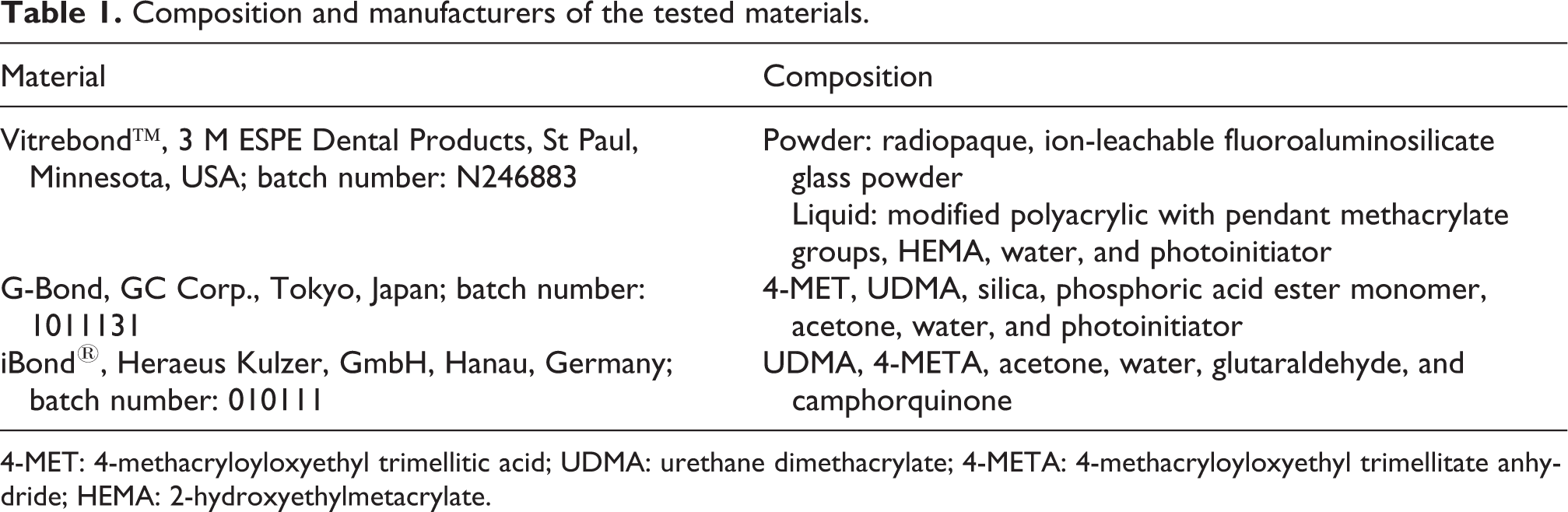
4-MET: 4-methacryloyloxyethyl trimellitic acid; UDMA: urethane dimethacrylate; 4-META: 4-methacryloyloxyethyl trimellitate anhydride; HEMA: 2-hydroxyethylmetacrylate.
A commercially available cell culture perfusion chamber (Minucells and Minutissue GmbH, Bad Abbach, Germany) made of polycarbonate with a base of 40 × 40 mm and a height of 36 mm was modified. The three-dimensional cultures were placed on Er:YAG laser-treated discs and normal dentin discs were held in place by a special biocompatible stainless-steel holder, resulting in a dentin barrier test situation. Thus, the cell culture chamber was separated into two compartments by the dentin disc. The cell culture tissues were placed in direct contact with the etched side of the dentin disc and held in place by the stainless-steel holder. All chambers were perfused with 0.3 ml assay medium (growth medium with 5.96 g/l HEPES (2-[4-(2-hydroxyethyl)piperazin-1-yl]ethanesulfonic acid) buffer, Merck, Germany) per hour for 24 h at 37°C. Perfusion was switched off; test materials were introduced into the upper compartment in direct contact with the “cavity” side of the dentin disc. Test materials were applied according to the manufacturers’ instructions. A nontoxic polyvinyl siloxane impression material (President, Coltene AG, Alstatten, Switzerland) was used as a negative control (100% cell viability). Cytotoxicity of test materials was recorded after the pulpal part of the in vitro pulp chamber was perfused with cell culture medium (2 ml/h) for 24 h of incubation at 37°C. Each material was tested six times. Cell viability of three-dimensional cultures was determined by enzyme activity. The tissues were removed from the pulp chambers, placed into 24-well plates containing 1 ml of pre-warmed MTT (3-(4,5-dimethylthiazol-2-yl)-2,5-diphenyltetrazolium bromide) solution (0.5 mg/ml in growth medium), and incubated for 2 h at 37°C. Then the cells were washed twice with PBS. The blue formazan precipitate was extracted from the mitochondria using 0.5 ml of dimethyl sulfoxide on a shaker at room temperature for 30 min. Of this solution, 200 µl was transferred to a 96-well plate, and the absorption at a wavelength of 540 nm (µQuant, Bio-Tek Instruments, Inc, Winooski, Vermont, USA) was determined spectrophotometrically. The mean values of control tissues (cell cultures exposed to the polyvinyl siloxane impression material) were set to represent 100% viability. Results of cytotoxicity experiments were expressed as a percentage of matching control tissue; differences between median values were statistically analyzed using the Kruskal–Wallis one-way analysis of variance and Mann–Whitney U test (α = 0.05; Statistical Package for the Social Sciences (SPSS) version 13.0; SPSS, Chicago, Illinois, USA).
SEM analysis
For scanning electron microscopy (SEM) analysis, 500 ± 20 µm dentin discs were cut from first incisors of freshly slaughtered bovines. To remove the smear layer, each dentin discs was submerged into a 17% ethylenediaminetetraacetic acid solution (pH = 7.8) for 5 min, rinsed with distilled water, immersed into a 5.25% sodium hypochlorite solution for 5 min, and then stored in distilled water until use. Dentine discs were irradiated by Er:YAG laser, as mentioned above. Untreated samples were used as control. Samples were gold sputtered and evaluated by SEM (Zeiss EVO Ls10, Jena, Germany).
Results
Cytotoxicity testing
The cytotoxicity evaluation of dentin contacting materials with dentin barrier test device using Er:YAG laser-treated dentin is summarized in Figure 1. A polyvinyl siloxane material (President) was used as a negative control material. Vitrebond™ was the most toxic material for both untreated dentin (24% cell survival) and Er:YAG laser-treated dentin (41% cell survival), when comparing with the cell cultures exposed to President (p < 0.05). G-Bond™ reduced the cell survival rate to 54% in the untreated dentin and to 75% in the Er:YAG laser-treated dentin and iBond® showed only a minor cell reduction (to 98% cell survival) in untreated dentin and 54% cell survival in the Er:YAG laser-treated dentin.

Cytotoxicity of dentin contacting materials on three-dimensional cultures of SV40 large T-antigen-transfected bovine pulp-derived cells using Er:YAG laser-treated dentin. Data are expressed as percentage of the negative-control cultures (n = 6). Er:YAG: erbium-doped yttrium, aluminum, and garnet.
G-bond was cytotoxic to the pulp-derived cells (p < 0.05), and iBond was similar to the negative control group (p > 0.05) on untreated dentin. However, G-Bond and iBond were not cytotoxic when they were applied to Er:YAG laser-treated dentin (p > 0.05).
SEM surface analysis
The most significant images obtained are shown in figures. The untreated areas in the control group presented a smooth appearance and opened tubule orifices. Moreover, no smear layer covering the dentin and no smear plugs closing the tubules were observed (Figure 2). SEM observation of the laser group showed a homogeneous area with less-exposed dentinal tubules, the diameter of which was also significantly decreased compared with the control group (Figures 3 and 4).

Control group showing open dentinal tubules before irradiation of the Er:YAG laser (magnification, ×1000). Er:YAG: erbium-doped yttrium, aluminum, and garnet.

Er:YAG laser-treated dentin, energy of 60 s/cm2 at 3 Hz, and 100 mJ (magnification, ×1000).
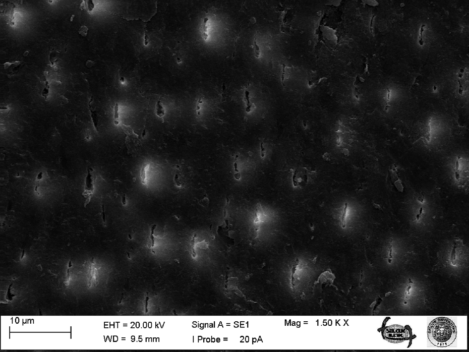
Er:YAG laser-treated dentin, energy of 60 s/cm2 at 3 Hz, and 100 mJ (magnification, ×1500).
Discussion
In this in vitro study, the use of Er:YAG laser on dentin discs occlude the open dentinal tubules that are believed to conduct cytotoxic extracts of dental materials, and laser treatment of dentin reduced the cytotoxic effects of dentin contacting materials to the three-dimensional bovine pulp-derived cells.
It was consistently demonstrated that dentin was an effective barrier, preventing cell damage from a great variety of materials and chemicals. Studies have shown that dentin can reduce the toxicity of resins and bonding adhesives by limiting diffusion of those substances from the cavity preparation to the pulp. 19 – 21 Dentin probably adsorbs substances in the tubules and further limits the traverse of substances. 7 Some adhesive components diffuse rapidly through dentin. 22,23 Thus, there are compelling reasons to question whether these adhesives can cause cytotoxicity by diffusion through the dentin. Bouillaguet et al. showed that the high permeability of dentin generally allowed more diffusion of the dental adhesives, but the effect of dentin depended on the material. 1 In the current study, Er:YAG laser-treated dentin showed more biocompatible results than those of normal dentin.
The in vitro system used in the current study mimics a clinical situation, and it is better than direct cell-material contact in vitro methods. 24,25 In the present study, we used bovine dentin discs for a dentin barrier test. The advantages of using bovine dentin are that large amounts are readily available, the dentin can be easily standardized with respect to permeability characteristics, and fewer variations in permeability characteristics are expected than in human dentin. One important prerequisite for using bovine dentin to replace human dentin is that bovine dentin must show permeability characteristics that are similar to human dentin. 25 Therefore, an immortalized bovine dental papilla-derived cell line may be a valuable tool for in vitro cytotoxicity testing, since no incompatibility between target cells and dentin due to different donor species needs to be anticipated. Furthermore, once complete characterization has been performed, the unlimited availability of bovine dental papilla-derived cells may be of value for the synthesis of larger amounts of tissue specific molecules. In addition, by maintaining specific characteristics, immortalized dental papilla-derived cells provide the possibility for the evaluation of tooth-specific cell metabolism and cell–cell interactions. 26
Laser irradiation is capable of occluding exposed dentin tubules. The occluding effect is proportional to the duration of irradiation while using the same energy and frequency settings. In this study, 60 s/cm2 of irradiation seemed sufficient for almost total occlusion of dentin tubules in the irradiated spot. With this duration, heat damage to the pulp is unlikely to occur with the energy settings used. 27,28 There are several different theories for explaining the effect of laser irradiation on dentin, but the most approved one suggests the sealing of dentinal tubules by the melting and recrystallization of dentin. 17
So far, no published data are available concerning the effectiveness of an Er:YAG laser in decreasing the cytotoxicity of dental materials, whereas the use of lasers has often been propagated for dentine hypersensitivity. 15,18 By sealing the tubule orifices below the ablation threshold, treatment of dentin hypersensitivity by Er:YAG laser revealed high efficacy in reducing the diameters of dentin tubules under some specific conditions. 29,30 As the SEM analyses in our investigations have shown, open dentinal tubules can be occluded using Er:YAG laser irradiation (Figures 3 and 4).
Vitrebond, which is frequently used as positive control in dentin barrier tests, was found to be the most toxic in this study, but higher cell survival was seen when it was applied on laser-treated dentin. This difference was statistically significant. 20,21,24 The positive control should reduce cell viability by approximately 50% after 24 h of exposure; the negative control should have no effect on cell viability. The dentin barrier tests revealed no toxicity of President, whereas Vitrebond was approximately 50% cytotoxic. 20 The dentin bonding agent G-bond, the other cytotoxic material for untreated dentin, was not cytotoxic on laser-treated dentin. This is consistent with other authors. 21 The iBond, showed no toxicity in either laser-treated and untreated dentin. However, in a previous study, this material was cytotoxic in a dentin barrier test device. 20
Conclusion
Er:YAG laser treatment of dentin may protect the pulp cells from the toxic substances of dentin contacting restorative material; however, this effect is material related. Taking into consideration the limitations of this in vitro study, the Er:YAG laser treatment of dentin before restoration might be an option for decreasing the cytotoxic effects of the dental materials. Further research is required for clinical application.
Footnotes
Funding
This research received no specific grant from any funding agency in the public, commercial, or not-for-profit sectors.