
Abstract
Thyroid hormones are recognized as the key metabolic hormones that play a critical role in the development of central nervous system (CNS) throughout life. The present study was designed to determine the changes in brain monoamine concentrations in 6-n-propyl thiouracil (PTU)-induced hypothyroid rats, in addition to the ameliorating role of folic acid treatment. Fifty male albino rats were equally divided into five groups; first and second groups were the control and folic acid groups, respectively, while the third group was the hypothyroid group in which the rats received PTU in drinking water for 6 weeks. The fourth and fifth groups were co- and post-treated folic acid groups with hypothyroid rats, respectively. Our results revealed that serotonin and norepinephrine concentrations were significantly decreased in the hypothalamus and cortex, while it significantly increased in the hippocampus of hypothyroid rats when compared with control group. Serotonin and norepinephrine concentrations were decreased in hypothalamus and cortex in co- and post-treated folic acid groups with hypothyroid rats, while the concentration of dopamine were significantly increased in the hypothalamus and hippocampus of the hypothyroid rats and co-treated folic acid group with hypothyroid rats. In cortex, the dopamine concentration was significantly increased in hypothyroid rats and post-treated folic acid group with hypothyroid rats, while it significantly decreased in co-treated folic acid group with hypothyroid rats when compared with the control group. Also, our results revealed that, folic acid treatment was better if it is administered as an adjuvant after returning to the euthyroid state by withdrawing PTU from the drinking water.
Introduction
Thyroid hormones have marked effects on the growth, development and metabolic function of virtually all organs and tissues.1,2 Thyroid hormones are recognized as key metabolic hormones that play a critical role in development of brain, mediating important effects within the central nervous system throughout the life,2–5 regulating the metabolism and function of various neurotransmitters, 6 their receptors, second messengers 7 and gene expression. 8 Normal brain development requires the presence of thyroid hormones that are essential for cell migration, dendrite and axon outgrowth, synapse formation, myelination and gliogenesis. 9 It is well established that thyroid hormones are essential for both the development and maturation of the human brain, affecting such diverse events as neuronal processing and integration, glial cell proliferation, myelination and the synthesis of key enzymes required for neurotransmitter synthesis. 10
Hypothyroidism refers to any state in which thyroid hormone production is below normal. Neurologically, hypothyroidism has been associated with cerebellar ataxia, confusion, delusions, memory impairment, hallucinations and psychotic behaviour.2,4,11 It has been shown that absence of thyroid hormone causes diminished axonal growth and dendritic arborization in the cerebral cortex, visual and auditory cortex, hippocampus, and cerebellum. 2 Galton et al. 12 reported that the absence of thyroid hormone in the cerebellum delays proliferation and migration of granule cells from the external to the internal granular layer. Interaction of the thyroid and monoamine neurotransmitter (norepinephrine, epinephrine, serotonin and dopamine) systems has been suggested as a potential underlying mechanism of action. Thyroid hormones are known to modulate a number of neurotransmitter systems.3,5 Thyroid hormones undoubtedly play an important or even leading role in the mechanisms of appearance and development of multiple behavioral impairments, including a variety of depressive, bipolar, panic and anxiety disorders, as well as schizophrenia. 13 Close relationship between thyroid hormones and monoaminergic systems of the brain is well known. 5 The monoamines, specifically norepinephrine and serotonin, play a major role in mood modulation. 3 Ito et al. 14 emphasized that the accumulation rates of serotonin and dopamine in hyperthyroidism increased in pons-medulla and mesodiencephalon, respectively, while in hypothyroidism, these monoamines decreased in cerebral hemispheres and mesodiencephalon of rats.
Hypothyroidism has been reported to cause mild hyperhomocysteinaemia. 15 The mechanism by which hypothyroidism causes mild hyperhomocysteinaemia is not clear but altered remethylation is likely to be involved. Folic acid exerts an important role on homocysteine catabolism, by the donation of methyl group to methionine in remethylation pathway. 16 Moreover, this vitamin also has antioxidant properties. 17 In addition, it has been reported that folic acid has an important role in DNA stability maintenance, preventing uracil misincorporation into DNA, single- and double-strand DNA breaks and micronucleus formation. 18
As most of the previous publications deal with the effect of thyroid hormones on the structural development of the brain and neurons, the present study aims to determine the changes in brain monoamine concentrations in 6-n-propyl thiouracil (PTU)-induced hypothyroid rats and the ameliorating role of folic acid in treatment.
Materials and methods
The experiments were performed on 50 male albino rats (Rattus norvigicus) weighing 120 g (±10 g) and aged 6–7 weeks. They were obtained from our laboratory farms for the Egyptian Organization for Vaccine and Biologic Preparations. The rats were kept in the laboratory for 1 week before the experimental work and maintained on a standard diet and water available ad libitum. The temperature in the animal room was maintained at 23 ± 2°C with a relative humidity of 55 ± 5%. Light was on a 12:12 h light–dark cycle. The experimental protocol was approved by Local Ethics Committee and Animals Research. The rats were randomly and equally divided into five groups (10 animals each). Group 1 (G1): Control group in which animals never received any treatment. Group 2 (G2): Folic acid group in which rats received folic acid orally (El Nasr Pharmaceutical Chemicals Co.; 0.011 μmol/g of body weight)
19
for 4 weeks (from 2nd week to 6th week). Group 3 (G3): Hypothyroid group in which the rats received 0.05% PTU in drinking water for 6 weeks.
20
Group 4 (G4): Co-treatment group in which the animals received 0.05% PTU in drinking water and oral folic acid simultaneously according to Matte et al.
19
The dose period of PTU was 6 weeks as in the hypothyroid group. However, folic acid was administered orally for 4 weeks (from 2nd week to 6th week) after evidence of hypothyroidism had been established at the end of the 2nd week. Group 5 (G5): Post-treatment group in which the animals received 0.05% PTU in drinking water for 6 weeks as in the hypothyroid group. Additionally, folic acid was administered for another 4 weeks (from 7th week to 10th week) while PTU was withdrawn after the 6th week to return to the euthyroid state.
At the end of the experimental period, five rats from each group were euthanized with intravenous sodium pentobarbital injection and subjected to a complete necropsy. Blood samples were individually collected from the inferior vena cava of each rat in non-heparinized glass tubes to estimate blood parameters. Serum was separated by centrifugation at 3000 rpm for 15 min. The collected serum was stored at −18°C until analysis. Blood serum was analyzed to determine the triiodothyronine (T3) and thyrotropin (TSH) levels. Serum T3 was assayed using commercial test supplied by the Diagnostic Systems Laboratories (DSL), TX, USA. Serum TSH was assayed using the commercial kit supplied by Coat-A-Count TSH IRMA, Los Angeles, USA.
Estimation of the brain monoamines concentrations by high-performance liquid chromatography (HPLC) method
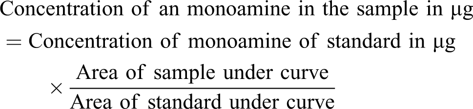
Five animals from each group were sacrificed by decapitation and the brain regions (hypothalamus, cortex and hippocampus) were rapidly removed and used for determination of monoamine by HPLC method. The tissues were weighed and homogenized in 1/10 weight/volume of 75% aqueous HPLC grade methanol. The homogenate was spun at 3000 rpm for 10 min and the supernatant was immediately extracted from the trace elements and lipids using the solid phase extraction CHROMABOND column NH2 phase Cat. No.730031. The sample was then injected directly to the AQUA column (150 mm × 4.6 mm, 5 μm and C18) which was obtained from phenomenex USA under the following conditions: mobile phase 97/3 20 mM potassium phosphate, pH 3.0/methanol, and flow rate 1.5 ml/min, UV 270 nm. Additionally, norepinephrine (NE), dopamine (DOP) and serotonin (5-HT) were separated after 12 min. The resulting chromatogram identifying each monoamine position and area under curve for each sample was compared to that of the standard curve made by Eurochrom HPLC Software, version 1.6. The content of each monoamine was measured as microgram per gram brain tissue3,21 using the following equation:
Statistical analysis
Data (n = 5) were expressed as mean values ± SE, and statistical analysis was performed using one-way analysis of variance (ANOVA) to assess significant differences among treatment groups. The criterion for statistical significance was set at p < 0.05. All statistical analyses were performed using SPSS statistical version 16 software package (SPSS® Inc., USA).
Results
In order to ensure the hypothyroid state, we regularly determined the serum T3 and TSH through the dose period. Table 1 shows that the serum T3 levels were significantly lower in hypothyroid rats (57.7 ± 6.89 ng/dl) compared with the control (147 ± 22.37 ng/dl), folic acid (148 ± 23.52 ng/dl) and post-treated folic acid (142.7 ± 22.11 ng/dl) groups and significantly higher than those found in the co-treated folic acid group (40 ± 6.89 ng/dl). On the other hand, serum TSH levels in hypothyroid rats (3.900 ± 0.513 μIU/ml) were significantly higher than those found in the control (0.014 ± 0.003 μIU/ml), folic acid (0.152 ± 0.124 µIU/ml) and post-treated folic acid (0.060 ± 0.026 μIU/ml) groups and significantly lower than those found in the co-treated folic acid group (5.367 ± 0.462 μIU/ml).
Serum T3 and TSH levels in different groups

G1: control group, G2: folic acid group, G3: hypothyroid rats group, G4: co-treatment group, G5: post treatment group. a–c Means in the same row with different superscripts are significantly different (p ≤ 0.05).
Concerning 5-HT concentration, HPLC analysis of 5-HT (Table 2 ; Figure 1 ) were significantly decreased in the hypothalamus, cortex and hippocampus (32.6%, 43.3% and 31.4%, respectively) of hypothyroid rats receiving PTU in drinking water for 6 weeks when compared with control group. Folic acid group showed that 5-HT significantly decreased in hypothalamus and cortex (20.5% and 24.9%, respectively) and significantly increased in hippocampus (37.7%), while in co-treated folic acid group of hypothyroid rats, the concentration of 5-HT was significantly decreased in the hypothalamus and cortex (18.4% and 29.6%, respectively) and significantly increased in hippocampus (10.95%) in comparison with the control group. Post-treated folic acid group of hypothyroid rats, as in G5, the concentration of 5-HT was significantly decreased in the hypothalamus, cortex and hippocampus (26.6%, 36.2% and 9.8%, respectively).
The percentage and concentrations of monoamine (μg/g tissues) in different brain regions of experimental groups compared to control group

G1: control group, G2: folic acid group, G3: hypothyroid rats group, G4: co-treatment group, G5: post treatment group, ↑: increased, ↓: decreased, NE: norepinephrine, DOP: dopamine, 5-HT: serotonin.
a–d Means in the same raw with different superscript are significantly different (p ≤ 0.05).

Serotonin (5-HT) concentration changes on rat hypothalamus, cortex and hippocampus in different groups.
As regard the results of NE, Table 2 and Figure 2 showed the HPLC analysis of NE that significantly decreased in the hypothalamus, cortex and hippocampus (30.3%, 22.5% and 29.3%, respectively) of hypothyroid rats when compared with the control group. Folic acid group showed a significantly decreased NE in hypothalamus (12.5%) and significantly increased NE in cortex and hippocampus (7.4% and 11.8%, respectively). In co-treated folic acid group of hypothyroid rats, the concentration of NE was significantly decreased in the hypothalamus and cortex (8.9% and 0.63%, respectively) and significantly increased in hippocampus (14.1%), while in the post-treated folic acid group of hypothyroid rats, the concentration of NE was significantly decreased in the hypothalamus and cortex (11.9% and 18.9%, respectively) and significantly increased in hippocampus (2.7%) in comparison with the control group.

Norepinephrine (NE) concentration changes on rat hypothalamus, cortex and hippocampus in different groups.
As regard the HPLC analysis results of DOP, Table 2 and Figure 3 show a significant increase in the hypothalamus, cortex and hippocampus (54.8%, 40.1% and 30.5%, respectively) of hypothyroid rats when compared with the control group. Folic acid group showed a significantly decreased DOP in cortex and hippocampus (18.3% and 6.22%, respectively) and significantly increased in hypothalamus (14.3%) in comparison with control group. In co-treated folic acid group of hypothyroid rats, the concentration of DOP was significantly increased in the hypothalamus and hippocampus (14.5% and 1.28%, respectively) and significantly decreased in cortex (29.3%), while in post-treated folic acid group of hypothyroid rats, the concentration of DOP was significantly decreased in the hypothalamus and hippocampus (13.7% and 17%, respectively) and significantly increased in cortex (39.1%) in comparison with control group.

Dopamine (DOP) concentration changes on rat hypothalamus, cortex and hippocampus in different groups.
Discussion
The present study was designed to investigate the role of thyroid hormone on the changes in monoamine rat brains. In order to achieve this target, we made a deficient state of thyroid hormones by a reversible goitrogen (PTUl). PTU is known to decrease the conversion of peripheral thyroxine (T4) to T3 and thereby reduces serum T3 concentration, and it was administered in the drinking water of the rats during a period of 6 weeks. 22 In order to ensure the hypothyroid state, we regularly determined the serum T3 and TSH through the dose period, wherein serum T3 concentration is depressed and serum TSH concentration is significantly elevated. This result coincides with several studies. Both young and aged rats received the antithyroid drug as PTU-induced hypothyroidism.19,22
Thyroid hormones are known to modulate a number of neurotransmitter systems. 3 Serotonin is a neurotransmitter that is produced by the brain to help regulate mood along with dopamine, another mood-elevating brain chemical. 6 They are responsible for keeping the individuals in a positive frame of mind and out of depression. The present results showed that hypothyroidism have marked adverse effect on 5-HT, NE, and DOP in different regions of the brain (hypothalamus, hippocampus and cortex). 5-HT and NE concentrations were found to be significantly decreased in the cortex, the hypothalamus and hippocampus, while the DOP concentration was found to be significantly increased in the hippocampus when compared with the control group. In co-treated folic acid group of hypothyroid rats, the concentrations of 5-HT and NE were significantly decreased in the hypothalamus and cortex but increased in hippocampus, while it significantly decreased in the hypothalamus, cortex and hippocampus in the post-treatment when compared with the control group; also in cortex, the DOP concentration was significantly increased in post-treated folic acid group of hypothyroid rats, while it significantly decreased in co-treated folic acid group of hypothyroid rats. Thyroid hormones regulate the development of monoaminergic neurotransmission system.3,5 These results are in concurrence with many other publications such as Ito et al. 14 who speculated that the accumulation rates of 5-HT and DOP decreased in cerebral hemispheres and mesodiencephalon of hypothyroid rat. Thus, the present study supports the suggestion that the development of dopaminergic system is delayed in hypothyroid rat. 23 It was also reported that the synthesis, turnover rate and steady levels of 5-HT are reportedly depressed in the brains of offspring and adult hypothyroid rats. 14 Application of thyroid hormone to euthyroid rodents increased cortical or whole brain concentrations of 5-HT, 5-Hydroxytryptophan (5-HTP) and 5-Hydroxyindoleacetic acid (5-HIAA).6,24 Serotonin stimulates hypothalamic thyrotropin-releasing hormone (TRH) production, leading to an increase in TSH production from the pituitary. Adequate serotonin production is necessary to maintain thyroid hormone levels. These results indicating increased cortical 5-HT turnover were consistent, despite changing technologies over a 25-year period, and may be considered robust. In only one study the whole brain 5-HT level did not increase after thyroid hormone administration. 25 Also, Sandrini et al. 6 and Portela-Gomes et al. 26 reported that thyroidectomy appears to reduce the serotonin content in the rat brain; whereas in the present study, the hyperthyroidism shows the opposite effect. Schatzberg and Schildkraut 27 studied norepinephrine synthesis in hypothyroid rats that appeared to be markedly hypoadrenergic. These investigators found that the rats showed a marked increase in the synthesis of norepinephrine in the heart, with a similar increase in the synthesis of norepinephrine in the brain. Despite the increased synthesis of norepinephrine, these animals were markedly hypoadrenergic. Coulombe et al. 28 indicate that the plasma NE secretion rate is normal in hyperthyroidism and is significantly elevated in hypothyroidism, thereby explaining the higher plasma NE concentrations seen in hypothyroidism. Dopamine is a biogenic amine synthesized in the hypothalamus, in the arcuate nucleus and various areas of the central and peripheral nervous system. It has been widely established that dopamine and its agonists play an important role in cardiovascular, renal, hormonal and central nervous system regulation through stimulation of alpha and beta adrenergic and dopaminergic receptors. 29 Crocker 30 reported that, hypothyroidism increases dopamine receptor sensitivity by increasing receptor concentration. Multiple studies that were addressed in revealing hormonal effects on the content and metabolism of catecholamines in brain structures have demonstrated that hormones change the activities of neurotransmitter systems in specific structures of the CNS.3,31 These changes are probably related to hormonal influences on the induction and activities of enzymes involved in biosynthesis and metabolism of transmitters. Hormones also affect neurotransmitter storage and the permeability of the blood–brain barrier, plasma membranes and membranes of intracellular organelles to transmitters as well as their precursors and metabolites. 31 Our results agreed with Fedotova and Masalova 32 reported that the concentration of NE, 5-HT and DOP were decreased in the hypothalamus and increased in the hippocampus in young thyroidectomized rat and that the changes in monoamine metabolism under conditions of thyroidectomy deficiency were more expressed in the brain and age dependent. In conclusion, this study indicates that prepubertal hypothyroidism in male rats is associated with monoamines concentration, where the present results showed that hypothyroidism have marked adverse effect on 5-HT, NE and DOP in different brain regions (hypothalamus, hippocampus and cortex).
Folic acid is essential for numerous body functions ranging from nucleotide biosynthesis to remethylation of homocysteine. In humans, body needs folate to synthesize DNA, repair DNA and methylate DNA as well as to act as a cofactor in the biological reactions involving folate; moreover, this vitamin also has antioxidant properties. 17 Matte et al. 19 showed that homocysteine induces oxidative stress in the brain of rats, reducing antioxidant defences and increasing lipid peroxidation. Our results show that folic acid can be used in treatment and/or as supplementation of hypothyroidism, where the concentrations of 5-HT and NE were significantly decreased, while the concentration of DOP was significantly increased in the hypothalamus and cortex in co- and post-treated folic acid groups of hypothyroid rats. Also, the concentration of 5-HT and NE were significantly increased in the hippocampus in the co-treated folic acid group of hypothyroid rats in comparison with the control group. Cousens 33 reported that folic acid can heighten serotonin function by slowing the destruction of brain tryptophan, and that it also acts as a cofactor for enzymes that convert tryptophan into serotonin, and for enzymes that convert tyrosine into norepinephrine/noradrenalin. Bottiglieri et al. 34 reported that folic acid is involved in the synthesis of the monoamine neurotransmitters and in serotonin, dopamine and noradrenergic systems. Zhu et al. 35 reported that the folic acid can be used in the treatment of Alzheimer’s disease, depression, anemia and certain types of cancer. Folic acid, with its antioxidant activity, is a little studied compound in the terms of human intervention trials. This water-soluble vitamin has gathered abundant attention because of its role in the pathogenesis of cardiovascular disease, neural tube defects and cancer prevention.35,36 Low levels of folate can also lead to homocysteine accumulation as a result of the impairment of one-carbon metabolism mechanism and methylation. Plasma homocysteine concentrations increase with age and remain an independent risk factor for vascular disease in the elderly. The marginal folate and other vitamin deficiencies known to be common in the elderly are likely to be the contributing factors to hyperhomocysteinemia. 37 Hypothyroidism has been reported to cause mild hyperhomocysteinaemia. 15 It was recently shown that homocysteine in vitro induced oxidative stress in cortex and hippocampus. 38 This study revealed that folic acid treatment was effective only if it was administered as an adjuvant after returning to the euthyroid state by withdrawing PTU from the drinking water.
Footnotes
This research received no specific grant from any funding agency in the public, commercial, or not-for-profit sectors.