
Abstract
Controversial studies from others suggested that alcohol intake could be associated with some deleterious effects in the uterus. Not all the effects of alcohol drinking on female reproductive organs can be explained in terms of endocrine disturbances. Deleterious effect of alcohol or its metabolites in situ could also play a role. Accordingly, we found a metabolism of alcohol to acetaldehyde in the rat uterine horn tissue cytosolic fraction mediated by xanthine oxidoreductase, requiring a purine cosubstrate and inhibited by allopurinol. This activity was detected by histochemistry in the epithelium and aldehyde dehydrogenase activity was detected in the muscular layer and in the serosa. There was a microsomal process, not requiring NADPH and of enzymatic nature, oxygen-dependent and inhibited by diethyldithiocarbamate, diphenyleneiodonium and partially sensitive to esculetin and nordihydroguaiaretic acid. The presence of metabolic pathways in the uterine horn able to generate acetaldehyde, accompanied by a low capacity to destroy it through aldehyde dehydrogenase, led to acetaldehyde accumulation in the uterus during ethanol exposure. Results suggest that any acetaldehyde produced in situ or arriving to the uterine horn via blood would remain in this organ sufficiently to have the opportunity to react with critical molecules to cause deleterious effects.
Introduction
Epidemiological studies made in different countries evidenced that alcohol misuse among women is an important and growing problem. As expected, increased incidence of alcohol use in health disorders were observed particularly in younger versus older cohorts of female drinkers. Consequently, the number and impact of older female drinkers is expected to increase in the next decades.1–6 It is important in this respect to consider that due to differences in metabolism of alcohol, women at old ages compared to men are at higher risk for negative consequences associated with at-risk and higher levels of alcohol consumption. 7
Further, the harmful consequences of alcohol misuse are particularly severe for women. For example, several studies have found higher risk of cervical, vulvar and vaginal cancers among alcoholics than in the general population. However, these findings have not been confirmed in population-based studies, in which confounding factors have been adjusted for. Endometrial, corpus uteri and ovarian cancers do not seem to be related to alcohol consumption. 8 Despite that, studies evaluating the role of alcohol intake in endometrial cancer risk have generally not been supportive of a relationship. However, an effect cannot be ruled out. 9 In contrast to these controversial findings concerning the effects of alcohol drinking on cancer incidence on women, described above, there is today a clear-cut evidence that even low to moderate amounts of alcohol increases the risk of breast cancer in women.10,11
Alcohol markedly disrupts normal estrous cycling in female humans and rats. Alcoholic women are known to have a variety of menstrual and reproductive disorders, from irregular menstrual cycles to complete cessation of menses, absence of ovulation and infertility and anticipation of menopause.12,13
Other harmful consequences of alcohol misuse for women were also reported. For example, Grodstein et al. investigated the cause of infertility in just 3,800 women and reported that moderate alcohol intake was associated with a significant, but small increased risk of ovulatory infertility and increased risk of endometriosis. 14
Further, Jensen et al. concluded that the chance of successful conception decreased with increasing alcohol intake. 15 Other prospective studies reached similar conclusions.16,17 A harmful effect on ovarian functions is one of several reasons for infertility.18,19 Alcohol may also affect implantation of early development of the blastocyst. 20
In addition, other data were provided suggesting that significant alcohol intake is associated with higher prevalence of uterine fibroids.21,22
Interestingly, Zaitseva et al. demonstrated the occurrence of a differential expression of aldehyde dehydrogenase 1 (AldDh1) between fibroids and myometrium in cell culture studies where endometrial cells were not simultaneously present. 23
All the above-described alcohol drinking-provoked reproductive disorders no doubt involve significant alterations of critical hormonal factors.12,13 Those alterations include an association with higher levels of circulating estrogens in pre and postmenopausal women, and with reduced progesterone levels in premenopausal women. 9
Notwithstanding, not all the effects of alcohol drinking on reproductive organs and associated tissues can fully be explained in terms of those endocrine disturbances. However, the possibility that direct effects of alcohol and of its products of biotransformation were also involved in the observed alterations on target reproductive organs deserves to be considered. That appeared to be the case for the toxic effects of alcohol intake in mammary tissue, prostate and ovarian function.19,24–29
Previous studies from Messiha showed that the female rat genital system has alcohol dehydrogenase and aldehyde dehydrogenase activities.30,31 In the present study, we attempt to verify whether the horns of rat uterus have own ability to metabolize alcohol to acetaldehyde and to detoxify the latter to less toxic metabolites. The attempt includes establishing whether any acetaldehyde produced tends to accumulate during sufficient time in uterine tissue to have reasonable possibilities to interact with critical molecules and lead to deleterious effects.
Materials and methods
Chemicals
Ethanol (analytical grade) was from Sintorgan (Villa Martelli, Argentina). Acetaldehyde was from Fluka (Buchs, Switzerland). Hypoxanthine, allopurinol, NAD+, NADP+ were from Sigma Chemical Co. (St. Louis, Missouri, USA). All other chemicals were of the best quality available.
Animals and Treatments
Non-inbred female Sprague-Dawley rats were used. The procedures used for breeding, housing and handling animals were those established by the Food, Drug and Medical Technology National Administration (ANMAT; Buenos Aires, Argentina). The starting breeding colony was from Charles River (Wilmington, Massachusetts, USA). For the studies on the metabolism in microsomal and cytosolic fractions, Sprague-Dawley female rats (220–250 g body weight) were used. Food was withdrawn 12−14 hours before sacrifice, but the animals had free access to water. For the ethanol single-dose administration, ethanol was administered p.o. in water (5 mL 200 g−1 b.w.) at a dose of 3.8 g kg−1. In the control groups, the calories provided by the alcohol were replaced by sucrose. After each time, the animals were killed for sample processing. 27
Isolation of uterine horn tissue cytosolic, microsomal and mitochondrial fractions
Animals were killed by decapitation and their uterine horns were rapidly excised, separated from ovary and oviduct and processed to obtain cytosolic and microsomal fractions. Cytosolic and microsomal fractions were obtained from whole uterine horn tissue homogenates by cellular fractionation procedures via ultracentrifugation at 4°C. 26 Mitochondrial fractions employed to determine AldDh activity were prepared according to Koivula and Koivusalo. 32
Determination of alcohol dehydrogenase and aldehyde dehydrogenase activities in rat uterine horn and liver tissue
Alcohol dehydrogenase (ADh) was measured in the cytosolic fraction of uterine horn tissue by the detection of the NADH formed, at 340 nm. Under an excess of alcohol, the rate of NADH formation is proportional to enzyme concentration. 33 AldDh activity in uterine horn tissue was measured by the method described by Koivula and Koivusalo, with minor modifications. 32 Cytosolic, microsomal or mitochondrial fractions were resuspended in pyrophosphate buffer pH 8, 1.67 mM pyrazole was added and the mixture (3 mL) was incubated at 37°C for 30 min. Immediately after adding 6 mM propanal and 0.67 mM NAD+, absorbance at 340 nm was measured at 15 seconds intervals in quartz cuvettes termostatized at 37°C. Values obtained for uterus tissue were compared to those in the liver of the same animals (five animals per group).
Histochemical procedure for alcohol dehydrogenase and aldehyde dehydrogenase activities detection in uterine horn tissue
Animals were killed by decapitation or by cervical dislocation and 5 mm cubes of tissues were taken and immediately frozen on metal chucks maintained at −70°C in freezing mixture of solid carbon dioxide and hexane. Cryostat sections, 16 µm thick, were cut at −20°C and mounted on clean glass microscope slides. The sections were allowed to equilibrate to room temperature for 30 min and then placed in the incubation medium. Prior to staining, sections were dried at room temperature for 30 min, then incubated for 1 hour at 42°C in the dark in a staining solution containing 50 mM sodium phosphate, pH 7.6; 5 mM NAD+, 11 mM pyruvic acid; 3.4 mM nitroblue tetrazolium and 500 mM ethanol as a substrate. After staining, the slides were washed with phosphate-buffered saline, fixed with 4% paraformaldehyde neutral solutions for 10 min, dehydrated in ethanol: water, cleared in xylene and moistened in permount. In the case of the histochemistry for acetaldehyde dehydrogenase, same steps were performed, except that ethanol was replaced by 500 mM acetaldehyde. In both cases, liver tissue was processed simultaneously as a control. 34
Histochemical procedure for xanthine oxidase activity detection in uterine horn tissue
Portions of uterine horn tissue from control rats (five animals) were frozen at −70°C in hexane in a mixture of solid carbon dioxide and absolute alcohol. Tissue blocks were stored at −80°C until further use. Sections 8 μm thick were cut on a cryostat at −24°C. The sections were picked up onto clean glass slides and incubated immediately for xanthine oxidase activity using the cerium capture method in the presence of polyvinyl alcohol described by Frederiks et al. 35 Briefly, the incubation mixture contained 100 mM Tris–maleate buffer, pH 8; 10 mM cerium chloride; 100 mM sodium azide; 0.5 mM hypoxanthine and 10% polyvinyl alcohol. Incubations lasted 60 min at 37°C. After that, sections were washed in hot distilled water (60°C). Visualization was performed by incubating sections for 30 min at room temperature in 50 mM acetate buffer, pH 5.3; 42 mM cobalt chloride; 100 mM sodium azide; 1.4 mM diaminobenzidine and 0.6 mM H2O2. After rinsing the sections were embedded in glycerol jelly. Liver tissue was processed simultaneously as a control.
Ethanol metabolism to acetaldehyde in the microsomal fraction
Preparations containing microsomes (0.53 ± 0.07 mg protein/mL), NADPH generating system (0.45 mM NADP+, 4 mM d,l-isocitric acid trisodium salt and 0.25 units of isocitric dehydrogenase) and 0.14 M ethanol in 50 mM KH2PO4, pH 7.4, 3 mL final volume, were incubated for 1 hour at 37°C under air. Three samples per group were run, each consisting of microsomes from a separate lot of pooled uterus tissue (twelve animals each). Incubations were performed in aluminum-sealed neoprene-septum-stoppered glass vials. The reaction was terminated by plunging in ice. After adding 1 mL of saturated NaCl solution, samples were kept at 37°C for 15 min and an aliquot (100 μL) of the headspace was analyzed by GC-FID. Chromatographic conditions were column, GS-Q PLOT, 25 m × 0.53 mm i.d. (J&W Scientific, California, USA); temperature 110°C isothermal, injection port temperature: 150°C, FID: 200°C. 25
Ethanol metabolism to acetaldehyde in the cytosolic fraction
Incubation mixtures containing cytosol (3.67 ± 0.63 mg protein/mL) in STKM buffer (0.25 M sucrose/50 mM Tris-HCl, pH 7.5/2.5 mM KCl /5 mM MgCl2); 0.25 mM hypoxanthine; 0.3 mM NAD+ and 0.14 M ethanol (3 mL final volume) were conducted for 1 hour at 37°C under air atmosphere. Three samples per group were run, each consisting of cytosol prepared from a separate lot of pooled uterus tissue (twelve animals each). Incubations were performed in aluminum-sealed neoprene-septum stoppered glass vials (15 mL). Samples were processed as described above. Acetaldehyde was quantified in the head space by GC-FID in the same conditions as above. 24
Determination of ethanol and acetaldehyde levels in rat uterine horn tissue after single dose ethanol administration
Animals were killed by decapitation and liver and uterus tissue were rapidly taken and frozen. Blood samples were obtained by bleeding and the plasma separated by centrifugation. In the case of tissues, frozen samples from liver (0.5 g) and uterine horn tissue (0.30 g) were rapidly chipped and placed in aluminum-sealed-neoprene-septum stoppered glass vials. For blood samples, 0.5 mL plasma was used in the case of alcohol and 1 mL for acetaldehyde determinations. Ethanol and acetaldehyde levels were measured by GC-FID. In the case of ethanol, after adding 1.5 mL phosphate buffer and 2 mL potassium carbonate solution, samples were kept at 37°C for half hour with shaking and then an aliquot (100 µL) of the headspace was analyzed by GC-FID, as described above. In the case of acetaldehyde determination, salting-out was performed by adding 1 mL phosphate buffer, 1 mL of saturated ZnSO4 solution and 1 mL saturated NaCl solution, then samples were processed as performed for the ethanol quantification.27,36
Protein concentrations
Protein concentrations were determined by the method of Lowry et al., using bovine serum albumin as standard. 37
Statistics
The significance of the difference between mean values was assessed by unpaired t-test (Student’s t-test). 38 Calculations were performed using GraphPad Software. Differences were considered significant when p < 0.05.
Results
Alcohol dehydrogenase and aldehyde dehydrogenase activities in rat uterine horn tissue
Alcohol dehydrogenase and aldehyde dehydrogenase activities in rat uterine horn tissue were significantly lower than those found in the liver of the same animals (see Table 1). In the case of AldDh, activity only was present in the mitochondrial fraction. By our histochemical studies, alcohol dehydrogenase activity was not detected in the rat uterine horn tissue. Instead, acetaldehyde dehydrogenase was revealed in the muscular and serous parts of the uterine horn (Figure 1).
Alcohol dehydrogenase and aldehyde dehydrogenase activities in rat uterine horn and liver tissue
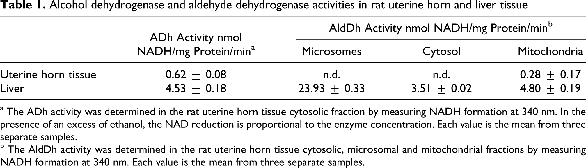
a The ADh activity was determined in the rat uterine horn tissue cytosolic fraction by measuring NADH formation at 340 nm. In the presence of an excess of ethanol, the NAD reduction is proportional to the enzyme concentration. Each value is the mean from three separate samples.
b

Rat uterine horn treated with phosphate-NAD-pyruvate-nitroblue tetrazolium. Acetaldehyde dehydrogenase activity is observed in the strome surrounding these structures (violet-blue staining), ×160.
Xanthine oxidase activity in rat uterine horn tissue
Histochemical studies showed that xanthine oxidase activity was present in high amounts in the rat uterine horn tissue epithelial cells (Figure 2).

Section of rat uterine horn tissue showing positive staining for the presence of xanthine oxidase activity. ×40.
Biotransformation of ethanol to acetaldehyde in the microsomal fraction
Results depicted in Table 2 show that the uterine horn microsomal fraction was able to biotransform ethanol into acetaldehyde in the presence (+NADPH) or absence of NADPH (−NADPH). There was no significant difference in the amount of acetaldehyde formed in the absence of NADPH or when the cofactor was present. Both processes were found to be partially enzymatic in nature, in light that about 85% of the +NADPH and about 44% of the –NADPH activities were suppressed by heating microsomes for 5 min at 100°C. Oxygen from air was required for a part of the +NADPH requiring process (87% of it) and for 75% of that –NADPH (Table 2).
Ethanol biotransformation to acetaldehyde in the microsomal fraction of rat uterine horn a

DPI: diphenyleneiodonium chloride, MMI: methylmercaptoimidazole, TBA: thiobenzamide.
a Incubation mixtures containing microsomal preparations (0.53 ± 0.07 mg of microsomal protein/mL), NADPH generating system (0.45 mM NADP+, 4 mM d,l-isocitric acid trisodium salt and 0.25 units of isocitric dehydrogenase) and 0.14 M ethanol were conducted for 1 hour at 37°C. Acetaldehyde was measured in the head space of each sample after adding 1 mL NaCl saturated solution. See Methods for details. Each result is the mean of ten separated lots of pooled uterine horn tissue samples for control group and three for the other experiments.
b p < 0.05 compared to −NADPH.
c p < 0.05 compared to control + NADPH.
d p < 0.05 compared to linoleic acid.
A known inhibitor of CYP2E1-catalyzed oxidations, like chlormethiazole, was not able to significantly deplete acetaldehyde production in microsomes. Desferrioxamine was only able to inhibit the oxidation to acetaldehyde in the absence of NADPH (63%). Sodium diethyldithiocarbamate inhibited the process significantly only in the absence of the cofactor (51%). Diphenyleneiodonium chloride (DPI), a potent inhibitor of flavoproteins, inhibited most of the ability of uterine horn microsomes to oxidize ethanol into acetaldehyde. In the absence of NADPH, DPI blocked 89% the biotransformation process under air and 80% in its presence.
Strong inhibitory effects of thiobenzamide (TBA) and methylmercaptoimidazole (MMI) were also observed for the +NADPH pathway (76% and 59% inhibition, respectively) and for the –NADPH-dependent metabolism of ethanol to acetaldehyde (82% and 87% inhibition; Table 2).
Aminotriazole and sodium azide were also able to significantly inhibit the oxidation of ethanol; in the absence of NADPH the production of acetaldehyde diminished 76% and 74%, respectively. In the presence of NADPH, aminotriazole inhibited 54% and azide a 41%.
Two inhibitors of lipoxygenases were able to decrease the formation of acetaldehyde; nordihydroguaiaretic acid (44%) and esculetin (44%). The effect of esculetin was also observed in the linoleic acid-mediated oxidation of alcohol to acetaldehyde (77% inhibition). Other known inhibitors of lipoxygenases, like 1-phenyl-3-pyrazolidone or baicalein did not inhibit the NADPH-dependent metabolism of ethanol to acetaldehyde.
Biotransformation of ethanol to acetaldehyde in the cytosolic fraction
Results on ethanol metabolism to acetaldehyde in incubation mixtures containing the cytosolic fraction of rat uterine horn tissue are summarized in Table 3. The reaction occurring in the presence of NAD+ was partially inhibited by pyrazole (30%), an inhibitor of alcohol dehydrogenase, and allopurinol (61%), an inhibitor of xanthine oxidoreductase. The ethanol metabolism to acetaldehyde was significantly enhanced by hypoxanthine in the presence of NAD+ and this process was also inhibited by allopurinol (76%).
Ethanol biotransformation to acetaldehyde in the cytosolic fraction of rat uterine horn a

a Incubation mixtures containing cytosol (3.67 ± 0.63 mg of cytosolic protein/mL) and 0.14 M ethanol, and when indicated, 0.3 mM NAD+, were conducted for 1 hour at 37°C. Acetaldehyde was measured in the head space of each sample after adding 1 mL NaCl saturated solution. See Methods for details. Each result is the mean of five separated lots of pooled uterine horn tissue samples for control group and three for the other experiments.
b p < 0.05, when compared to NAD ++ hypoxanthine.
Ethanol levels in rat uterine horn tissue after a single dose ethanol administration
After a single dose of ethanol, uterine horn, plasmatic and liver contents of alcohol lead to curves of similar shape, with a maximum at 3 hours after ethanol administration, decreasing slowly until almost disappearing at 10 hours (Figure 3). In control animals ethanol levels were very low, at any time for the dose of sucrose employed. Concentrations never exceeded 0.24 µmol/g in uterus tissue, 0.09 µmol/g in liver or 0.07 µmol/mL in plasma.

Ethanol levels in uterine horn tissue, liver and plasma, after a single dose of 3.8 g/kg alcohol. Animals (six per group) received alcohol p.o. as a solution in water (5 mL/200 g b.w.). In control groups, alcohol was isocalorically replaced with sucrose. Ethanol levels in control groups were (expressed as a range): liver, no detected-0.09 μmol/g; plasma, no detected-0.07 μmol/mL; uterine horn, no detected-0.24 μmol/g.
Acetaldehyde levels in rat uterine horn tissue after single dose ethanol administration
The results obtained show that acetaldehyde concentrations in uterine horn tissue remained significantly higher than in plasma during several hours following ethanol administration (Figure 4). In control animals, acetaldehyde levels were very low, at any time for the dose of sucrose. Concentrations never exceeded 2.3 nmol/g in uterus tissue, 1.4 nmol/g in liver or 0.5 nmol/mL in plasma.

Acetaldehyde levels in uterine horn tissue, liver and plasma, after a single dose of 3.8 g/kg alcohol. Animals (six per group) received alcohol p.o. as a solution in water (5 mL/200 g b.w.). In control groups, alcohol was isocalorically replaced with sucrose. Acetaldehyde levels in control groups were (expressed as a range): liver, 1.0
Discussion
It is well established that alcohol deleterious effects are linked to its metabolic activation to acetaldehyde and free radicals that upon interaction with critical cellular components initiate the toxic manifestations. 39 Several enzymes were described as involved in those ethanol metabolic activation pathways in different tissues, from either humans or rodents. They included alcohol dehydrogenases; CYP2E1; catalase; NADPH oxidase; xanthineoxidoreductase (XOR); peroxidase-like enzymes and a variety of tissues from the upper aerodigestive and digestive tract to pancreas, brain, testes, prostate, placenta and breast.26,29,40–45
In the present study, we provide evidence that the rat uterine horn has own ability to metabolize alcohol to acetaldehyde, in the different purified subcellular fractions tested. Our results are in agreement with previous findings of Messiha, who detected the presence of alcohol dehydrogenase and aldehyde dehydrogenase in the female rat genital system. 30 Now, we found that in addition, the uterine horn cytosolic fraction evidenced to have a XOR-mediated pathway for the metabolism of alcohol to acetaldehyde. That was clearly shown using hypoxanthine as cosubstrate and by inhibiting this pathway with allopurinol, ellagic acid or folic acid, that are potent inhibitors of XOR at low concentrations. 46 Our histochemical studies evidenced that this enzymatic activity was localized essentially in the epithelial cells.
In contrast, no alcohol dehydrogenase activity was detected by histochemistry in the uterine horn, despite that a minor activity was biochemically measured in the cytosolic fraction. This activity was significantly lower than the one determined in liver.
We also provide evidence that the microsomal fraction from the rat uterine horn has the ability to metabolize ethanol to acetaldehyde. A significant portion of that metabolism was of enzymatic nature as evidenced by heating the incubation mixtures containing the microsomal fractions at 100°C during 5 min.
The microsomal metabolic transformation to acetaldehyde proved to require oxygen from air to proceed. No NADPH-generating system was needed for the microsomal bioactivation of alcohol to acetaldehyde and chlormethiazol was not inhibitory of this metabolism. That behavior completely excludes a participation of a CYP2E1-mediated process as the one well-known to occur in liver microsomes. 39 Interestingly, the microsomal enzymatic process was significantly inhibited by DPI at low concentrations. DPI is a very well-known inhibitor of flavoenzymes and was repetitively employed to prove the presence of them or a given role in a process. 47 To prove the participation of NADPH oxidase was the most frequent use of this inhibitor. However, other flavine-containing enzymes can be inhibited as well (e.g. cytosolic XOR). 46
In the present experiments, the microsomal enzymatic system did not evidence a NADPH requirement to proceed and that excludes NADPH oxidase as the enzyme involved. However, the potency of the DPI inhibitory effect still suggests that a flavoenzyme would be involved in the process. We still are not in a position to define its nature.
Notwithstanding the behavior of the microsomal metabolic process against given inhibitors might give a clue about the role this flavoenzyme could have in the transformation of ethanol to acetaldehyde in the uterine horn microsomal fraction. For example, we found that the presence of aminotriazole or azide in concentrations known to inhibit catalase also significantly inhibited the production of acetaldehyde in microsomes. 48 The metabolic process by which catalase can produce acetaldehyde from ethanol has been thoroughly described and requires the participation of hydrogen peroxide. 39 Catalase proved to be present in uterine tissue. 49
Concerning the source of the needed hydrogen peroxide for the overall metabolic process to proceed, we envisaged a potential additional role for the above-described flavoenzyme.
In effect, according with this hypothesis, the putative flavoenzime should be a flavoprotein oxidase. Flavoprotein oxidases convert their substrates (e.g. ethanol to acetaldehyde in our case) with the concomitant reduction of molecular oxygen, to hydrogen peroxide. 50 This hydrogen peroxide would supply the catalase with the necessary cosubstrate to further metabolize ethanol to acetaldehyde.
As expected, levels of ethanol present in the uterine horn after an acute dose of alcohol tend to be closer to those occurring in plasma but higher than those found in its liver counterpart. That might reflect the fact that the liver has far more ability to metabolize ethanol to acetaldehyde and to detoxify it to less harmful compounds than the uterus. Liver AldDh plays a critical role in that respect. In the case of the uterine horn, we found that the AldDh activity present in the mitochondrial fraction is almost negligible and not detectable at all in the microsomal or cytosolic fractions. That minor AldDh activity proved to be present, in our histochemical studies, in the muscular and serous parts of the uterine horn.
The presence of metabolic pathways in the uterine horn able to oxidize ethanol to acetaldehyde in its microsomal and cytosolic fractions and its low capacity to handle it through AldDh suggest a potential tendency for this tissue to accumulate very toxic acetaldehyde during ethanol exposure. That proved to be the case in our experiments. This strongly suggests that any acetaldehyde produced in situ or arriving to the uterine horn via blood would remain in this organ sufficiently to have the opportunity to react with critical molecules to cause deleterious effects.
If consideration is made to the fact that acetaldehyde proved to be a carcinogenic and a highly toxic chemical and able to react with many cellular constituents including DNA, proteins, lipids, glutathione and others, that acetaldehyde accumulation merit concern.39,51–54 This working hypothesis is at present being explored in our laboratory.
Footnotes
This work was supported by grants from ANPCyT (PICT-2004 25354), CONICET (PIP 2005 5158), and the University of San Martín, Argentina.