
Abstract
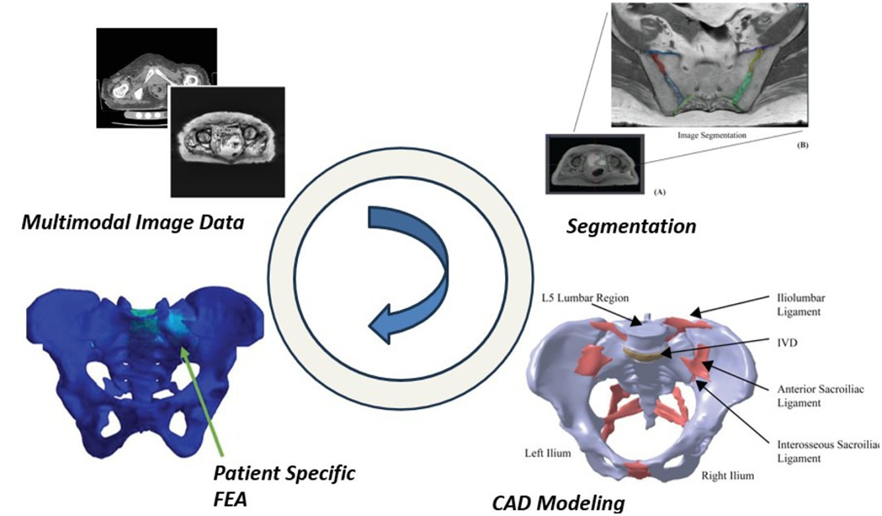
Numerical techniques in the context of osteoporosis and osteoporotic pelvic fracture could potentially contribute toward the development of patient-specific diagnosis. In most finite element simulations of the pelvis the associated ligaments are often neglected due to the modeling complexities involved. This study aims to develop a 3D volume-based continuum approach for these ligaments. The pelvic ligaments were generated based on segmentation of magnetic resonance imaging data from specific patients. Closed volume models were generated based on segmentation and assembled with the 3D models of the corresponding pelvic bones, which themselves were generated from computed tomography data. The resulting pelvic assembly with the ligamentous boundary conditions was numerically simulated under two specific loading conditions: the double-leg stance and double-leg stance with an additional lateral bending moment. The stress state under the force of simulated upper body weight showed a maximum deformation of 0.16 mm at the center of the sacral promontory; this shifted toward the periphery of the sacral promontory and closer to the sacroiliac joint with the addition of the bending moment as well as the contact space between the sacrum and the ilium. The results demonstrated that some of the critical deformation zones are seen in the ligaments and also near their contact regions with the pelvic bones. The approach used for modeling these ligaments, when limited to using 1D springs or force-based boundary conditions, cannot fully factor-in these critical stress concentration zones. As such, this study highlights the necessity of incorporating accompanying ligaments into pelvic bone numerical models.
Keywords
Get full access to this article
View all access options for this article.