
Abstract
Dental malocclusions are commonly encountered in horses. The objective of this study was to report the normal cephalometric values from skull computed tomography (CT) scans of adult Straight Egyptian Arabian (SEAR) and thoroughbred (TB) horses and to compare differences in the measurements between the two breeds. Standing skull CTs were performed on 29 clinically normal adult horses (15 SEAR; 14 TB) and cephalometric measurements were taken. All 9 cephalometric measurements, as well as the interincisal angle, were found to be significantly greater in TB than SEAR (P < 0.05). TB were found to have significantly greater facial line: mandibular length (FL/ML) ratios compared to SEAR (P = 0.002) whereas for the maxillary cheek teeth length: ethmoidal line ratio (Mxa/EtL), SEAR were found to have significantly greater values than TB (P = 0.0007). SEAR cephalometric measurements, as well as certain ratios, differ significantly from TB and could have an impact on the development of dental malocclusions in this breed. Further investigation is needed to ascertain whether these cephalometric differences between the two breeds predispose the SEAR to the development of dental malocclusions.
Introduction
A variety of growth abnormalities of craniofacial bones in horses have been reported. Such abnormalities can result in dental malocclusion and possible permanent dental malfunction. 1 These abnormalities can have a significant impact on the Arabian show horse industry due to disqualification from showing, as well as having implications for breeding programs. Overjet (“overshot” jaw) occurs when the incisal surfaces of maxillary incisors project rostral to the incisal surfaces of mandibular incisors, and this can progress to overbite. 2 Although this condition is commonly referred to as “mandibular brachygnathism,” it is not understood whether it occurs due to a relative shortness of the mandible or a relative lengthening of the maxilla. 2 Overbite (“parrot mouth”) occurs when the maxillary incisors occlude rostral to and directly in front of mandibular incisors. 2 The maxillary incisors may mechanically trap the mandibular incisors behind them, restricting mandibular growth, and resulting in further disparity in length. 2 Although they rarely cause difficulty in prehension, both forms are aesthetically undesirable in show horses. 2 They also can result in concurrent cheek teeth disorders due to maxillary cheek teeth rows being positioned rostrally relative to mandibular cheek teeth, leading to focal overgrowths of maxillary and mandibular teeth. 2 Conversely, underjet (“sow mouth”) can also occur (mandibular prognathism) where the mandibular incisors occlude rostral to their maxillary counterparts, although it is considered rare. 2 Again, it is unknown whether it occurs due to a relative lengthening of the mandible or shortening of the maxilla.
Mandibular brachygnathism is the most common form of incisor malocclusion, and the literature suggests a prevalence in the general horse population of 2% to 5%.1,3,4 More specifically, 3.4% in Franches-Montagnes breeds, and 8.5% in Warmbloods. In one large-scale study of 450 mixed-breed horses in Egypt, 23.6% with incisor disorders: 7.8% (n = 35) had mandibular brachygnathism (i.e., 1.8% total population) and 0.7% (n = 3) had prognathism (i.e., 0.17% total population). 3 The prevalence of both forms in Arabian breeds is unknown. There is some evidence that suggests that malocclusions are heritable in equids as they are in other species with certain genes and chromosomal regions having been identified as being associated with both mandibular prognathism and brachygnathism.5,6 In horses, a region on the distal chromosomal region of the ECA13 has been identified as possibly being associated with maxillary prognathism. 6 In addition, in donkeys MATN1 polymorphisms have been implicated as playing a role in mandibular prognathism. 5 It has been reported that Arabians are predisposed to underjet/prognathism due to their dished face phenotype. 2 It has been shown that one factor contributing to the Arabian's distinct phenotypic skull appearance is that this breed has a significantly shorter nasal length compared to thoroughbreds (TBs) and Standardbreds. 7 However, the Straight Egyptian Arabian subgroup (SEAR) has been shown to have significantly shorter head lengths and facial crest lengths compared to TBs, as well as smaller craniofacial angles. 8 Potentially this could have implications for the development of malocclusions, resulting in a predisposition for the development of underjet/prognathism. According to one study, certain cephalometric ratios can be useful to predict whether a young warmblood foal born with a physiological malocclusion may spontaneously resolve or progress into an overjet later in life. 4 To date no data relating to such cephalometric measurements are available for either the TB or SEAR.
A variety of treatments for dental malocclusions exist in horses, including both dental rasping and surgical techniques, such as dental wiring and osteodistraction.2,9,10 Selection of the correct technique relies on a complete understanding of the disease mechanism. In human orthodontics, cephalometric analysis is considered a vital tool to investigate the underlying cause of malocclusions and to formulate appropriate therapeutic plans. 11 Traditionally, a 2-dimensional lateral skull radiograph was used for this purpose, however more recently there has been a move toward the use of computed tomography (CT) due to its 3-dimensional nature providing more accurate and comprehensive images for cephalometric measurements.12,13 Cephalometry has been used to provide measurements on laterolateral radiographs to demonstrate the development of malocclusions in warmblood foals. 1 A CT 3D cephalometric approach has also been investigated for the purpose of age determination in horses by measuring the angulation between incisor clinical crowns but has not been found to be a reliable tool for this purpose. 14
The objective of this study is to report the normal cephalometric values from skull CTs of adult SEAR and TB horses and to compare differences in the measurements between the two breeds. The authors hypothesize that the SEAR has significant differences in cephalometric measurements compared to the TB.
Materials and Methods
Study Design, Sample Size, and Study Population
The study was a prospective cohort study. Twenty-nine clinically normal, nonpregnant, adult horses (aged 5 years and over), without signs of malocclusions, were included comprising 15 SEAR and 14 TB horses. Quantitative data was collected. The animal study was reviewed and approved by the Institutional Animal Care and Use Committee of the Equine Veterinary Medical Center, a member of Qatar Foundation, Doha, Qatar, under the protocol number EVMC- 2021-1160.
Data Collection
The study was performed on-site at the Equine Veterinary Medical Clinic, Doha, Qatar. Physical examinations determined that each horse was healthy for inclusion in the study. Brief oral examinations were performed to ensure the animals did not have obvious incisor malocclusions. Body weights (kg) were recorded using walk-on digital scales. Horse height at the withers (cm) was measured using a measuring stick.
To ensure patient compliance and welfare, horses were sedated with intravenous injections of xylazine HCL (0.1-1 mg/kg) with or without detomidine (0.004-0.02 mg/kg), butorphanol (0.01-0.04 mg/kg), and acepromazine (0.02-0.05 mg/kg) to effect.
CT of the skull was performed with the horse during standing sedation, using a 64-slice Siemens Definition AS Sliding Gantry CT systema. One-millimeter helical images processed by using a high-frequency convolution kernel were acquired (parameters 35 Ma, 140 KV, 0.6 mm slice thickness).
CT Assessment
CT images were assessed by a board-certified diplomate of the European College of Veterinary Diagnostic Imaging and American College of Veterinary Radiology using diagnostic imaging viewing software.
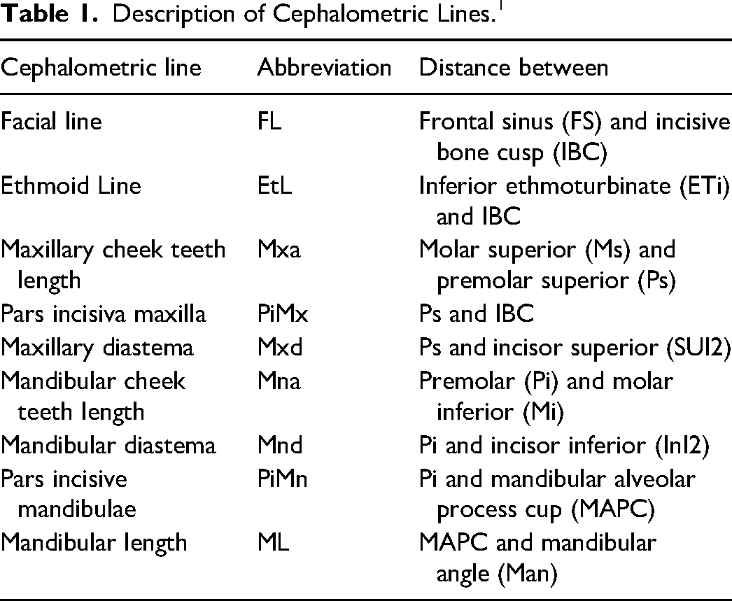
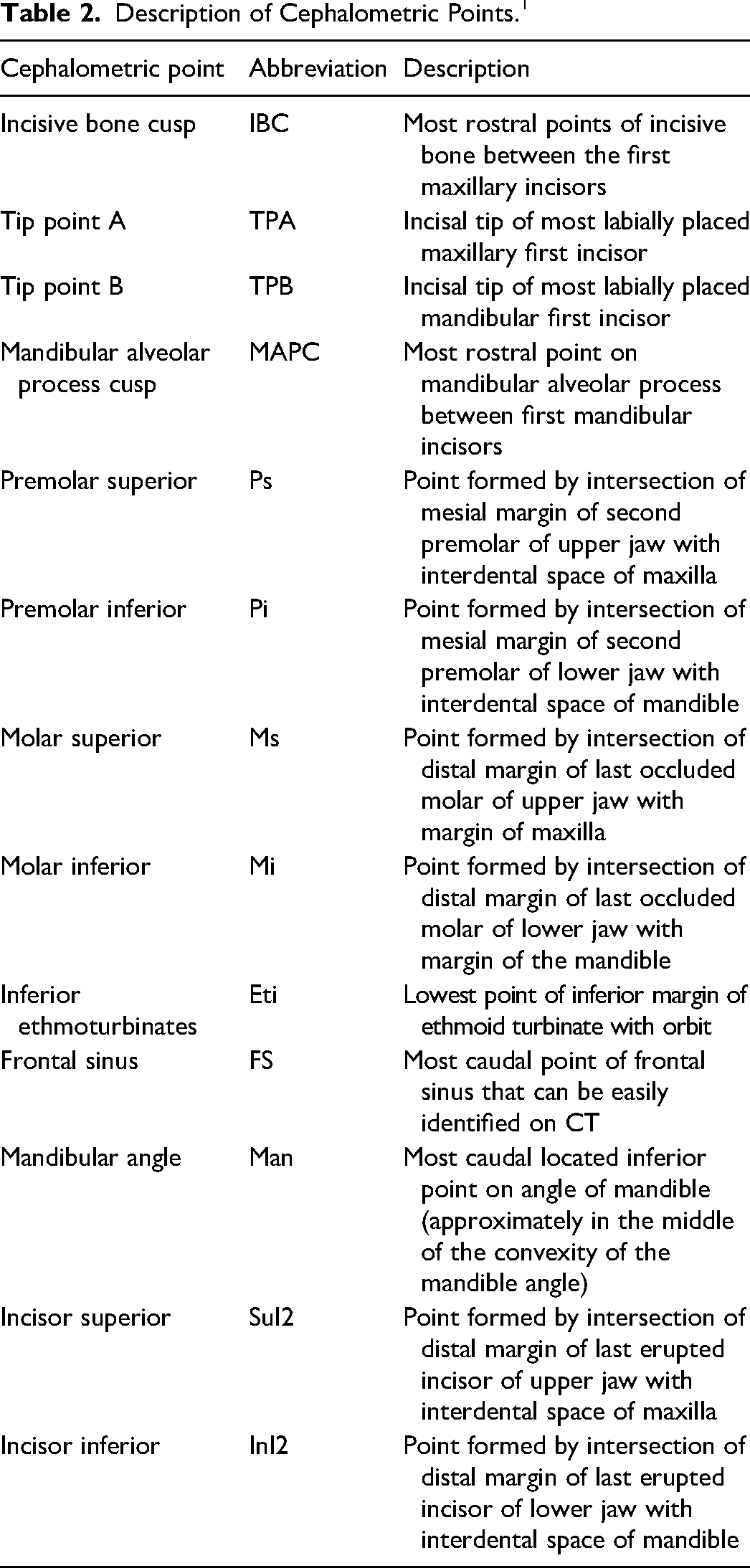
Quantitative CT measurements were taken according to the description of the cephalometric points and lines as previously published 4 (Tables 1 and 2) (Figure 1). The high-frequency reconstruction algorithm study from each CT examination was viewed in the bone window using image analysis softwareb. The study was viewed in a sagittal plane, with the axis aligned with the midline of the head in Multi-Planar Reconstruction. In this sagittal plane, each point was chosen, and the angle function was used to measure the angles made by lines intersecting at the points of interest. The superior incisal line was one connecting the incisive bone cusp to tip point A, and the inferior incisor line was one connecting tip point B to the mandibular alveolar process cusp. The interincisal angle was then measured between the intersection of the superior incisal and inferior incisal angles (Figure 2).
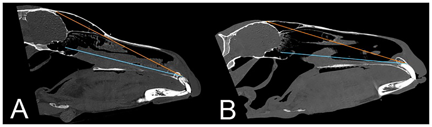
CT scans showing facial line (FL), measured from the frontal sinus and the incisive bone cusp, and the ethmoid line (EtL), measured from the inferior ethmoturbinate and the incisive bone cusp, for (A) Straight Egyptian Arabian and (B) Thoroughbred horses.
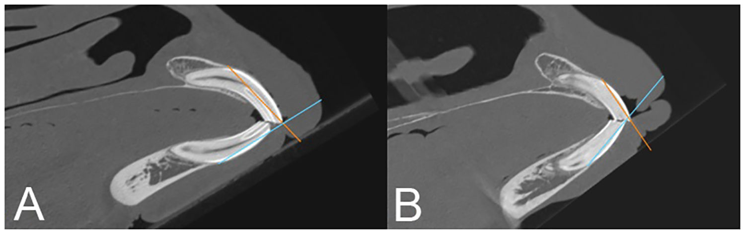
CT scans showing interincisal angle (IIA) for (A) Straight Egyptian Arabian and (B) thoroughbred horses. The interincisal angle was measured between the intersection of the superior incisal and inferior incisal angles.
Description of Cephalometric Lines. 1
Description of Cephalometric Points. 1
Data Analysis
The Shapiro–Wilk test was used to examine the normality of the data for each group, and normality was not rejected (P > 0.05) for all the data. Nineteen ratios were calculated from the cephalometric lines (Table 3). The Shapiro–Wilk test was also used to check for normality and was not rejected (P > 0.05) for all ratios. Age and height for the two breeds were compared with an equal variance 2-sided t-test. While age and height differed significantly between the 2 breeds, only age influenced cephalometric measurements and neither age nor height influenced the ratios. For consistency, the 2 breeds were compared for all variables using a linear model with breed as a fixed factor and age as a co-factor. Least-squares means (SEM) were presented for the 2 breeds.
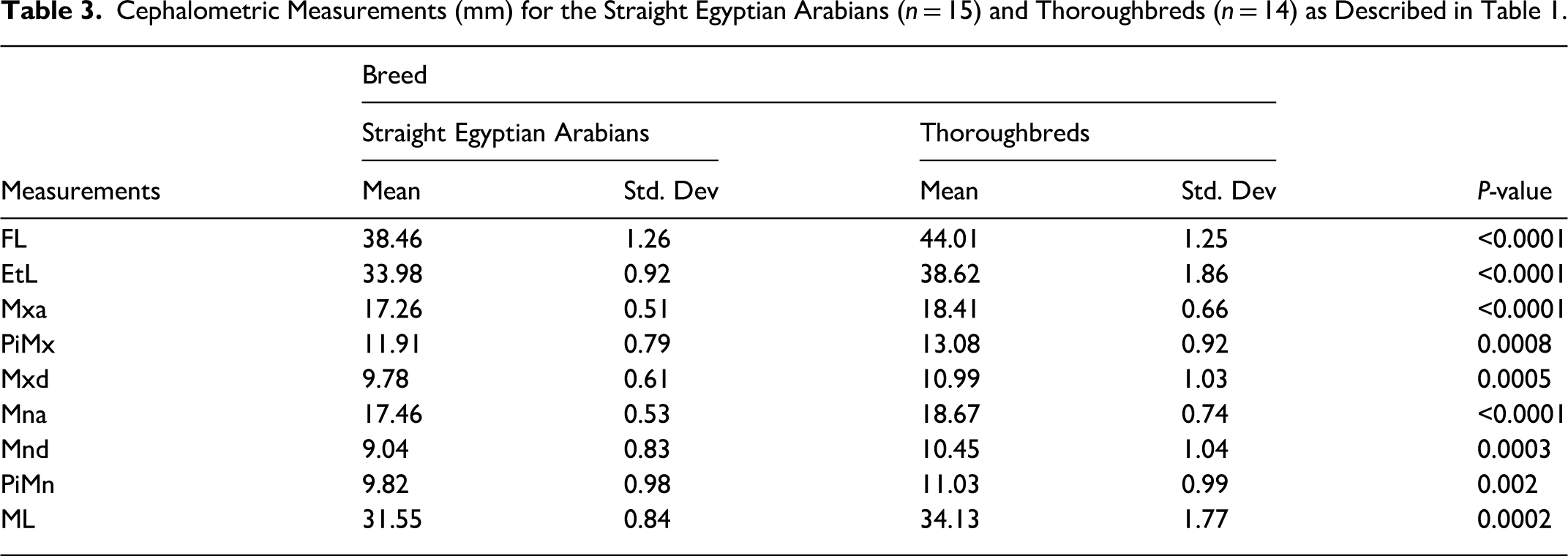
Cephalometric Measurements (mm) for the Straight Egyptian Arabians (n = 15) and Thoroughbreds (n = 14) as Described in Table 1.
Results
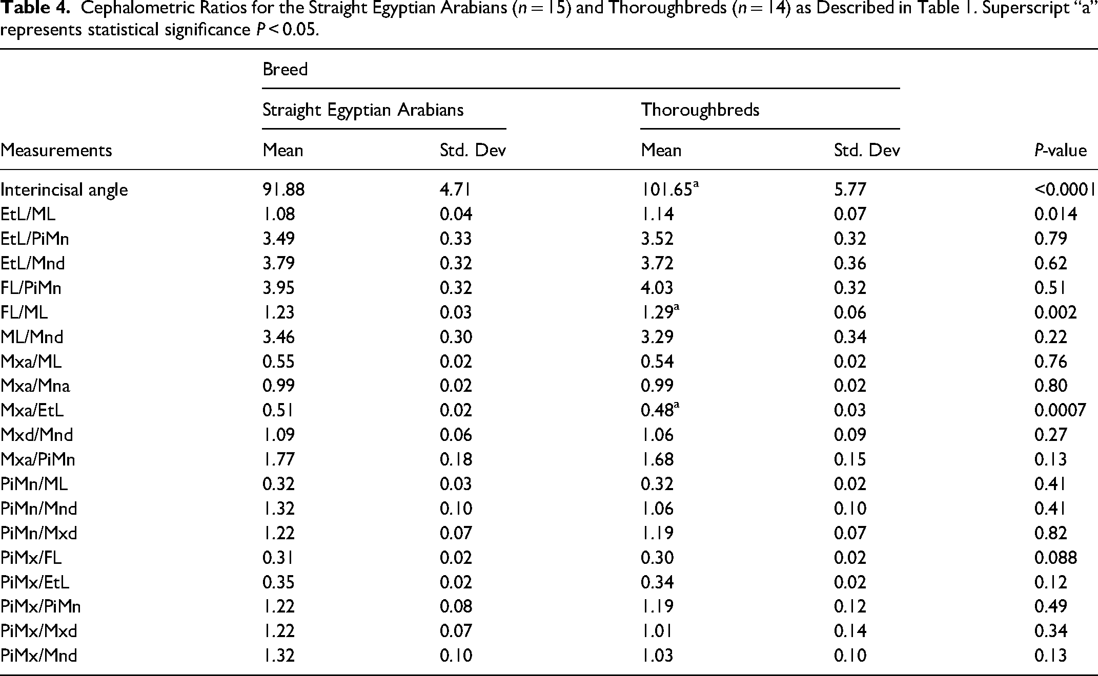
Measurements were taken from 29 clinically normal, nonpregnant, adult horses (5 years and over), including 15 SEAR horses (all were mares; mean age 10.74 ± 3.82 years; mean body weight 405.33 ± 29.91 kg; mean height at withers 147.95 ± 3.11 cm) and 14 TB (12 mares, 2 geldings; mean age 8.19 ± 2.35 years; mean body weight 502.79 ± 44.36 kg; mean height at withers 160.28 ± 4.02 cm), without gross signs of incisor malocclusions. In addition, CTs were incomplete for 6 horses (2 TB and 4 SEAR) meaning that mandibular length measurements could not be ascertained for them, and subsequently, ErL/ML, FL/ML, ML/Mnd, and Mxa/ML ratios could not be calculated for these individuals, however, the remainder of the measurements could be made. Descriptive statistics were performed for all variables and all data were normally distributed (Table 3). All nine cephalometric measurements were significantly greater in TB than SEAR, in addition to the interincisal angle (P < 0.05) (Table 3). Of the 19 ratios, statistically significant differences between the 2 breeds were only found in two of the variables. TB were found to have significantly greater FL/ML ratios compared to SEAR (P = 0.002) whereas, for Mxa/EtL, SEAR was found to have significantly greater values than TB (P = 0.0007). The equal variance t-test revealed a significant difference between the two breeds for mean age (P = 0.04). The mean age was larger for the AR breed than for the TB breed. The equal variance t-test revealed a significant difference between the 2 breeds for mean height (P < 0.0001). The mean height was smaller for the AR breed than for the TB breed. Using a linear model with breed as a fixed factor, and age and height as cofactors, differences in age and height between the 2 groups were investigated. The model revealed a statistically significant effect of breed (P < 0.0001) controlling for the effect of age and height. Even when controlling for the effect of age and height, the effect of breed remained, with TB having significantly greater measurements for all 9 cephalometric measurements (Table 3). Regarding the effect of age, it had a significant negative effect (with measurements being smaller in older individuals) for Mxa and Mna (Table 3). The effect of height was not found to have a significant effect on any of the variables. For interincisal angle, the effect of age was not significant (P = 0.07).
Discussion
The results of this study reported the normal cephalometric values from skull CTs of normal adult SEAR and from TB horses and highlighted some significant differences in the measurements between the 2 breeds.
All 9 cephalometric measurements were found to be significantly greater in TB than SEAR, in addition to the interincisal angle (P < 0.05). 8 Even after controlling for horse height, the effect of breed remained the same with TB > SEAR for all measurements (P < 0.05). It is worth noting the statistically significant differences in 2 of the reported ratios, FL/ML and Mxa/EtL. Regarding the FL/ML ratio, this was found to be significantly greater in TB compared to SEAR (P = 0.002). The SEAR has been shown to have significantly shorter head lengths, as well as significantly smaller facial crest length, compared to TB, which likely explains this finding. 8 This could possibly provide an explanation for why it has previously been anecdotally reported that Arabians are predisposed to underjet/prognathism. 2 The relatively shorter facial-to-mandibular length could contribute to the Arabian's dished-face phenotype. Furthermore, the relatively longer mandibular length may result in a greater prevalence of mandibular prognathism. While it is commonly referred to as “prognathism,” meaning a relatively longer mandibular arcade compared to the maxillary one, it is unknown whether this condition occurs due to a lengthening of the mandible or a shortening of the maxilla. 2 The current findings suggest that the SEAR has a relatively longer mandibular length, thus potentially explaining the reason for their apparent predisposition to the underjet/prognathism condition. This knowledge can have clinical implications for our management of this condition and confirms our current treatment methods, whereby a focus on growth retardation of the mandible (as is often performed with dental wiring in horses) is used to compensate for the disparity in lengths. 2
The Mxa/EtL ratio was found to be significantly greater in SEAR compared to TB. Previous findings indicate that there is no significant difference in the lengths of the caudal or rostral maxillary sinuses between the 2 breeds. 8 While this is not directly comparable to the Mxa length in the present study, which was measured as the distance between the superior molar and superior premolar, it does suggest that the difference between the 2 breeds may be attributable to the SEAR having a shorter EtL distance. Again, while not directly comparable, it has been shown that absolute distances from the maxillary septal bulla to the rostral aspect of the cribriform plate are significantly shorter in SEAR compared to TB. 8 It could be theorized that this may have an impact on the length of the EtL in the current study, potentially causing the SEAR to have a shorter EtL and thus a significantly greater Mxa/EtL ratio. However, further investigation is required to understand these differences in more detail.
The results of the present study also indicated a significant difference in the interincisal angle (IIA) between the 2 breeds, with TB being greater than SEAR. The mean IIA in the SEAR was 91.9° compared to 101.7° in TB. Previous research has shown that horses 8 to 15 years of age typically have IIA of approximately 90°, which is consistent with the SEAR findings in the current study.14,15 With the mean age of the TB group (7.47 years) being significantly younger than the mean age of the SEAR group (10.4 years), it is logical to assume that this difference in age could explain the difference in IIA between the 2 breeds. However, it is interesting to note that although it is widely accepted that IIA decreases with age, the results of the current study did not find age to have a significant effect on IIA (P = 0.12).14,15 After controlling for the effect of breed on the 9 cephalometric measurements, age was then found to have a significant negative effect on Mxa and Mna (P < 0.05), with measurements being smaller in older individuals. One possible explanation for this is that the maxillary and mandibular cheek teeth lengths decrease with age due to the hypsodont dentition of the horse, as a result of the progressive eruption of the crowns over time (Table 4).
Cephalometric Ratios for the Straight Egyptian Arabians (n = 15) and Thoroughbreds (n = 14) as Described in Table 1. Superscript “a” represents statistical significance P < 0.05.
Previous radiographic cephalometric studies relating to the development of overjet in Warmblood foals have been performed, and have highlighted a number of useful measurements for the identification of those foals which would achieve spontaneous regression of malocclusions over the first 4 months of life. 4 It is difficult to make comparisons with the results of the present study for several reasons. One major weakness of the present study was the inconsistent head positioning for the purpose of performing the skull CTs, which was typically done with the horses’ heads in extension. It has been demonstrated that head position can influence the relative rostrocaudal position of the mandible with respect to the maxilla. 16 For the radiographic cephalometric study in the Warmblood foals, a custom-made head-holding device, akin to a human cephalostat, which fixed the heads in a standard, reproducible position allowing consistent measurements to be taken was used.1,4 For the radiographic cephalometry, the cephalostat was used to achieve an angle of 90° between the ventral margin of the mandible and the ventral border of the neck. 1 Foals were positioned with the forelimbs square and vertical to the ground, the top of the neck horizontal to the withers and the foal's mouth level with the elbow. 1 In adult horses, this position would not be possible to achieve a standing skull CT in an adult horse due to the gantry size using standard CT techniques. While standardized methods for head positioning during standing skull CTs in horses have been described, they were not used in the present study. 17 Furthermore, the horses in the current study were all sedated to allow the CT scans to be performed. It has been theorized that the muscle relaxation induced by sedation results in an “unnatural” head position which can affect the results of cephalometric measurements. 4 The results of one study indicated two radiographic cephalometric measurements (PiMx/PiMn and Mna/ML ratios) which can be useful to predict whether a foal with a physiological incisor malocclusion would subsequently develop an overjet. 4 In the present study, there were no significant differences between SEAR and TB for these 2 ratios. However, given that the current study involved adult horses with no gross evidence of malocclusions, no comparisons can be made.
Other limitations of this study include the small number of horses, as well as the fact that the majority of them were mares meaning that sex distribution could not be analyzed. Furthermore, the SEAR is the only Arabian horse subgroup which has been shown to differ significantly from the others. It has also been shown to have a relatively high degree of inbreeding. 18 Therefore, it is likely that the SEAR group in this study, which came from 2 breeding farms, had a relatively high degree of relatedness and probably lacked individual variation. Given the high degree of inbreeding in this breed subgroup, finding unrelated individuals would present a challenge. 18 Finally, having reported the normal values for these breeds, further investigation should be performed in clinical cases of malocclusions to compare them with “normal” horses to ascertain whether significant differences in cephalometric measurements exist between the 2 groups.
Conclusion
SEAR cephalometric measurements, as well as certain ratios, differ significantly from those of the TB, and could have an impact on the development of dental malocclusions in this breed. Further investigation is needed to ascertain whether these cephalometric differences between the 2 breeds predispose the SEAR to the development of dental malocclusions.
Footnotes
Materials
Siemens Definition AS, Siemens Healthineers, Malvern, PA, USA OsiriX, Version 6.0, Pixmeo, Switzerland
Acknowledgments
The authors would like to express their gratitude to the supporting hospital staff of the Equine Veterinary Medical Center for their precious help. We also would like to thank Dr Guy Beauchamp DVM, PhD, for his biostatistics expertise, as well as Al Shaqab and Al Rayyan farms, for their support with this project.
Declaration of Conflicting Interests
The authors declared no potential conflicts of interest with respect to the research, authorship, and/or publication of this article.
Funding
The authors disclosed receipt of the following financial support for the research, authorship, and/or publication of this article: This work was supported by the Equine Veterinary Medical Center Intramural Funding (grant number RG21_JJ1 (Dr Jessica Johnson)).
Ethics Statement
The animal study was reviewed and approved by the Institutional Animal Care and Use Committee of the Equine Veterinary Medical Center, a member of Qatar Foundation, Doha, Qatar, under the protocol number EVMC- 2021-1160.