
Abstract
We share an unusual presentation of fungal (Aspergillus) peritoneal dialysis catheter infection diagnosed by a color change in a patient's PD transfer set. Recognizing and sharing uncommon presentations of fungal infection is important to help others diagnose and intervene early, which may reduce the development of fungal peritonitis, which is associated with high rates of mortality and technique failure.
Keywords
Images in PD
Clinical context and findings
Fungal peritonitis is a rare complication of peritoneal dialysis (PD) but is associated with a high risk of mortality and technique failure.1,2 While infection with Aspergillus is not the most common causative organism of fungal peritonitis, it is associated with higher mortality.3–5 Early diagnosis and treatment is correlated with improved outcomes. 4 Diagnosis involves identifying fungal elements on a gram stain of peritoneal fluid or on peritoneal fluid culture. 1 As fungal culture is challenging and slow, recognition of presentation patterns and risk factors for infection is essential for prompt diagnosis and treatment. Risk factors include previous bacterial peritonitis, prolonged antibiotic use, immunosuppression, recent hospitalization, and autoimmune disease. 3 Peritoneal eosinophilia should also raise suspicion of fungal infection. 6 Treatment includes antifungal agents, including amphotericin B or azoles, and prompts removal of the PD catheter.6,7 Previous case reports have described peritoneal catheter discoloration in association with Aspergillus species on culture.8,9
We share a case of asymptomatic Aspergillus catheter infection identified by discoloration of a PD catheter transfer set.
A 71-year-old male with a history of liver transplant secondary to non-alcoholic steatohepatitis and hepatocellular carcinoma, diabetes mellitus, and end-stage kidney disease due to calcineurin inhibitor toxicity presented for his monthly PD clinic visit. The patient began PD 7 years prior and was currently on APD. He had a history of peritonitis and tunnel tract infections, including pseudomonas peritonitis 2 years prior, with associated tunnel tract infection, MRSA peritonitis with exit site abscess requiring catheter removal and replacement 1 year prior, culture-negative peritonitis 8 months, and, most recently, pseudomonas peritonitis 7 months prior to his clinic visit. He did not have antibiotic exposure for the 6 months prior to presentation. His transfer set was most recently changed at the clinic visit the month before.
An area of brown discoloration was noted at the proximal portion of his catheter transfer set (see Figure 1). He noted the color change for three days without associated abdominal pain or fever.
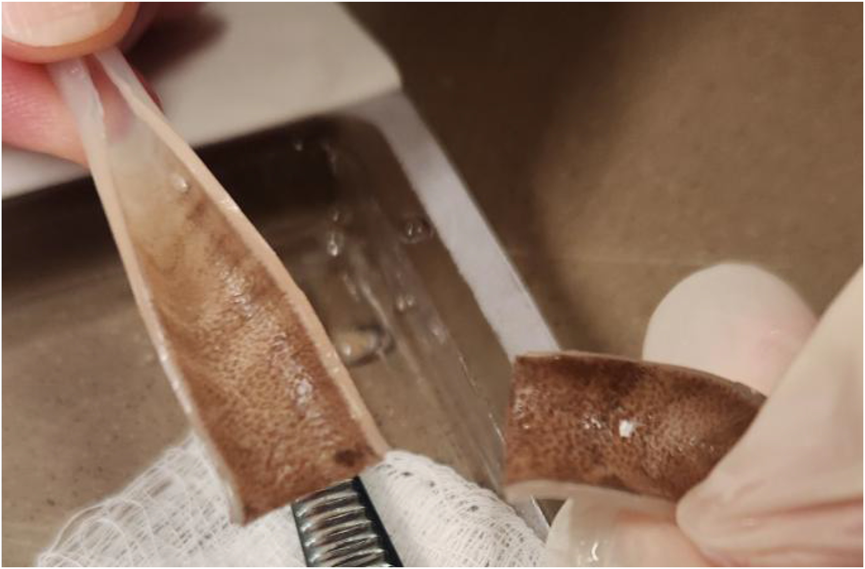
Bisected transfer set with brown discoloration in the internal lumen.
The transfer set was changed, and the old transfer set was sent to anatomical pathology for analysis. PD fluid was sent for cell count and culture. Results showed an elevated white cell count: 432 cells/µL with 54% neutrophils and 11% eosinophils with a hazy appearance. Empiric peritonitis treatment was started with intraperitoneal vancomycin and ceftazidime and was narrowed to ceftazidime monotherapy when peritoneal fluid cultures remained negative.
The pathologist's evaluation of the catheter revealed septate, acute-angle branching hyphal elements consistent with mold.
Given concern for fungal infection, he was admitted to the hospital and remained without fever, abdominal pain, or systemic leukocytosis. Peritoneal fluid studies on admission showed a nucleated cell count of 358 with 68% neutrophils, 15% lymphocytes, 12% monocytes/macrophages, and 5% eosinophils. He was treated with oral voriconazole (450 mg twice a day X 2 loading dose followed by 300 mg BID maintenance dose for a target dose of 4 mg/kg adjusted body weight) and underwent PD catheter removal with transition to hemodialysis. Given his clinical stability, voriconazole was chosen over amphotericin B for its lower side effect profile. His peritoneal fluid culture eventually grew Staphylococcus epidermidis (thought to be a contaminant), while cultures from his removed PD catheter grew Aspergillus species, non-fumigatus.
His voriconazole levels were followed in the infectious disease clinic at 1-week intervals, and he completed 2 months of treatment. Per preference, he remains on hemodialysis.
Footnotes
Acknowledgments
None.
Authorship
MG, TR, and MS researched literature and composed the manuscript. All authors reviewed and edited the manuscript and approved its final version.
Declaration of conflicting interests
The authors declared no potential conflicts of interest with respect to the research, authorship, and/or publication of this article.
Ethical approval
No ethical approval was provided for the manuscript content (Images in PD submission).
Funding
The authors received no financial support for the research, authorship, and/or publication of this article.
Informed consent to participate
No informed consent was obtained as there are no patient images or trial data included in the submission.
Informed consent to publish
No informed consent to publish was sought as there are no patient images included in the submission.
Trial registration
There is no trial registration to disclose.