
Abstract
A novel biocomposite with poly(methyl methacrylate) as resin containing Salvadora persica powders was elaborated. In this study, for the first time, S. persica was used to enhance the bioactive performance of dental restoration materials. Material characterization was carried out both on bare materials and on the elaborated biocomposite (30 wt% of S. persica). X-ray diffraction, Fourier transform infrared spectroscopy, differential scanning calorimetry, and high-performance liquid chromatography techniques were conducted to perform material characterization. The obtained results linked to dental material showed the presence of the organic chemical compounds of S. persica, which are responsible for biological activities, and the presence of mineral chemical compounds of S. persica, which are useful for dental applications and health. They also revealed the absence of toxic residual monomers. In addition, they proved the antioxidant activities proof of elaborated composite related to total polyphenol flavonoid content. Finally, they exhibited the antibacterial activity of S. persica and the composite.
Introduction
Dental composites have been used as restorative materials for decades. Many studies have focused on reducing breakages by the reinforcement of the acrylic resin. The reinforcement has been previously undertaken by using fibers such as glass, carbon/graphite, calcium oxide nanoparticles, and ultrahigh-modulus polyethylene fibers.1,2 However, only few research works have expounded the antibacterial dental resin-based materials. These materials can efficiently reduce oral biofilm formation and prevent bacterial micro leakage.3,4 In fact, in recent studies, novel polymers containing quaternary ammonium monomer dimethylaminododecyl methacrylate have been synthesized with antibacterial activities. 5 Furthermore, in order to improve the biological activity and antibacterial properties of polyetheretherketone (PK) as bone implants, Tang et al. fabricated nano zinc-magnesium silicate/PK antibacterial biocomposites (nZPC). 6 Furthermore, Tahriri et al. report that the addition of graphene and its derivatives with biocompatible polymers led to obtain an antibacterial biocomposite. 7 In addition, Varoni et al. have investigated the role of the bioactive plant in enhancing the bioactive performance of dental biomaterials. 8 In some works, some natural products, originating from medicinal and food plants, have been reported to have a beneficial role against periodontal disease and in promoting periodontal healing, that is, Cissus quadrangularis, Carthamus tinctorius, and Glycine. 8
In this work, the medical plant Salvadora persica was interesting to study due to its chemical, physical, and biological properties to elaborate bioactive dental composite. Therefore, we used the natural S. persica for its clinical importance that arises from a number of mechanisms, including its anti-plaque, anti-caries, anti-inflammatory, antioxidant, antiviral, and antibacterial properties. 9 Moreover, these effects of S. persica are attributed to various chemicals contained in its stems. The S. persica contains the following compounds: flavonoids, stearic acid, linoleic acid, salvadorine, salvadourea, lignans, cyanogenic glycosides, alkaloids, saponins, tannins, silica, vitamin C, and different salts which have a significant antimicrobial activity. Calcium (Ca) promotes remineralization of tooth enamel. 9 Furthermore, the antioxidant activity of S. persica was confirmed by the important concentration of the polyphenol and the flavonoid in the extracts. 10
To the best of our knowledge, it is for the first time that the effects of S. persica incorporation on acrylic-based material were investigated. The popularity of poly(methyl methacrylate) (PMMA) as denture base material has been attributed to its ease of processing and cost efficiency. 11
Thus, given these beneficial therapeutic properties of S. persica, we elaborated a biocomposite PMMA/S. persica for dental restoration to determine the properties of S. persica, specially its antioxidant and antibacterial activities.
Materials and methods
Materials and process
A denture base material that was commercially available, self-cure acrylic resin (Nic Tone; MDC Dental, Gardena, CA, USA), was supplied by MDC Dental (Manufacturera Dental Continental S.A. de C.V.) in the form of powder and liquid. The powder contains approximately 97% of PMMA polymer, while the liquid was MMA mixed with ether glycol as cross-linking agent. The acrylic resin (Nic Tone (R)) Basic Vita Shades 4 × 1 was used as polymer/monomer ratio of 2:1.
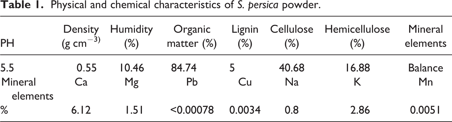
The raw S. Persica (Al Baraka, KSA) of the family Salvadoraceae was obtained. The biomaterial was dried at a room temperature. Then, the dried sticks were blended in a food-processing blender (coffee grinder; Moulinex, China). The mean size of the obtained particles powder was inferior to 40 µm. The characteristics of the S. persica powder are listed in Table 1.
Physical and chemical characteristics of S. persica powder.
In the present study, only a PMMA composite filled with 30 wt% of untreated S. persica powder was investigated. The optimization of the content of S. persica powder in composite will be the subject of future work. It is to be noted that the self-cure denture base resin powder and liquid were mixed according to the conditions specified in the instruction manual and polymerization procedure. The required quantity of S. persica powder was mixed into the monomer liquid and stirred. After that, the PMMA powder was added to the mixture with continued stirring until the filler was uniformly distributed in the resin. It was poured into the gypsum mold of required dimensions needed for testing samples. Self-cure composites acrylic resins with the content of 30 wt% S. persica were conducted in a pressure device (Mini Major 2000, Major, Italy) at 60°C for 10 min under the pressure of 0.5 MPa.
Materials characterization process
The measurement of pH
The pH of S. persica suspension was measured to investigate the change in pH due to any soluble species from the filler material in water. A suspension was prepared by mixing a volume of S. persica (2 mL) with five times its volume of distilled water (10 mL). The suspension was shaken for 5 min and let stand for at least 2 h but no more than 24 h. The pH was measured by pH-Meter (632 pH-Meter; Metrohom, Switzerland).
The flame atomic absorption spectroscopy
The flame atomic absorption spectroscopy was also used for the determination of the metal contents in S. persica. The solution of samples of 1000 mL was prepared by an acid attack. The ash of 2 g of S. persica powder was mixed with 5 mL hydrochloric acid (37%) and 5 mL nitric acid (65%). The solution was heated to boiling. Afterward, 15 mL of distilled water was added and heated to boiling. The filtration was achieved with filter papers. The distilled water was added to the filtrate to 1000 mL. It is to be noted that the mineralogy of this content depends on the geography of the sample’s regions. 12 A flame atomic absorption spectrometer (ICE 3000 Series; Thermo Scientific, England) is equipped with a hollow-cathode-lamp and an acetylene/air burner. The iCE SOLAAR AA software package was used. The elements’ concentrations are listed in Table 1.
X-ray diffraction
The X-ray diffractions (XRDs) of samples (composite (30 wt% of S. persica powder), S. persica powder, ash of S. persica, PMMA) were collected on X-ray diffractometer (D8 XRD, DIFFRAC. SUITE; Bruker, Germany) using Ni-filtered Cu Kα1 radiation (λ = 1.540600 Å) in the 2θ range of 5–6° at a voltage of 40 kV and a current of 40 mA and a scanning rate of 1.2° min−1.
Infrared spectroscopy
The characterization of the chemical structure of the investigated samples (composite (30 wt% of S. persica powder), S. persica powder, ash of S. persica, PMMA) was carried out using the Fourier transform infrared spectroscopy (FTIR) spectrometer (Cary 630 FTIR; Agilent Technologies) in the wave region between 3800 and 600 cm−1 with a resolution of 4 cm−1.
Scanning electron microscopy
Scanning electronic microscope (SEM) was employed to characterize the morphology of the S. persica powder and the resulting composite (30 wt% of S. persica powder) via the fracture section of samples. The samples were firstly coated with gold and examined using a scanning electron microscope (Jeol JSM T330A, Electron Optical Laboratory, Tokyo, Japan) at an accelerating voltage of 25 kV.
Differential scanning calorimetry
Differential scanning calorimetry (DSC) analysis was performed with a DSC 204F1 (DSC 204 F1 Phoenix, NETZSCH, Germany) apparatus equipped with a thermal analysis processing system “Proteus.” A sample of composite (30 wt% of S. persica) of 15 mg was placed in an aluminum pan at room temperature (20°C) and then heated at 15°C min−1 up to 220°C in air (first run). It was then cooled to room temperature at 20°C min−1 and heated again at 15°C min−1 to 220°C (second run). 13
High-performance liquid chromatography
High-performance liquid chromatography (HPLC) analysis was used to quantity the residual MMA content in the composite (30 wt% of S. persica). A sample of 50 mg was dissolved in 1 mL of acetone and then 10 mL of methanol was added to the solution to precipitate the polymer. The supernatant of the solution was filtered through a 0.45-µm pore Millipore filter. HPLC analysis was performed using a Thermo Fisher Scientific system (Ultimate 3000; Thermo Fisher Scientific, Germany), equipped with a CAPCELL PAK C18 column (4.6 × 250 mm2, Supelco, USA) and a diode array detector (Ultimate 3000; Thermo Science, USA; detection at λ = 230 nm). An amount of 10 µL of the sample solution was injected and analyzed at 40°C at a flow rate of 1.0 mL min−1 with acetonitrile/water (50/50%). This procedure was previously adopted by Ohyama and Imai. 13
Antioxidant property analysis
The total antioxidant capacity (TAC) of the extracts of fresh S. persica powder and S. persica powder (T = 60°C, P = 0.5 MPa, t = 10 min) was evaluated using the phosphomolybdenum method described by Prieto et al. 14 It is worthwhile to note that S. persica powder was firstly introduced in the gypsum mold, and the latter was immersed in a pressure device (Mini Major) according to the previously described polymerization conditions. The phosphomolybdenum method is based on the reduction of Mo(IV) present in the form of molybdate ion MoO42− to Mo(V) in the presence of MoO2+ extracted to form a green phosphate/Mo(V) complex at acid pH. The extract was prepared by dissolving 1 g of S. persica powder in 10 mL of artificial saliva according to Huang 15 (Table 2). The suspension was macerated for 12 h at 37°C and 125 r min−1 stirring. A volume of 100 µL of the sample solution (five replicates) was mixed with 1000 µL of the reagent solution (0.6 M sulfuric acid, 28 mM sodium phosphate, and 4 mM ammonium molybdate). For the blank, 100 µL of H2O was mixed with 1000 µL of the reagent. The tubes were capped and incubated in a thermal block (marine bath) at 95°C for 90 min. The samples had cooled to room temperature. Then, the absorbance of the aqueous solution of each one was measured against a blank at 695 nm. The antioxidant activity of S. persica was expressed for the samples as gallic acid equivalents (GAE; µg of GAE/mL of extract).
Composition of artificial saliva.
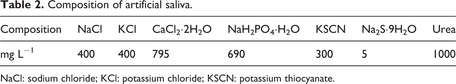
NaCl: sodium chloride; KCl: potassium chloride; KSCN: potassium thiocyanate.
Total polyphenol and flavonoid content
The ethanol extracts (EEs) of S. persica (EES) and of composite (EEC; 30 wt% of S. persica) were prepared with 50 mL of 80% ethanol per 0.5 g of powder, kept until total dissolution and subsequently filtered to obtain the EE. The concentration of EE is 10 mg mL−1. 16
The total polyphenol concentration in the EE was determined according to the Folin–Ciocalteu colorimetric method. 17 For this purpose, 0.5 mL of EE (diluted 2×) was mixed with 2.5 mL of Folin–Ciocalteu reagent (diluted 10×) and 2.0 mL of 7.5% sodium carbonate (sodium carbonate). The absorbance was read at 760 nm after 2-h incubation at room temperature in the dark. Gallic acid (250 mg mL−1) was used as a standard to produce a calibration curve. The average of three readings was used to determine the total polyphenol content, which was expressed as the mg of GAE/g of powder.
The flavonoid content in EEC was determined according to the method described by Meda et al., 17 with minor modifications. For this purpose, 400 µL of EE (diluted 2×) was mixed with 120 µL of NaNO2 (5%). After 5 min, 120 µL of aluminum chloride (10%) was added, and 6 min later, the mixtures were neutralized with 800 µL of 1 M sodium hydroxide solution. The absorbance was immediately read at 510 nm. Quercetin (120 mg mL−1) was used as a standard to produce a calibration curve. The average of three readings was used to determine the flavonoid content, which was expressed as the mg of quercetin equivalents (QE)/g of powder.
Antibacterial activity of elaborated composite
The following oral cavity pathogen strains were selected for antimicrobial activity testing of the composite: Streptococcus mutans (CECT 479), Streptococcus pneumonia (CIP 49619), and Haemophilus influenza (ATCC 10211).
S. mutans and S. pneumonia were grown on blood agar and H. influenza on chocolate agar with 5% carbon dioxide (CO2) at 37°C.
The antibacterial activity of S. persica and the composite were evaluated using an agar diffusion assay. The sticks of S. persica were cut into pieces. The piece’s weight was equal to the weight of the S. persica which was incorporated into the composite. The pieces were treated at 60°C, the temperature of polymerization. The disc of composite with 30% of S. persica was immersed in 400 µL of sterile water for 1 day, in order to allow the composite to release the antibacterial substances. The required numbers of wells, each 6 mm in diameter, were cut out of the agar using a sterile glass capillary ensuring proper distribution of holes in the periphery of agar plate. Then, wells were filled with 100 µL of the S. persica-immersed water. For the S. persica sticks, each piece was placed into a well. After incubating the plates at 37°C with 5% CO2 for 24 h, the diameters of inhibition zone around the S. persica samples were measured. The experiments were performed in triplicate.
Statistical analysis
Data are expressed as mean ± standard deviation (n = 5). One-way analysis of variance was used to determine the differences among various groups. Values were considered statistically significant when p < 0.05.
Results and discussions
Materials characterization
The beneficial effects of S. persica are attributed to their chemical compounds contained in its stems. For that reason, it was important to identify the composition of this plant as well as show the interaction and the adhesion between PMMA and S. persica (matrix and filler).
X-ray diffraction
The composition of the samples was identified using the XRD. Figure 1(inset d) shows the X-ray diffractograms of PMMA. A characteristic broad amorphous hump was observed around 2θ = 13°, 30°, and 41°. 18
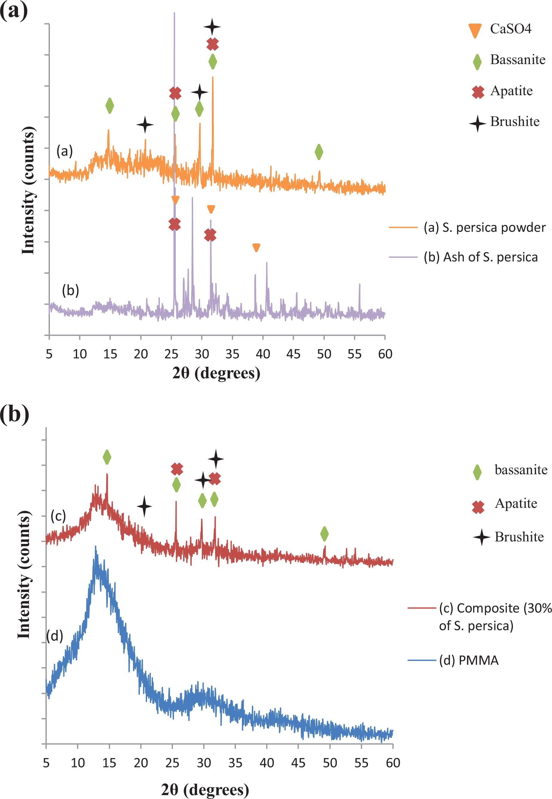
XRD patterns: (a) (inset a) S. persica powder and (inset b) ash of S. persica. (b) (inset c) Composite (30 wt% of S. persica) and (inset d) PMMA. XRD: X-ray diffraction; PMMA: poly(methyl methacrylate).
The powder of S. persica was ignited at 650°C, for 48 h until maintaining constant weights. Figure 1(inset b) presents the X-ray diffractograms of mineral compounds. The observed scans of ash of S. persica present the major peaks. They were most probably corresponding to anhydrite CaSO4 (25.1° (310), 31.5° (202), and 38.4° (312)) 19 and the apatite (26° (002) and 32°(112)). 20
Figure 1(inset a) shows the peaks of the S. persica powder and presents its mineral and organic compounds. A characteristic amorphous hump was observed around 2θ = 15° and 25°, which present the cellulose.
For the mineral compounds, the scan can prove the presence of apatite, brushite, and bassanite. The characteristic peaks of bassanite (CaSO4·0.5H2O) were 2θ = 15° (101), 25.26° (002), 29.9° (202), 32.5° (222), and 49.7° (442) according to Dogan et al. 21 It has been shown that the bassanite, also called, plaster of Paris, is safe and effective for a variety of clinical applications, such as medical and dental orthopedics. 22
Moreover, the XRD of S. persica powder showed the presence of characteristic peaks of brushite (CaHPO4·2H2O) at 21°, 29.18°, and 31.5° corresponding to Miller indices (12-1), (14-1), and (21-1), respectively.20,23 It is worth noting that crystals of brushite exist in abundance in bones and dental salts. 24
Considering the above results, the S. persica powder contains the hydrated calcium sulfate (CaSO4·xH2O) and the calcium phosphate compounds which contain the orthophosphate group PO43−. These calcium phosphate compounds reveal excellent bioactivity and biocompatibility in the physiological conditions due to their chemical similarities to the inorganic part of bone tissue, tooth enamel, and dentin.25,26
This finding is in agreement with previous research works. Ahmad and Rajagopal have proved the presence of flavonoids, the organic sulfur compounds, and carbohydrates in S. persica. 27 The antioxidant activity occurs due to the presence of several biologically active chemical constituents such as flavonoids. 14 The different compounds in S. persica, such as polyphenol, flavonoids, saponins, salvadorine, and alkaloids, have antioxidant and antibacterial effects.9,10 S. persica has significant antimicrobial activity facing aerobic and anaerobic bacteria collected from teeth with inflamed gums and necrotic pulps. 27 Chlorine, trimethyl amine, alkaloid resin, and sulfur compounds present in S. persica have antimycotic effect. 27 Thus, S. persica contains mineral and organic compounds that can be used in dental applications. The organic compounds were responsible for many mechanisms, such as antimycotic effects, antimicrobial, and antioxidant properties.
All these compounds, of very different classes, can be present in S. persica powder, and they probably contribute to the biological activity of the plant, which makes us think that they act synergistically.
Ultimately, the major peaks of the composite (30 wt% of S. persica) were recorded at 2θ values of 14.73°, 25.62°, 29.78°, 31.84°, and 49.21° corresponding to the characteristic peaks of S. persica (Figure 1(inset c)).
Infrared spectroscopy
It should be noted, firstly, that FTIR spectra are used not only to indicate the details of functional groups present in materials but also to investigate the incorporation of S. persica powder into the PMMA biopolymer matrix. Figure 2(a) shows FTIR spectra of Nic Tone dental resin. The observed peaks at 2948 and 1720 cm−1 can be assigned to C–H stretching vibrations and C=O stretching vibrations in PMMA, respectively. The two double bands at (1140 and 1190 cm−1) and (1237 and 1265 cm−1) correspond to the C–O stretching vibrations of ester groups. The peak at 963 cm−1 can be attributed to C–O stretching. The absorptions around 1431 and 1474 cm−1 characterize the asymmetric bending vibrations of (C–CH3) and (C–CH2) bonds in PMMA, respectively. Similar reflectance bands were observed in FTIR of pure PMMA in the literature.28,29
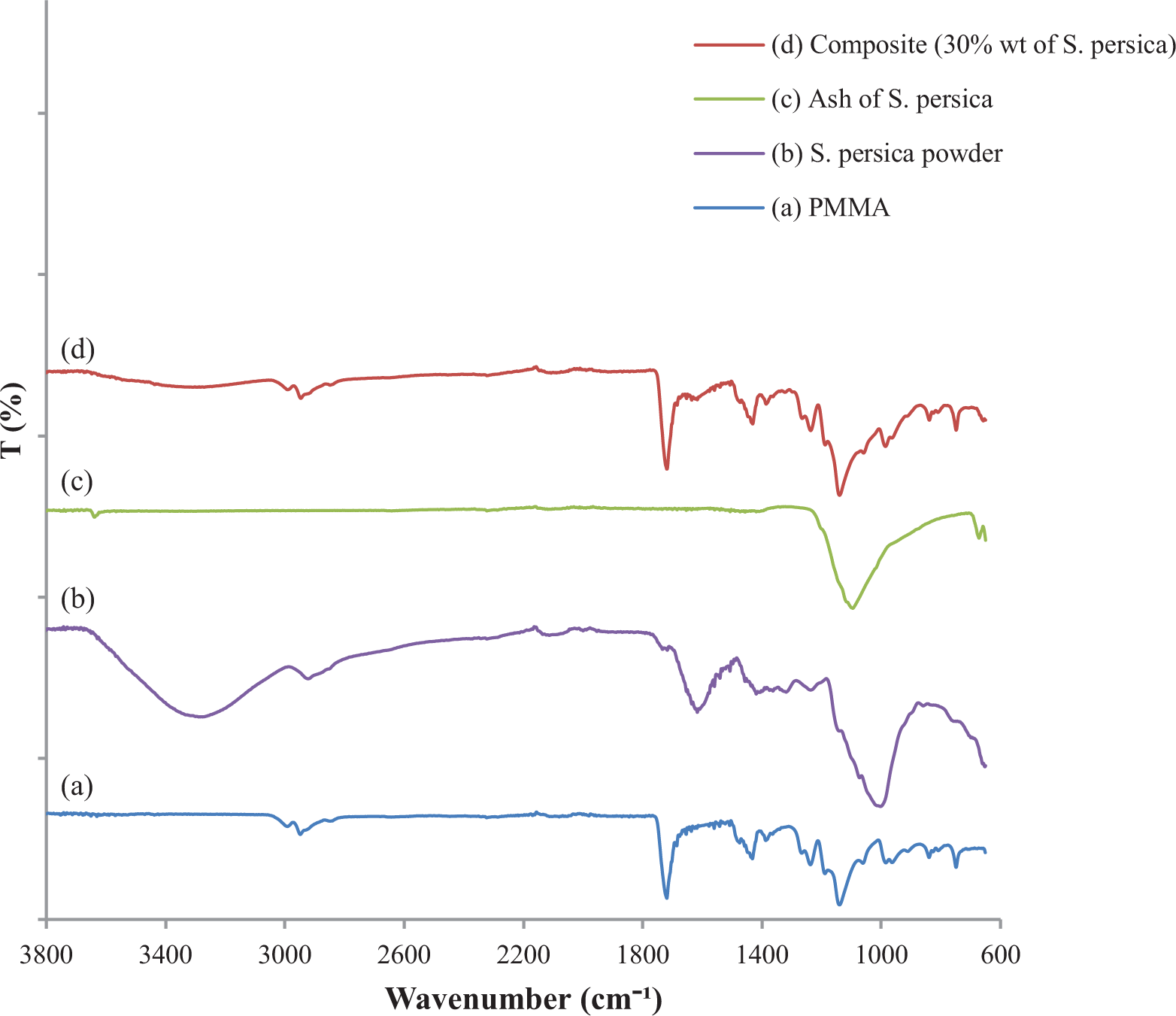
The FTIR spectra of (a) PMMA, (b) S. persica powder, (c) ash of S. persica, and (d) composite (30 wt% of S. persica). FTIR: Fourier transform infrared spectroscopy; PMMA: poly(methyl methacrylate).
The FTIR spectrum of S. persica (Figure 2(b)) indicates the details of functional groups present in the filler. The broad strong absorption band between 3600 and 3000 cm−1 is due to the hydrogen bonded OH stretching vibrations and –NH2 stretching. The shoulder at 2924 cm−1 most probably arises from CH stretching vibrations. The band at 1718 cm−1 corresponds to carbonyl stretching vibration of carboxylic acids of hemicellulose. The band at 1669, 1615, and 1513 cm−1 is due to the benzene ring and –C=O group of quercetin. 30 The band near 1420 cm−1 is due to CH3 deformation in lignin and xylan, as well as CH2 bending in xylan. A weak band at 1237 cm−1 is possibly ascribed to C–O–C band in the cellulose chain. The broad band between 1160 and 900 cm−1 would correspond to the asymmetrical bridge C–O–C stretching, C–O stretching, or O–H bending of the C–OH group in the cellulose and hemicellulose.12,31 The band at 1100 cm−1 may be due to the absorption of PO43− ions as reported by Bahabri. 31 According to Mao et al., 32 the band at 1005 cm−1 is due to (SO42−) stretching mode. The CH stretching of the phenyl ring is assigned at 848 and 726 cm−1. 33 The FTIR spectra of S. persica powder prove the presence of hydrated calcium sulfate and the calcium phosphate compounds.
The IR spectrum of the ash of S. persica is shown in Figure 2(c). The infrared spectra of the inorganic portion revealed that the spectra of the samples exhibit absorption bands at the frequencies of 1100 and 674 cm−1. According to Bahabri 31 and Wu et al., 34 the band at 1100 cm−1 may be due to the absorption of PO43− ions. The band at 674 cm−1 is the characteristic band of anhydrite CaSO4 as reported by Bahabri. 31
As shown in Figure 2(d), compared with the spectra of PMMA, the spectra of the obtained composite (30 wt% of S. persica) reveal that some bands have their intensity changed and a new absorption peak appeared. The peaks at 2107, 1140, and 963 cm−1 intensity slightly increased and a small peak at 1617 cm−1 appeared.
Finally, we can conclude from Figure 2 that the spectra of pure PMMA and the composites exhibit some difference, indicating that the inclusion of S. persica powder in the polymer matrix is rather physical with some chemical bonds.
Scanning electron microscopy
Figure 3 shows the SEM images of the morphology of S. persica powder and the fracture surface of elaborated composite. As can be observed, the microstructure of S. persica shows that the cells are nearly spherical and irregular in size and distribution. Furthermore, it is important to point out the presence of some crystals in the S. persica powder (Figure 3(a)) and in the resulting composite (Figure 3(b)) corresponding to bassanite (calcium sulfate hemihydrate: CaSO4·½H2O) crystals of biogenic origin ranged from 10 µm to 20 µm in size. 21 The existence of this biomineral, extracted from the stems of S. persica, was already confirmed by the powder XRD analysis. The use of a thin window energy dispersive X-ray spectroscopy (EDS) enabled the detection of oxygen (O), and the EDS data showed that the mineral was composed of Ca, S, and O (Table 3). According to Dogan et al., the beneficiary effects of S. persica are the biomineral bassanite. 21
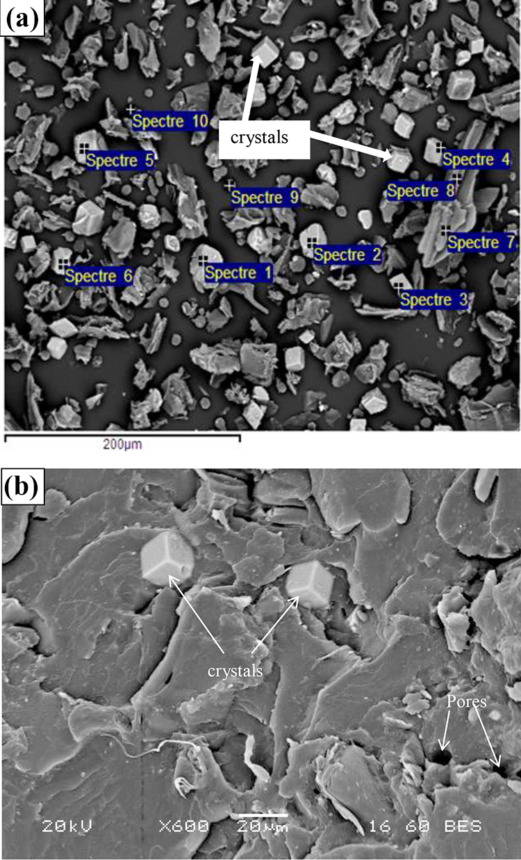
SEM images of the S. persica powder (a) and the resulting composite (30 wt% of S. persica) (b). SEM: scanning electronic microscope.
EDS of the S. persica powder.
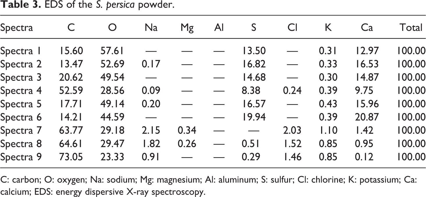
C: carbon; O: oxygen; Na: sodium; Mg: magnesium; Al: aluminum; S: sulfur; Cl: chlorine; K: potassium; Ca: calcium; EDS: energy dispersive X-ray spectroscopy.
Figure 3(b) demonstrates a good adhesion between PMMA and S. persica and proves the presence of pores, which locate in the matrix and not in the interface between matrix and filler. The pores can be taken into account for the stress concentration, which plays an important role in the mechanical properties.
The contents of residual monomers in the elaborated composite
The reaction of polymerization between the polymer and the monomer is more or less incomplete, leaving part of the free monomers able to diffuse in the oral medium. These residual monomers are regarded as poison when its proportion is more than 0.4% of the finished product. Residual monomers in polymerized denture base polymers cause the oral mucosa inflammatory symptoms and the irritations of the mucous membranes. 35 Therefore, it is necessary to determine the percentage of the residual monomers using DSC and HPLC.
Thermal analysis
Figure 4 shows typical DSC thermograms of the composite. The thermogram obtained on the scan (heating curve) reveals no exothermic peak but only a glass transition pattern (Tg). A possible explanation for the absence of polymerization peak is the absence of residual monomers in the composite. 13
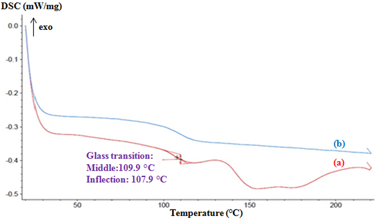
DSC thermograms of the resulting composite (30 wt% of S. persica). DSC: differential scanning calorimetry.
To confirm this hypothesis, we try to determine the percentage of residual monomers using HPLC.
High-performance liquid chromatography
Figure 5 presents the chromatogram of the reference which contains the MMA and a low percentage of glycol ether. Two peaks have been distinguished. As for the first, it is between 7 min and 8 min, which is a very important peak, and the second is between 11.5 min and 12 min, which is a low peak. According to Viljanena et al., 36 the peak of MMA was among 7–8 min. However, the chromatogram obtained on the second scan (Figure 6) shows no significant peak between 7 min and 8 min. Therefore, we can conclude that the composite analyzed in the present study does not present residual monomers. This reveals a positive effect on the biocompatibility of the composite as reported by Viljanena et al. 36 The migration of residual monomers, which are toxic, was avoided in the body and the oral environment.
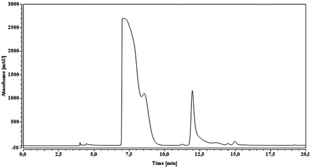
Chromatogram of the sample “Reference.”
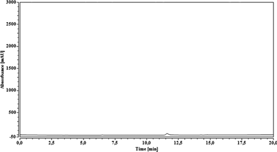
Chromatogram of the resulting composite (30 wt% of S. persica).
Antioxidant activity of elaborated composite
Dental amalgam, which contains mercury, has an oxidative action on brain neurons. This phenomenon is an etiological factor of Alzheimer’s disease. 37 For that, the development of dental material, which has antioxidant activity, has a positive effect on oral hygiene and helps the body to protect itself against various types of oxidative damage.
Antioxidant property analysis
The purpose of this test is to evaluate the antioxidant activity of S. persica powder and the influence of polymerization conditions on the antioxidant activity of S. persica powder. The phosphomolybdenum method is routinely applied in laboratory to evaluate the TAC of plant extracts. TAC was determined in the extract of artificial saliva. The total antioxidant evaluation of the target compounds was performed through the formation of a green phosphomolybdenum complex by the reduction of Mo(VI) to Mo(V). The antioxidant activity of fresh S. persica was found to be about 677.72 ± 52 µg of GAE/mL of extract and 630.75 ± 116 µg of GAE/mL of extract for S. persica powder (T = 60°C, P = 0.5 MPa, t = 10 min). These results were important comparing to the results of Noumi et al. (528 µg of GAE/g of DW). 38 Thus, these finding results confirm that the S. persica has antioxidant activity and one applying the polymerization conditions, the antioxidant activity of S. persica powder remains constant (p > 0.05) (Figure 7). Furthermore, previous research works have shown that the polyphenol and flavonoid are responsible for various biological activities, especially its antioxidant and antimicrobial activity. 10 That is why, the determination of total polyphenol and flavonoid content in S. persica and the resulting composite can prove the antioxidant activity of the resulting composite.
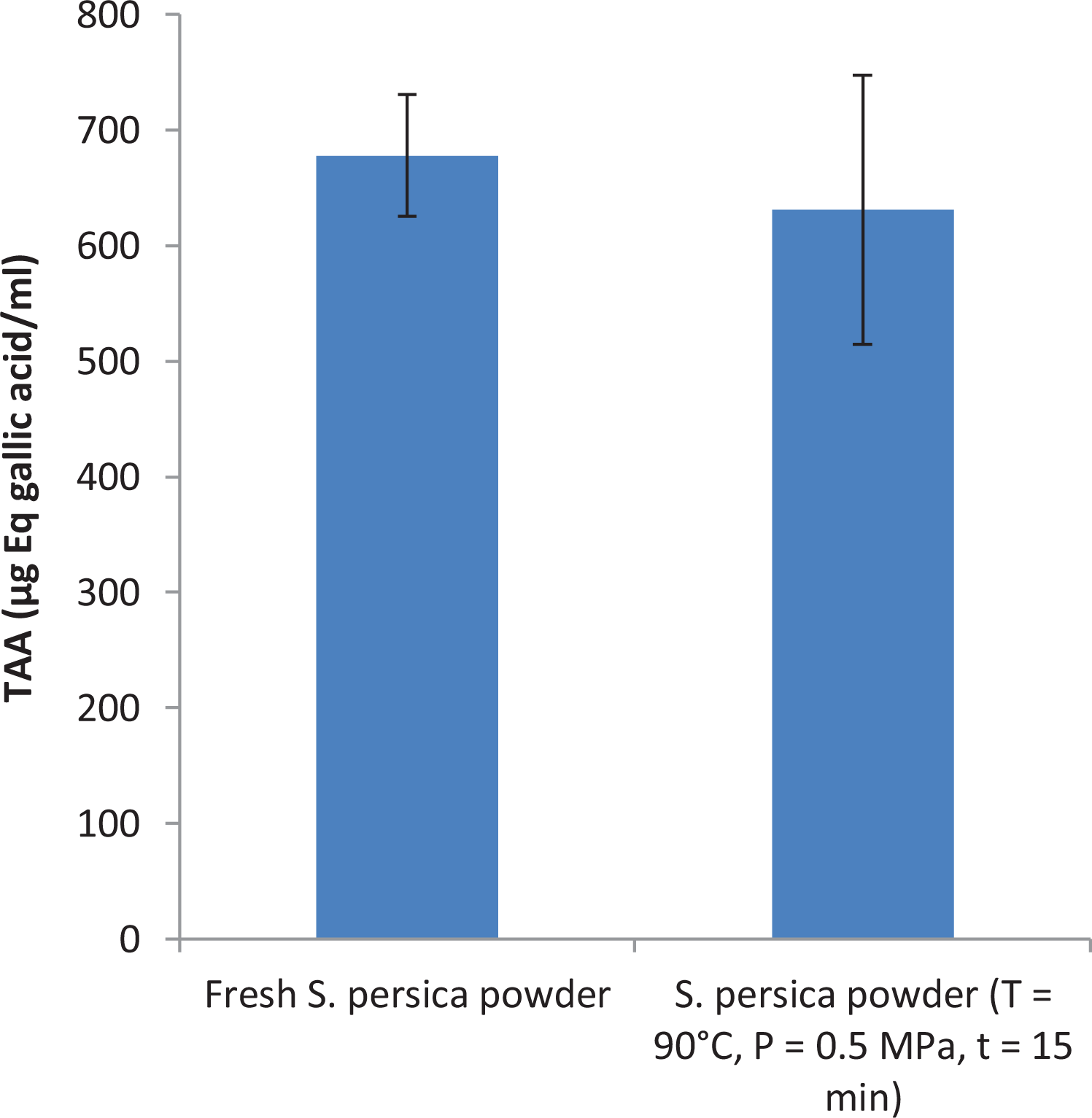
Total antioxidant activity of S. persica powder.
Total polyphenol and flavonoid content
The total polyphenol concentrations present in the EES and EEC were 87.79 ± 1.32 mg of GAE/g of S. persica and 71.37 ± 4.89 mg of GAE/g of composite, respectively. These results were comparable to those found in other studies. In fact, Nadia et al. revealed that the content of the total polyphenol content from the leaves of S. persica was 70 mg of GAE/g of DW. 39 Concerning Taha et al., they found that the amount of the total extractable polyphenol from the bark and leaves of S. persica was 52.6 and 166.7 mg of GAE/g of DW, respectively. 40 The total flavonoid concentrations present in the EES and EEC were 52.12 ± 2.32 mg of QE/g of S. persica and 43.62 ± 5.90 mg of QE/g of composite, respectively. The ratios of total flavonoids/total polyphenol in S. persica (0.59) and in the composite (0.61) indicate high proportions of flavonoids. These results are in agreement with the study carried out by Ibrahim et al. 10 who attributed the antioxidant activities to the presence of flavonoids and polyphenolic compounds in S. persica. The important concentrations of polyphenol and flavonoid in EES can establish the antioxidant activity of S. persica. The total polyphenol and flavonoid concentrations in EEC were decreased compared to the total polyphenol and flavonoid concentrations in EES, but they remain important and prove the antioxidant activity of the resulting composite (30 wt% of S. persica powder).
Antibacterial activity of elaborated composite
The development of bacteria on the surface of the dental prosthesis, producing acid layer, accelerates the damage and the failure of dental composite prosthesis. To overcome this problem, we aimed to develop a bioactive material that can limit the prosthesis aggression caused by bacteria. Therefore, we elaborated a biocomposite reinforced with 30% of S. persica and we tested its antibacterial activity.
Figure 8 shows the antibacterial activity of S. persica and the S. persica composite against S. pneumonia, S. mutans, and H. influenza. Both materials had antibacterial activity. A previous study indicated that S. persica exhibited strong antibacterial activity.9,41,42 But, the S. persica composite had less inhibitory effects on the growth of bacteria than S. persica alone. This fact can be explained by the effect of the matrix, which decreases the release of the antibacterial agent. However, we succeeded to elaborate an antibacterial dental composite. The demonstration of S. persica composite antibacterial activity against the test bacteria is an indication that alternative antibiotic substances in S. persica could be used for the development of an antibacterial dental composite in future.
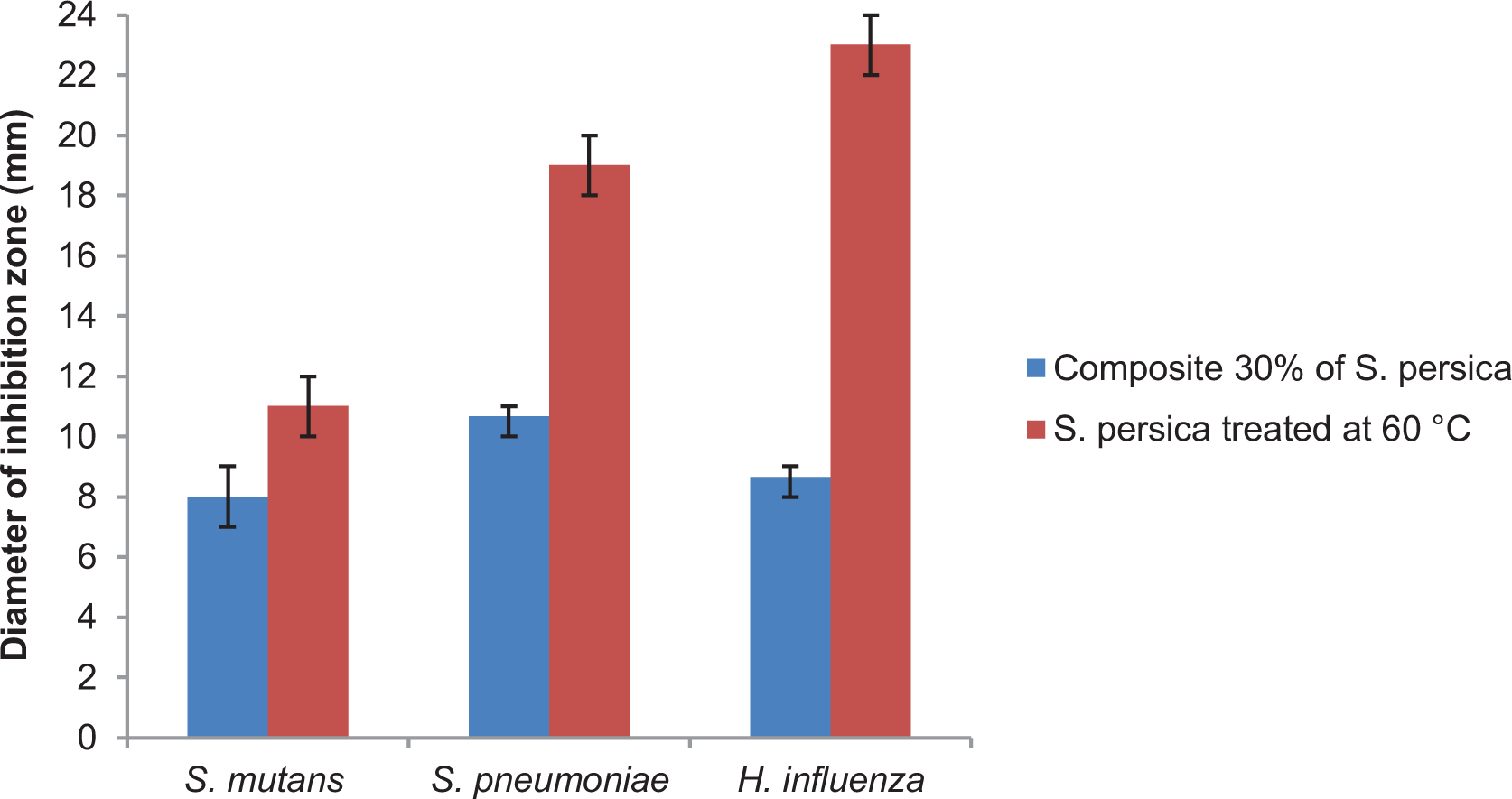
Antibacterial activity of S. persica and the composite (30% of S. persica).
Conclusion
The work presented in this article focused on the elaboration and characterization of a biocomposite material based on PMMA/S. persica. From the main results, the following conclusion can be drawn:
XRD revealed the presence of the hydrated calcium sulfate and the calcium phosphate compounds in S. persica, which are used in dental applications. In addition, the organic compounds were responsible for many mechanisms, such as antimicrobial and antioxidant properties. The FTIR analysis of S. persica powder proves also the presence of hydrated calcium sulfate and the calcium phosphate compounds. Moreover, they indicated the presence of quercetin (antioxidant agent). In fact, the inclusion of S. persica powder in the PMMA matrix is rather physical with some chemical bond. SEM images and EDS analysis show the presence of bassanite crystals of biogenic origin in the S. persica powder and in the resulting composite, besides a good adhesion between PMMA and S. persica was illustrated. The obtained composite did not present residual monomers that cause the oral mucosa inflammatory symptoms and the irritations of the mucous membranes. The antioxidant and the antibacterial tests prove the antioxidant and antibacterial activities of S. persica and the resulting composite.
Footnotes
Acknowledgements
The authors would like to express our special thanks of gratitude to Pr. Emna Ammar (National Engineering School in Sfax, University of Sfax) as well as Mme. Wafa Gargouri (Research Group of Agri-Food Processing Engineering, National School of Engineers of Sfax, University of Sfax) for their assistance in laboratory analyses and Mme. Leila Mahfoudhi (Faculty of Sciences, Sfax) for proof-reading our manuscript.
Funding
The author(s) received no financial support for the research, authorship, and/or publication of this article.