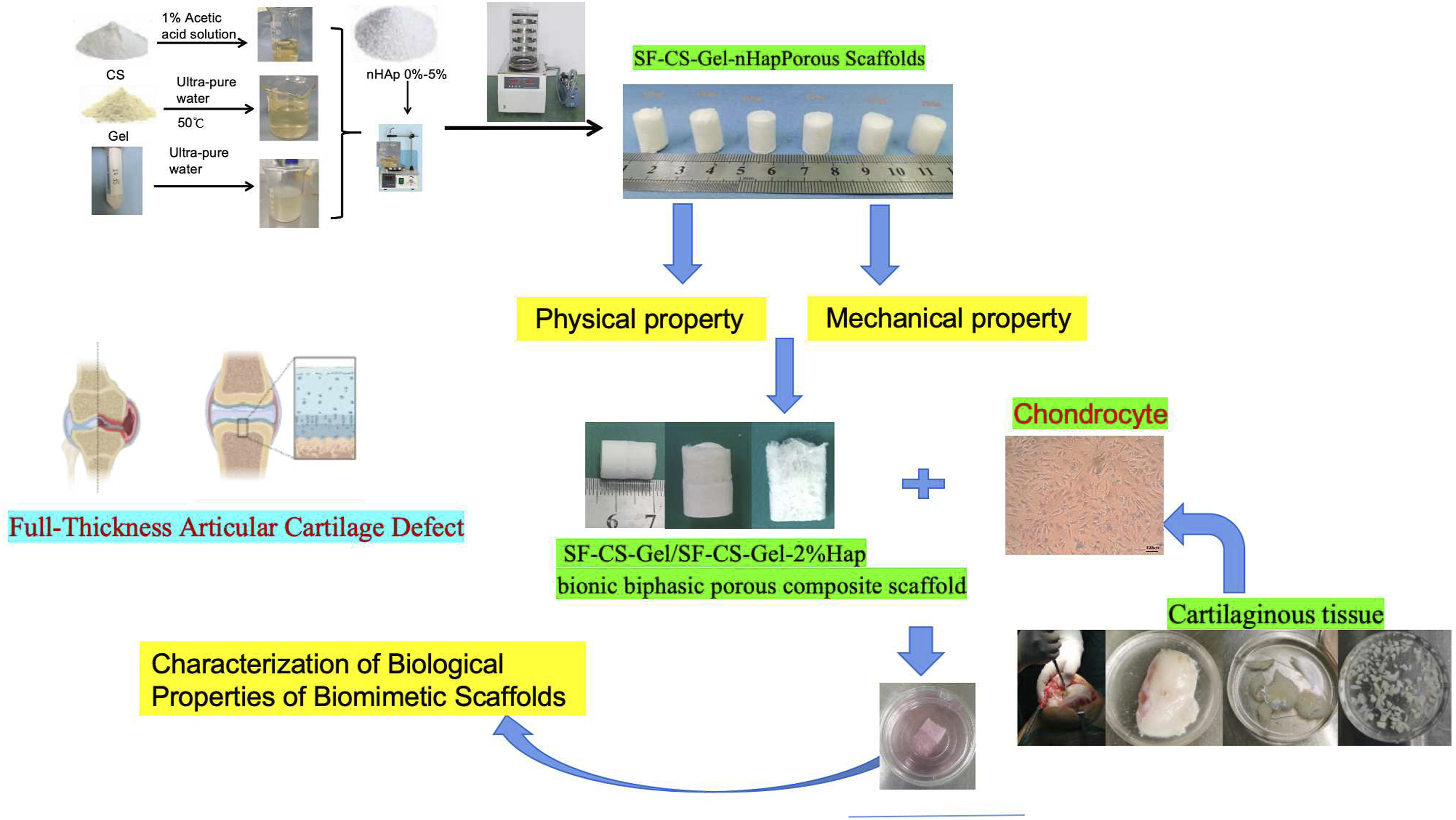
Objective: Full-thickness cartilage defect are usually accompanied by subchondral bone damage, which is difficult to self-repair once damaged due to the lack of vascularization and innervation. In this study, a biphasic composite scaffold was developed by combining vacuum freeze-drying and iterative freeze-thawing with gelatin, chitosan, silk fibroin, and hydroxyapatite as the basic materials to explore the feasibility of using them for the repair of total cartilage defects. Methods and Results: Six groups of SF-CS-Gel-nHap porous scaffolds (Hap-0%, Hap-1%, Hap- 2%, Hap-3%, Hap-4%, Hap-5%) were prepared by vacuum freeze-drying and chemical cross-linking using filipin protein (SF), gelatin (Gel), chitosan (CS) and hydroxyapatite (Hap) as the base materials. A series of characterization methods were used to systematically analyze and test the morphological features as well as physical and mechanical properties of the scaffolds. Then a novel bionic biphasic porous scaffold was developed by a combination of freeze-drying and freeze-thawing using the SF-CS-Gel as the cartilage phase and the SF-CS-Gel-2%Hap as the subchondral bone phase. Finally, it was co-cultured with chondrocytes to verify the biological properties of the SF-CS-Gel/SF-CS-Gel-2%Hap bionic biphasic porous composite scaffold in vitro. The results showed that the SF-CS-Gel/SF-CS-Gel-2%Hap biphasic scaffolds had a highly porous mesh structure, with an average pore size of 156.06 ± 42.36 μm in the cartilage phase and 214.38 ± 65.82 μm in the subchondral bone phase. Co-cultured with chondrocytes, the live and dead cells stained, cck-8 growth and proliferation curves showed that the bionic scaffolds had good biocompatibility and cytotoxicity. Cytoskeletal staining showed that the morphology of chondrocytes in the bionic scaffolds could maintain three-dimensional growth as in vivo. Conclusion: The results showed that SF-CS-Gel/SF-CS-Gel-2%Hap biphasic scaffolds have good biocompatibility, biodegradability, stability, appropriate mechanical properties and porosity, and are suitable for repairing articular cartilage and subchondral bone. It is expected to be used as a repair material for articular cartilage in clinical applications.
DaouFCochisALeighebM, et al.Current advances in the regeneration of degenerated articular cartilage: a literature review on tissue engineering and its recent clinical translation. Materials2021; 15(1): 31.
2.
ShantoPCParkSFahadMAA, et al.3D bio-printed proteinaceous bioactive scaffold loaded with dual growth factor enhanced chondrogenesis and in situ cartilage regeneration. Bioact Mater2024; 46: 365–385.
3.
ChangLRMarstonGMartinA. Anatomy, cartilage. 2022 oct 17. In: StatPearls [Internet]. Treasure Island (FL). StatPearls Publishing, 2024.
4.
ThunsiriKPitjamitSPothacharoenP, et al.The 3D-printed bilayer's bioactive-biomaterials scaffold for full-thickness articular cartilage defects treatment. Materials2020; 13(15): 3417.
5.
BaoWLiMYangY, et al.Advancements and frontiers in the high performance of natural hydrogels for cartilage tissue engineering. Front Chem2020; 8: 53.
6.
PogliacomiFSchiaviPParaskevopoulosA, et al.When is indicated viscosupplementation in hip osteoarthritis?Acta Biomed2018; 90(1-S): 67–74.
7.
LeighebMBosettiMDe ConsoliA, et al.Chondral tissue engineering of the reumatoid knee with collagen matrix autologous chondrocytes implant. Acta Biomed2017; 88(4S): 107–113.
8.
WangHWangZLiuH, et al.Three-dimensional printing strategies for irregularly shaped cartilage tissue engineering: current state and challenges. Front Bioeng Biotechnol2022; 9: 777039.
9.
TsanaktsidouEKammonaOKiparissidesC. Recent developments in hyaluronic acid-based hydrogels for cartilage tissue engineering applications. Polymers2022; 14(4): 839.
10.
RadulescuDMNeacsuIAGrumezescuAM, et al.New insights of scaffolds based on hydrogels in tissue engineering. Polymers2022; 14(4): 799.
11.
XiaoHHuangWXiongK, et al.Osteochondral repair using scaffolds with gradient pore sizes constructed with silk fibroin, chitosan, and nano-hydroxyapatite. Int J Nanomed2019; 14: 2011–2027. DOI: 10.2147/IJN.S191627.
12.
ShalumonKTLaiGJChenCH, et al.Modulation of bone-specific tissue regeneration by incorporating bone morphogenetic protein and controlling the shell thickness of silk fibroin/chitosan/nanohydroxyapatite core-shell nanofibrous membranes. ACS Appl Mater Interfaces2015; 7(38): 21170–21181.
13.
SunWGregoryDATomehMA, et al.Silk fibroin as a functional biomaterial for tissue engineering. Int J Mol Sci2021; 22(3): 1499. DOI: 10.3390/ijms22031499.
14.
LiYZhangYWeiY, et al.Preparation of chitosan-based injectable hydrogels and its application in 3D cell culture. J Vis Exp2017; 29(127): 56253.
15.
LiJWangQGuY, et al.Production of composite scaffold containing silk fibroin, chitosan, and gelatin for 3D cell culture and bone tissue regeneration. Med Sci Monit2017; 23: 5311–5320.
16.
ShangLMaBWangF, et al.Nanotextured silk fibroin/hydroxyapatite biomimetic bilayer tough structure regulated osteogenic/chondrogenic differentiation of mesenchymal stem cells for osteochondral repair. Cell Prolif2020; 53(11): e12917.
17.
MedvedevaEVGrebenikEAGornostaevaSN, et al.Repair of damaged articular cartilage: current approaches and future directions. Int J Mol Sci2018; 19(8): 2366.
18.
ZhangTZhangHZhangL, et al.Biomimetic design and fabrication of multilayered osteochondral scaffolds by low-temperature deposition manufacturing and thermal-induced phase-separation techniques. Biofabrication2017; 9(2): 025021.
19.
MellatiAFanCMTamayolA, et al.Microengineered 3D cell-laden thermoresponsive hydrogels for mimicking cell morphology and orientation in cartilage tissue engineering. Biotechnol Bioeng2017; 114(1): 217–231.
20.
ZhangBHuangJNarayanRJ. Gradient scaffolds for osteochondral tissue engineering and regeneration. J Mater Chem B2020; 8(36): 8149–8170.
21.
WangMLuoYYuY, et al.Bioengineering approaches to accelerate clinical translation of stem cell therapies treating osteochondral diseases. Stem Cell Int2020; 2020: 8874742.
22.
BecerraJRodriguezMLealD, et al.Chitosan-collagen-hydroxyapatite membranes for tissue engineering. J Mater Sci Mater Med2022; 33(2): 18.
23.
HafeziMNouri KhorasaniSZareM, et al.Advanced hydrogels for cartilage tissue engineering: recent progress and future directions. Polymers2021; 13(23): 4199.
24.
DaiWSunMLengX, et al.Recent progress in 3D printing of elastic and high-strength hydrogels for the treatment of osteochondral and cartilage diseases. Front Bioeng Biotechnol2020; 8: 604814.
25.
QiJYuTHuB, et al.Current biomaterial-based bone tissue engineering and translational medicine. Int J Mol Sci2021; 22(19): 10233.
26.
ArmientoARStoddartMJAliniM, et al.Biomaterials for articular cartilage tissue engineering: learning from biology. Acta Biomater2018; 65: 1–20.
27.
FuNDongTMengA, et al.Research progress of the types and preparation techniques of scaffold materials in cartilage tissue engineering. Curr Stem Cell Res Ther2018; 13(7): 583–590.
28.
QasimMChaeDSLeeNY. Advancements and frontiers in nano-based 3D and 4D scaffolds for bone and cartilage tissue engineering. Int J Nanomed2019; 14: 4333–4351.
29.
NgadiminKDStokesAGentileP, et al.Biomimetic hydrogels designed for cartilage tissue engineering. Biomater Sci2021; 9(12): 4246–4259.
30.
LeeSChoiJYounJ, et al.Development and evaluation of gellan gum/silk fibroin/chondroitin sulfate ternary injectable hydrogel for cartilage tissue engineering. Biomolecules2021; 11(8): 1184.
31.
ZhengACaoLLiuY, et al.Biocompatible silk/calcium silicate/sodium alginate composite scaffolds for bone tissue engineering. Carbohydr Polym2018; 199: 244–255.
32.
Grabska-ZielińskaSSionkowskaACarvalhoÂ, et al.Biomaterials with potential use in bone tissue regeneration-collagen/chitosan/silk fibroin scaffolds cross-linked by EDC/NHS. Materials2021; 14(5): 1105.
33.
AsadpourSKargozarSMoradiL, et al.Natural biomacromolecule based composite scaffolds from silk fibroin, gelatin and chitosan toward tissue engineering applications. Int J Biol Macromol2020; 154: 1285–1294.
34.
YePYuBDengJ, et al.Application of silk fibroin/chitosan/nano-hydroxyapatite composite scaffold in the repair of rabbit radial bone defect. Exp Ther Med2017; 14(6): 5547–5553.
35.
JabbariFHesarakiSHoushmandB. The physical, mechanical, and biological properties of silk fibroin/chitosan/reduced graphene oxide composite membranes for guided bone regeneration. J Biomater Sci Polym Ed2019; 30(18): 1779–1802.
36.
RoginaARicoPGallego FerrerG, et al.In situ hydroxyapatite content affects the cell differentiation on porous chitosan/hydroxyapatite scaffolds. Ann Biomed Eng2016; 44(4): 1107–1119.
37.
DengLLiYZhangA, et al.Nano-hydroxyapatite incorporated gelatin/zein nanofibrous membranes: fabrication, characterization and copper adsorption. Int J Biol Macromol2020; 154: 1478–1489.
38.
LohQLChoongC. Three-dimensional scaffolds for tissue engineering applications: role of porosity and pore size. Tissue Eng Part B2013; 19(6): 485–502.
39.
KumarSSDAbrahamseH. Advancement of nanobiomaterials to deliver natural compounds for tissue engineering applications. Int J Mol Sci2020; 21(18): 6752.
40.
XiaPLuoY. Vascularization in tissue engineering: the architecture cues of pores in scaffolds. J Biomed Mater Res B Appl Biomater2022; 110(5): 1206–1214.
41.
RatnayakeJTBMucaloMDiasGJ. Substituted hydroxyapatites for bone regeneration: a review of current trends. J Biomed Mater Res B Appl Biomater2017; 105(5): 1285–1299.
42.
ZhaoZFanCChenF, et al.Progress in articular cartilage tissue engineering: a review on therapeutic cells and macromolecular scaffolds. Macromol Biosci2020; 20(2): e1900278.
43.
HuangBChenMTianJ, et al.Oxygen-carrying and antibacterial fluorinated nano-hydroxyapatite incorporated hydrogels for enhanced bone regeneration. Adv Healthcare Mater2022; 11(12): e2102540.
44.
ZhangYLiJMouserVHM, et al.Biomimetic mechanically strong one-dimensional hydroxyapatite/poly(d,l-lactide) composite inducing formation of anisotropic collagen matrix. ACS Nano2021; 15(11): 17480–17498.
45.
VolpiMParadisoACostantiniM, et al.Hydrogel-based fiber biofabrication techniques for skeletal muscle tissue engineering. ACS Biomater Sci Eng2022; 8(2): 379–405.
46.
CostantiniMTestaSFornettiE, et al.Engineering muscle networks in 3D gelatin methacryloyl hydrogels: influence of mechanical stiffness and geometrical confinement. Front Bioeng Biotechnol2017; 5: 22.
47.
SongJETripathyNLeeDH, et al.Quercetin inlaid silk fibroin/hydroxyapatite scaffold promotes enhanced osteogenesis. ACS Appl Mater Interfaces2018; 10(39): 32955–32964.
48.
JunIHanHSEdwardsJR, et al.Electrospun fibrous scaffolds for tissue engineering: viewpoints on architecture and fabrication. Int J Mol Sci2018; 19(3): 745.
49.
PereiraDRReisRLOliveiraJM. Layered scaffolds for osteochondral tissue engineering. Adv Exp Med Biol2018; 1058: 193–218.
50.
ShimomuraKMoriguchiYMurawskiCD, et al.Osteochondral tissue engineering with biphasic scaffold: current strategies and techniques. Tissue Eng Part B2014; 20(5): 468–476.
51.
KangHZengYVargheseS. Functionally graded multilayer scaffolds for in vivo osteochondral tissue engineering. Acta Biomater2018; 78: 365–377.
52.
FilardoGPerdisaFGelinskyM, et al.Novel alginate biphasic scaffold for osteochondral regeneration: an in vivo evaluation in rabbit and sheep models. J Mater Sci Mater Med2018; 29(6): 74.
53.
SartoriMPaganiSFerrariA, et al.A new bi-layered scaffold for osteochondral tissue regeneration: in vitro and in vivo preclinical investigations. Mater Sci Eng C2017; 70(Pt 1): 101–111.