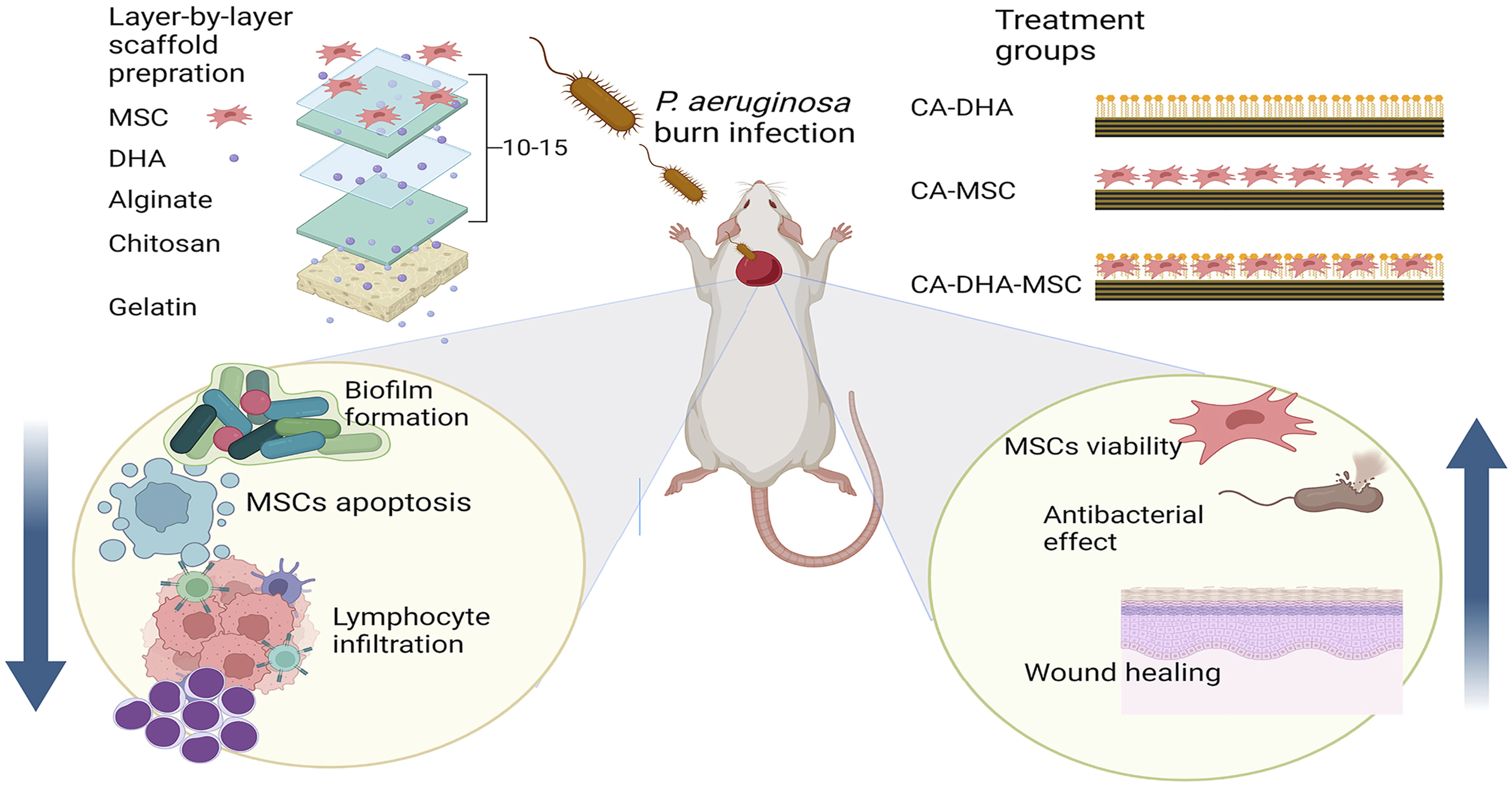
Aims: Chitosan, like docosahexaenoic acid (DHA) and mesenchymal stem cells (MSCs), is used in medicine as a wound healing accelerator. Thus, in this study, chitosan-alginate (CA) membranes containing DHA and MSCs were produced, and their antibacterial and antibiofilm activities against burn infections caused by Pseudomonas aeruginosa were investigated.
Methods: Physicochemical properties were assessed by SEM, Fourier transform infrared (FTIR), and X-ray diffraction (XRD). Porosity, cytocompatibility, and antibacterial and antibiofilm activities were evaluated both in vitro and in vivo. The viability and apoptosis of MSCs were studied using flow cytometry. Wound healing effects were analyzed based on histopathological features, the wound contraction rate (WCR) ratio, and bacterial clearance.
Results: The CA membranes showed antibiofilm activity both in vivo and in vitro, accompanied by reduced lasI and rhlI expressions and pyocyanin production. The membranes were highly porous and biocompatible and showed favorable physicochemical properties. Docosahexaenoic acid incorporation to CA membranes improved their antibacterial and antibiofilm activities, as well as MSCs’ viability by reducing crystallinity and increasing porosity (p = .008). Treatment with CA-DHA-MSC accelerated burn wound healing (with complete healing being observed after 14 days, WCR = 85%) and augmented antibacterial and antibiofilm activities in vivo compared to CA-DHA and CA-MSC. The CA-DHA-MSC group delivered a significantly higher WCR and lower inflammation than the CA-MSC group (p = .0001).
Conclusion: In combination with DHA-loaded CA membranes, MSCs reduced the healing time of burn wounds, offering a viable option for designing effective wound dressings.
JiangBTangYWangH, et al.Down-regulation of long non-coding RNA HOTAIR promotes angiogenesis via regulating miR-126/SCEL pathways in burn wound healing. Cell Death Dis2020; 11(1): 61.
2.
EskandariniaAKefayatAAghebM, et al.A novel bilayer wound dressing composed of a dense polyurethane/propolis membrane and a biodegradable polycaprolactone/gelatin nanofibrous scaffold. Sci Rep2020; 10(1): 3063.
3.
MaticaMAAachmannFLTøndervikA, et al.Chitosan as a wound dressing starting material: ntimicrobial properties and mode of action. Int Journal Molecular Sciences2019; 20(23): 5889.
4.
BagherZEhteramiASafdelMH, et al.Wound healing with alginate/chitosan hydrogel containing hesperidin in rat model. J Drug Deliv Sci Technol2020; 55: 101379.
5.
MoranHBTTurleyJLAnderssonM, et al.Immunomodulatory properties of chitosan polymers. Biomaterials2018; 184: 1–9.
6.
TorkamanSRahmaniHAshoriA, et al.Modification of chitosan using amino acids for wound healing purposes: a review. Carbohydr Polymers2021; 258: 117675.
7.
OlivaresEBadel-BerchouxSProvotC, et al.Clinical impact of antibiotics for the treatment of pseudomonas aeruginosa biofilm infections. Front Microbiol2019; 10: 2894.
8.
DumontMVilletRGuirandM, et al.Processing and antibacterial properties of chitosan-coated alginate fibers. Carbohydr Polym2018; 190: 31–42.
9.
ThayaRVaseeharanBSivakamavalliJ, et al.Synthesis of chitosan-alginate microspheres with high antimicrobial and antibiofilm activity against multi-drug resistant microbial pathogens. Microb Pathog2018; 114: 17–24.
10.
SilvaJRBurgerBKuhlCMC, et al.Wound healing and omega-6 fatty acids: from inflammation to repair. Mediators Inflamm2018; 2018: 2503950.
11.
ChenYQiuXYangJ. Comparing the in vitro antitumor, antioxidant and anti-inflammatory activities between two new very long chain polyunsaturated fatty acids, docosadienoic acid (dda) and docosatrienoic acid (dta), and docosahexaenoic acid (DHA). Nutr Cancer2021; 73(9): 1697–1707.
12.
Casillas-VargasGOcasio-MalaveCMedinaS, et al.Antibacterial fatty acids: an update of possible mechanisms of action and implications in the development of the next-generation of antibacterial agents. Prog Lipid Res2021; 82: 101093.
13.
SunYSongLZhangY, et al.Adipose stem cells from type 2 diabetic mice exhibit therapeutic potential in wound healing. Stem Cell Res Ther2020; 11(1): 298.
14.
TamamaKKerpedjievaSS. Acceleration of wound healing by multiple growth factors and cytokines secreted from multipotential stromal cells/mesenchymal stem cells. Adv Wound Care (New Rochelle)2012; 1(4): 177–182.
15.
KongYXuRDarabiMA, et al.Fast and safe fabrication of a free-standing chitosan/alginate nanomembrane to promote stem cell delivery and wound healing. Int J Nanomedicine2016; 11: 2543–2555.
16.
LiXZhangYQiG. Evaluation of isolation methods and culture conditions for rat bone marrow mesenchymal stem cells. Cytotechnology2013; 65(3): 323–334.
17.
LawrieGKeenIDrewB, et al.Interactions between alginate and chitosan biopolymers characterized using FTIR and XPS. Biomacromolecules2007; 8(8): 2533–2541.
18.
KarunathilakaSRChoiSHMossobaMM, et al.Rapid classification and quantification of marine oil omega-3 supplements using ATR-FTIR, FT-NIR and chemometrics. J Food Compost Anal2019; 77: 9–19.
19.
ČernáMBarrosASNunesA, et al.Use of FT-IR spectroscopy as a tool for the analysis of polysaccharide food additives. Carbohydr Polym2003; 51(4): 383–389.
20.
PetrovaVAGolovkinASMishaninAI, et al.Cytocompatibility of bilayer scaffolds electrospun from chitosan/alginate-chitin nanowhiskers. Biomedicines2020; 8(9): 305.
21.
Abou-OkeilAAlyAAAmrA, et al.Biocompatible hydrogel for cartilage repair with adjustable properties. Polym Adv Tech2019; 30(8): 2026–2033.
22.
NiLKuangZGongZ, et al.Dihydroartemisinin promotes the osteogenesis of human mesenchymal stem cells via the ERK and Wnt/β-catenin signaling pathways. Biomed Res Int2019; 2019: 3456719.
23.
ShinSYBajpaiVKKimHR, et al.Antibacterial activity of bioconverted eicosapentaenoic (EPA) and docosahexaenoic acid (DHA) against foodborne pathogenic bacteria. Int J Food Microbiol2007; 113(2): 233–236.
24.
WuSWangSWangL, et al.Docosahexaenoic acid supplementation represses the early immune response against murine cytomegalovirus but enhances NK cell effector function. BMC Immunol2022; 23(1): 1–16.
25.
HuebingerRMStonesDHde Souza SantosM, et al.Targeting bacterial adherence inhibits multidrug-resistant Pseudomonas aeruginosa infection following burn injury. Sci Rep2016; 6(1): 39341.
26.
Guillamat-PratsR. The role of MSC in wound healing, scarring and regeneration. Cells2021; 10(7): 1729.
27.
HirakawaMPTjahjonoNLightYK, et al.Upregulation of CD14 in mesenchymal stromal cells accelerates lipopolysaccharide-induced response and enhances antibacterial properties. iScience2022; 25(2): 103759.
28.
HirakawaMPTjahjonoNLightYK, et al.Augmentation of antibacterial activity in mesenchymal stromal cells through systems-level analysis and crispr-mediated activation of CD14. Available at SSRN 3732415. 2020.
29.
Castañeda-TamezPRamírez-PerisJPérez-VelázquezJ, et al.Pyocyanin restricts social cheating in Pseudomonas aeruginosa. Front Microbiology2018; 9: 1348.
30.
de Cássia OliveiraVSteixnerSdo NascimentoC, et al.Expression of virulence factors by Pseudomonas aeruginosa biofilm after bacteriophage infection. Microb Pathogenesis2021; 154: 104834.
31.
WoodCRAl DhahriDAl DelfiI, et al.Human adipose tissue-derived mesenchymal stem/stromal cells adhere to and inhibit the growth of Staphylococcus aureus and Pseudomonas aeruginosa. J Med Microbiol2018; 67(12): 1789–1795.
32.
LiL-lZhuY-gJiaX-m, et al.Adipose-derived mesenchymal stem cells ameliorating Pseudomonas aeruginosa–induced acute lung infection via inhibition of NLRC4 Inflammasome. Front Cellular Infection Microbiol2021; 10: 830.
33.
ÇankiriliNKKartDÇelebi-SaltikB. Evaluation of the biofilm formation of Staphylococcus aureus and pseudomonas aeruginosa on human umbilical cord CD146+ stem cells and stem cell-based decellularized matrix. Cell Tissue Banking2020; 21(2): 215–231.
34.
HongSLuY. Omega-3 fatty acid-derived resolvins and protectins in inflammation resolution and leukocyte functions: targeting novel lipid mediator pathways in mitigation of acute kidney injury. Front Immunol2013; 4: 13.
35.
MendtMRezvaniKShpallE. Mesenchymal stem cell-derived exosomes for clinical use. Bone Marrow Transplant2019; 54(2): 789–792.