
Abstract
To identify the effectiveness of polymer-induced liquid precursors (PILP) on dentin remineralization and the assessment approaches used to evaluate remineralization. The analyses were done into six stages: (1) Identifying the research question; (2) Identifying the studies; (3) Selecting relevant studies; (4) Plotting the data; (5) Reporting results; and (6) Risk of bias. The searches were carried out in the following databases: PubMed, Web of Science, and Scopus. A total of 247 articles were identified in the electronic database search. After applying the eligibility criteria, only 12 articles were included for data extraction. The outcomes of dentin remineralization patterns were plotted in association to PILP agent type and application mode. The intrafibrillar mineralization occurred in 78% of studies where polyaspartic acid (pAsp) was used, and 50% when the polyacrylic acid (PAA) was used. All the studies where PILP was used in a restorative material (resin composite, glass ionomer, and adhesive system)—extra and intrafibrillar mineralization pattern was found. The association of PILP agents with other materials with potential for mineralization showed beneficial results in remineralization, since they provide a medium to high concentration of calcium and phosphate. The pAsp showed better results for interfibrillar remineralization when compared to PAA. The association with Ca/P release materials or solution Ca P are essentials for mineralization via PILP agent. Further studies are needed to assess the effectiveness of remineralization through PILP agents because the level of evidence of the studies was low.
Introduction
The caries-affected dentin (CAD) presents altered physical and chemical properties, because of successive cycles of des- and remineralization. CAD has reduced inter-tubular mineral content 1 revealing the loss of calcium, phosphate, and magnesium, which make the substrate porous and with low mechanical properties. 2 Transmission electron microscopy (TEM) observations have revealed that when the adhesive systems are applied on this substrate, a porous zone appears beneath the hybrid layer, leaving the collagen fibrils unprotected. 3
Dentinal collagen exposed by etch-and-rinse systems has been reported to be highly vulnerable to hydrolytic and enzymatic degradation processes. 4 In addition, self-etch adhesives use calcium to chemically bond to molecules like methacryloyloxydecyl dihydrogen phosphate (MDP) of the adhesive systems and form stable salts. As the mineral content on CAD is reduced, it has been pointed out as one of the possible reasons for the low bond strength reported on long-term basis. 5 Breschi et al. 6 have also reported less bond strength, long-term degradation, and a compromised longevity of the restorations on CAD. For that reason, the remineralization of demineralized dentin should represent a favorable impact on the adhesion to resin monomers.
The strategies to remineralize dentin consist of two main mechanisms. The first is the classical remineralization, where calcium and phosphate ions are deposited on the existing apatite crystals. 7 However, with the traditional ion-based pathway, remineralization does not occur in places where crystals are absent. 8 It was observed that the deposit of mineral occurs superficially or only on the extrafibrillar portion of the dentin, without achieving intrafibrillar mineralization.9 –11 The second mechanism, the non-classical pathway, is based on biomimetic agents analogous to non-collagenous proteins that mineralize the intrafibrillar collagen. This mineralization increases the mechanical properties of dentin,11,12 and also protects collagen molecules from external challenges—such as temperature, endogenous enzymes, bacterial acids, and other chemical factors. 13 The Gower group suggested a model of remineralization using a PILP (polymers-induced liquid precursors) process based on the formation of a liquid precursor system induced by polymer 14 and were successful in remineralizing a variety of organic matrices. 15
The PILP process consists of adding anionic polymers to a supersaturated mineralization solution, in order to induce the formation of an amorphous calcium phosphate (ACP) precursor that mineralizes the intrafibrillar collagen.9,14,16 PILP agents aim to stabilize the calcium phosphate ions, preventing spontaneous nucleation, and mineral precipitation before reaching the desired location.14,17,18 In the lumen of collagen fibrils, PILP agents have the ability to deposit their content, forming an amorphous calcium phosphate. Then, the ACP will transform into apatite crystallites oriented in a similar way to the nanocrystals observed in mineralized tissues.14,19
Some molecules such as osteopontin, 20 poly acrylic acid (PAA), 21 and polyaspartic acid (pAsp) 16 are considered PILP agents. Aspartic acid was the first PILP agent used for biomimetic remineralization in dentin, 16 and most of the studies available to date evaluate it. However, there is still some controversy as to whether PILP agents can actually remineralize apatite in collagen tissues, mainly whether it is able to promote intrafibrillar remineralization.
There are several ways to evaluate the effects of biomimetic agents on demineralized dentin: chemically, by crystallographic parameters and by the mechanical properties of dentin—but there is still no consensus on which is more suitable. 18 Thus, this review aimed to evaluate the available scientific evidence on the effectiveness of PILP agents on dentin remineralization, and the results related to the chemical, crystallographic, and mechanical properties in the treated dentin.
Materials and methods
Study design
The “scoping review” was the research methodology selected. This methodology maps the existing literature on a complex topic being reviewed for the first time in terms of volume, nature, and characteristics of primary research. We assess the literature on polymer-induced liquid precursors (PILP) for dentin remineralization. The methodology was divided into six stages according to Arskey and O’Malley’s 22 : (1) Identification of the research question; (2) Identification of studies; (3) Selection of relevant studies; (4) Charting the Data; (5) Report of results; and (6) Risk of bias.
Stage I: “Research question.”
Is there scientific evidence that PILP agents induce intrafibrillar and extrafibrillar mineralization in dentin?
Stage II: Identification of studies
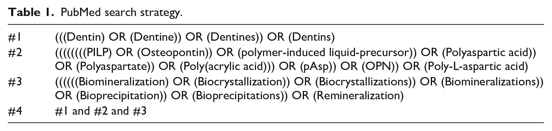
PubMed, Web of Science, and Scopus databases were searched. The search strategy developed for PubMed (Table 1) was used as a blueprint and adapted to the other databases. Two hundred and forty-seven articles were found and grouped with the EndNote program (Clarivate Analytics—Online Version), then duplicates were excluded leaving 209 articles. The flowchart for the search and screening strategy can be seen in Figure 1.
PubMed search strategy.
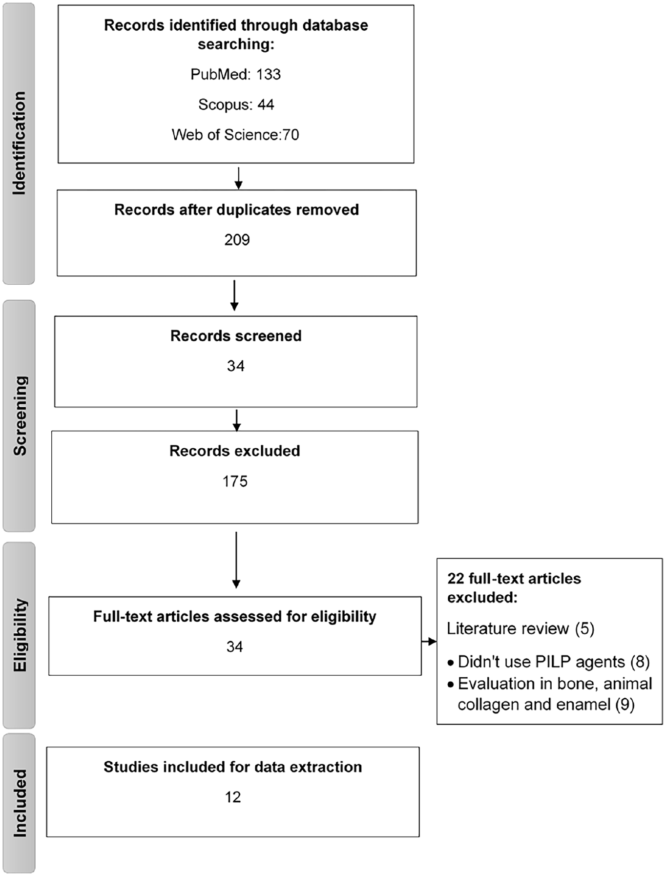
PRISMA flowchart of literature search.
Stage III: Selection of relevant studies
Abstracts of the 209 studies found were revised, and 175 abstracts that not corresponding to the research question of this review were excluded. The selection of the studies was performed by two evaluators separately. In case of doubt, a third evaluator was included.
After the selection phase, the articles were assessed according to the following criteria:
Inclusion criteria: Studies that evaluate dentin remineralization using PILP; in-vitro and in-vivo studies; articles in English; publication date from 2008 to 2020.
Exclusion criteria: Studies that evaluate remineralization in bone; animal collagen and enamel; articles in languages other than English; literature reviews.
Stage IV: Charting the data
The data obtained from all studies were allocated in a spreadsheet (Microsoft Excel) to perform the data extraction regarding the type of PILP agent, association with restorative material, assessment methodology, type of mineralization, objective, main results, and conclusion. This stage was performed by two calibrated authors.
Stage V: Report of results
The extracted data were organized according to: (1) Type of PILP; (2) PILP Application method; (3) Biomimetic remineralizing effect on dentin. In addition, a table was prepared with the main information for each article (Table 2).
Description of objectives, type of PILP, method of application, method of assessment, and principal results of the studies.
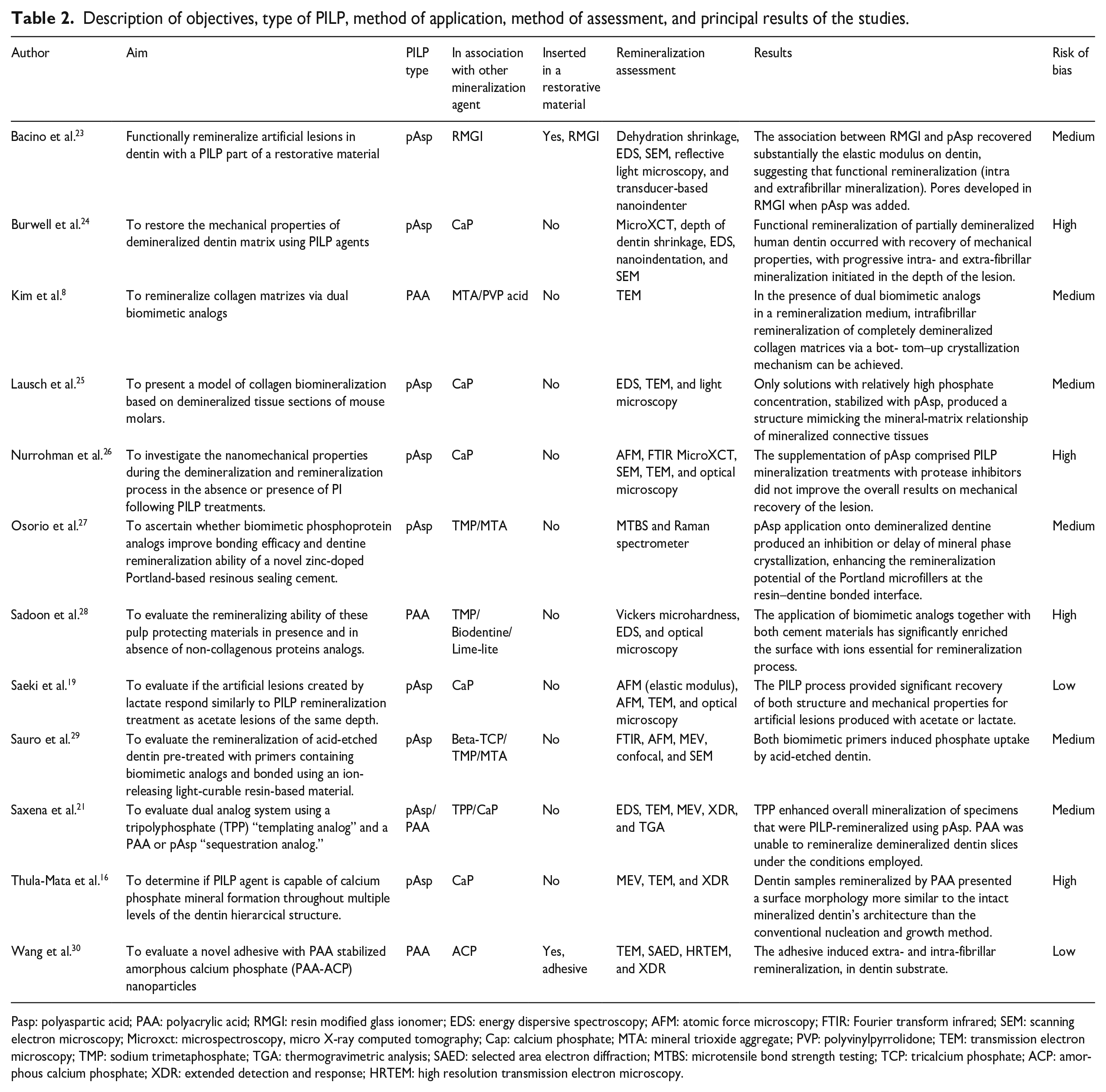
Pasp: polyaspartic acid; PAA: polyacrylic acid; RMGI: resin modified glass ionomer; EDS: energy dispersive spectroscopy; AFM: atomic force microscopy; FTIR: Fourier transform infrared; SEM: scanning electron microscopy; Microxct: microspectroscopy, micro X-ray computed tomography; Cap: calcium phosphate; MTA: mineral trioxide aggregate; PVP: polyvinylpyrrolidone; TEM: transmission electron microscopy; TMP: sodium trimetaphosphate; TGA: thermogravimetric analysis; SAED: selected area electron diffraction; MTBS: microtensile bond strength testing; TCP: tricalcium phosphate; ACP: amorphous calcium phosphate; XDR: extended detection and response; HRTEM: high resolution transmission electron microscopy.
Stage VI: Risk of bias
All included studies were assessed for risk of bias. The following topics were evaluated: Sampling bias-sample size, Sampling Bias-Blindness, Conflict Bias, Sampling statistic, Assessment Bias and Funding Bias (Table 3). The risk of bias was assessed by two evaluators (MAAF and PANS) considering the patterns described in Table 3, in case of disagreement a third evaluator (CSG) was recruited to define the analysis.
Quality assessment list.
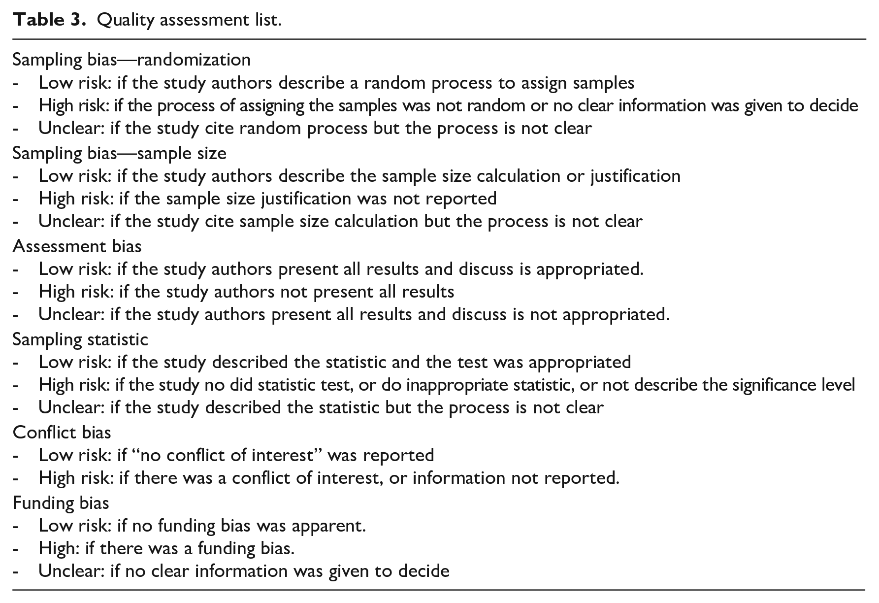
All evaluators judged each item as “low,” “unclear,” or “high.” The risk of bias for each study was established according to the number of items marked with “low bias.” Studies that presented between 6 and 5 items at risk of “low bias” were classified as “low risk”; 4–3 items at risk of “low bias” were classified as “unclear”; and 2 or less items at risk of “low bias” were classified as “high risk.”
Results
A total of 247 articles were identified in the electronic database search. After removing the duplicated studies, 209 remained. Among them, 175 were excluded based on the title and abstract. The 34 full-text studies assessed for eligibility, 12 remained and were included in this scoping review (Figure 1). These studies ranged in publication date from 2010 to 2020.
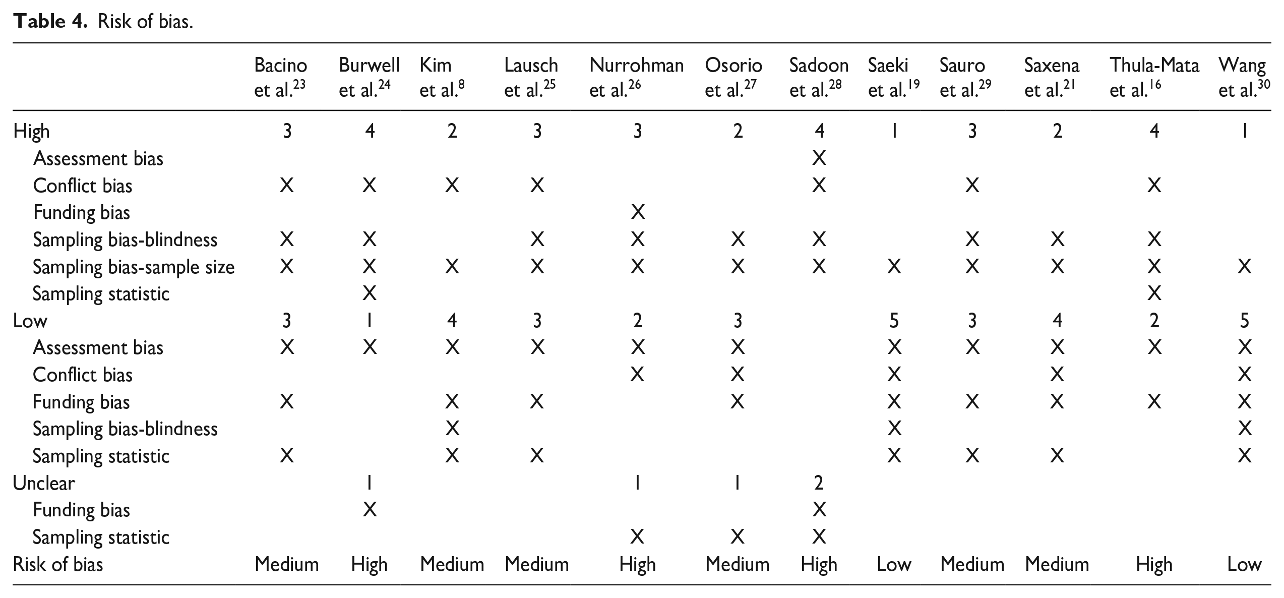
The risk of bias for included studies can be observed in Table 4. Only two articles showed low risk of bias. Six articles showed a medium risk of bias, and four had a high risk of bias. For all studies, the sample size was neglected (high risk)—being an important factor for the quality of the studies.
Risk of bias.
The principal findings of this study are shown in Table 2. The outcomes of dentin mineralization pattern were plotted in association with PILP type and application mode (Figures 2 and 3). The intrafibrillar mineralization occurred in 78% of studies (seven articles16,21,23,24,26,27,29) where polyaspartic acid was used. When polyacrylic acid was used the intrafibrillar mineralization was 50% (two articles8,30; Figure 2).
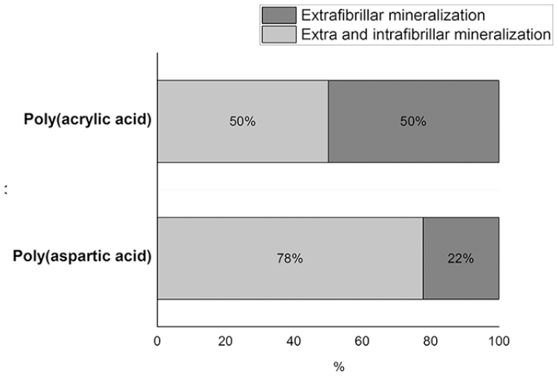
Dentin mineralization pattern according to the type of PILP.
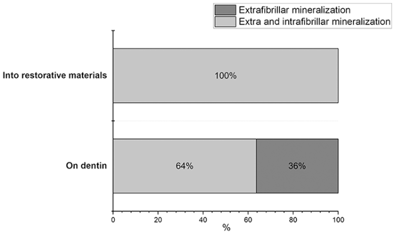
Dentin mineralization pattern according to the PILP application mode.
All two studies23,30 where PILP was applied together with the restorative material showed extra and intrafibrillar mineralization (Figure 3). When the material was applied on dentin, 64% of the studies (seven articles8,16,21,24,26,27,29) demonstrated extra and intra-fibrillar mineralization. Of these, one article 8 used polyacrylic acid and 616,21,24,26,27,29 articles polyaspartic acid. The 36% of the works where there was extrafibrillar mineralization when the material was applied on the dentin were composed of two articles that used polyacrylic acid21,28 and two article that used polyaspartic acid19,25 (Figure 3).
Discussion
Our findings revealed that in most of the studies PILP agents promoted intra and extrafibrillar mineralization in dentin where pAsp was used in 69% of the studies, and 78% of them demonstrated to promote intrafibrillar remineralization.8,16,21,23,24,26,27,29,30
A PILP is an anionic polymer that acts as an analog of non-collagenous extracellular matrix proteins, regulating mineral deposition, and resulting in biomimetic mineralization. 31 This means that remineralization occurs from the bottom to the top of the cavity (in nanoscale) following the non-classical theory of mineralization. 8 Due to the polymers size, it is able to penetrate in the intrafibrillar spaces of dentin. Then, the PILP sequesters calcium ions that build up a positive charge to sequester phosphate (the opposite charge) to induce the formation of a nanoprecursor of ACP and promote mineralization. 32
Two types of PILP have been used for this purpose, PAA and pAsp—the latter being used in most studies and obtaining the best results. This can be explained because pAsp will avoid crystal precipitation, while allowing calcium and phosphate to remain available to diffuse in the dentin collagen. 27 Thus, the remineralization initiation process occurs more slowly (7 days) with pAsp than with other agents, and at the same time it is possible to obtain a greater mineral recovery of the tissue. 24
For the PAA, the intrafibrillar remineralization happened in 50% of the studies (Figure 2).8,21,28,30 A study that compared PAA with pAsp, showed less mineralization for PAA. 21 The results were probably related to the lower molecular weight of PAA (1.8 kDa) compared to pAsp (27 kDa). 21 According to the “concept of molecules selective permeability in collagen,” molecules larger than 40 kDa are excluded from the intrafibrillar water compartments of the fibrillar collagen, whereas molecules smaller than 6 kDa cannot remain in the intrafibrillar spaces. 18 Thus, nucleation was overly inhibited in the PAA groups. 21 Demonstrating that mineralization is facilitated by a range of molecular weights both above and below the critical 6 kDa level. Another possible explanation is that, in contrast to polyacrylic acid (PAA), polyaspartic acid (pAsp) possesses terminal amine groups, allowing it to closely mimic the structure of non-collagenous proteins essential to the remineralization process. This unique feature of pAsp potentially facilitates stronger interactions with the mineral phase. 8 Additionally, a comprehensive exploration of other characteristics, including the stereochemistry and molecular weight of the polymer, could shed light on how specific properties of polyaspartic acid contribute to its enhanced mineralization performance. On the other hand, the intrafibrillar mineralization was efficient when PAA was incorporated with ACP nanoparticles, being a promising strategy for future research. 30
Another approach is the association of tripolyphosphate (TPP) with a PILP where an improved mineralization process was showed. 21 TPP acts as a dual-analog system, where the TPP serves as a “templating analog,” mimicking the highly phosphorylated domains within many non-collagenous proteins. In contrast, the PAA is referred to as the “sequestration analog” because it contains repeated carboxylic acid side chains that mimic the acidic domains on many non-collagenous proteins—which are thought to play a role in stabilizing amorphous calcium phosphate precursors. 21 Saxena et al. 21 used PAA and pAsp after pretreatment of collagen with TPP and obtained promising results—increasing general mineralization. TPP seemed to have the function of inhibiting mineralization on the substrate surfaces, leaving the tubules open, allowing the PILP to penetrate. Besides, it may be inhibiting mineral formation by chelating Ca2+ ions and preventing them from associating with free phosphates. However, neither PILP alone nor PILP with TPP pretreatment could fully remineralize these substrates at all depths.
Additionally, some studies also associated PILP agents with other materials with mineralization potential, like supersaturated Ca P solutions, sodium trimetaphosphate (TMP), Portland cement (MTA), and beta tricalcium phosphate (β-TCP; Table 2). The studies showed that these associations are essential in producing biomimetic mineralized tissue since a medium with relatively high calcium-phosphate (CaP) concentration is necessary for mineralization, 25 which may be the reason most studies assessed hypersaturated CaP solutions. 27 Other studies8,27,29 reported that the use of doped resin composites with Portland cement and β-TCP over the PILP layer were important for calcium and phosphate release. In addition, it was shown that when PILP was not associated with these ion-releasing materials, mineralization did not occur. 29
TMP is a substantial source of P ions and it was used in three studies.27 –29 Although in the study by Osorio et al. 27 the association between TMP and pAsp was not favorable, other studies have shown that TMP can contribute to intrafibrillar mineralization.28,29
PILP agents have only been associated with restorative materials in two studies (Resin-Modified Glass-Ionomer (RGMI) and adhesives), which showed no influence on the mechanism of PILP extra and intrafibrillar remineralization.23,30 Bacino et al. 23 promoted remineralization adding up to 40% wt of pAsp powder in glass ionomer. However, pAsp does not integrate well within the resin matrix of the cement—instead it separates from the matrix phase forming pores, which negatively impacted on the mechanical properties of the cement. Wang et al. 30 found that PAA added to an adhesive was able to remineralize intra and extrafibrillar zones. This is of great advantage since during a restorative procedure the regular adhesives cannot penetrate the intrafibrillar areas, and then they are filled with water. PILP agents are able to penetrate into those regions and promote remineralization. 8 However, more studies are needed—especially in vivo.
Nanomechanical properties, mineral composition (Ca and P), and images showing direct evidence of mineral deposition and crystal growth were the most common properties assessed, and should be employed to have reliable remineralization outcomes and conclusions. The methods used to assess mineral density, such as energy-dispersive X-ray spectroscopy (EDS) and 3D X-ray computed tomography (XCT), not always corresponded to the results for mechanical properties, 24 but are probably more accurate than solely image analyses like Scanning Electron Microscopy (SEM). Saxena et al. 21 observed remineralization in their specimens using SEM; however, when evaluated through EDS, the intrafibrillar mineral deposition was much lower than expected. 21 Transmission Electron Microscopy (TEM) was the most used methodology because its high magnification allows examining the presence, orientation, or dislocations of crystals. 8 However, the quality of the equipment is crucial, as Kim et al. 8 showed problems to interpret their results for such reasons. X-ray diffraction (XDR) was also very useful to identify the nature and crystallinity of the mineral formed by PILP treatments. 24
Two methodologies that were less used were dehydration shrinkage and nanomechanical tests of the remineralized dentin lesion; however, they are suitable indicators of functional remineralization of dentin matrices. Burwell et al. 24 found that the PILP used recovered the full mineral density, but the modulus (nano-indentation) was not fully reestablished since it is believed that intrafibrillar mineralization is the one that increases the mechanical properties.11,16 In this test, it is worthy to highlight that the nanoindentations should be performed on hydrated tissue, since dehydration alters the mechanical response of the collagenous matrix and provides little information about the functionality of dentin. So, when carious or partially demineralized dentin is measured dry, the demineralized portion collapses and its measurement gives erroneously high values as showed by Bertassoni et al. 11
The studies evaluated in this review showed several limitations. Most studies were in-vitro, and the caries lesions were produced by chemical methods. In vivo and in vitro studies inducing caries by the biological method are necessary. Only two studies presented low risk of bias, and none reported the sample calculation. Future studies should describe with more detail the methodologic aspects.
Thus, the available information on biomimetic mineralization by PILP in dentin remains limited. Further studies, particularly in vivo, employing rigorous methodologies, are needed to assess the effectiveness of remineralization through PILP agents, considering the low level of evidence found in the existing studies
Conclusion
The pAsp showed better results for interfibrillar remineralization when compared to PAA. The association with materials with Ca P release or Ca P solutions are essentials for mineralization via PILP agent. Further studies are needed to assess the effectiveness of remineralization through PILP agents since the level of evidence of the studies was low.
Footnotes
Declaration of conflicting interests
The author(s) declared no potential conflicts of interest with respect to the research, authorship, and/or publication of this article.
Funding
The author(s) disclosed receipt of the following financial support for the research, authorship, and/or publication of this article: The authors thank the Coordination for the Improvement of Higher Education Personnel (CAPES) by the financial support (Code 001).