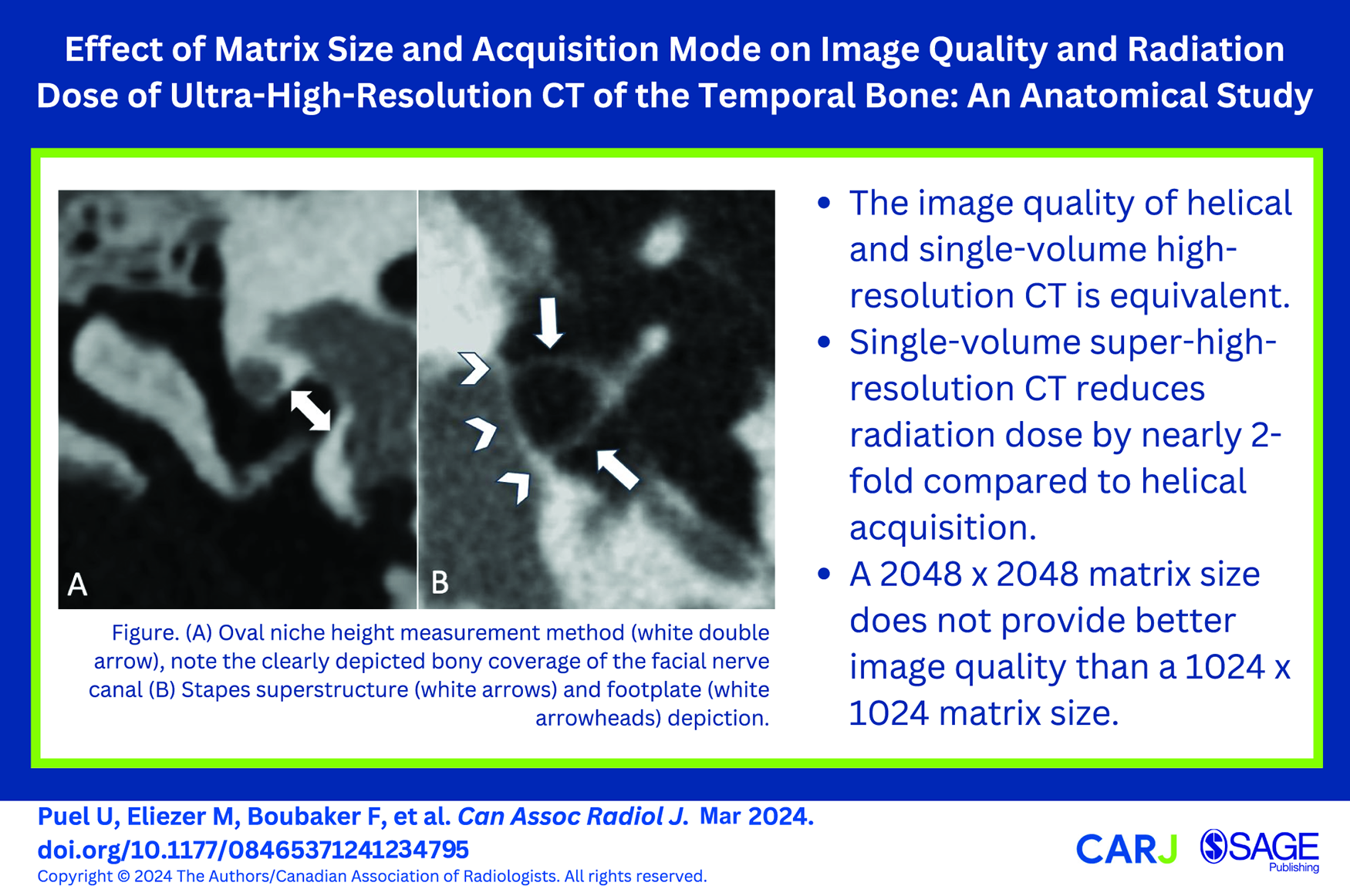
Purpose: To compare image quality and radiation exposure between super- and ultra-high-resolution helical and super-high-resolution volumetric CT of the temporal bone. Methods: Six cadaveric temporal bone specimens were used to evaluate key temporal bone structures using the following CT reconstruction and acquisition modes: helical and single-volume acquisition modes in super-high resolution (0.25-mm slice thickness, 10242 matrix), and helical mode in ultra-high resolution (0.25-mm slice thickness, 20482 matrix). Two observers performed 5 previously described preoperative measurements, measured noise and signal-to-noise ratios for air, and noise for bone, and rated the visualization of 5 anatomical structures on a 4-point scale, for each reconstruction mode. Radiation dose exposure was recorded for each examination. Results: There was no significant difference between any of the quantitative or qualitative measurements in any of the reconstruction and acquisition modes. There was a slight increase in noise and a decrease in signal-to-noise ratio in the air using the single-volume mode (115 ± 13.1 HU and 8.37 ± 0.91, respectively) compared to the helicoidal super-high-resolution (92.4 ± 11.8 HU and 10.8 ± 1.26, respectively) and helicoidal ultra-high-resolution (91.1 ± 10.7 HU and 10.9 ± 1.39, respectively) modes (P < .002). The volumic CT dose index was 50.9 mGy with helical acquisition and 29.8 mGy with single-volume acquisition mode (P < .0001). Conclusion: The single-volume super-high-resolution acquisition mode allows a reduction in radiation dose exposure without compromising image quality compared to helical scanning, but with a slightly lower signal-to-noise ratio in air with the single-volume mode, while there was no difference in image quality between the helical super- and ultra-high-resolution modes.
DournesGBarreauXFranco-VidalVDarrouzetVDoussetV. Pre- and postoperative CT appearance of superior semicircular canal dehiscence syndrome. Diagn Interv Imaging. 2012;93:612-616.
2.
GentricJ-CRoussetJGaretierMSalemDBMériotP. High-resolution computed tomography of isolated congenital anomalies of the stapes: a pictural review using oblique multiplanar reformation in the “axial stapes” plane. J Neurorad. 2012;39:58-64.
3.
GuyaderESavéanJClodicCLetellierPMeriotPMarianowskiR. Three-dimensional reconstruction of the temporal bone: comparison of in situ, CT, and CBCT measurements. Eur Ann Otorhinolaryngol Head Neck Dis. 2018;135:393-398.
4.
MainnemarreJHautefortCToupetM, et al. The vestibular aqueduct ossification on temporal bone CT: an old sign revisited to rule out the presence of endolymphatic hydrops in Menière’s disease patients. Eur Radiol. 2020;30:6331-6338.
5.
HenrotPIochumSBatchTCoffinetLBlumARolandJ. Current multiplanar imaging of the stapes. AJNR Am J Neuroradiol. 2005;26:2128-2133.
6.
RoussetJGaretierMGentricJ-C, et al. Biometry of the normal stapes using stapes axial plane, high-resolution computed tomography. J Laryngol Otol. 2014;128:425-430.
7.
OharaAMachidaHShigaHYamamuraWYokoyamaK. Improved image quality of temporal bone CT with an ultrahigh-resolution CT scanner: clinical pilot studies. Jpn J Radiol. 2020;38:878-883.
8.
PhamNRaslanOStrongEB, et al. High-resolution CT imaging of the temporal bone: a cadaveric specimen study. J Neurol Surg B Skull Base. 2022;83:470-475.
9.
AkazawaYGanahaAHigaT, et al. Measurement of stapes footplate thickness in otosclerosis by ultra-high-resolution computed tomography. Acta Otolaryngol. 2020;140:899-903.
10.
FujiwaraMWatanabeYKashiwagiN, et al. Improved visualization of the chorda tympani nerve using ultra-high-resolution computed tomography. Acta Radiol Open. 2021;10:205846012110614.
11.
HiraumiHObaraMYoshiokaKEharaSSatoH. Detectability of minute temporal bone structures with ultra-high resolution CT. Auris Nasus Larynx. 2019;46:830-835.
12.
YamashitaKHiwatashiATogaoO, et al. Ultrahigh-resolution CT scan of the temporal bone. Eur Arch Otorhinolaryngol. 2018;275:2797-2803.
13.
BensonJCRajendranKLaneJI, et al. A new frontier in temporal bone imaging: photon-counting detector CT demonstrates superior visualization of critical anatomic structures at reduced radiation dose. AJNR Am J Neuroradiol. 2022;43:579-584. doi:10.3174/ajnr.A7452
14.
BoubakerFTeixeiraPAGHossuG, et al. In vivo depiction of cortical bone vascularization with ultra-high resolution-CT and deep learning algorithm reconstruction using osteoid osteoma as a model. Diagn Interv Imaging. 2024;105:26-32.
15.
GilletRBoubakerFHossuG, et al. Computed tomography bone imaging: pushing the boundaries in clinical practice. Semin Musculoskelet Radiol. 2023;27:397-410.
16.
Gondim TeixeiraPAVillaniNAit IdirM, et al. Ultra-high resolution computed tomography of joints: practical recommendations for acquisition protocol optimization. Quant Imaging Med Surg. 2021;11:4287-4298.
17.
PirimogluBSadeRSakatMSPolatGKantarciM. Low-dose non-contrast examination of the temporal bone using volumetric 320-row computed tomography. Acta Radiol. 2019;60:908-916.
18.
JwairSvan EijdenJJMBlijlevenEEDankbaarJWThomeerHG. Radiological and surgical aspects of round window visibility during cochlear implantation: a retrospective analysis. Eur Arch Otorhinolaryngol. 2022;279:67-74.
19.
ElzayatSElfarargyHHLotfyR, et al. Validation of the radiological detection of the chorda-facial angle: impact on the round window accessibility during pediatric cochlear implantation. Eur Radiol. 2023;33:144-151. doi:10.1007/s00330-022-08953-7
20.
GosselinEElblidiAAlhabibSFNaderMEWannaGSalibaI. Predictable prosthesis length on a high-resolution CT scan before a stapedotomy. Eur Arch Otorhinolaryngol. 2018;275:2219-2226.
21.
YildizSBalıkAÖZer TorosS. Is ossicular chain fixation predictable for tympanosclerosis on preoperative temporal bone computed tomography?Eur Arch Otorhinolaryngol. 2021;278:2789-2794.
22.
Ukkola-PonsEAyacheDPonsYRatajczakMNiocheCWilliamsM. Oval window niche height: quantitative evaluation with CT before stapes surgery for otosclerosis. AJNR Am J Neuroradiol. 2013;34:1082-1085.
23.
BaratMJannotA-SDohanASoyerP. How to report and compare quantitative variables in a radiology article. Diagn Interv Imaging. 2022;103:571-573.
24.
BenchoufiMMatzner-LoberEMolinariNJannotASSoyerP. Interobserver agreement issues in radiology. Diagn Interv Imaging. 2020;101:639-641.
25.
GuptaRCheungACBartlingSH, et al. Flat-panel volume CT: fundamental principles, technology, and applications. Radiographics. 2008;28:2009-2022.
26.
Dahmani-CausseMMarxMDeguineOFraysseBLepageBEscudéB. Morphologic examination of the temporal bone by cone beam computed tomography: comparison with multislice helical computed tomography. Eur Ann Otorhinolaryngol Head Neck Dis. 2011;128:230-235.
27.
DebeaupteMHermannRPialatJ-BMartinonATruyELtaief BoudriguaA. Cone beam versus multi-detector computed tomography for detecting hearing loss. Eur Arch Otorhinolaryngol. 2019;276:315-321.
28.
LiktorBRévészPCsomorPGerlingerISziklaiIKarosiT. Diagnostic value of cone-beam CT in histologically confirmed otosclerosis. Eur Arch Otorhinolaryngol. 2014;271:2131-2138.
29.
HernandezAMWuPMaheshMSiewerdsenJHBooneJM. Location and direction dependence in the 3D MTF for a high-resolution CT system. Med Phys. 2021;48:2760-2771.
30.
ZhouWLaneJICarlsonML, et al. Comparison of a photon-counting-detector CT with an energy-integrating-detector CT for temporal bone imaging: a cadaveric study. AJNR Am J Neuroradiol. 2018;39:1733-1738.
31.
GreffierJFrandonJDurandQ, et al. Contribution of an artificial intelligence deep-learning reconstruction algorithm for dose optimization in lumbar spine CT examination: a phantom study. Diagn Interv Imaging. 2023;104:76-83.