
Abstract
Introduction
Born around 1974, nanotechnology is a quickly developing field for assembling novel materials from 1 to 100 nm1–3 with applications in numerous fields including science, agriculture, and anti-infection treatment. 4
Nanoparticles have physicochemical characteristics superior to bulk materials due to their enormous surface to volume proportion, higher reactivity, stability, bioactivity, bioavailability, controlled particle size, controlled release of loaded drugs, and site-specific targeting.5–8
Furthermore, nanoparticles have an incredible potential for medication delivery due to their capacity to enter cells, tissues, and organs, improving on the poor bio-accessibility and high toxicity of present drugs. 9 Drugs might be incorporated inside the nanoparticles or attached to their surface. 10 That allows not only overcoming issues associated with current medicines, but also provides new avenues of treatment for various ongoing issues.8,11
Nano medicines are based on the use of different apparatuses dependent on nanotechnology to widen snappier and additional responses for scientific issues or infection control.
The economy of most nations is reliant on animals. In spite of the rise of numerous illnesses new indicative and helpful tools are created by time to recognize and treat animal sicknesses with the end goal of expanded protein supply for human nourishment. Nanotechnology has an incredible potential role in the improvement of drug delivery9,12,13 in veterinary medicine
Newly synthetic atoms can provide new medicinal drugs against certain diseases, protecting animals/humans from viral or bacterial diseases and improving wound healing. Furthermore, a combination with nanoparticles could transport drugs into cells for successful treatment.14–16 Nano-theragnostics is considered a treatment strategy combining medications and diagnostics; it aims to monitor the treatment response and increase drug efficacy and safety. In addition, it allows to design and develop combination agents, allowing the delivery of therapeutics and the detection modality that used before and throughout the treatment regimen. 17
Wounds pose highly complicated issues to medical care due to their high susceptibility to microbial infection. Moreover, fast and satisfactory wound healing with less undesirable scarring is needed. Nanoparticles allow a wide range of biomedical applications that provide advanced treatment for several kinds of wounds. 18
Silver nanoparticles (Ag-NPs) are synthesized by different methods as precipitation, sonochemical, and solvothermal methods.19,20 They are effective on bacteria such as E. coli, S.aureus, Klebsiella, and Pseudomonas. While attacking their respiratory chain and cell division resulting in cell death, at low concentrations they are nontoxic for humans.21,22
Neomycin is a poorly absorbed bactericidal aminoglycoside antibiotic. Neomycin is hydrophilic while silver nanoparticles are hydrophobic. Thereby, silver nanoparticle–bound antibiotics can be easily delivered to cells. 23 The present study aimed toward preparation, characterization, and investigation of the wound healing activity of a neomycin silver nano-composite gel compared to neomycin or silver nano gel alone.
Material and methods
Chemical used for synthesis
Trisodium citrate (TSC), Carbopol 940, Trimethylamine, and silver nitrate (AgNO3) were manufactured by Sigma Chemical Co. (St Louis, MO, USA). Neomycin powder was obtained from pharma-Swede company, Egypt as neomycin sulfate in pure powdered form (65%). Fusidic acid was used as fusidin cream 2%, produced by Minapharm Company, Egypt under license of Leo Pharmaceutical Products Ballerup, Denmark.
Synthesis of neomycin silver nano-composite gel
The first step is the synthesis of silver nanoparticles by precipitation with assistance of ultrasonication.
22
Typically, 125 mL of 0.002 M AgNO3 was heated to boil; then, 10 mL of 1% trisodium citrate were added drop by drop. Subsequently, the solution was subjected to ultrasonication using Hielscher UP400S (400 W) at an amplitude of 73% and a cycle of 0.81 for 15 min at 90°C until the color changed to pale yellow. Then, the solution was cooled to room temperature avoids light incidence. The following equations illustrate the formation of silver nanoparticles:
The second step is the preparation the neomycin silver nano gel. Typically, 0.75 g of Carbopol 940 were dissolved in 350 mL doubled deionized water, and added to 100 mL of Silver nanoparticles (50 ppm) with neomycin (50 ppm) and sonicated in a device by Hielscher Company for 400 s with an amplitude of 71 and 91% cycle; then, 75 mL of trimethylamine were added drop by drop with continuous sonication until pale yellow gel formation.
Characterization
To this end, we characterized the physical and chemical properties of the neomycin silver nano gel to evaluate its wound healing capability. The microscopic characterization served to determine the shape and surface topography of the neomycin silver nano gel and carried out with an atomic force microscope (AFM) (5600LS, Agilent, USA) and transmission electron microscope (TEM) (Jeol, JEM-2100 high-resolution, Japan). Identification was achieved by X-ray diffraction (XRD) with the Bruker D8 Discover to identify neomycin silver nano gel crystals and adequate preparation without contamination from the synthesis process. The index aimed to obtain information about the ability to dispersion in solution by zeta potential and size using dynamic light scattering (DLS; Malvern, UK).
Experimental design
Animals
Forty-five adult healthy Wistar rats (150–200 g each) were included in the present study. Animals were housed for ≥2 days before and during the experiments under hygienic conditions at a room temperature of 22°C and 55% humidity with a 12 h light/12 h dark schedule. The rats were fed standard rat pellets and water was provided ad libitum. The study protocol was approved by The Institutional Animal Care and Use Committee, Faculty of Veterinary Medicine, Cairo University; all animal experiments were carried out in accordance with the ethical guidelines of animal welfare (Vet CU08032022466).
Rats were anesthetized prior to and during wound excision by intraperitoneal ketamine injection (5 mg/kg body weight) and xylazine (5 mg/kg body weight). The dorsal fur of all animals was shaved using an electric clipper and disinfected with 70% alcohol. A uniform circular wound of approximately 100 mm2 was carefully excised on the dorsal side of each rat to avoid injuring the muscle layer as described. 24 The wounding day is considered as day zero. Animals were divided into nine groups (W, St, N, NS1, NS2, NS3, S1, S2, S3) till the wounds were completely healed; the percentage of wound contraction was assessed at days 0, 3, 6, 9, 12, and 15 post wounding.
Experimental groups
W: wound untreated group
St: wound group treated with fusidic acid cream
N: wound group treated with neomycin gel
NS1: wound group treated with neomycin silver nano-composite gel 1: 1
NS2: wound group treated with neomycin silver nano-composite gel 1: 2
NS3: wound group treated with neomycin silver nano-composite gel 1: 3
S1: wound group treated with silver nano gel 1
S2: wound group treated with silver nano gel 2
S3: wound group treated with silver nano gel 3
Parameters for wound healing evaluation
1. Wound healing %
2. Histopathological examination
• (Re-epithelization, granulation tissue, inflammation, and angiogenesis)
• Photomicrographs of wounded areas
• Photomicrographs of the dermal content of collagen bundles
Wound healing (%)
The wound area was measured in each individual of each group as previously described 25 and calculated according to the following equation:
Histopathological examination
Wound skin samples were collected and preserved in 10% neutral buffered formalin, then routinely processed, stained, and examined under a light microscope. Histologic lesion scoring was performed as described. 26 Further tissue slides were stained by Masson’s trichrome stain (MTC) for evaluation of collagen fiber deposition in the dermal layer. Collagen fibers were quantified and statistically analyzed as area percentage.
Statistical analysis
Data were analyzed using IBM SPSS statistics 20 software using one way analysis of variance followed by the Duncan multiple comparisons test for post hoc analysis. A p < 0.05 was considered statistically significant.
Results
Characterization
The XRD pattern of the silver-neomycin nano gel illustrates the XRD fingerprint pattern for nano silver, according to JCPDS file No. 04-0783, while the gel matrix and neomycin do not show any peaks due to its amorphous nature. Atomic force microscope and TEM images illustrate the spherical shape of silver and neomycin nanoparticles without aggregation, homogenously dispersed in the gel matrix. Dynamic light scattering showed a 4 nm size for nano silver particles, which agrees with AFM image data analysis but not with TEM images, due to the good coating of the gel matrix to silver nanoparticles. The zeta potential was −21 mV, illustrating the high bioactivity of the silver-neomycin nano gel (Figure 1). (A) Transmission electron microscopy (TEM), (B) Zeta potential, (C) Dynamic light scattering (DLS), (D) Atomic force microscopy (AFM), and (E) X-ray diffraction (XRD) of neomycin silver nano-composite gel.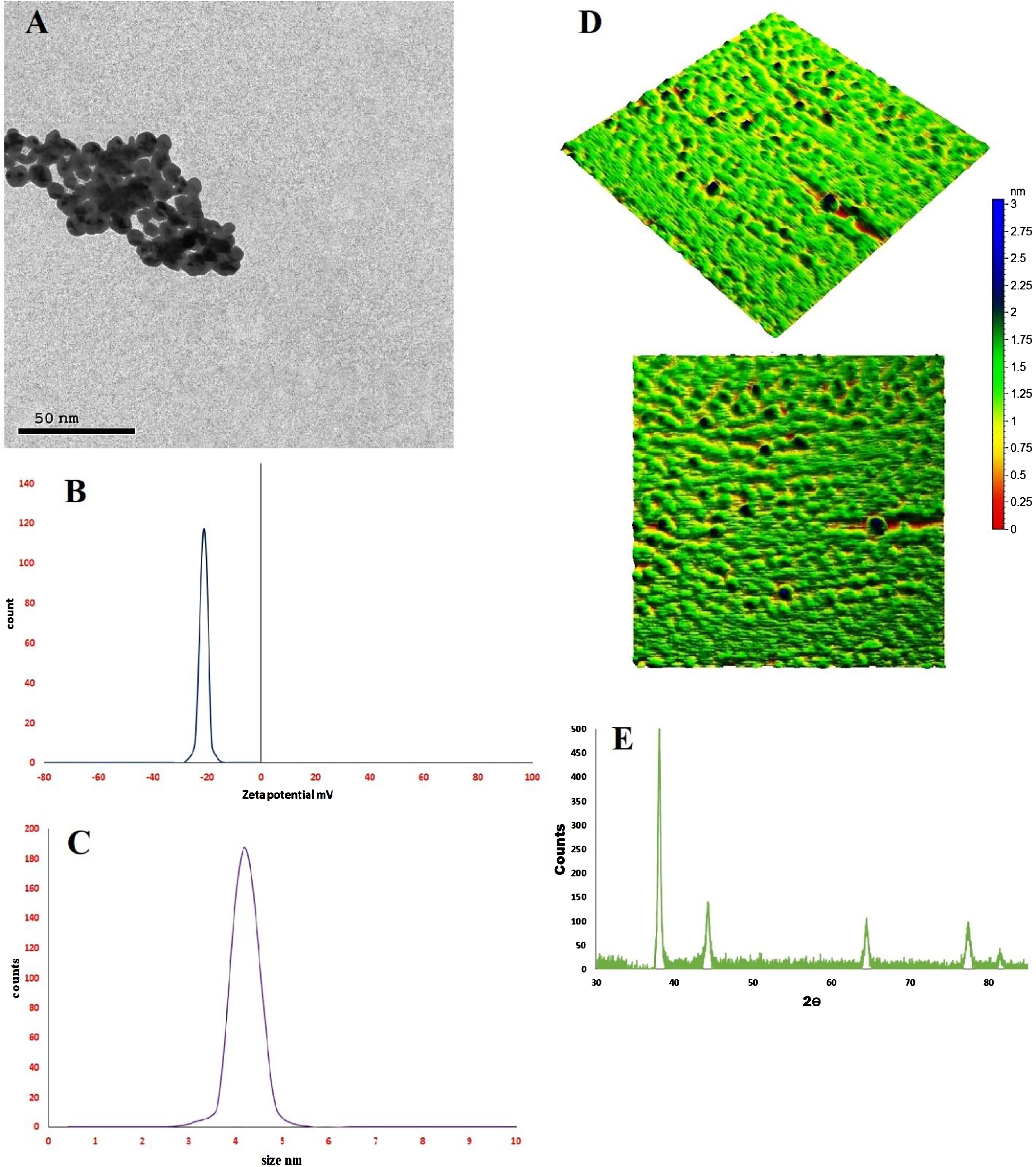
Wound healing
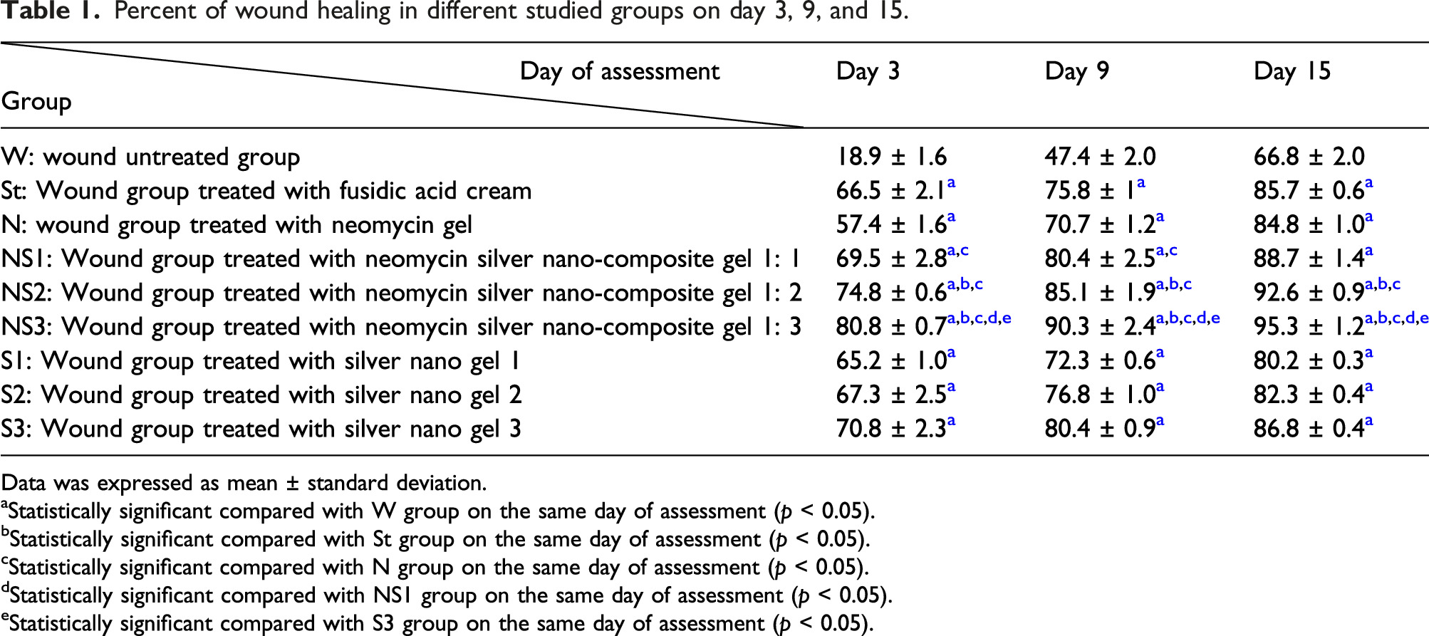
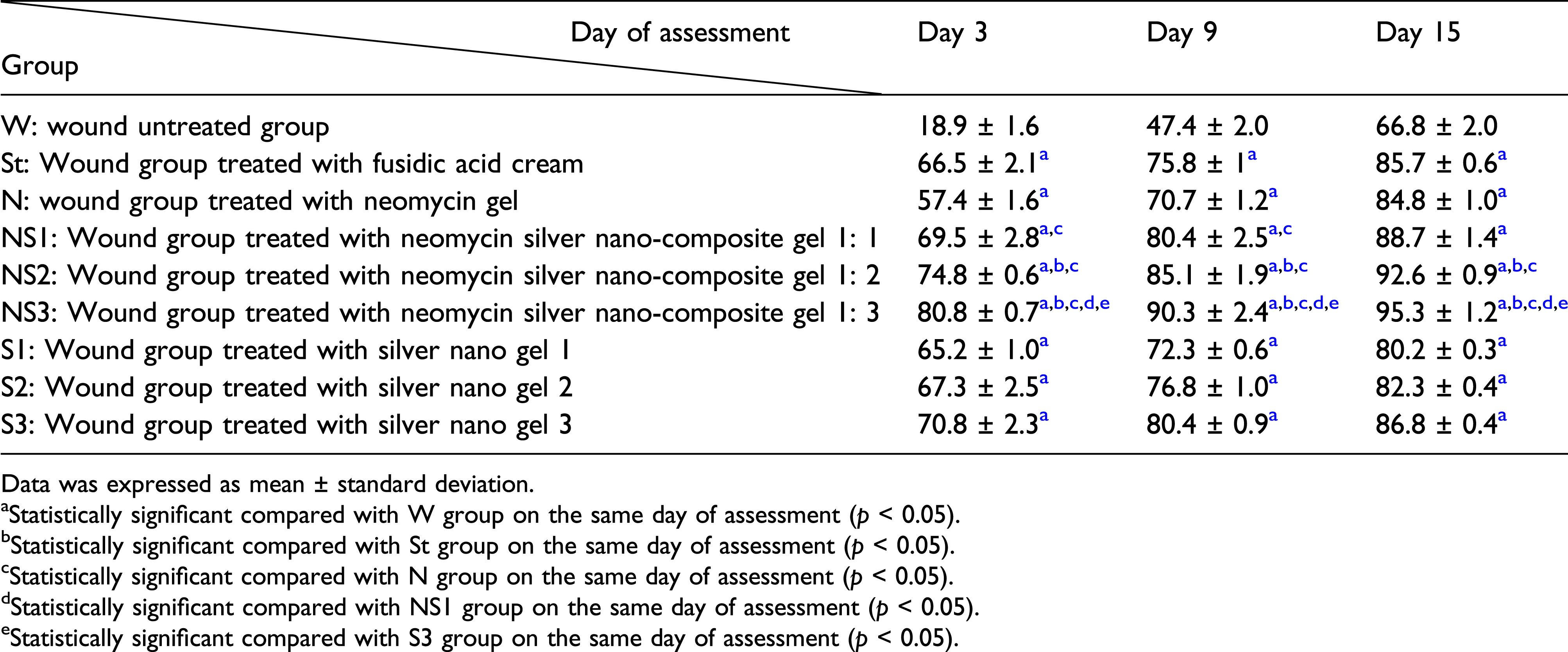
Percent of wound healing in different studied groups on day 3, 9, and 15.

Photographs of wound repair at different time interval in excision wound model in rats in different studied groups. (W: wound untreated group, St: wound group treated with fusidic acid cream, N: wound group treated with neomycin gel, NS1: wound group treated with neomycin silver nano-composite gel 1: 1, NS2: wound group treated with neomycin silver nano-composite gel 1: 2, NS3: wound group treated with neomycin silver nano-composite gel 1: 3, S1: wound group treated with silver nano gel 1, S2: wound group treated with silver nano gel 2 and S3: wound group treated with silver nano gel 3).
Histopathological examination
Poor wound healing was observed in the W group, showing an uncovered wound surface associated with a persistent necrotic serocellular crust containing numerous bacterial colonies and intense neutrophilic infiltration. The filling granulation tissue showed a haphazard arrangement with excessive inflammatory cell infiltration and poor vascularization.
Moderate wound closure was observed in the St and N groups, displaying complete epidermal remodeling in various individuals. Organized granulation tissue occupied the wound gap with minimal inflammation.
Concerning the silver administrating groups (S1, S2, and S3); minimal wound healing was observed in the S1 group. Meanwhile, wound healing was markedly enhanced in the S2 and S3 groups. Perfect wound healing closure was observed in the nano-composite groups (NS1, NS2, and NS3) with the highest closure observed in the NS3 group, which showed a marked decrease in wound area with complete re-epithelization, evidence of keratinization, and organized tissue filling in the wound gap, rich in collagen bundles and little to no inflammatory cell infiltration (Figure 3). Photomicrograph of wounded areas of different studied groups on day 15 (H&E) (a) Wound untreated group showing serocellular crust covering with inflamed granulation tissue filling the wound gap, (b) wound group treated with fusidic acid cream showing abundant collagen bundles filling the wound gap, (c) wound group treated with neomycin gel showing complete epidermal remodeling with fibrovascular tissue filling the wound gap, (d) wound group treated with silver nano gel 1 showing incomplete re-epithelization with moderate inflamed granulation tissue, (e) wound group treated with silver nano gel 2, (f) wound group treated with silver nano gel 3 showing enhanced wound healing, (g) wound group treated with neomycin silver nano-composite gel 1, (h) wound group treated with neomycin silver nano-composite gel 1: 2 showing advanced wound healing with complete re-epithelization, and (i) wound group treated with neomycin silver nano-composite gel 1: 3 showing marked wound closure with complete re-epithelization and evidence of keratinization.
The statistical analysis of wound healing parameters showed a significant decrease in re-epithelization, granulation tissue, inflammation, and angiogenesis in the W group compared to other groups. Meanwhile, a significant increase in granulation tissue, inflammation, and angiogenesis was recorded in NS3 group in comparison with all other experimental groups (Figure 4). Re-epithelization, granulation tissue, inflammation and angiogenesis of wound healing evaluation on day 15 in different groups (W: wound untreated group, St: wound group treated with fusidic acid cream, N: wound group treated with neomycin gel, S1: wound group treated with silver nano gel 1, S2: wound group treated with silver nano gel 2, S3: wound group treated with silver nano gel 3, NS: wound group treated with neomycin silver nano-composite gel 1: 1, NS2: wound group treated with neomycin silver nano-composite gel 1: 2 and NS3: wound group treated with neomycin silver nano-composite gel 1: 3). (Data Expressed as means ± standard error. Different letter indicates a significant difference at p ≤ 0.05 compared to wound of untreated group).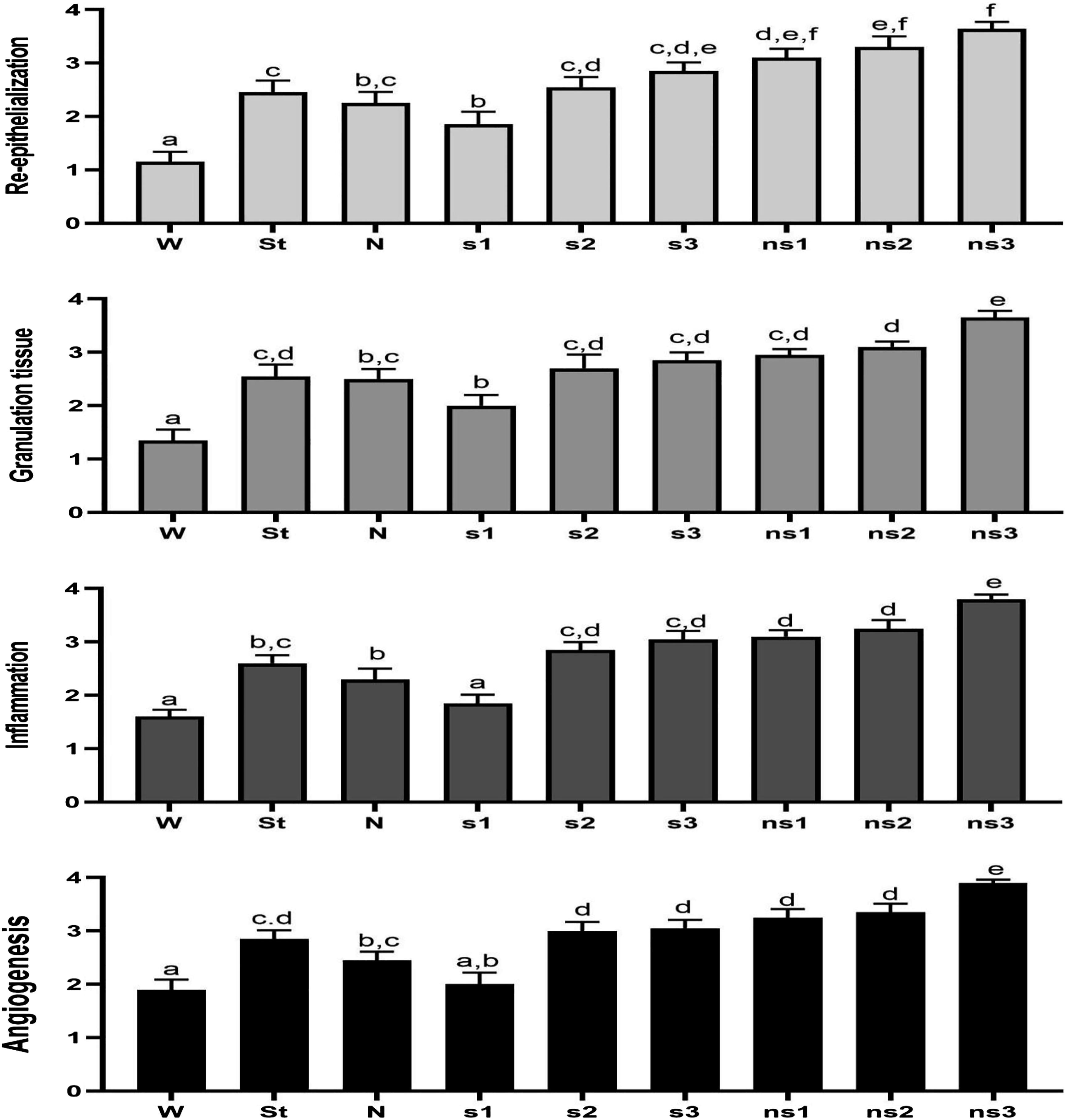
The evaluation of collagen bundles deposition was examined in MTC stained sections. Few collagen fibers were deposited in the W group, exhibiting a significant decrease compared to other groups. However, the highest collagen bundle deposition was observed in the NS3 group, which showed a significant increase in the area (%) of collagen bundles in MTC stained sections compared to other groups (Figure 5). Photomicrograph of dermal content of collagen bundles in different groups on day 15 (MTC) (a) W group (wound untreated group), (b) St group (wound group treated with fusidic acid cream), (c) N group (wound group treated with neomycin gel), (d) S1 group (S1: wound group treated with silver nano gel 1), (e) S2 group (wound group treated with silver nano gel 2), (f) S3 group (wound group treated with silver nano gel 3), (g) NS1 group (wound group treated with neomycin silver nano-composite gel 1: 1), (h) NS2 group (wound group treated with neomycin silver nano-composite gel 1: 2), and (i) NS3 group (wound group treated with neomycin silver nano-composite gel 1: 3). (Data Expressed as means ± standard error. Different letter indicates a significant difference at p ≤ 0.05 compared to wound of untreated group).
Discussion
Wound healing is a normal biological process. Wound healing occurs in four successive and highly programmed phases: hemostasis, inflammation, proliferation, and remodeling. Any factor interfering with ≥1 of these phases leads to impaired wound healing. 27 Tissue formation and tissue remodeling consist of various sequential controlled stages including angiogenesis, cellular proliferation, and collagen synthesis followed by formation of granulation tissue and matrix degradation followed by collagen replacement, wound contraction, and scar tissue formation.28–31
Wound healing is controlled by different factors, such as cytokines, mitogens, and chemotactic factors including platelet-derived growth factors, insulin like growth factors, epidermal growth factors, and fibroblast growth factors. These factors control cell migration and proliferation as well as production of extracellular matrix proteins, essential for granulation tissue formation.32–34
When the skin is injured, bacteria normally present on the skin surface can access the underlying tissues. Both bacteria and endotoxins can lead to elevated pro-inflammatory cytokines, such as interleukin-1 and TNF-α. At this point, the wound may enter a chronic state and fail to heal. 35 Nanoparticles can simultaneously kill microorganisms and stimulate skin regeneration. Among various nanoparticles, Ag-NPs are one of the most efficient. Their unique properties suggest that they can both effectively prevent wound infections and improve the healing process of the damaged tissues compared with traditional topical treatments.36,37
The present results showed that daily topical application of neomycin or a silver nano-composite together with fucidin cream promoted wound healing compared to the non-treatment group. Wound healing occurs in the healthy skin surrounding the wounds which coats or covers the naked area. These processes may be due to myofibroblasts, while epithelialization or epithelial regeneration following damage, require the proliferation and immigration of epithelial cells to the wound center. 35 In addition, there is significant concentration dependent promotion of wound healing in the groups topically treated with a silver nano-composite (S1, S2, and S3) compared with untreated group and that treated with fucidin cream only. Silver nanoparticles could effectively inhibit and kill the bacteria in a concentration and time dependent manner.
Similarly, Zhang et al. (2016) and Agnihotri et al. (2014) reported that the small size of Ag-NPs could cause more toxicity to the bacteria, having a better bactericidal effect, and killing multidrug resistant bacteria compared to larger particles, as they have a larger surface area.38,39 The healing of any wound depends on the wound itself, the presence or absence of infection, age, dietary supply, and health conditions. All induced wounds were performed by the same surgeon in an identical way, and all other factors, including age, health conditions, and daily dietary supply, were the same.
Silver nanoparticles release silver ions that kill microbes.
40
Due to their attraction and affinity to sulfur proteins, silver ions adhere to the cell wall and cytoplasmic membrane. The adhered ions enhance the permeability of the cytoplasmic membrane and lead to bacterial envelope disruption.
41
Uptake of free silver ions into cells leads to deactivation of respiratory enzymes and generation of reactive oxygen species (ROS). ROS are a principal agent in cell membrane disruption. As sulfur and phosphorus are important DNA components, the interaction of silver ions with the sulfur and phosphorus of DNA can interrupt DNA replication, cell reproduction, or even death of the microorganisms. Moreover, silver ions can inhibit protein synthesis by denaturing cytoplasmic ribosomes.
42
In addition, some membrane proteins that regulate antibiotic tolerance, ion binding, pore-forming, membrane stabilization, and flagellum assembly are controlled by Ag-NPs
Infections with antibiotic-resistant microorganisms can result in healing failure and predicted mortality. 44 Ag-NPs can be an alternative to conventional chemical antimicrobial agents to overcome multidrug resistance microorganisms, as bacteria are less likely to develop resistance to metal nanoparticles compared to conventional antibiotics. 45 The antimicrobial effects of Ag-NPs against resistant E. coli, 46 multidrug resistant strains of Pseudomonas aeruginosa, 47 methicillin-resistant S.taphylococcus aureus (MRSA), 48 and extended-spectrum β-lactam producing bacteria 49 have been described. Ag-NPs’ anti-inflammatory role in burns and other wounds is due to reducing inflammatory cell infiltration and inhibiting the development of inflammatory cytokines. 50
The present results agree with Pyun et al. (2015), who confirmed the role of Ag-NPs in the enhancement and acceleration of wound healing 18 and with Liang et al. (2016) who found that Ag-NPs could accelerate wound healing via enhancing re-epithelialization, granulation tissue formation, cell proliferation, and controlling inflammatory responses. 51 In addition, Masood et al. (2019) reported that Ag-NP impregnated hydrogels of chitosan–polyethylene glycol accelerated wound healing in diabetic wounds in rabbits. 52
In addition, Wasef et al. (2020) confirmed the healing properties of Ag-NPs in burns induced in mouse model.
53
Furthermore, Ag-NPs did not have cytotoxic effects on human cells at <30 mg/L
Nanoparticle-antibiotic combinations have numerous benefits, including a reduction of the concentration used and toxicity of both agents while increasing the antimicrobial properties. 55 Such a combination may increase antibiotic concentrations at the point of antibiotic-microbe contact, promoting increased affectivity. Thus, synergistic effects may occur because of effective drug transport of Ag-NPs to the cell, as Ag-NPs may encourage extensive damage to the cell wall and facilitates transfer of hydrophilic antibiotics to the cell surface. 56 The large surface area of nanoparticles can facilitate the interaction with active antibiotic groups, as hydroxyl and amine groups, resulting in conjugation of both molecules (antibiotic-Ag-NP complexes), thus increasing the antibiotic concentration at the injection site. 57 Another potential mechanism that may lead to increased antibiotic activity due to the combination with Ag-NPs is the inhibition of bacterial enzymes responsible for bacterial tolerance to antibiotics. 58
The results of the present study agree with those of Khalil et al. (2021), who reported that a combination of Ag-NPs significantly improved the antibacterial efficacy of neomycin as evidenced by an increase of up to eight times in the inhibition zone diameter against multiple drug resistance (MDR) of P. aeruginosa-infected burn wounds. 58 Related research by Panáček et al. (2016) demonstrated a synergism of Ag-NPs with ampicillin against multi-resistant strains of P. aeruginosa, Enterobacter aerogenes, and Methicillin-resistant Staphylococcus aureus MRSA. 59 Moreover, zinc–aluminum layered double hydroides (LDH), curcumin, and curcumin nanohybrids revealed good tissue repair in acute and chronic wounds with good biocompatibility and healing activity with collagen formation, in addition to prolongation of the duration of action of the loaded materials or drugs with LDH nanomaterial in a controlled release manner. 60
Our results agree with the findings by Jamaran and Zarif (2016) who investigated the synergistic wound healing activity of neomycin and silver within the gel matrix. 61 Kumar et al. (2016) reported that tetracycline conjugated Ag-NPs increased the antibacterial action of tetracycline due to enhanced Ag+ accumulation around bacterial cell membranes. 62 In addition, Katva et al. (2017) recorded the synergistic antimicrobial activity of Ag-NPs with chloramphenicol and gentamicin against MDR Enterococcus faecalis compared to antibiotics alone. 54 The nano silver and neomycin gel caused both antibacterial and antibiofilm movement against multiple bacterial strains especially for Pseudomonas aeruginosa and Streptococcus mutans, two opportunistic bacteria often related with human and animal infections or diseases. 62
Limitation
The main limitation of this study was the lack of a power analysis to calculate the sample size selected for this study.
Conclusion
According to the present findings, a neomycin silver nano-composite gel may be promising for wound management. It is cheap, nontoxic, and more effective than either silver nanoparticles or neomycin alone. However, further research is needed on human volunteers with skin wounds to confirm the efficacy of Ag-NPs alone or in combination with an antibiotic(s), especially after the demonstrated success of Ag-NPs and that of their combination with neomycin in the treatment of wounds in experimental animal models.
Footnotes
Author contribution
This study was designed, directed and coordinated by H.A. El-Banna1 and H. ElZorba 1 as the principal investigators, provided conceptual and technical guidance; G.G. Mohamed2 and S. H. Ismail3 planned and performed the preparation and characterization of the used materials; the data analyzed with F. Sayed1, A. Galal1 performed the application experiments and A. H. El-Banna6 and Afaf, S. Osman5 performed the statistical analysis. M R. Mousa4 performs histopathological investigation. All authors contributed to the editing of the revised article, and approved the final article.
Declaration of conflicting interests
The author(s) declared no potential conflicts of interest with respect to the research, authorship, and/or publication of this article.
Funding
The author(s) received no financial support for the research, authorship, and/or publication of this article.
Availability of data and materials
All data and materials are available and can be submitted when needed.
Ethics approval
Ethical approval for this study was obtained from *NAME OF ETHICS COMMITTEE OR INSTITUTIONAL REVIEW BOARD (APPROVAL NUMBER/ID)*. Vet CU08032022466: The Institutional Animal Care and Use Committee (IACUC).
Animal welfare
The present study followed international, national, and/or institutional guidelines for humane animal treatment and complied with relevant legislation. The protocol for this study was confirmed by Animal Research Ethical Committee, Faculty of Veterinary Medicine, Cairo University (Vet. CU. IACUC V, dated 08 March 2022) and the authors of this manuscript observed ethical issues. Animals were handled according to the International Guidelines for Care and Handling of Experimental Animals.