
Abstract
Brucellosis, especially caused by Brucella melitensis, is considered the most-widespread zoonosis in the world, particularly in developing countries. This study was planned to develop an accurate test for diagnosis of ovine brucellosis using a specific hot saline extracted soluble Brucella melitensis periplasmic proteins (SBPPs). The efficacy of the latex agglutination test (LAT) using SBPPs compared to the Rose Bengal test (RBT), buffered plate agglutination test (BPAT), serum agglutination test (SAT), and an indirect enzyme-linked immunosorbent assay (i-ELISA) was evaluated in the field diagnosis of ovine brucellosis. The test performance was evaluated by estimating sensitivity (Se), specificity (Sp), positive predictive value (PPV), negative predictive value (NPV), disease prevalence (DP), positive likelihood ratio (PLR), and negative likelihood ratio (NLR) using test agreement and bacteriological culture in 1777 samples. The false-positive result was significantly (P ⩽0.05) lower in LAT than RBT, BPAT, SAT, and i-ELISA. With reference to test agreement, the Se, Sp, PPV, and PLR were highest (P ⩽0.05) in LAT 99.33%, 99.88%, 98.68%, and 827.25%, respectively. With reference to bacteriological culture, the LAT and i-ELISA tests showed a significant difference in Se with SAT. However, no significant difference in specificity was detected. The DP was 8.44% in the five tests. In conclusion, LAT using SBPPs of B. melitensis could be a suitable serodiagnostic field test for ovine brucellosis, with high sensitivity and specificity.
Introduction
Brucella melitensis (biovars 1, 2, or 3), the causative agent of caprine and ovine brucellosis, is highly pathogenic for humans, causing one of most serious zoonosis in the world, and is mainly endemic in the Mediterranean and Middle East regions with recent prevalence up to 42%. 1 The clinical diagnosis of B. melitensis infection is based on the observation of clinical manifestations, which may include abortion, retained placenta, orchitis, and epididymitis. 1 The clinical diagnosis is confirmed by isolation of Brucella from aborted material, udder secretions, and/or positive serology. 1 Primary Brucella isolation from field samples requires 4–7 days of incubation that might lead to contamination that reduced the sensitivity of bacteriological diagnosis.2,3 Accordingly, the indirect diagnosis of the disease based on serological tests is of the approach of choice, especially for eradication and control. 4
The conventional serological tests that are widely used for diagnosis of brucellosis in small ruminants are the buffered plate agglutination test (BPAT), the Rose Bengal test (RBT), the complement fixation test (CFT), an indirect enzyme-linked immunosorbent assay (i-ELISA), a fluorescence polarization assay (FPA), and the serum agglutination test (SAT).1,4–6 All of these tests are prescribed for international trade. 1 These serological tests depend on the detection of B. abortus whole bacterial cells or smooth lipopolysaccharide (sLPS) as antigens to detect antibodies in serum that give rise to false-positive (FP) reactions because of cross-reactivity with LPS from other bacteria. 7 This and other drawbacks of anti-LPS antibodies have led to an increased interest in the detection of antibodies to alternative antigens, mainly outer membrane proteins and cytoplasmic proteins. 8 No reference test is currently available. Therefore, the availability of more accurate diagnostic tests is necessary for the diagnosis and control of B. melitensis in sheep.
A periplasmic protein has been identified as an immunodominant antigen of the cytosoluble protein extract of B. melitensis in sheep. 8 In humans, latex agglutination assay was used for the serodiagnosis of brucellosis by coating colored latex beads with Brucella lipopolysaccharides. 9 The major advantages of the latex agglutination assay are that the test reagent is highly stable, the test is simple to perform, and the test result is obtained in less than 30 s. 9
The current study aimed at: (1) developing practical agglutination test for diagnosis of brucellosis in ovine; (2) evaluating the efficacy of latex agglutination test (LAT) for the diagnosis of brucellosis in ovine compared to other serological tests; and (3) determining the prevalence of brucellosis and the best classification of the gold standard among the five validated tests.
Materials and methods
Animals and sampling
This study was conducted between February 2013 and January 2015 in nine sheep flocks located in the central and western regions of the Kingdom of Saudi Arabia. The western region was historically designated as areas of concern for ruminant brucellosis and five sheep flocks from Turrabah and Taif governorates were selected. The naturally infected animals from these flocks were used as positive reference populations. Four flocks from the center region, located in Riyadh governorate, were historically Brucella-free and used as negative reference populations. Consequently, this population provided information about test specificity. All treatments of the animals and the collection of samples were in compliance with the guidelines for the welfare of animals and those of the concerned ethical authorities.
The flocks were subjected to careful clinical and laboratory investigations for application of the inclusion and exclusion criteria. The inclusion criteria included: (1) flocks that have never been vaccinated and were considered as free or not free of infection, so any positive reaction in the serological tests was considered to be due to infection with a field strain of Brucella; (2) animals with a history of abortion or stillbirth; (3) a group of clinically healthy animals to be used as a control in bacteriological examination. The exclusion criteria included: (1) flocks vaccinated with any type of Brucella vaccines; and (2) animals with surgical interference. All animals of the selected herds were sampled. A total of 1777 blood samples were collected from sheep of both sexes (females, n = 1420; males, n = 357) at different ages, that represented the applied criteria in the western (n = 1130) and central (n = 647) regions. However, in some instances, access to animals and flocks was difficult or restricted resulting in small sample sizes in some areas. The sample sizes according to governorates were as follows: Turrabah (n = 630), Taif (n = 500), and Riyadh (n = 647). Approximately 3 mL of blood was collected from each animal by jugular venipuncture with disposable needles and venoject tubes. The tubes were labeled and the blood was allowed to clot at ambient temperature before transport to the laboratory on ice. The sera were separated by centrifuging at 5000×g for 7 min. Serum samples were heated at 56°C for inactivation of non-specific antibodies. Each sample was identified by a code number and stored in aliquots at −20°C until used for Bayesian and test agreement analysis in all the five tests (LAT, RBT, BPAT, SAT, and i-ELISA). Sera from both Brucella-infected and Brucella-free individuals act as positive and negative reference samples. A pretested questionnaire designed to collect animal and flock level data during blood sampling was administered. Whole blood (3 mL), milk samples (20 mL), and vaginal swabs (after abortion) were collected from all animals (n = 150) in flocks with clinical infection in the western region and submitted to bacteriological study. All infected tissues, cultures, and potentially contaminated materials handled at containment level 3.
Bacteriological examination
Infection confirmed individually in all cases by culture on Brucella selective media (Oxoid) and the pathogen isolated from whole blood, milk samples, and vaginal swabs. 10 The species and biovars of Brucella were identified by cultural and serological criteria as previously described. 1
Diagnostic serological tests
We evaluated the SBPPs antigen in LAT by comparison with four other validated well established serological tests (RBT, BPAT, SAT, and i-ELISA). All tests were carried out in accordance with the Office International des Epizooties (OIE) Manual of Standards for Diagnostic Tests. 1 Tests were performed by personnel blind to the results of the other tests.
LAT
The SBPPs were prepared as previously described 11 with some modifications. Five milliliters of B. melitensis serotype 3 suspension (1010/mL) were added to 800 mL of sterile Trypticase soy broth (BBL Microbiology Systems, Cockeysville, MD, USA) and incubated at 37°C in a Co2 incubator for 48 h. Brucella melitensis was harvested and washed once with normal saline. Hot saline extracts were obtained by suspending B. melitensis in normal saline and autoclaving at 121°C for 20 min. The autoclaved suspension was centrifuged at 12,000×g for 20 min at 4°C. Then the ammonium sulphate was added to supernatant and the solution was centrifuged at 10,000×g for 20 min. The precipitate was dissolved in 0.01 M phosphate buffered saline (PBS) (pH 7.2). Dialysis occurred against PBS (pH 7.2) in a dialysis bag overnight at 4°C. This preparation was designated SBPPs. The protein concentration was determined by a commercial kit using bovine serum albumin as standard (Pierce, Rockford, IL, USA). The SDS-PAGE was done for determining the different pattern of protein bands in SBPPs. The SBPPs were stored at −80°C until use.
The procedure for the sensitization of latex microspheres was a slight modification of a published method. 12 A latex polystyrene microsphere particle with a diameter of 0.81 µm (LB8, Sigma) was used in the covalent binding reaction. Prior to use, the beads were suspended in deionized water and vigorously vortexed to ensure even distribution, to break up any large particles, and to obtain a 10% suspension. An equal volume of latex particles and SBPPs antigen (0.6 mg protein/mL) was incubated over night at 37°C. The mixture was centrifuged at 6000×g for 10 min. The supernatant was removed and the pellet was washed and brought to an original volume in glycine sodium chloride buffer (final pH 8.2) containing 1:500 bovine serum albumin to keep the latex suspended (7.505 g of glycine, 5.85 g of sodium chloride, and 0.1% sodium azide in 1 L of deionized water, adjust the pH to 8.2 with 1 N NaOH). The latex particles were colored by methylene blue (0.005%), and the latex suspension was kept at 4°C.
Agglutination reactions were performed using the following procedure. A drop of the sensitized latex solution and sample was placed on a glass slide. The reaction was mixed by a side-to-side movement. The agglutination reaction appears in less than 2 min at room temperature in positive cases. 9
i-ELISA
LPS from B. melitensis serotype 3 was extracted 1 by heating 5 g dry weight (or 50 g wet weight) of cells suspended in 170 mL distilled water to 66°C followed by the addition of 190 mL of 90% (v/v) phenol at 66°C. The mixture was stirred continuously at 66°C for 15 min, cooled, and centrifuged at 10000 g for 15 min at 4°C. The brownish phenol in the bottom layer was removed with a long cannula and large cell debris was removed by filtration. The sLPS was precipitated by the addition of 500 mL cold methanol containing 5 mL methanol saturated with sodium acetate. After a 2-h incubation at 4°C, the precipitate was removed by centrifugation at 10,000 g for 10 min. The precipitate was stirred with 80 mL of distilled water for 18 h and centrifuged at 10,000 g for 10 min. The supernatant solution was kept at 4°C. The precipitate was resuspended in 80 mL distilled water and stirred for an additional 2 h at 4°C. The supernatant solution was recovered by centrifugation as above and pooled with the previously recovered supernatant. Next, 8 g of trichloroacetic acid was added to the 160 mL of crude sLPS. After stirring for 10 min, the precipitate was removed by centrifugation and the translucent supernatant solution was dialyzed against distilled water (two changes of at least 4000 mL each) and then freeze-dried. The freeze-dried sLPS was weighed and reconstituted to 1 mg/mL in 0.05 M carbonate buffer, pH 9.6, and sonicated in an ice bath using approximately 6 watts three times for 1 min each. The sLPS was then freeze-dried in 1 mL amounts and stored at room temperature. The freeze-dried sLPS was reconstituted to 1 mL with distilled water and was further diluted 1/1000 (or to a dilution predetermined by titration against the OIE ELISA Standard Sera) in 0.05 M carbonate buffer, pH 9.6.
The test procedure was performed as previously described. 1 The results were recorded on an ELISA reader (Reader 270, BioMerieux, France) at 490 nm. The color development is directly proportional to the bound antigen-antibody complex. Samples with a value double the optical density of the mean negative control were considered positive.
BPAT
Antigen for the BPAT is prepared from B. melitensis according to the procedure previously described. 1 Serum samples (80 µL of each) was placed on a glass plate marked in 4 × 4 cm squares. Then, 30 µL of antigen was placed near each serum spot and the serum and antigen was thoroughly mixed to produce a circular zone approximately 3 cm in diameter. The plate was rotated three times in a tilting motion to ensure even dispersion of the reagents, and then incubated for 4 min in a humid chamber at ambient temperature. The plate was removed and rotated as above, and then returned for a second 4-min incubation. Agglutination was read immediately after the 8-min period is completed. Any visible reaction was considered to be positive. A control serum that gives a minimum positive reaction was tested before each day’s tests to verify the sensitivity of test conditions.
RBT
This test is a simple spot agglutination test using antigen stained with rose Bengal and buffered to a low pH, usually 3.65 ± 0.05. Serum samples (25–30 µL of each) were placed on a WHO hemagglutination plate. Equal volume of antigen was placed near serum spots. The serum and antigen were mixed thoroughly to produce an oval zone approximately 2 cm in diameter. The mixture was agitated gently for 4 min at ambient temperature on a rocker. The agglutination was read after the 4-min period. Any visible reaction was considered to be positive. A control serum that gives a minimum positive reaction was tested before each day’s tests to verify the sensitivity of test conditions.
SAT
The antigen represents a bacterial suspension in phenol saline (NaCl 0.85% [w/v] and phenol at 0.5% [v/v]). The ethylene diamine tetraacetic acid was added to the antigen suspension to 5 mM final test dilution to reduce the level of FP results. Subsequently, the pH of 7.2 was readjusted in the antigen suspension. The sensitivity of the antigen was standardized in relation to the OIE. Serum agglutination test was performed in microtiter plates. 1 The mixture of antigen and serum dilutions was incubated for 6 h at 37°C. At least three dilutions were prepared for each serum in order to refute prozone negative responders. The degree of Brucella agglutination in a serum was expressed in IU per mL. Samples showing more than 30 IU/mL were considered positive.
Diagnostic test evaluation and validation
The performance and validation of the various tests was analyzed using classical means of calculation with respect to the test agreement and bacteriological culture.10,13 With respect to the test agreement, 13 sera from both Brucella-infected and Brucella-free individuals act as positive and negative reference samples. The test sample was considered a true positive if a positive result was given by at least three tests. Similarly, a sample was considered a true negative if a negative result was given by at least three tests. The number of true-positive (TP) and false-negative (FN) cases in the diseased group were counted. The number of false-positive (FP) and true-negative (TN) cases in the non-diseased group were counted. From these counted number of cases, we calculated sensitivity (Se), specificity (Sp), positive likelihood ratio (PLR), negative likelihood ratio (NLR), positive predictive value (PPV), negative predictive value (NPV), and disease prevalence (DP) using the following equations: Se=TP/(TP+FN); Sp=TN/(TN+FP); PPV=TP/(TP+FP); NPV=TN/(TN+FN); PLR=Se/(100-Sp); NLR=(100-Se)/Sp; and DP=(TP+FN)/(TP+FP+FN+TN). Sensitivity means probability that a test result will be positive when the disease is present. It is also called the TP rate. Specificity means probability that a test result will be negative when the disease is not present. It is also called the TN rate. PLR is the ratio between the probability of a positive test result given the presence of the disease and the probability of a positive test result given the absence of the disease. NLR is the ratio between the probability of a negative test result given the presence of the disease and the probability of a negative test result given the absence of the disease. PPV means the probability that the disease is present when the test is positive. NPV means the probability that the disease is not present when the test is negative. Sensitivity, specificity, PPV, and NPV as well as DP are expressed as percentages for ease of interpretation.
With respect to bacteriological culture, 10 the infected animals that gave positive results in bacteriological culture were compared to their serological results in the selected five tests to calculate the sensitivity of each test. Therefore, sensitivity with reference to bacteriological culture was calculated by dividing the number of animals showing positive results in the serological tests with the number of the same animals showing positive results in the bacteriological test. The non-infected animals who gave negative results in the bacteriological culture were compared to their serological results in the five selected tests to calculate specificity of each test. Therefore, specificity with reference to bacteriological culture was calculated by dividing the number of animals showing negative results in the serological tests with the number of the same animals showing negative results in the bacteriological tests.
Statistical analysis
SPSS 17 statistical software (SPSS v. 17.0, SPSS Inc., Chicago, IL, USA.) was used to analyze the collected data. Comparisons among serological tests were performed using the χ2 (Chi-square) test. A difference was considered to be significant when P ⩽0.05.
Results
Clinical picture
Some clinical signs appeared in naturally infected sheep (n = 150) in the Turrabah and Taif governorates including reduced appetite, abortion, retained placenta, or stillbirth.
General serological results
A total of 1777 serum samples were tested for the presence of B. melitensis antibodies using RBT, BPAT, SAT, i-ELISA, and LAT. An example of the LAT assay is shown in Figure 1. The number of animals that were positive or negative in the various serological tests and had or did not have the disease is shown in Table 1, which is based on the two-by-two contingency table. The number of total positive sera were significantly lower than the total negative sera for all five serological tests. The false positive and negative results were significantly (P ⩽0.05) lower in LAT than RBT, BPAT, SAT, and i-ELISA. Additionally, the FN result was significantly (P ⩽0.05) lower in i-ELISA than RBT. No significant differences in the FP results were detected between i-ELISA, SAT, BPAT, and RBT. The cross-classified test results were used to calculate the indices of agreement between the RBT, BPAT, SAT, i-ELISA, and LAT. There was agreement among the five tests in 1.724 cases (1.588 negative and 136 positive), with some disagreement in 53 cases.

Latex agglutination test using the SBPPs antigen. A clear agglutination reaction appears in less than 2 min at room temperature in the case of a positive control serum (a), with no agglutination in the case of a negative control serum (b), and latex SBPPs alone (c).
Evaluation of RBT, BPAT, SAT, i-ELISA, and LAT for diagnosis of ovine brucellosis.
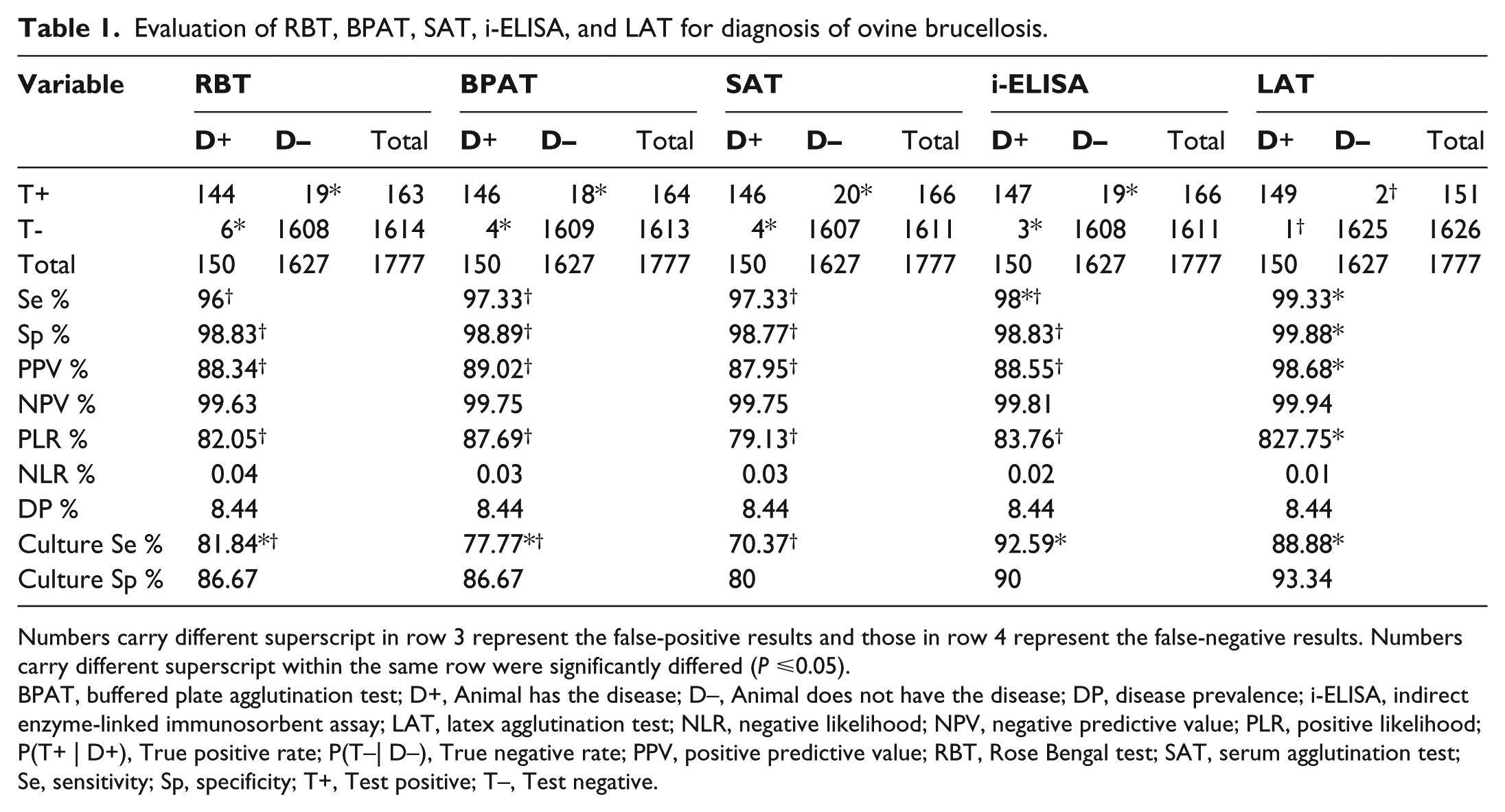
Numbers carry different superscript in row 3 represent the false-positive results and those in row 4 represent the false-negative results. Numbers carry different superscript within the same row were significantly differed (P ⩽0.05).
BPAT, buffered plate agglutination test; D+, Animal has the disease; D–, Animal does not have the disease; DP, disease prevalence; i-ELISA, indirect enzyme-linked immunosorbent assay; LAT, latex agglutination test; NLR, negative likelihood; NPV, negative predictive value; PLR, positive likelihood; P(T+ | D+), True positive rate; P(T–| D–), True negative rate; PPV, positive predictive value; RBT, Rose Bengal test; SAT, serum agglutination test; Se, sensitivity; Sp, specificity; T+, Test positive; T–, Test negative.
Diagnostic test performance and validation results
Test agreement results. The estimated values of Se, Sp, PPV, NPV, PLR, NLR, and DP that based on the test agreement analysis are shown in Table 1. For diagnostic Se and Sp, LAT was significantly (P ⩽0.05) higher than i-ELISA, SAT, BPAT, and RBT. Additionally, the diagnostic specificity did not differ for i-ELISA, SAT, BPAT, and RBT. The PLR was significantly (P ⩽0.05) higher in LAT than other tests. The NLR was not significantly different between all the tests. The PPV was significantly (P ⩽0.05) higher in LAT than BPAT, i-ELISA, RBT, and SAT. No significant difference in the NPV existed for all five serological tests.
Bacteriological culture results
Natural infections were confirmed by the isolation and identification of B. melitensis serotype 3 from 27 of 150 sheep (18%). The 27 bacterial isolates were recovered from seropositive whole blood (n = 7), milk (n = 7), and vaginal swabs (13). The 27 isolates were identified as B. melitensis serotype 3. The 30 seronegative sheep were negative by cultue and serology. The sensitivity and specificity of the serological tests with respect to culture are illustrated in Table 1. The LAT and i-ELISA tests showed a significant difference in sensitivity with SAT. However, no significant difference in specificity was detected.
Discussion
This study describes the set-up of a latex agglutination test (LAT) for the serodiagnosis of B. melitensis infection in sheep. LAT consider as an improvement of traditional serological tests and based on coupling of a periplasmic protein of antigen, without LPS fraction, to the latex beads. Thus, it has a significant less cross-reactivity with infections mediated by other pathogens. In addition, the stability of the test reagent and the simplicity of the assay procedure make LAT suitable as field diagnostic test without need to laboratory. A similar assay was developed for the serodiagnosis of human brucellosis, 9 but that assay used Brucella LPS as an antigen and gave FP reactions because of cross-reactivity with LPS from other bacteria. 1
In this study, we prepared the SBPPs from B. melitensis and used it to coat colored latex beads for use as the antigen in LAT (Figure 1). The efficacy of LAT using SBPPs was evaluated by comparison with well-established serological tests (RBT, BPAT, SAT, and i-ELISA), as shown in Table 1. A major goal in immunological studies of brucellosis has been the identification of non-lipopolysaccharide antigens, which could be useful to circumvent drawbacks of the LPS antigens. 1 The FP result was significantly (P ⩽0.05) lower in LAT than RBT, BPAT, SAT, and i-ELISA (Table 1). This may be because the other tests mainly detect antibodies directed against Brucella whole bacterial cells or sLPS portion of the cell membrane. 1 This could lead to increased FP results because of cross-reactivity with other Gram-negative bacteria, such as Escherichia coli,7,14 Y. enterocolitica O:9,7,15 and Salmonella enterica serovar Urbana. 16
Test performance can be evaluated by different approaches. The first approach that used to evaluate the test performance and validation is the test agreement analysis (Table 1). The test agreement analysis is helpful to estimate test Se, Sp, prevalence, and other diagnostic variables. 17 This approach requires representative sera from both Brucella-infected and Brucella-free individuals to act as positive and negative reference samples. 18
Another objective of this study was to investigate the strains of Brucella spp. found in sheep in Saudi Arabia, and to verify the suitability of the antigen used in the serological tests. The 27 isolates collected throughout the country were identified as B. melitensis serotype 3. Culture results represent the second approach that used to evaluate the test performance and validation (Table 1), but it is provided a lower measure of test performance than test agreement analysis. The reduced sensitivity of bacteriological diagnosis might be attributed to the frequent contamination of culture plates.2,3 The primary Brucella isolation from field samples requires 4–7 days of incubation that might lead to overgrow of fungi as well as commensal and environmental bacteria.2,3
Conclusion
In conclusion, this is the first study, to the authors’ knowledge, to identify serum reactivity to a purified hot saline extract SBPPS of B. melitensis in ovine brucellosis. Moreover, we have developed a simple and rapid screen field test for the serodiagnosis of sheep brucellosis, LAT, using SBPPS of B. melitensis as antigen. This test may be suitable for use in the field as: (1) it is very easy to perform and to read; (2) it does not require expensive equipment and electricity; (3) it does not require access to a diagnostic laboratory and can be used in places where there is no access to veterinary care; and (4) it is as sensitive and specific as the four other validated tests. 3 The identification of protein components of Brucella that elicit an antibody response in the majority of infected animals would improve the diagnosis of the disease and contribute to the development of new vaccine strategies. Hence, further studies will be needed to evaluate the SBPPs of B. melitensis as vaccine candidate using the safe molecular methods. 19
Footnotes
Declaration of conflicting interests
The authors declared no potential conflicts of interest with respect to the research, authorship, and/or publication of this article.
Funding
The authors appreciate and thank the Collage of Food and Agriculture Sciences, Agriculture Research Center and Deanship of Scientific Research, King Saud University, Saudi Arabia, for financial support for this research study.