
Abstract
Background:
Shoulder stabilization surgery has evolved over time, and bony augmentation procedures on the glenoid side are being performed more often. The Latarjet procedure modifies subscapularis anatomy because the conjoined tendon divides the subscapularis muscle fibers through a split/takedown, which has structural and functional implications. Arthroscopic anatomic glenoid reconstruction (AAGR) re-creates anatomy. This technique uses the Halifax portal to deploy and fix a distal tibial allograft (DTA) through the rotator interval, thus preserving the subscapularis anatomy.
Purpose/Hypothesis:
The purpose was to analyze the radiographic properties of the subscapularis muscle after AAGR. It was hypothesized that the subscapularis muscle structure remains preserved postoperatively.
Study Design:
Case series; Level of evidence, 4.
Methods:
A retrospective review was performed comprising a consecutive series of patients treated with AAGR with DTA between November 2012 and April 2021 for traumatic anterior shoulder instability with glenoid bone loss. Patients were excluded if they had posterior instability, glenoid fracture, missing pre- or postoperative computed tomography (CT) scans, or only CT arthrogram available. Radiographic variables measured on CT scans included estimates of subscapularis muscle volume, subscapularis/infraspinatus muscle ratio, and fatty infiltration according to the Goutallier classification. Pre- and postoperative Western Ontario Shoulder Instability index scores were collected as a secondary outcome of this study.
Results:
Ninety-three patients were included in the study with a clinical follow-up of 2.3 ± 1.5 years (mean ± SD). The subscapularis volume increased from 185.91 ± 45.85 mL preoperatively to 194.1 ± 49.0 mL postoperatively (P = .006). The subscapularis to infraspinatus muscle ratio showed a significant increase from 0.96 ± 0.27 to 1.05 ± 0.30 after surgery (P = .002). All patients had a Goutallier stage of 0 before and after surgery. The Western Ontario Shoulder Instability scores showed a significant improvement from 64.8 ± 15.5 preoperatively to 28.2 ± 24.0 postoperatively (P < .001).
Conclusion:
Patients who undergo AAGR with DTA for traumatic shoulder instability with glenoid bone loss have a preserved subscapularis muscle volume with no fatty infiltration, while showing a significant improvement in clinical outcomes.
Keywords
Glenohumeral instability is a common health problem, which has an estimated incidence density rate in the United States of 23.9 to 26.9 per 100,000 person-years. 40 After a traumatic anterior glenohumeral dislocation, a range of soft tissue and bony injuries can occur. Glenoid bony injuries are found in 50% to 86% of patients with recurrent dislocation.22,39 Bone loss on the glenoid and/or humeral head is the diverging point in the shoulder instability treatment algorithm.11,12 Any bony disturbance modifies the glenoid track: this concept takes into account the dynamic interaction of the glenoid and humeral head through range of motion (ROM)—specifically, when bone loss is present, there is a higher risk of having an engaging lesion and therefore recurrent instability episodes.14,15,27,37,38
The Latarjet procedure has been the gold standard to treat bony deficiencies on the anterior glenoid owing to its low recurrence rate,10,23,29 but this procedure has a high incidence of complications, such as infection, nerve injury, graft malposition, and arthritis.7,13,30 As part of the Latarjet technique, in open and arthroscopic approaches, the coracoid process is transferred to the anterior glenoid through a subscapularis muscle split or takedown.
Arthroscopic anatomic glenoid reconstruction (AAGR) using a distal tibial allograft (DTA) was designed to obtain shoulder stability by re-creating anatomy. This technique uses a far medial portal called the Halifax portal, which goes through the rotator interval to attach the bone graft, thereby preserving the subscapularis muscle anatomy. 36
AAGR differs from the Latarjet procedure in that the Latarjet requires a subscapularis muscle split or takedown,17,19 which could contribute to reported functional changes, including decreased strength, 2 endurance, 2 limitation of ROM, 34 and radiographic changes such as thinning 31 and fatty infiltration.18,31 In contrast, AAGR does not disturb the subscapularis muscle anatomy, preserving its structure and function.
The purpose of this study was to analyze and compare the structural properties of the subscapularis muscle, including volume, subscapularis/infraspinatus muscle ratio, and fatty infiltration measured with Goutallier classification. Computed tomography (CT) images obtained before and after the AAGR procedure using DTA were used to obtain these measurements. We hypothesized that patients treated with this arthroscopic, subscapularis muscle-sparing, glenoid reconstruction would not show significantly changed subscapularis muscle structural properties postoperatively.
Methods
Design
This was a retrospective study of a consecutive series of patients. This study was approved by the Nova Scotia Health Research Ethics Board.
Participants
Patients were eligible for the study if they were treated with AAGR with DTA between November 2012 and April 2021. The demographic information of the patients was recorded, including age, sex, and body mass index. The indication for surgery was symptomatic traumatic anterior shoulder instability with glenoid bone loss >15% as estimated on preoperative CT scans using the best-fit circle method described by Sugaya et al. 33 Patients were excluded if they had symptoms of posterior instability, multidirectional instability, or a glenoid fracture; if patients missed their pre- or postoperative CT scan; or if their CT scans were CT arthrograms. All surgical procedures were performed by the senior author (I.W.).
Surgical Technique
AAGR is an all-arthroscopic technique that was conducted with the patient under general anesthesia. Details of the surgical technique, graft preparation, and portal placement were published by Wong and Urquhart 36 (Figure 1). The procedure was done with the patient in a lateral decubitus position with a vacuum beanbag beneath the patient and with a pneumatic arm positioner. At the beginning of every case, a routine diagnostic arthroscopy following the 15-point routine described by Snyder et al 32 was performed through the posterior and anterior portals. The rotator interval was excised, identifying the coracoacromial ligament, the tip of the coracoid process, and the origin of the conjoined tendon. The anteroinferior aspect of the glenoid was prepared to obtain a flat, healthy bleeding bone bed to receive the graft. The Halifax portal was established with an inside-out technique using a Wissinger rod parallel to the glenoid, superior to the subscapularis tendon, and lateral to the conjoined tendon. 19 A frozen DTA (readily available in the hospital bone bank) was prepared on the back table with the following approximate dimensions: anterior-posterior, 10 mm; superior-inferior, 20 mm; and medial-lateral, 15 mm (Figure 2). The graft was secured using a double-barrel cannula and was inserted into the joint through the Halifax portal without any disruption to the subscapularis muscle integrity. The graft was positioned with arthroscopic visualization and fixed with two 3.5-mm partially threaded cannulated screws (DePuy Mitek). The remaining capsule-labral complex was repaired to native glenoid bone with at least two 1.8-mm all-suture anchors (Q-Fix; Smith & Nephew), as a routine Bankart procedure.
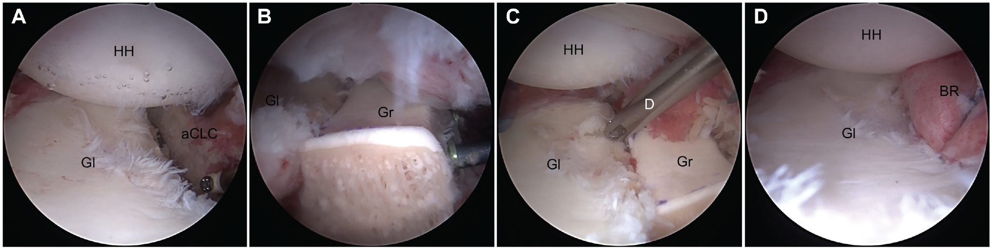
Intraoperative images of an arthroscopic anatomic glenoid reconstruction procedure in a left shoulder viewing from the anterosuperior portal. (A) The humeral head is seen on the top; the glenoid is visualized on the bottom with a significant anterior bony defect; and the anterior capsule–labrum complex is detached from the anterior face of the glenoid. (B) The distal tibial allograft is placed with the cartilage of the graft in line with the native glenoid cartilage. (C) The graft is fixed to the glenoid with 2 cannulated screws. A drill guide is used to place the first all-suture anchor to start the Bankart repair. (D) Final view of the construct with the Bankart repair completely covering the graft. The humeral head is balanced over the glenoid. aCLC, anterior capsule–labrum complex; BR, Bankart repair; D, drill guide; Gl, glenoid; Gr, graft; HH, humeral head.
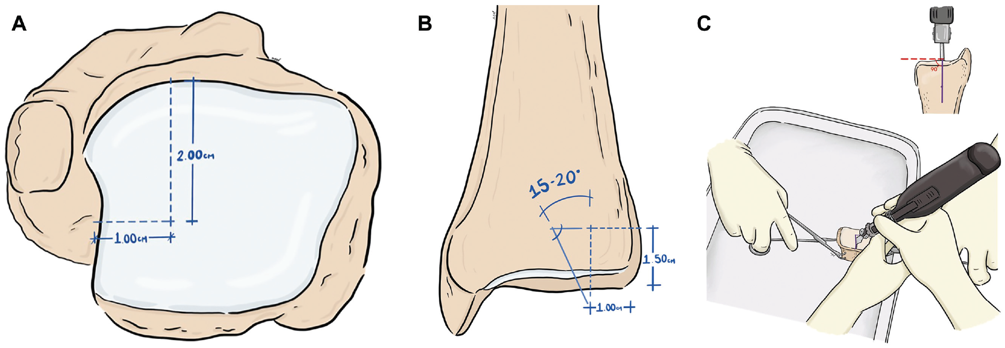
Illustration of a distal tibial allograft preparation highlighting the mean dimensions used to harvest the graft freehanded with a microsagittal saw. (A) View of the distal articular surface of a tibia showing the markings done in the cartilage before harvesting. (B) Lateral view of the same distal tibia showing the subsequent markings made before harvesting. (C) Illustration of the freehand graft harvesting. The assistant holds the graft while the surgeon uses the outlined marks to create the cuts. The articular surface is used to create a perpendicular 90° cut.
Rehabilitation
Patients were instructed to use a postoperative brace (SlingShot 3; Breg) that kept the arm in neutral rotation and 15° of abduction for 6 weeks; cradle pendulums were started the day after surgery. The first physical therapy visit was on days 3 to 5 postoperatively. The protocol was divided into 4 phases: phase 1, protection (0-2 weeks); phase 2, mobility (2-6 weeks); phase 3, neuromuscular retraining (6-12 weeks); phase 4, strength and function (+12 weeks). The timing of progression to the subsequent stage was an estimate, as the patient must fulfill the goals of each phase before advancing. ROM was progressed throughout the phases, with no restriction of external rotation. Passive ROM was initiated during phase 1. Active assisted ROM can be started as soon as week 2 if passive ROM is at 90%. Full active ROM was expected between weeks 6 and 8. In phase 4, the program was focused on scapular stabilization, periscapular strength, rotator cuff strengthening, and proprioception. Return-to-sport criteria were typically met 6 to 12 months after surgery.
Imaging Protocol and Measurements
Every patient included in the analysis received a pre- and postoperative CT scan. The CT scans were conducted in one of the hospitals across the province according to availability. Two-millimeter axial cuts and sagittal, coronal, and 3-dimensional reconstructions were performed. The CT images were retrieved from the provincial imaging database in DICOM (Digital Imaging and Communications in Medicine) format. The imaging field included the affected glenohumeral joint and rotator cuff muscles. All the radiographic measurements were done by a sports medicine fellowship-trained orthopaedic surgeon (J.C.) and reviewed by the senior author (I.W.).
The subscapularis muscle volume was calculated in accordance with the methodology described by Henninger et al. 8 DICOM stacks were imported into Horos (version 3.3.6), using the 3-dimensional multiplanar reconstruction viewer mode. The images were oriented in the scapular plane, and 3 landmarks—the center of the glenoid, the trigonum spinae, and the scapular inferior angle—were used to establish this scapular plane, as seen in Figure 3C.
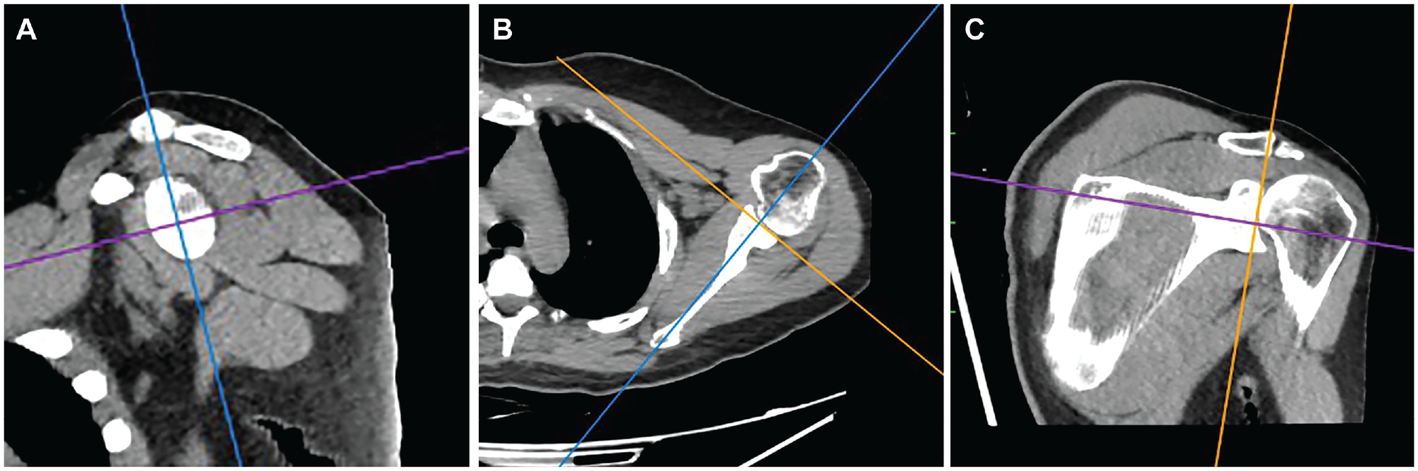
Multiplanar reconstruction of the shoulder computed tomography scan of a patient with recurrent shoulder instability. (A) Sagittal cut of a left shoulder at the level of the joint in the center of the glenoid. (B) Axial cut of the same patient at the center of the glenoid in line with the scapular axis. (C) Coronal cut showing the 3 landmarks used for all patients: center of the glenoid, trigonum spinae, and scapular inferior angle.
The area of the subscapularis muscle (Figure 4) in squared centimeters was manually outlined in 2 sagittal cuts: glenoid face (area A) and midscapular body (area B). These measured areas were then inputted into the formula published by Henninger et al 8 :

This formula uses the measured areas to estimate the subscapularis volume. 8
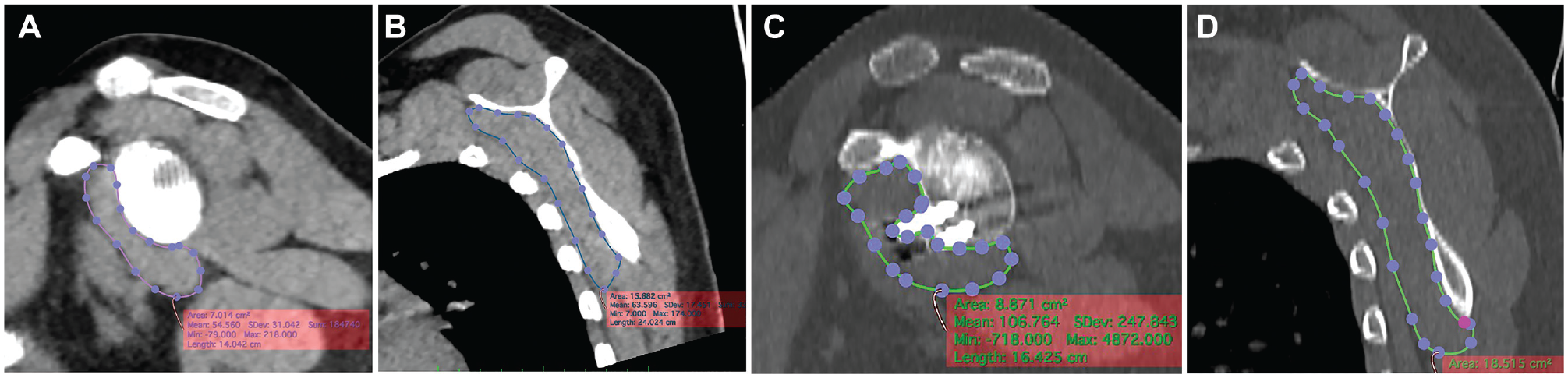
The subscapularis volume was estimated with the formula [0.06 (area A + area B)] – 13.02. The measurements were performed on pre- and postoperative CT imaging. (A) Area A was estimated with a sagittal cut perpendicular to the scapular axis at the level of the glenoid, outlining the subscapularis muscle periphery. (B) Area B was estimated in a sagittal cut perpendicular to the scapular axis at the midglenoid point, outlining the subscapularis muscle. (C) Area A in the postoperative CT scan was measured excluding the hardware. (D) Area B measurement in the postoperative CT scan. CT, computed tomography.
The subscapularis to infraspinatus muscle ratio was measured at the midglenoid point on the transverse plane using a line tangent to the articular surface and parallel to this line. The measurement was done from the anterior-to-posterior margin of the subscapularis and infraspinatus muscles, as originally described by Maynou et al 18 and seen in Figure 5. Fatty infiltration was classified according to the description of Goutallier et al 6 from stage 0 (normal) to stage 4 (more fat than muscle). This was evaluated in the same cut where area B was measured, as shown in Figure 4, B and D.
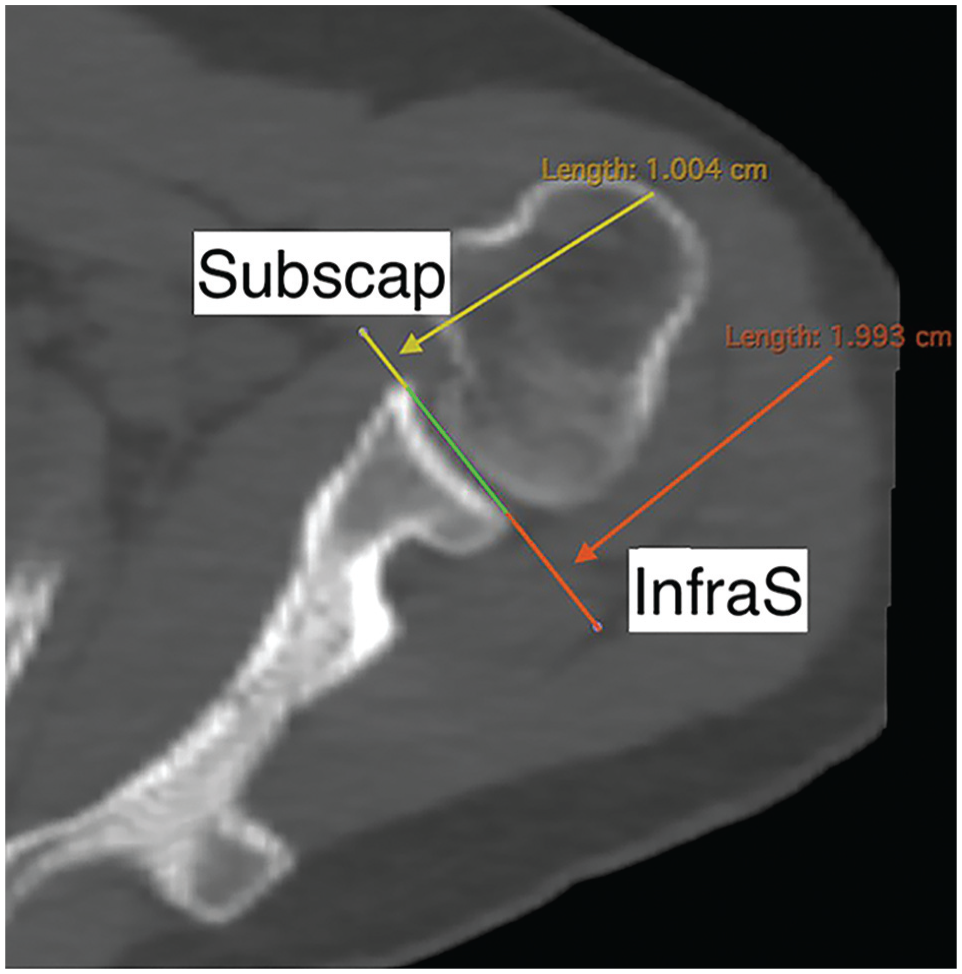
The subscapularis (Subscap) to infraspinatus (InfraS) muscle ratio was measured in a computed tomography scan axial cut at the midglenoid point. The anterior-posterior lengths of the subscapularis (1.004 cm) and infraspinatus (1.993 cm) muscles were measured perpendicular to the scapular axis.
Clinical Outcomes
The clinical outcomes were analyzed as a secondary outcome. Pre- and postoperative patient-reported outcomes were measured using the Western Ontario Shoulder Instability (WOSI) index. WOSI scores were reported as a percentage, with lower scores indicating better outcomes. The scores were obtained at 6 months, 1 year, and yearly after this, with the score reported being the most recent score obtained in clinical follow-up. The minimal clinically important difference of WOSI scores used in this study was 10.4. 16
Data Analysis
The analysis was conducted using IBM SPSS Statistics (Version 26). Patient characteristics were summarized and presented using descriptive statistics. Continuous variables—including pre- and postoperative subscapularis muscle volume, subscapularis to infraspinatus muscle ratio and WOSI scores—were compared using paired 2-sample t test or Wilcoxon matched-pair signed rank test, depending on the results of the normality test and Levene test. The correlations between pre- and postoperative variables, including subscapularis muscle volume and the subscapularis to infraspinatus muscle ratio, were evaluated using Pearson bivariate correlation tests. The significance level was .05 in the aforementioned analyses.
Results
A total of 231 patients received AAGR between November 2012 and April 2021, and 138 patients were excluded per the exclusion criteria (Figure 6). Ninety-three patients were included in the analysis. The characteristics of the 93 eligible patients are summarized in Table 1.
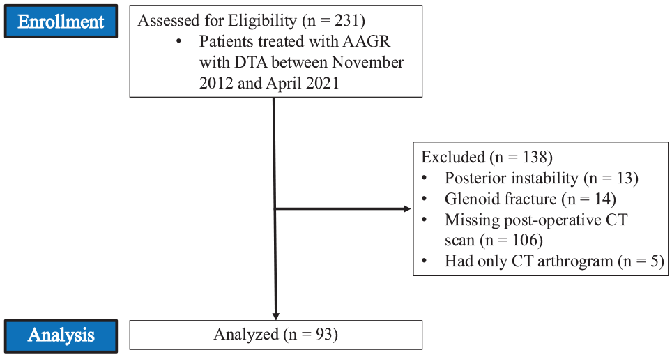
Flowchart of patient selection. AAGR, arthroscopic anatomic glenoid reconstruction; CT, computed tomography; DTA, distal tibial allograft.
Characteristics of the Study Population (N = 93) a
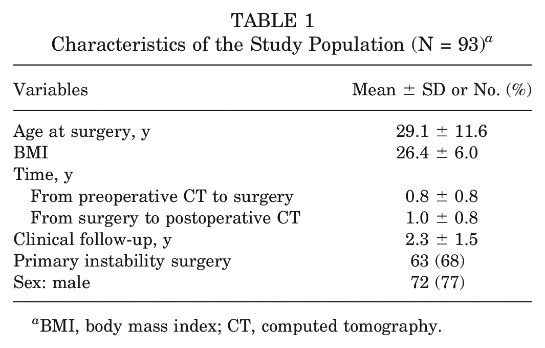
BMI, body mass index; CT, computed tomography.
The subscapularis muscle volume after AAGR with DTA increased significantly from 185.91 ± 45.85 mL (mean ± SD) preoperatively to 194.1 ± 49.0 mL postoperatively (P = .006) (Table 2). Also, the mean subscapularis to infraspinatus muscle ratio significantly increased from 0.96 ± 0.27 preoperatively to 1.05 ± 0.3 postoperatively (P = .002). All patients maintained the same fatty infiltration levels pre- and postoperatively (ie, Goutallier grade 0). There were significant correlations between pre- and postoperative subscapularis muscle volumes and of the subscapularis to infraspinatus muscle ratio. As shown in Figure 7, the WOSI scores significantly improved pre- to postoperatively (P < .001) with a mean improvement of 37.5% ± 22.6%. Ninety percent of patients met the minimal clinically important difference for WOSI.
Pre- and Postoperative Subscapularis Volume and Subscapularis to Infraspinatus Ratio (N = 93) a

Data are presented as mean ± SD unless noted otherwise.
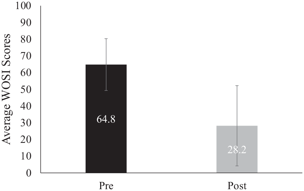
Pre- and postoperative WOSI scores (mean ± SD, 64.8 ± 15.5 and 28.2 ± 24.0, respectively). WOSI, Western Ontario Shoulder Instability.
Discussion
The main finding of this study was that patients who received AAGR with DTA had a preserved subscapularis muscle volume and a significant improvement in WOSI scores after surgery when compared with the preoperative assessments. Although no clinical parameters (ie, ROM, strength, and endurance) were included, the promising results of this study set the foundation for further clinical investigations. The subscapularis to infraspinatus muscle ratio also significantly increased from pre- to postoperative measurements.
Owing to a relatively high complication rate in the Latarjet procedure, alternative open and arthroscopic bone-grafting techniques have been explored. 7 Autografts such as iliac crest, scapular spine, or clavicle distal end and allografts such as distal tibia have been used.9,20,24,25 DTA has been used successfully, decreasing the harvesting site morbidity.5,35 DTA is one of the preferred techniques because it offers a surface with articular cartilage and with similar anatomy to the glenoid. 24 The use of DTA in shoulder instability was first explored by Provencher et al, 24 but this technique still had the drawbacks of being an open approach. AAGR avoids subscapularis muscle split/takedown with similar patient-reported outcomes and recurrence rates as the Latarjet procedure.12,35
Although the literature claims that the subscapularis muscle volume is maintained after the Latarjet procedure, there are limitations to their conclusions. Caubère et al 2 investigated a series of 20 patients treated with open Latarjet, but they evaluated only postoperative 1-year magnetic resonance imaging scans. The observer subjectively classified the trophicity of the muscle as normal or atrophic in a single coronal image as compared with the rest of the rotator cuff muscles. 2 Valencia et al 34 studied a series of 40 patients who underwent arthroscopic Latarjet. An objective cross-sectional measurement of the subscapularis muscle was conducted, and the authors claimed that the subscapularis muscle suffered no atrophy; yet, there was no comparison with a baseline in the study, and only postoperative CT scans were analyzed. Ernstbrunner et al 4 demonstrated volume preservation after Latarjet by assessing the subscapularis muscle volume on magnetic resonance imaging in a series of 42 patients treated with open Latarjet. They found not only preservation but also a significant volume increase in the subscapularis muscle compared to the contralateral side (177 vs 169 mL respectively; P = .022). However, this study did not include a baseline of the same shoulder in the comparison, and the subscapularis volume estimation was made using a different method, which could explain the difference as compared with our measurements.
There are no known studies in the literature reporting the volume of the subscapularis muscle before or after surgery with the methodology used in this study in the setting of shoulder instability. Henninger et al 8 described the formula to estimate the subscapularis muscle volume used for our study, and they reported smaller subscapularis muscle volumes (115 ± 52 mL) as compared with the ones in our population (185.91 ± 45.85 mL). This may be because of the mean age of the population: ours was aged 29.1 ± 11.6 years and theirs was aged 65 ± 6 years, and older individuals generally have smaller subscapularis muscle volumes.8,26
In our study population, we identified a significant increase in the subscapularis muscle volume preoperatively (185.91 ± 45.85 mL) to postoperatively (194.1 ± 49.0 mL; P = .006). Maynou et al 18 demonstrated greater muscle atrophy in patients who underwent open Latarjet with an L-shaped subscapularis muscle tenotomy versus a subscapularis muscle split. This volume increase in our population may be explained by the fact that patients often have decreased activity levels after shoulder instability, which may lead to initial muscle atrophy. After surgery and postoperative rehabilitation, patients resume their typical activities and sports approximately 1 year postoperatively, which may result in subsequent hypertrophy of the subscapularis muscle. Even though this increase is statistically significant, it is potentially not clinically significant. Overall, this finding suggests that we do not have any worsening of the subscapularis muscle volume after AAGR.
Fatty infiltration has been consistently reported in patients who undergo the Latarjet procedure. The Latarjet can be performed using a variety of techniques, including open subscapularis muscle tenotomy and repair,18,21,28 open subscapularis muscle split,1-3,18,31 and arthroscopically.21,34 Regardless of which technique is used, each study reports fatty degeneration of the subscapularis muscle, particularly in the upper part of the muscle. Fatty degeneration might not be only a marker of poor muscle quality and function. In the series presented by Maynou et al, 18 the patients who had Goutallier grades >2 after undergoing open Latarjet had persistent apprehension in 35.3% of cases. In our patient series, there was no fatty infiltration or progression, which was expected because the muscle structure is not violated; therefore, there was no negative change in volume.
The subscapularis to infraspinatus muscle ratio was first described by Maynou et al 18 to control some confounding variables while assessing the subscapularis volume. In their study, 102 patients were treated with open Latarjet with 2 technical variations: subscapularis muscle L-shape tenotomy and subscapularis muscle split. Interestingly, results described only anterior-posterior measurement of the subscapularis muscle but not the ratio. This anterior-posterior measurement was statistically lower in the operative side as compared with the contralateral shoulder (0.9 ± 0.37 vs 1.16 ± 0.24 cm; P = .0001). Siegert et al 31 conducted a study derived from a randomized controlled trial of 2 open stabilization techniques: Latarjet and iliac crest. 20 In their study, the subscapularis to infraspinatus muscle ratio was measured pre- and postoperatively on CT scans in both study groups. In the baseline measurement, groups had comparable findings (Latarjet, 1.03 ± 0.03; iliac crest, 0.97 ± 0.3; P = .338), similar to the value in our baseline measurement (0.96 ± 0.27). After surgery, this ratio was slightly increased in the iliac crest group and significantly lower in the Latarjet group (1.00 ± 0.2 vs 0.70 ± 0.3, respectively; P < .001), each similar again to that of our population where the postoperative ratio was 1.05 ± 0.3. One inference from these data is that keeping the conjoined tendon through the subscapularis muscle to create the sling effect might negatively affect the subscapularis muscle.
Strengths of this study include the fact that we measured baseline characteristics with postoperative changes and that we had a large sample size when compared with previously published studies. However, this study also has limitations. The nature of the study design is retrospective with its inherent limitations, and surgery was conducted by a single surgeon in a single center. It is important to mention that a substantial number of patients were excluded from analysis as they were lacking adequate postoperative CT images. Additionally, even though this study is lacking a control group to a Latarjet population, a baseline comparison was performed, which is unique to previous literature and can be used as a control. Finally, the mean radiographic follow-up is relatively short, as volumetric changes in the muscle could be expected later in time.
The clinical relevance of the findings of this study is that by using the Halifax portal to avoid a subscapularis muscle split/takedown, the subscapularis muscle belly volume, quality, and structure are preserved. This study demonstrated the benefit of using a Halifax portal to perform AAGR, allowing preservation of the subscapularis muscle when reconstructing the glenoid bone in shoulder instability with glenoid bone loss.
Conclusion
Patients who undergo AAGR with DTA for traumatic shoulder instability with glenoid bone loss have a preserved subscapularis muscle structure, volume, and no fatty infiltration, while showing a significant improvement in WOSI scores.
Footnotes
Acknowledgements
Thank you to Felicia Licht for manuscript editing and submission formatting.
Submitted September 25, 2023; accepted August 29, 2024.
One or more of the authors has declared the following potential conflict of interest or source of funding: P.N.C. has received consulting fees from Medical Device Business Services. I.W. has received consulting fees from DePuy Mitek Inc, Smith & Nephew, CONMED Corp, and Bioventus. AOSSM checks author disclosures against the Open Payments Database (OPD). AOSSM has not conducted an independent investigation on the OPD and disclaims any liability or responsibility relating thereto.
References
Supplementary Material
Please find the following supplemental material available below.
For Open Access articles published under a Creative Commons License, all supplemental material carries the same license as the article it is associated with.
For non-Open Access articles published, all supplemental material carries a non-exclusive license, and permission requests for re-use of supplemental material or any part of supplemental material shall be sent directly to the copyright owner as specified in the copyright notice associated with the article.