
Abstract

Wilhelm Fabry was the first physician to give serious attention to the surgical management of burns. Later preferring his Latin name, Fabricus Hildanus, he is regarded as the founder of German scientific surgery. In 1607, he divided burns into first, second and third degree, starting a classification debate that would last for centuries. Although he revolutionised the treatment of contractures with dynamic splints and creative operations, he had little to offer for the pain of acute burn injuries: ‘it scatters and consumes the vital spirits and causes unquietness and fevers, and is followed by a defect of the mind’. 1
Over the ensuing centuries, the surgical treatment of burns slowly improved, as did pain management, but the pathophysiology remained poorly understood and treatments were often counterproductive. By the 19th century, the prevailing belief was that burns prevented perspiration, leading to detrimental fluid accumulation. The first priority of treatment, therefore, was to prevent this by the use of diuretics, ‘guarded blood letting, or by encouraging the process of suppuration’. 2 Unsurprisingly, death rates remained high in patients with significant burns.
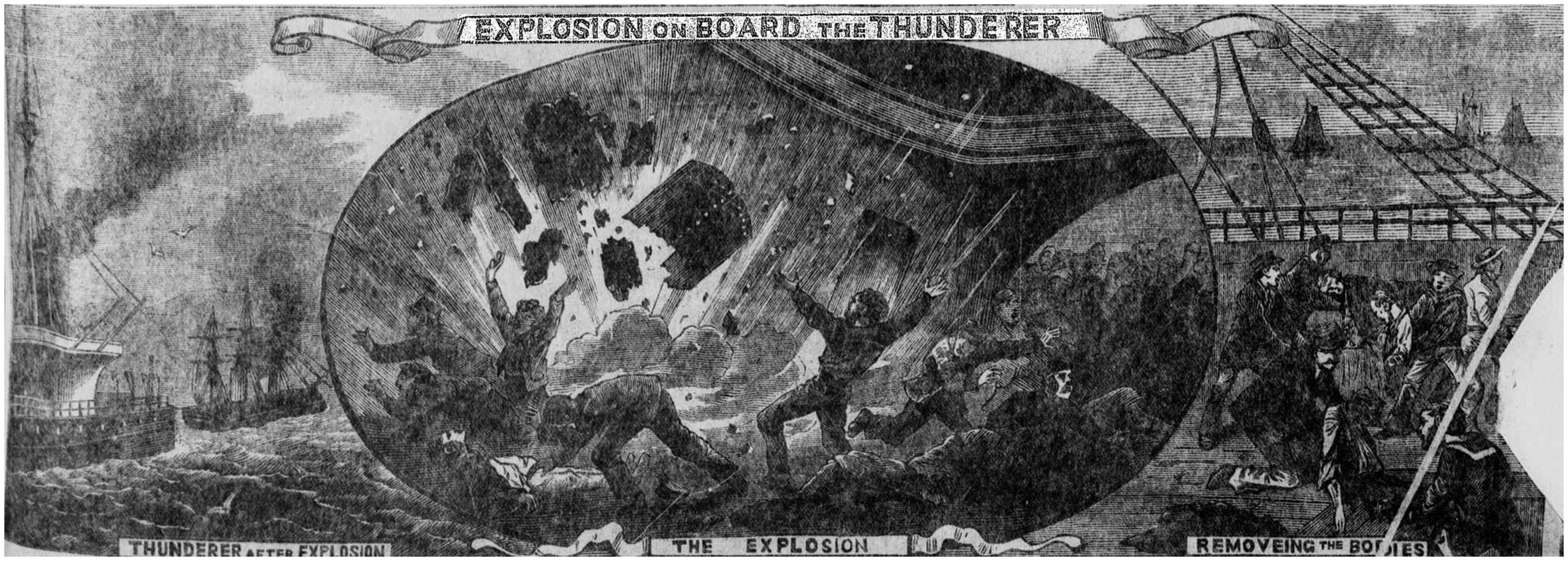
The greatest impetus to research into the medical management of burns subsequently came from a number of mass casualty events. On 14 July 1876, while en route from Portsmouth Harbour to Stokes Bay in England, the HMS Thunderer suffered a massive boiler explosion. Nineteen people were killed instantly. 3 A further 58 survived, 11 of whom died within 30 hours of ‘primary shock’. Many were noted to have burns to the throat and airways. Medical staff painstakingly measured the surface area of burns on the initial survivors. In his report, Inspector-General Smart concluded that 350 square inches of scald or 250 square inches of gunpowder burn were equally fatal injuries. Apart from various local dressings, patients were given opium and chloral hydrate for pain and lime water with milk to prevent ulceration of the stomach.
Smart was the first to suggest a patient’s prognosis may be related to the burnt surface area. Karl Meeh, from Tübingen, Germany, subsequently produced a complicated analysis of body surface area using graph paper, which proved too complex for others to emulate. 4 In 1905, in Vienna, Stefan Weidenfeld and Leo von Zumbusch developed a more simplified system linking survival and treatment to percentage body surface area. 5 They concluded that if less than a 12th of the surface area was burnt, patients would survive whatever treatment was administered, but at one-seventh, intravenous infusions of normal salt solution would save the patient’s life. Those with burns from a third to half their surface area were sometimes saved with intravenous fluids and early debridement, but anything more than that was fatal. Intravenous fluids were still a relatively new concept in any setting and suggesting they should be used routinely in burns was controversial.
During World War I, Frank Underhill and his colleagues at Yale University began studying the measurement of blood concentration as a marker of resuscitation. They noticed a rise in haematocrit in patients with gas injuries and fulminating pulmonary oedema, commenting: ‘Marked concentration of the blood means a failing circulation, an inefficient oxygen carrier, oxygen starvation of the tissues, fall of temperature, and finally, suspension of vital activities’. 6 They reasoned that the inflammatory response to superficial burn injuries led to the same physiological derangement.
When the Rialto Theatre in New Haven, Connecticut, caught fire on 27 November 1921, they were able to test their hypothesis.6,7 In total, 21 patients were admitted to New Haven Hospital overnight, 15 with serious burns. Haematocrit measurements were taken regularly and active administration of fluids commenced. Mostly fluids were given orally but some patients required fluids per rectum or subcutaneously; only one was given intravenous fluids. Generally treatment resulted in lowering of the haematocrit and improvement in symptoms. On average, patients received four to eight litres of fluid a day for several days. Four patients died, two in the first few hours and two later from pneumonia. They concluded that blood concentration was a useful marker of burn severity and a guide to fluid resuscitation.
Twenty years later, the experiences of World War II were evident in the more sophisticated treatment of patients from the horrific Cocoanut Grove Fire. The fire occurred in a nightclub in Boston on 28 November 1942. 8 Overall, 498 people died, some at the scene and some in hospital; a further 131 survived, many with significant burns. All but 11 of the patients were taken to the Boston City Hospital (BCH) and Massachusetts General Hospital (MGH). Because it was the middle of the war, plans for mass casualties had been made and a mock exercise with 300 casualties had been run in Boston only one week earlier. As a result, triage was swift and treatment well coordinated. Nurses and medical students greeted patients on arrival with pain relief: ‘Each patient on the floor was immediately given morphia gr ¼ [15 mg] and marked with an “M” in lipstick on the left anterior chest wall’. 9
At MGH, fluid was administered according to a formula, with 500 ml of plasma in 500 ml of saline for every 10% burn in the first 24 hours. Repeat dosing was dependent on serial haematocrit and protein measurements over the next few days. 10 At BCH, plasma, saline and oral fluids were administered in the first 24 hours according to pulse and blood pressure and afterwards according to blood test results, as at MGH. 11 Overall the BCH patients were given more fluid but both hospitals had good results.
Henry Beecher, anaesthetist to MGH, was heavily involved in the treatment of the casualties. He noticed many of the patients were hyperactive, despite often quite minor injuries. He was judicious in his pain management, assessing every patient carefully and administering sedation when he felt anxiety, not pain, was the underlying problem. Alcohol and hypoxia were also found to be important contributors to agitation.
Airway burns and respiratory injuries were common as the fire had been in a large, enclosed space with essentially only one exit. Three patients were intubated—one for morphine overdose, one for aspiration and another for respiratory failure immediately prior to death. Uncuffed tracheal tubes were used to prevent pressure injuries to the trachea and were removed within hours. Five patients later required tracheostomies and three of these patients died. This event was prior to the development of intensive care units and mechanical ventilators so any patient with obvious hypoxia was treated with a positive pressure oxygen mask. 12 Anaesthetic involvement in this event led to a more nuanced approach to airway management and analgesia. Over subsequent decades, airway management and respiratory support in acute burn injury evolved in parallel with the development of intensive care units and more sophisticated airway equipment.
The Cocoanut Grove fire focused attention on body surface area measurements and formulas for fluid replacement. Charles Lund and Newton Browder at BCH examined all the available measuring scales before developing the Lund-Browder chart in 1944. 13 This chart accounts for the age of the patient and is particularly useful in paediatric burns. Scottish surgeon, Alexander B Wallace later developed the ‘Rule of 9s’, which is a much quicker way of assessing the surface area of burnt skin. 14 It is less accurate in children but both systems remain useful to this day.
In 1952, Everett Evans and colleagues in Richmond, Virginia published an extensive trial of a simple formula consisting of 75 ml of colloid and 75 ml of crystalloid per percentage of burns in the first 24 hours. Overall they found it useful but noted in patients over 50 or with large burns, hourly observations and blood measurements may require deviation from the formula. 15 In the 1960s, Charles Baxter and Tom Shires, at Texas University, conducted animal trials of resuscitation using crystalloid as the sole resuscitation fluid. 16 Based on the success of these trials, they developed their formula of 4 ml per kg per percentage burns in the first 24–48 hours. Subsequently they applied this formula to 277 consecutive burns patients at Parkland Hospital in Dallas, demonstrating ‘an acceptable early mortality and a favourable total mortality rate compared to other burn series’. 16 A slightly modified version of this became known as the Parkland formula and is still in use today.
Despite vast improvements in the treatment of burns, they remain a significant cause of death and disfigurement with an estimated 180,000 people dying yearly from burn injury worldwide. 17
Footnotes
Declaration of conflicting interests
The author(s) declared no potential conflicts of interest with respect to the research, authorship, and/or publication of this article.
Funding
The author(s) received no financial support for the research, authorship, and/or publication of this article.