
Abstract
Many musicians live with music performance anxiety (MPA), which may affect their psychological and physiological functioning. Mindfulness, being aware in the present moment without judgment, has been found to help ease anxiety. Mindfulness may also help alleviate the negative effects of MPA, but what is the neurophysiological basis for this effect? Core components of mindfulness, including emotional processing and acceptance, are related to specific patterns of brain activity. In the current study, 20 musicians with MPA underwent a resting-state functional magnetic resonance imaging (MRI) scan (Time 1), a method to examine the communication between brain regions at rest. Notably, 10 musicians then underwent 2 weeks of mindfulness training, while 10 did not. The same scan sequence was repeated in all participants 2 weeks later (Time 2). Compared with Time 1, participants in the mindfulness group exhibited decreased resting-state functional connectivity between areas of the prefrontal cortex and the vermis-6 and crus-II at Time 2. These two areas of the cerebellum are related to emotional processing and acceptance. Changes in communication between these brain regions and the prefrontal cortex suggest the neurophysiological influences of mindfulness and how mindfulness can be used to strengthen emotion regulation networks in musicians with MPA.
Mindfulness is “paying attention in a particular way: on purpose, in the present moment, and nonjudgmentally” (Kabat-Zinn, 1994, p. 4). It comprises two components. The first component is self-regulation of attention, which refers to one’s awareness of the present moment and one’s ability to shift one’s attention from one aspect of the experience to another (Bishop et al., 2004). The second component is being open and accepting of the current experience, which refers to experiencing the moment nonjudgmentally, without preoccupation or suppression of the experience (Bishop et al., 2004; Keng et al., 2011). Mindfulness has been used by Buddhists and other spiritual groups for centuries and has been used in Western contexts mostly since the 1970s (Kabat-Zinn, 1982; Keng et al., 2011). It has been used for a wide range of applications, including to reduce stress, anxiety, and depression in healthy individuals and various clinical groups, and it demonstrates moderate to high effectiveness in these applications (Khoury et al., 2015; Zhang et al., 2021). The study of mindfulness to reduce music performance anxiety (MPA)—“the experience of marked and persistent anxious apprehension related to musical performance. . .” (Kenny, 2010, p. 433)—has been of particular interest in recent years, with some studies demonstrating that increased mindfulness is associated with decreased MPA (Czajkowski et al., 2022; Diaz, 2018; Moral-Bofill et al., 2022; Rodríguez-Carvajal et al., 2017).
MPA is a significant problem for musicians as it is pervasive and can be psychologically and physiologically demanding (Fernholz et al., 2019; Guyon et al., 2020). MPA may affect the quality of performance and may lead to psychological distress (Osborne & Kirsner, 2022). To cope with MPA, many musicians turn to medications, recreational drugs, and alcohol (Kenny et al., 2014). In addition, musicians are three times more likely than the general population to undergo psychotherapy (Vaag et al., 2016). As previously mentioned, studies have examined mindfulness’ efficacy as a coping tool for MPA; however, few studies have examined the neurophysiological effects of mindfulness in relation to MPA.
Resting-state fMRI is a brain imaging technique that provides valuable information on how brain regions communicate with each other when no task is required. This type of imaging provides evidence of neural networks working together for specific outcomes, referred to as functional connectivity (FC). Some networks have been shown to alter with mindfulness interventions (Rahrig et al., 2022), most notably the default mode network (DMN), the frontoparietal network (CEN), and the salience network (SN). The DMN includes the medial prefrontal cortex (PFC), the posterior cingulate cortex (PCC), the posterior/lateral parietal cortices, and the parahippocampal gyrus (Buckner et al., 2008; Malinowski, 2013) and is involved in mind wandering (Hasenkamp et al., 2012). Meanwhile, the SN consists of the anterior cingulate cortex (ACC), the anterior insula, and the anterior PFC, and is involved in detecting relevant and salient stimuli (Hasenkamp et al., 2012). Finally, the CEN consists of the PFC and the posterior parietal cortex (PPC), and is involved in attentional disengagement and in re-orienting or directing attention while maintaining a goal (Hasenkamp et al., 2012). These three networks work together during mindful attentional awareness. The DMN is activated when the mind wanders, the SN recognizes the mind wandering, and the CEN lets go of the distracting thought through attentional disengagement (Malinowski, 2013).
Previous studies have demonstrated the effect mindfulness has on the FC of these networks. A recent meta-analysis found significantly greater resting-state functional connectivity (rsFC) between the left middle cingulate (SN) and the PCC (DMN) among 12 studies employing mindfulness-based training interventions (Rahrig et al., 2022). Among the studies included in the meta-analysis, one found stronger coupling in experienced meditators between the PCC (DMN), dorsal ACC (SN), and dorsolateral PFC (CEN), at baseline and during meditation (Brewer et al., 2011), while another study found increased rsFC between regions of the DMN and SN (Kwak et al., 2019). They conducted a 4-day meditation intervention with a meditation group and a relaxation (control) group. They found increased rsFC between the left rostral ACC (SN) and the dorsomedial PFC, precuneus, and angular gyrus (all DMN regions) immediately post-intervention in the meditation group compared with the relaxation group. They also found increased rsFC between the right caudate, ACC (SN), and the PCC (DMN) in the meditation group post-intervention. Finally, a recent study by Bremer et al. (2022) found increased rsFC between DMN and SN regions and between SN and CEN regions in healthy participants after 1 month of mindful meditation training.
Although MPA is often referred to as a social anxiety disorder, a recent study by Wiedemann et al. (2022) found that generalized anxiety disorder (GAD) is actually the best predictor of MPA among all major Diagnostic and Statistical Manual of Mental Disorders, 5th Edition (DSM-5) anxiety types. When examining the effects of mindfulness on rsFC in individuals with GAD, distinct patterns emerge (Zhao et al., 2019). Mainly, increased rsFC between the PCC (DMN) and the ACC (SN) and insula (SN) was observed. Another study by Kennedy et al. (2022) examined the effects of a 10-week mindfulness intervention on subjective anxiety and attention in children with subclinical anxiety-related attention impairments. They found increased rsFC of regions in the SN (i.e., ACC and insula) with regions in the DMN (i.e., supramarginal gyrus, ventromedial PFC, PCC, amygdala, and hippocampus) and regions in the CEN (i.e., dorsolateral PFC and inferior frontal gyrus). They also found increased rsFC between regions of the CEN and the caudate pre-intervention, which was associated with subjective anxiety, suggesting that this coupling is related to increased anxiety. Post-intervention, however, they did not find this relationship, suggesting regulation of participants’ rsFC with decreased subjective anxiety scores.
As demonstrated, many studies have examined the relationship between mindfulness and rsFC, and some have even examined this relationship in individuals with GAD; however, the current study examined rsFC of musicians with MPA before and after a brief mindfulness training program. Taken together, results from previous studies demonstrate that mindfulness affects rsFC in areas of the DMN, SN, and CEN and that these results are associated with decreases in subjective anxiety. Therefore, it was hypothesized that the mindfulness group would demonstrate increased rsFC in regions of the DMN, SN, and CEN, whereas participants in the control condition would show no changes.
Methodology
Participants
In total, 20 participants with MPA were recruited for this study. Participants were between the ages of 18 and 28 years (M = 23.4, SD = 2.9) and played a range of instruments. Of these, 10 participants identified as female and 10 identified as male. Participants were recruited through posters at a Canadian university’s music building, through convenience sampling via email, and by contacting private instructors to ask for participation. The first 10 participants who registered for the study were allocated to the experimental (mindfulness) condition and the next 10 were allocated to the control condition. This study was approved by the Canadian university’s research ethics board. These 20 participants were recruited from a larger study (Stanson et al., 2022), which demonstrated that participants in the mindfulness group decreased in self-reported anxiety from Time 1 to Time 2, while participants in the control group showed no change. However, this was not the focus of the current study.
Participants were included in the study if they had successfully completed Grade 7 examinations of the Royal Conservatory of Music or if they had completed or were currently completing an undergraduate or graduate degree program in music. Participants were also included if they had normal or corrected vision and had normal hearing. Participants were excluded if they engaged in any kind of mindfulness training currently or in the past. Participants were also excluded if they were taking medication for any psychological disorders, if they had experienced loss of consciousness for more than 10 min at any point in the past other than during surgery, if they had non-removable metal in their body or implanted devices, if they were currently pregnant or breastfeeding, if they had back problems, or if they were claustrophobic.
Measures
Demographic and health questionnaire
Prior to experimentation, demographic and health information was collected, including participants’ gender, age, experience with mindfulness activities (e.g., Mindfulness-Based Stress Reduction [MBSR], yoga, meditation), type of instrument, and whether they had pre-existing psychological conditions.
Procedure
The first step for each participant was to perform a solo repertoire in front of a panel of two judges. Each participant’s performance was video-recorded, which was used for an anxiety-provoking task during the fMRI session (more information about this task can be found in the study by Boileau et al., in review). Within 6 days (M = 6.7, SD = 2.3) of their performance, participants underwent a 1-hr MRI scan, which included, in order, a high-resolution structural scan, a resting-state fMRI scan (described below), a working memory n-back task, an anxiety-provoking task, and a diffusion tensor imaging sequence. The experimental group then underwent 2 weeks of mindfulness training. The control group did not practice any mindfulness activities throughout the duration of the study. At the end of the 2-week training period, all participants took part in post-training data collection, which had an identical protocol to the pre-training session. Within 2 days (M = 2.2, SD = 2.9) of the post-training performance session, participants underwent another 1-hr MRI session, which included the same imaging sequences as the first scanning session.
Mindfulness training
Participants in the experimental condition underwent a 2-week mindfulness training program, which was a condensed version of the traditional 8-week MBSR training program (Kabat-Zinn, 1982). The mindfulness training program comprised 10 1-hr sessions over the course of 2 weeks, and participants were required to attend a minimum of six sessions. Each session was conducted in a group format and consisted of a variety of meditation, breath awareness, body scanning, mindful walking, and qigong. Each session also consisted of discussion groups, in which participants were encouraged to talk about their experience with MPA in the past, their experiences with mindfulness training so far, and their daily stressors. Each session was taught by a registered mindfulness-based cognitive therapy facilitator. Participants in this condition were asked not to participate in any additional mindfulness activities (e.g., MBSR, yoga, meditation) throughout the duration of the training to ensure a similar level of exposure to mindfulness training across participants; however, factors such as exercise, diet, the number of hours practiced, and whether the participant performed during the training were not controlled for.
FMRI procedure
Participants underwent the MRI scans at a 3.0 Tesla Siemens Biograph MR-PET scanner. Participants were asked to lie on the bed of the scanner with their head immobilized in a 12-channel head coil. To examine participants’ brain activity at rest, participants laid immobile in the scanner for 5 min and 9 s. They were told, “Relax and try not to think about anything in particular. Keep your eyes closed until the scan is over. The scan will take about 5 minutes.” The resting-state fMRI scan was performed with an echo planar pulse sequence, TR/TE 3000/27 ms, flip angle 90°, field of view (FOV) 24 × 24 cm2, 64 × 64 matrix, slice thickness 3 mm, 48 axial slices, bandwidth 62.5 kHz. A conventional T1-weighted spin echo localizer was acquired to confirm positioning. This localizer was used to prescribe the subsequent T1-weighted 3D multi-echo magnetization prepared rapid acquisition gradient echo (MEMPRAGE) sequence, TR/TE 11.2/21 ms, flip angle 60°, FOV 26 × 26 cm2, 256 × 256 matrix, slice thickness 1.5 mm, for further structural analyses. All participants underwent the same scanning procedure.
Data analysis
FC analysis
According to the literature, three resting state networks were investigated, including the DMN, the SN, and the Frontoparietal network (CEN; Melis et al., 2022). A seed-based FC approach was applied using the CONN toolbox v21.0 (http://www.nitrc.org/projects/conn,RID:SCR_009550). For each network, the hub regions were selected as the seed (Table 1).
Networks and Seed Selection.
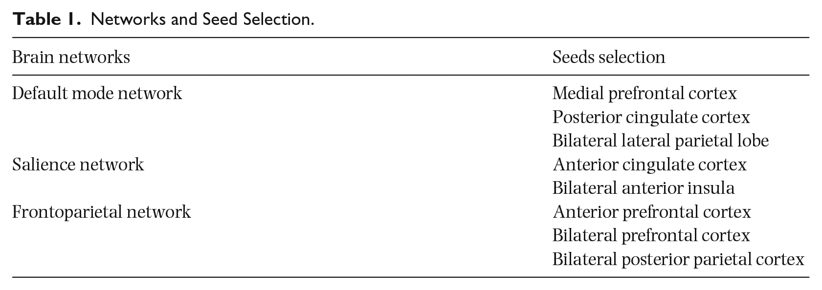
Functional image preprocessing was performed using the default pipeline in the CONN toolbox, including realignment, co-registration, and smoothing (6-mm full-width at half-maximum [FWHM] kernel). The default denoising pipeline in the CONN toolbox was applied to remove nuisance signals. Specifically, the signals from white matter and cerebrospinal fluid were regressed out by estimating five potential noise components (Chai et al., 2012). The Friston 24-parameter model was used to regress out head motion effects from the realigned data. Scrubbing was performed using artifact detection tools with the threshold for global signal above 5 (z-value) and for subject motion above 0.9 mm. In addition, linear and quadratic trends were also included as regressors since the BOLD signal exhibits low-frequency drifts. Temporal filtering (0.008–0.09 Hz) was then performed on the time series. To control for the effects of head motion differences across groups, the head motion and scrubbing parameters were included in the first level as covariates of interest. Using the seed-to-voxel FC analysis approach, the correlation coefficients between the time series of these seed regions and all other voxels in the whole brain were calculated and converted to z-scores using Fisher’s r-to-z transformation.
Group analysis
A 2 × 2 mixed-model analysis of variance (ANOVA) was conducted to examine the Group effect (i.e., Control vs. Mindfulness), Time effect (i.e., Pre-training vs. Post-training), and Group × Time interaction on the resting-state networks’ FC for each seed. The significance threshold was set as uncorrected p < .005 at the whole-brain level, and false discovery rate (FDR)-corrected p < .05 at the cluster level. Post hoc simple t-tests were performed using an ROI-to-ROI approach for the regions that showed significant main effects or interaction effects.
Results
Behavioral and sociodemographic
There were no significant group differences in gender (five females and five males in each group), age, pre-existing psychological conditions, experience with mindfulness activities, age since starting music, number of years of music lessons, and number of hours of practice in adolescence or as a young adult. However, there were differences in the number of hours of practice currently, with the mindfulness group practicing more than the control group, t(17) = 2.30, p = .034.
DMN
MPFC-seed FC
As shown in Figure 1(a), at the whole-brain level, a significant group × time interaction effect was found for the mPFC-seed FC in the left vermis 6, −8, −58, −20; t(18) = 5.76; pFDR-cluster = .017. Further post hoc simple tests revealed that the mindfulness group had decreased post-training FC, t(9) = −8.00; p < .001, while the control group had increased post-training FC, t(9) = 6.34, p < .001, compared to the pre-training FC between the mPFC seed and the left vermis-6 (Figure 1(b)). Furthermore, during the post-training, the mindfulness group showed significantly lower FC than the control group, t(18) = −10.47, p < .001.
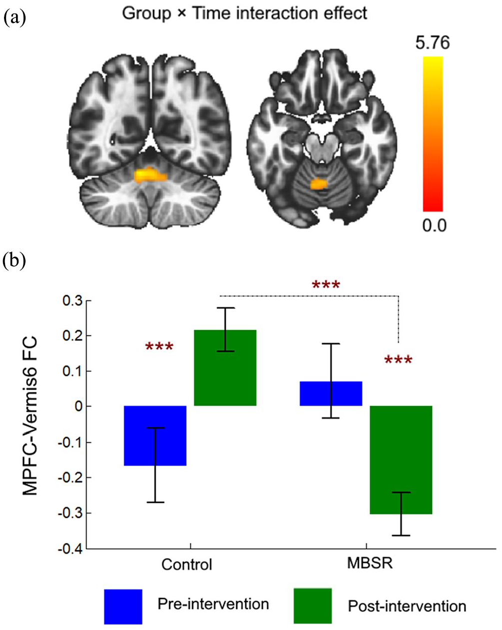
Group × Time Interaction Effect of FC Between the mPFC Seed and the Vermis-6. (a) The interaction effect at the whole-brain level (whole-brain uncorrected p < .005; pFDR-cluster < .05). (b) Post hoc simple tests on each level.
In addition, a significant group effect showed that the mindfulness group had greater FC between the mPFC seed and the right precentral gyrus (r. PreCG), 42, −10, 44; t(18) = 4.86; pFDR-cluster = .048, compared to the control group (Figure 2(a)). Further post hoc tests revealed that the mindfulness group showed greater mPFC-r. PreCG FC than the control group at both pre-training, t(18) = 4.37, p < .001, and post-training, t(18) = 4.37, p < .001; Figure 2(b).
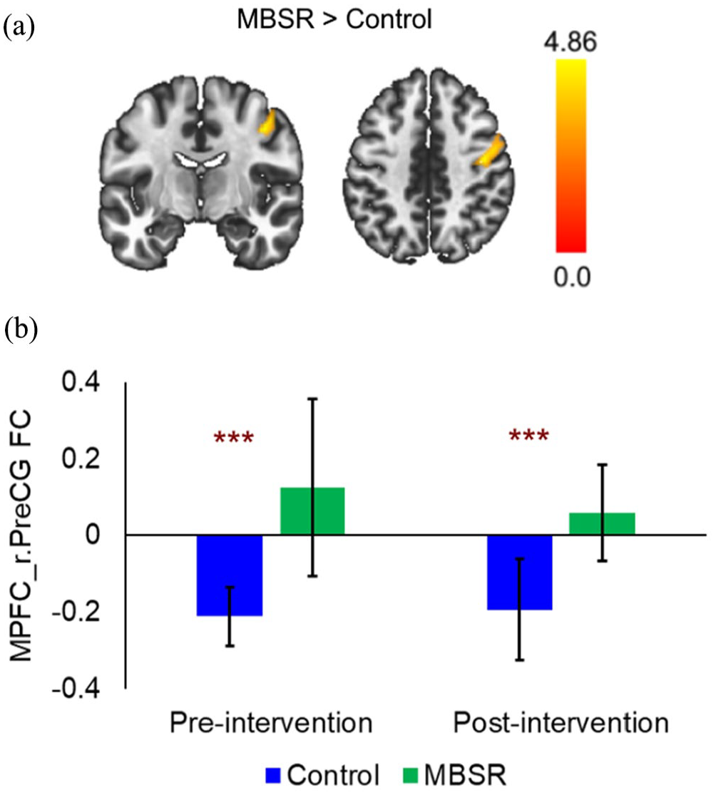
Group Effect of the FC Between the mPFC Seed and the Right Precentral Gyrus. (a) The mindfulness group showed greater FC than the control group at the whole-brain level. (b) Post hoc simple tests to examine the group effect for each scan session.
Finally, significant time effects were also found for the mPFC-seed FC. As shown in Figure 3(a), the time effects showed that post-training FC was greater than the pre-training FC in the right superior parietal lobule, r.SPL; 8, −68, 56; t(18) = 4.20, pFDR-cluster = .015. Further post hoc tests revealed that both the control group, t(9) = 2.82, p = .02, and mindfulness group, t(9) = 5.42, p < .001, showed greater mPFC-r.SPL FC in the post-intervention scan compared to the pre-intervention scan (Figure 3(b)).
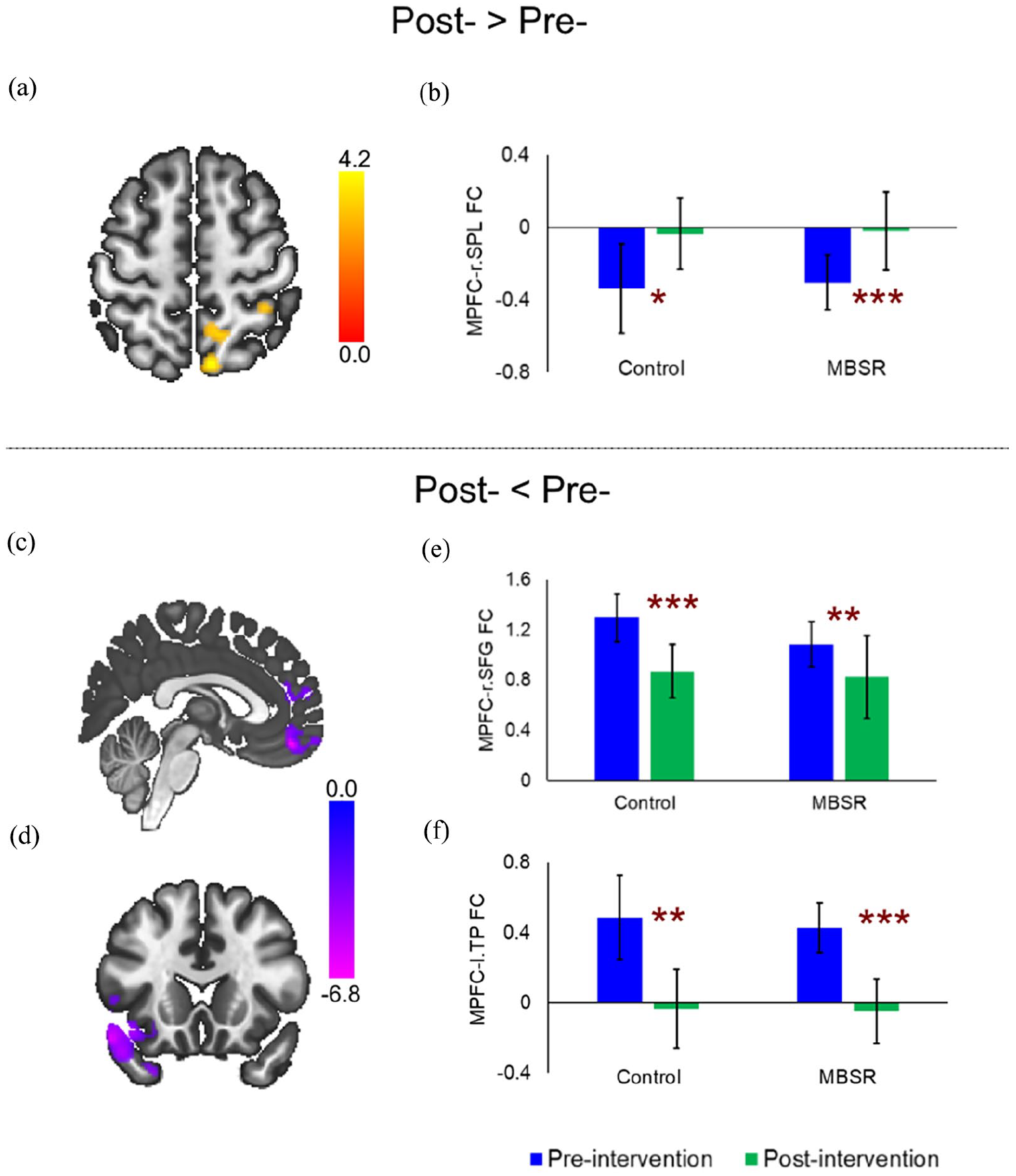
Time Effects of the mPFC-Seed FC. (a) Greater post-training (vs. pre-training) FC between the mPFC seed and the r.SPL at the whole-brain level. (b) Post hoc tests showed both the Control and Mindfulness groups had greater post-training (vs. pre-training) FC between the mPFC seed and r.SPL. Decreased post-training (vs. pre-training) FC between the mPFC seed and (c) r.SFG and (d) left temporal pole (l.TP) at the whole-brain level; post hoc tests showed both the Control and Mindfulness groups had decreased post-training (vs. pre-training) FC between the mPFC seed and (E) the r.SFG and (F) l.TP.
Conversely, compared to the pre-training condition, the post-training condition showed decreased FC in the right superior frontal gyrus (r.SFG), 2, 46, −16; t(18) = −6.81, pFDR-cluster < .001; Figure 3(c)) and the left temporal pole (l.TP), −52, 16, −16; t(18) = −6.72, pFDR-cluster < .001; Figure 3(d)). Further post hoc tests revealed that both the control group (t(9) = −6.47, p < .001) and mindfulness group (t(9) = −4.06, p = .003) showed decreased mPFC-r.SFG FC in the post-training scan (Figure 3(e)). For the FC between the mPFC and l.TP, both the control group (t(9) = −4.77, p = .001) and the mindfulness group (t(9) = −6.24, p < .001) showed significantly decreased FC during the post-training scan compared to pre-training scan (Figure 3(f)).
PCC-seed FC
For the PCC-seed FC, only a significant time effect was found in the left central opercular cortex (l.C-Op; −52, −34, 6), with decreased FC during the post-training scan compared to the pre-training scan (t(18) = −4.97; pFDR-cluster = .013; Figure 4(a)). Post hoc tests showed that both the control group (t(9) = −5.32, p < .001) and the mindfulness group (t(9) = −6.51, p < .001) had decreased post-training FC (vs. pre-training; Figure 4(b)).
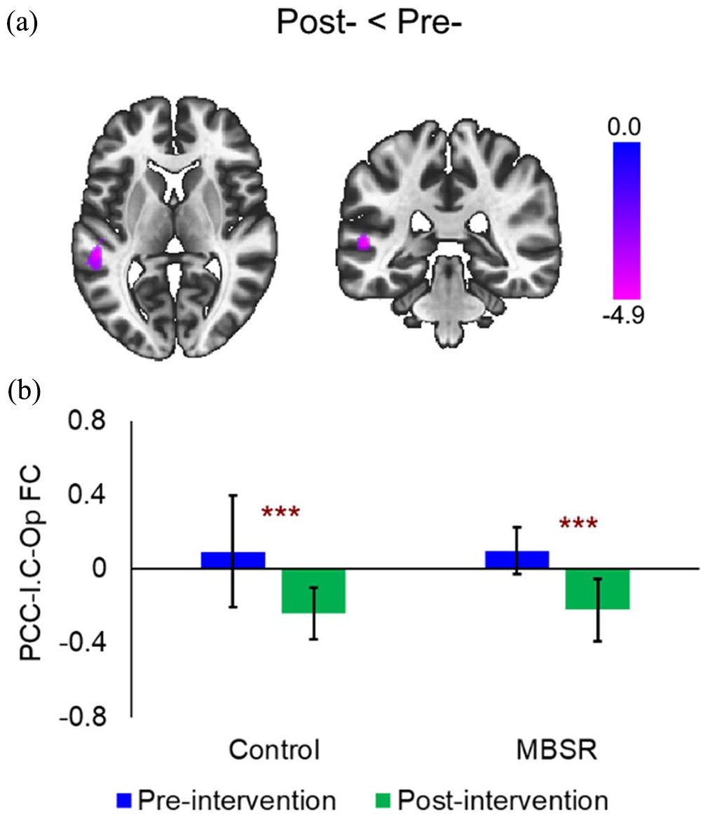
Time Effect of the PCC-Seed FC. (a) Decreased post-training (vs. pre-training) FC between the PCC seed and the left central opercular cortex (l.C-Op). (b) Post hoc tests showed both the Control and Mindfulness groups had decreased post-training (vs. pre-training) FC between the PCC seed and l.C-Op.
LP-seed FC
Neither a significant main effect nor an interaction effect was found for the LP-seed FC.
Salience network
Among three seeds within the SN, only a significant time effect was found for the bilateral insula-seed FC in the right frontal pole (r.FP; 28, 50, 24). Specifically, the post-training FC was lower than the pre-training FC (t(18) = −5.61; pFDR-cluster = .002; Figure 5(a)). Post hoc tests revealed lower post-training FC in both the control group (t(9) = −5.42, p < .001) and the mindfulness group (t(9) = −3.38, p = 0. 008) compared to the pre-training FC (Figure 5(b)).
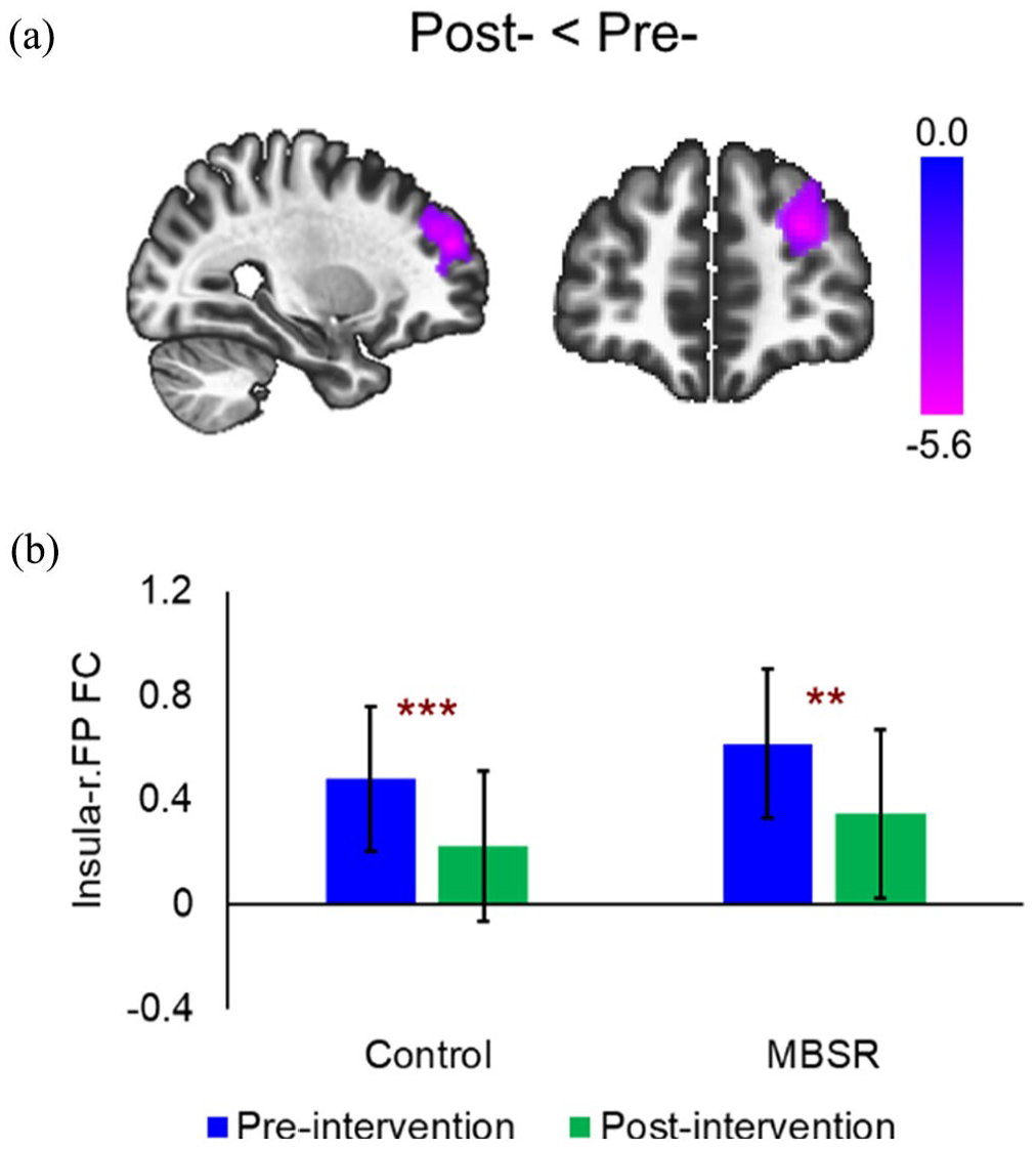
Time Effect of the Bilateral Insula-Seed FC. (a) Decreased post-training (vs. pre-training) FC between the bilateral insula seed and the right frontal pole (r.FP). (b) Post hoc tests showed both the Control and Mindfulness groups had decreased post-training (vs. pre-training) FC between the bilateral insula seed and the r.FP.
CEN network
Lateral PFC-seed FC
A significant group × time interaction effect was found for the bilateral lateral PFC-seed FC in the bilateral cerebellum crus II (0, −78, −34; t(18) = 2.28; pFDR-cluster = .047; Figure 6(a)). Post hoc analyses revealed that the control group (t(9) = 7.04, p < .001) showed increased FC, while the mindfulness group (t(9) = −4.38, p = .002) showed decreased FC during the post-training scan compared to the pre-training scan. Furthermore, during the post-training scan, the mindfulness group showed significantly lower FC than the control group (t(18) = −6.24, p < .001). No such group difference was found for the pre-training scan (t(18) = 1.32, p = .20; Figure 6(b)).
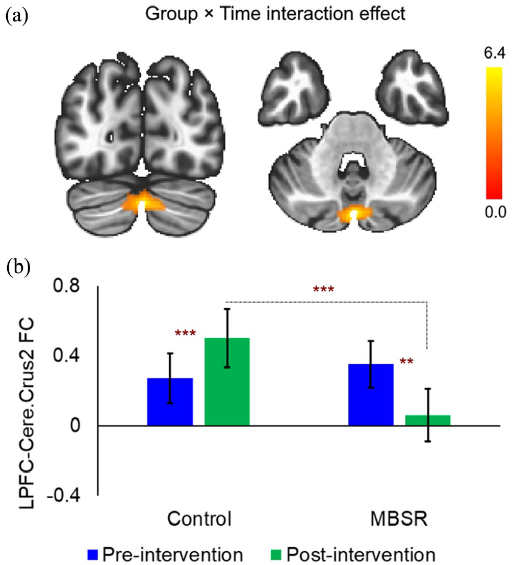
Group × Time Interaction Effect of FC Between the Bilateral Lateral PFC Seed and the Cerebellum Crus2 (Cere. Crus2). (a) The interaction effect at the whole-brain level. (b) Post hoc simple tests on each level.
In addition, the significant time effects showed decreased post-training FC (vs. pre-training) for the bilateral lateral PFC seed in the superior frontal gyrus (SFG; 4, 48, 34; t(18) = −5.01, pFDR-cluster = .022; Figure 7(a)) and the right middle frontal gyrus (r.MFG; 42, 20, 34; t(18) = −5.89, pFDR-cluster = .022; Figure 7(b)). Post hoc tests revealed that both the control (t(9) = −4.98, p < .001) and mindfulness group (t(9) = −5.67, p < .001) showed decreased post-training lateral PFC-SFG FC compared to the pre-training scan (Figure 7(c)). Similarly, both the control (t(9) = −3.64, p = .005) and mindfulness group (t(9) = −4.03, p = .003) showed decreased post-training lateral PFC-r.MFG FC compared to the pre-training scan (Figure 7(d)).
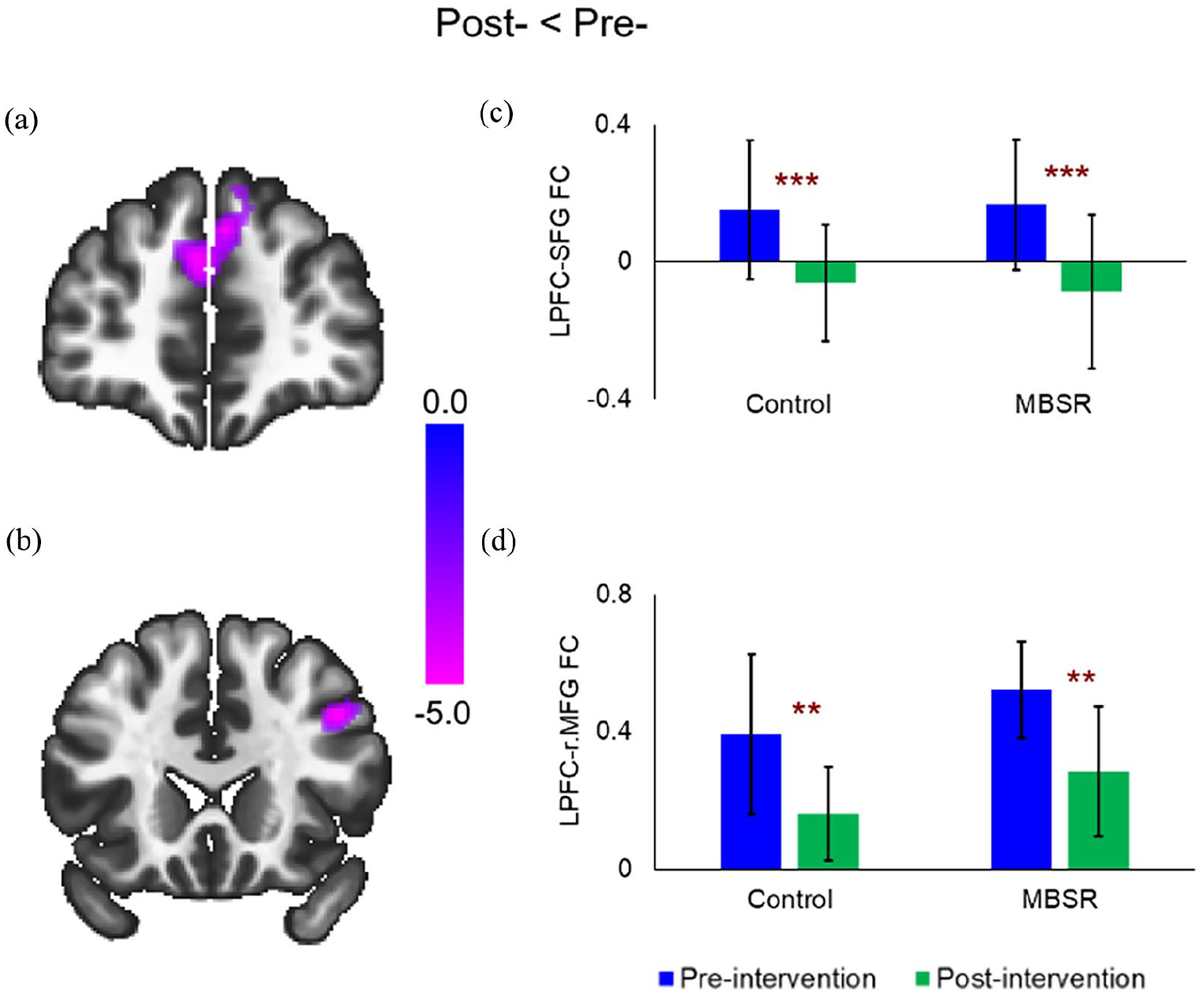
Time Effects of the Lateral PFC-Seed FC. Decreased post-training (vs. pre-training) FC between the lateral PFC seed and the (a) SFG and (b) r.MFG at the whole-brain level; post hoc tests showed both the Control and Mindfulness groups had decreased post-training (vs. pre-training) FC between the lateral PFC seed and the (c) SFG and (d) r.MFG.
PPC-seed FC
For the PPC-seed FC, only a significant time effect was found in the left inferior frontal gyrus (l.IFG; −50, 26, −6), with decreased FC during the post-training scan compared to the pre-training scan (t(18) = −5.27, pFDR-cluster < .001; Figure 8(a)). Post hoc tests showed that both the control group (t(9) = −5.79, p < .001) and the mindfulness group (t(9) = −3.09, p = .013) had decreased post-training FC (Figure 8(b)).
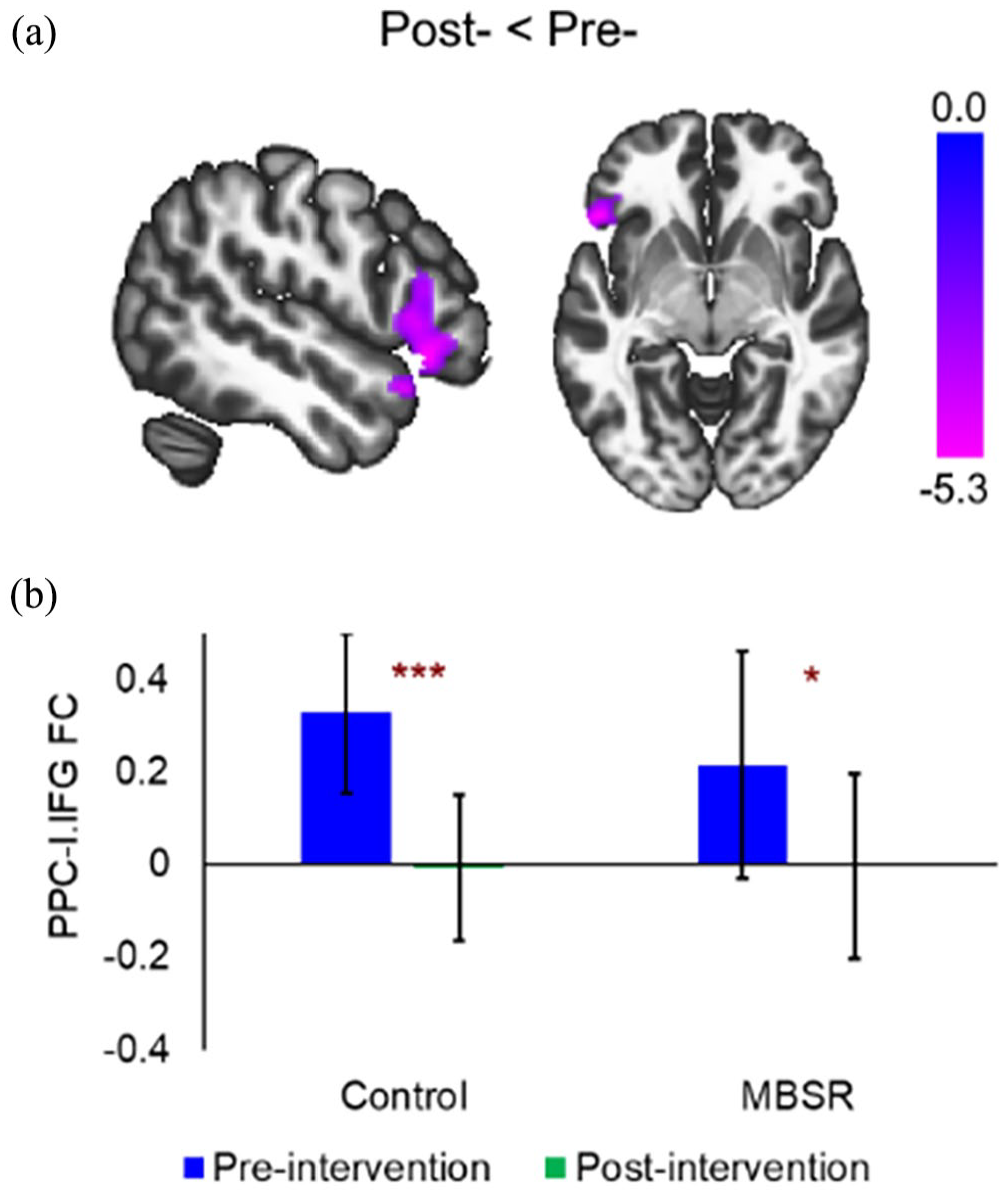
Time Effect of the Bilateral PPC-Seed FC. (a) Decreased post-training (vs. pre-training) FC between the PPC seed and the r.IFG. (b) Post hoc tests showed both the Control and Mindfulness groups had decreased post-training (vs. pre-training) FC between the PPC seed and r.IFG.
Discussion
This study examined the rsFC of areas of the DMN, SN, and central executive network (CEN) in musicians with MPA before and after a brief mindfulness training program. FC changes were found in both the mindfulness and control groups in various regions within these networks, with the most notable changes in the mindfulness group within the cerebellum.
The cerebellum has been shown to play a role in several sensorimotor, autonomic, cognitive, and emotional functions (Beuriat et al., 2022; Petrosini et al., 2024). The core emotional processing system includes connections within cortico–striatal–thalamic loops with structures, such as the PFC, amygdala, and hippocampus, with other subcortical structures, including the basal ganglia, thalamus, and striatum (Beuriat et al., 2022). Each of these regions is functionally and anatomically directly or indirectly connected to the cerebellum, which may underlie the cerebellum’s ability to influence affective processing.
The cerebellar vermis, especially the posterior vermis (which includes vermis-6), has been shown to be involved in more “limbic” tasks through these feedback loops involving cortical, subcortical, limbic, and cerebellar structures (Benagiano et al., 2018; Stoodley & Schmahmann, 2010). Previous studies have demonstrated the cerebellum’s ability to modulate the firing patterns of limbic structures, such as the amygdala and hippocampus, and vermis activation is exhibited in response to intense emotional states, such as panic and grief (Stoodley & Schmahmann, 2010).
In this study, participants who underwent mindfulness training exhibited decreased rsFC between the mPFC and the vermis-6, while the control participants exhibited increased rsFC between the mPFC and the vermis-6. The cerebellum, specifically the vermis, has been linked to anxiety through its connections to the limbic system. For example, previous studies have documented that FC between the amygdala and cerebellum is increased in patients with GAD and other anxiety-related disorders, such as social anxiety disorder and obsessive-compulsive disorder (Chin & Augustine, 2023). rsFC has also been examined between the amygdala and mPFC. A study by Kim et al. (2011) demonstrated decreased rsFC between the dmPFC and the amygdala in a low anxiety group, suggesting that this represents “efficient mPFC-amygdala crosstalk . . . mitigating the generation of anxious states.” Taken together, perhaps decreased FC between the mPFC and cerebellum in the mindfulness group and increased FC in the control group are a result of a cerebellar–amygdala–cortical feedback loop, in which there is less communication between these structures within the mindfulness group and more communication within the control group. These findings may demonstrate the influence of mindfulness on emotional processing through cortical and limbic brain regions. Lower subjective MPA levels from pre-training to post-training in the mindfulness group support this finding (Boileau et al., in review). There were no changes in subjective MPA from pre-training to post-training in the control group, perhaps demonstrating consistent levels of high MPA and “inefficient mPFC-amygdala crosstalk” without intervention.
rsFC changes were also found between the lateral PFC (CEN) and the bilateral cerebellum crus II from pre- to post-training in both groups. More specifically, the mindfulness group showed decreased FC between these two regions, while the control group showed increased FC between these regions. Increased acceptance as a result of undergoing mindfulness training may have altered the FC between these regions. To support this interpretation, Goldin et al. (2019) examined participants’ neural activity by asking participants to react to negative self-beliefs and to then implement acceptance strategies. They found that acceptance (a fundamental component of mindfulness), in comparison to reacting to negative self-beliefs, was associated with less neural activity in the lateral PFC and cerebellum. On the contrary, participants who reacted to negative self-beliefs exhibited more activity in the frontopolar medial PFC and bilateral cerebellum. The control group in the current study may have exhibited negative self-appraisals as opposed to accepting themselves and the situation with nonjudgement. Subjective MPA levels may elucidate these findings: The mindfulness group exhibited decreased subjective MPA levels from pre-training to post-training, whereas the control group showed no differences (Boileau et al., in review). As a result of mindfulness training, the mindfulness group may have learned acceptance strategies in response to music performance. The control group, however, may have continued to exhibit negative self-appraisals.
This study also found decreased FC between the mPFC (DMN) and the right SFG and left temporal pole; between the PCC (DMN) and the left opercular cortex; between the bilateral insula (SN) and the right frontal pole; between the bilateral lateral PFC (CEN) and the SFG and right middle frontal gyrus; and between the PPC (CEN) and the left inferior frontal gyrus in both the mindfulness and control groups at post-training. Decreased FC between these regions during the second scan may be due to being more comfortable in the MRI environment. The first MRI scan involved a novel experience that can elicit anxiety, but the participants would know what to expect during the second scan, perhaps reducing the communication between these regions.
In support of this interpretation, there was increased FC between the mPFC (DMN) and the right superior parietal lobule from pre-to post-training in both the mindfulness and control groups. The increased FC between these two regions at post-training may indicate that undergoing the second MRI scan was a more pleasant experience, as both the mPFC and right superior parietal lobule are activated in response to pleasant emotional states (Campagne et al., 2016; Sambuco et al., 2022).
In this study, a significant group effect was also found. The mindfulness group had greater FC between the mPFC and the right precentral gyrus than the control group at both pre-training and post-training. This may be due to lack of randomization to the mindfulness and control groups. It is possible that the first 10 participants who enrolled in the study, who were allocated to the mindfulness group, were more motivated to participate in the study to reduce their performance anxiety by becoming more mindful. A study by Seo et al. (2013) demonstrated that the right precentral gyrus is activated in response to positive, rewarding stimuli, which have been shown to elicit increased motivation in relation to appetitive goals and increased psychomotor action toward achieving these goals (Lang, 2010; Roesch & Olson, 2003). Considering this, future studies should randomly allocate participants to the conditions. Although the most significant limitation of this study was sample size—there were only 10 participants in each condition—this study is in line with similar studies that examine the effects of mindfulness training on MPA and brain activity (Gómez-López & Sánchez-Cabrero, 2023; Young et al., 2018). In addition, participants in the mindfulness condition completed the mindfulness training as a group, whereas those in the control condition did not. Therefore, some of the changes in rsFC in the mindfulness group may be partly due to the influence of group cohesion and social support, which has been suggested as a possible change mechanism of mindfulness-based interventions (Blanck et al., 2018). Future studies should include an active control group, as this study only employed a passive control group. There were also differences in the number of hours that the musicians currently practice music, with the mindfulness group practicing more than the control group. However, a recent study demonstrated no effect of hours practiced weekly on MPA in a global sample of adolescents, which is consistent with previous studies (Papageorgi, 2022). It must also be noted that this study recruited young adult musicians. Some studies have found that younger, undergraduate musicians report higher levels of MPA than professionals (Fernholz et al., 2019; Papageorgi et al., 2013); thus, the results from this study should not be generalized to other groups. Finally, the performance quality was not measured in this study. Lin et al. (2008) observed that daily individual meditation practice reduced MPA and increased performance quality in higher education students; however, the relationship between MPA, performance quality, and rsFC remains unexplored.
Although this study has limitations, it also has several strengths. First, the study design is a strength because it is in line with other studies employing a longitudinal mindfulness training program (Tang et al., 2015). The mindfulness intervention’s brevity is also a strength. Neurophysiological changes in rsFC were observed after only 2 weeks of mindfulness training. This is important to note because typical mindfulness training programs are 8 weeks long. Although an 8-week program may offer different benefits, this study demonstrates that brief mindfulness training may be an accessible and cost-efficient alternative. Follow-up of these participants to observe continued benefits of the mindfulness intervention is also important. MPA is debilitating; however, it is sometimes viewed as “inherent to the profession and/or musical training” (Gómez-López & Sánchez-Cabrero, 2023). Musicians below 30 years of age are more likely to experience MPA, perhaps as a result of less performing experience (Biasutti & Concina, 2014) or burnout (Bernhard, 2010). However, there is little focus on the diagnosis and prevention of MPA. This may lead young musicians to abandon their musical careers as their MPA increases without intervention. In addition, the normalization of using drugs to alleviate MPA points to the need for healthier options. Mindfulness may help address these issues.
Conclusion
Mindfulness, concisely, is paying attention to the present moment nonjudgmentally. The findings from this study demonstrate that even brief mindfulness training influences communication between regions of several important neural networks (DMN, SN, and CEN) within musicians with performance anxiety. Most notably, brief mindfulness training leads to FC changes between the areas of the PFC and areas of the cerebellum. These changes may indicate increased acceptance of the present moment as a result of the mindfulness training. This suggests that further research is needed to examine the potential relationship between different facets of mindfulness, such as acceptance and brain communication. It also suggests that mindfulness may be a useful tool to address the deleterious effects of MPA.
Footnotes
Acknowledgements
The authors thank the Brain Imaging Centre, including Rahim Ismaili, Katie Dinelle, and Reggie Taylor, for their support.
Authors’ note
Due to the nature of this research, participants of this study did not agree for their data to be shared publicly, so supporting data are not available.
Declaration of conflicting interests
The author(s) declared no potential conflicts of interest with respect to the research, authorship, and/or publication of this article.
Funding
The author(s) received no financial support for the research, authorship, and/or publication of this article.