
Abstract
Introduction
During Cone beam computed tomography (CBCT) imaging in the head and neck region of patients who are undergoing fixed orthodontic treatment, the different components of fixed orthodontic appliances can produce various metal artifacts which may degrade the diagnostic image quality in the region of interest. Therefore, the metal artifact reduction software (MARS) system can be used in most CBCT units to reduce the amount of artifacts generated from various fixed orthodontic appliance components including skeletal anchorage devices.
Aims and Objectives
The main purpose of this study was to quantitatively evaluate the effectiveness of MARS system of a CBCT unit in reducing the amount of artifacts generated by various fixed orthodontic appliance components during CBCT imaging.
Materials and Methods
Four dental arch shaped phantoms that were fabricated along with different fixed orthodontic appliance components (metal bracket, molar band, transpalatal arch, mini-implant) underwent CBCT scanning without and with the MARS setting option. After using the MARS system of a CBCT unit, the reduction in the amount of artifacts that were generated by various fixed orthodontic appliance component was calculated by using Google Colab software that utilized Python code and OpenCV system to generate a binary mask image. The resulting mask image highlights the areas where artifacts are detected, allowing for further analysis or visual inspection. The contours found within the mask image represent the boundaries of the artifact regions.
Results
After calculating the percentage decrease in artifact area for each component of fixed orthodontic appliance, one-way analysis of variance (ANOVA) test was performed to compare the percentage decrease in artifact area for each component of fixed orthodontic appliances. *P-values are calculated using one-way analysis of variance (p < 0.05). The percentage decrease in artifact area for different components of fixed orthodontic appliance ranged from 39.67% to 48.58%. The stainless steel bracket showed the significant artifact area reduction with a mean percentage decrease of 48.58% as compared to other components.
Conclusion
The effect of MARS system in reducing the amount of metal artifacts generated varied according to the component type of fixed orthodontic appliance and, MARS system was more efficient for stainless steel brackets as compared to other components. CBCT imaging of head and neck region can be carried out without taking off the orthodontic appliances by applying MARS system which can significantly decrease the artifact area.
Introduction
Cone beam computed tomography (CBCT) imaging provides detailed, high-resolution 3D images that allow clinicians to visualize the anatomy of the head and neck region in greater detail compared to traditional 2D X-rays. CBCT scans are also faster and involve less radiation exposure as compared to traditional CT scans, making them a valuable tool for diagnosing and planning treatment for a variety of conditions in head and neck region. CBCT can produce images in all the three planes, that is, axial, coronal and sagittal planes, but CBCT imaging is more susceptible for artifact production due to cone shaped beam of X-ray with its low radiation dose.1–3
However, during CBCT imaging various metallic objects present in the patient’s oral cavity, such as dental restorations, prosthetic crowns and bridges, dental implants and different metallic components of fixed orthodontic appliances which includes metal brackets, molar bands, archwires, transpalatal arches and different types of skeletal anchorage devices like mini-implants can create artifacts in the CBCT images, which can make it difficult to interpret the diagnostic images properly. All these metallic materials used in dentistry are having high atomic number and are more susceptible to artifact production during CBCT imaging in the head and neck region.
Metal artifacts occur when the X-rays from the CBCT machine interact with metal objects present in the patient’s oral cavity, causing scattered radiation to be detected by the machine. This scattered radiation can create streaks (alternate light and dark bands), shadows, or other distortions in the CBCT images, which can obscure important details and make it difficult to accurately diagnose and treat dental conditions.4, 5
To minimize the effects of metal artifacts in CBCT images, several techniques have been developed. These include using specialized imaging protocols that reduce the dose of radiation and filter out the scattered radiations from the metal objects. In addition, the use of metal artifact reduction software (MARS) can help to improve the quality of CBCT images by removing or reducing the artifacts caused by various metal objects.6–8 During image reconstruction process, the MARS system can be used to reduces some dark areas which are caused by loss of gray values and bright streaking artifacts per se.5–9 The amount of artifact generated depends on both, that is, the type, size and shape of dental material in metal form and parameters of CBCT unit like tube voltage, tube current, voxel size and field of view (FOV).10–13 Yet the relation between these parameters and MARS system have not been studied and evaluated in much detail, so the exposure parameters in CBCT units should be adjusted according to the patients conditions and diagnostic purposes, and optimal exposure along with MARS system should be elucidated to enhance the diagnostic quality of CBCT images when using MARS system.14, 15
So, in this study we prepared four dental arch shaped phantoms and equipped each phantom with different fixed orthodontic appliance components and quantitatively evaluated the performance of MARS system in reducing the metal artifacts that affected the diagnostic quality of CBCT images in head and neck region by using Google Colab Software.
Materials and Methods
Fabrication of Dental Arch Shaped Phantoms
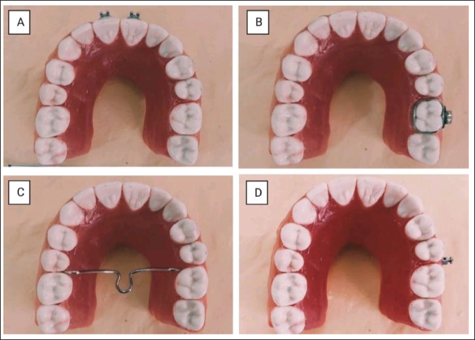
Forty dental arch shaped phantoms simulating the patient’s oral structures were fabricated using resin material. Each phantom was equipped with one of the four components of fixed orthodontic appliances in the form used by an orthodontist during the treatment of a particular malocclusion: (a) stainless steel brackets; (b) stainless steel molar band on the left side first molar; (c) a transpalatal bar from right side to left side on first molar; and (d) a temporary anchorage device (mini-implant) in the interdental area between second premolar and first molar on the left side as shown in Figure 1.
The dental arch-shaped phantoms with four different types of fixed orthodontic appliance components. (A) Stainless steel metal brackets on the central incisors, (B) A stainless steel molar band on the maxillary left first molar, (C) A transpalatal arch from right side to left side on first molar, and (D) A temporary anchorage device (mini-implant) in the interdental area between second premolar and first molar on the left side.
CBCT Imaging
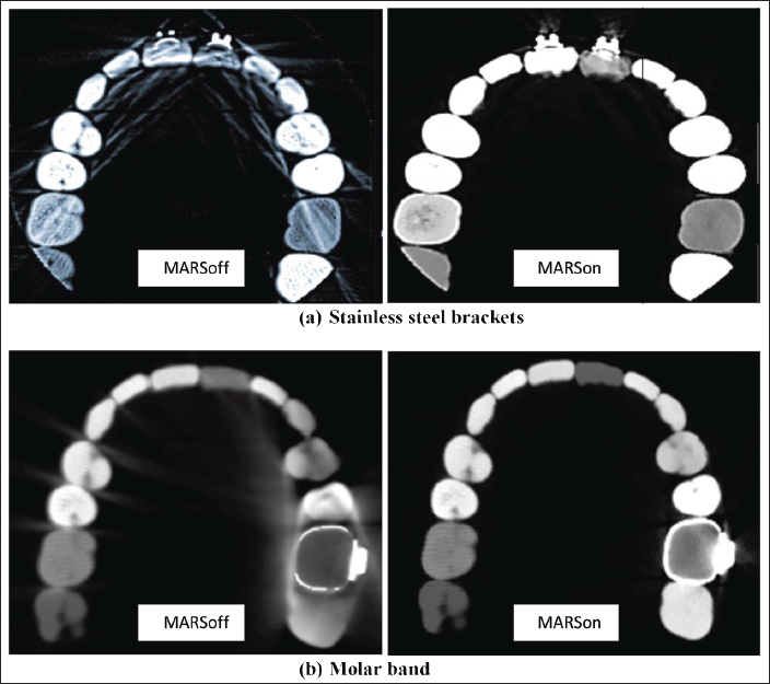
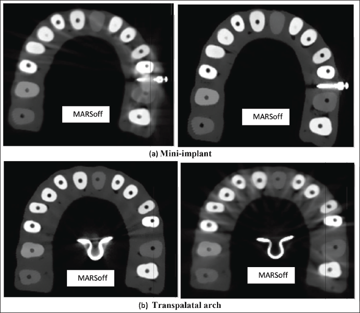
The 40 dental arch shaped phantoms along with different fixed orthodontic appliance components (metal bracket, molar band, transpalatal arch, mini-implant) were scanned and CBCT images were taken with NewTom Giano HR 3D CEPH CBCT machine. The CBCT images were taken with the medium FOV, height: min 1,650 mm (65 in) - max 2,410 mm (95 in), scan time: 18 s, 90 kVp, 1–10 mAs (pulsed mode) 0.5 mm focal spot, voxel size (micrometer): minimum slice thickness 75 μm. CBCT images were generated in all three sections, that is, axial, coronal and sagittal planes. Images were first generated with MARS setting off and second set of images were generated by applying the MARS system (Figures 2 and 3).
Axial images of phantoms with stainless steel brackets and molar band without and with metal artifact reduction (MARS) setting as shown in the (a) and (b), respectively. The scan parameters were as follows: tube voltage, 90 kVp; voxel resolution, 0.5 mm; field of view, height- min 1,650 mm (65 in) - max 2,410 mm (95 in); tube current, 1–10 mAs; scan time, 18.0 s.
Axial images of phantoms with mini-implant (TAD) and transpalatal arch without and with metal artifact reduction (MARS) setting as shown in the (a) and (b), respectively. The scan parameters were as follows: tube voltage, 90 kVp; voxel resolution, 0.5 mm; field of view, height- min 1,650 mm (65 in) - max 2,410 mm (95 in); tube current, 1–10 mAs; scan time, 18.0 s.
Software-based Image Processing and Analysis
To quantify and measure the amount of metal artifacts generated by various components of fixed orthodontic appliances, and to quantitatively evaluate the performance of MARS system of a CBCT unit in reducing the amount of artifacts generated, images were generated in all the axial, sagittal and coronal planes, and the images were first taken in a conventional manner with CBCT unit, and the second set of images was generated by using the MARS setting of the same CBCT unit. For the image analysis, only axial slices with components of the fixed orthodontic appliances were included in the study and axial slices without any components were excluded, and 20 axial slice images with minimum slice thickness of 75 μm were selected for each component.
The images which are captured with artifact reduction setting off and on, were analyzed by using Google Colab software; Google Colab is an online development environment that provides a Jupyter notebook interface. It allows to write and execute Python code (Python is a popular programming language widely used for various tasks, including image processing and computer vision), access pre-installed libraries like OpenCV (Open Source Computer Vision Library is a powerful open-source library that provides a wide range of functions and algorithms for image and video processing. It is commonly used for tasks such as image manipulation, feature detection, object recognition, and more), and utilize powerful computing resources provided by Google, including GPUs and TPUs. The Google Colab software was applied for quantitatively evaluating the performance of MARS system of a CBCT unit in reducing the amount of artifacts generated, by following sequence of methods:
Load the Images
The images were loaded into memory of the Google Colab software. One image was captured with the artifact reduction setting ON, and the other image was captured with the artifact reduction setting OFF, and we import the necessary libraries into the Google Colab software,
Find the Difference between the Images
The absolute difference between the two images was calculated. This highlights the pixels that differ between the two images, which may correspond to the artifact.
Threshold the Difference Image
Thresh applies a binary thresholding operation to the different images by using
Find Contours in the Thresholded Image
Contours are the boundaries of connected white regions in the binary image. The contours represent the shape of the artifact. The
Find the Contour with the Largest Area
The contour with the largest area is likely to correspond to the artifact. The area of each contour was calculated using
Filter Out Small Contours
Small contours that are likely to be noise or irrelevant to the artifact were filtered out based on a minimum area threshold. Contours with areas below the threshold were discarded, and only the larger contours were considered.
Find the Contour Closest to the Largest Contour
The contour that was closest to the largest contour was likely to be the most accurate representation of the artifact. The similarity distance between contours was calculated using
Calculate the Area of the Artifact Contour
The area of the artifact contour was calculated using
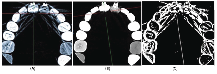
By following these steps, the code of the software identified and measured the artifact in the images. It quantified the decrease in artifact by comparing the areas of the artifact contours between the image with the artifact reduction setting ON and the image with the artifact reduction setting OFF (Figure 4). The percentage decrease was then calculated based on the difference in areas as described in the following section of statistical analysis, and this method is quite reliable and provides the same results when utilizing the MARS system of different CBCT units with this inbuilt software function.
(A). Image with Artifact Reduction Setting OFF, (B). Image with Artifact Reduction Setting ON, (C). Binary Mask Image: The image displayed is the binary mask image obtained after thresholding and morphological operations. The white regions in the image represent the detected artifacts or differences between the “ON” and “OFF” images. The mask is generated by calculating the absolute difference between the “ON” and “OFF” images and applying a threshold to identify regions with significant differences. The thresholded image is further processed using morphological operations (erosion and dilation) to refine the artifact regions. The resulting mask image highlights the areas where artifacts are detected, allowing for further analysis or visual inspection. The contours found within the mask represent the boundaries of the artifact regions.
Statistical Analysis
The normal distribution of data was carried out by using the Shapiro–Wilk test which provided the normal distribution of the data. The percentage decrease in artifact contour area for each component of fixed orthodontic appliance after applying the MARS system setting of the CBCT unit was obtained with Google Colab software. The following equation was used to calculate the percentage decrease in artifact area:
The numerator in the above equation, that is, decrease in the artifact area = Artifact area with MARS off − Artifact area with MARS on.
After calculating the percentage decrease in artifact area for each component of fixed orthodontic appliance, one-way analysis of variance (ANOVA) test was performed to compare the percentage decrease in artifact area for each component of fixed orthodontic appliances. *P-values are calculated using one-way analysis of variance (p < 0.05).
Results
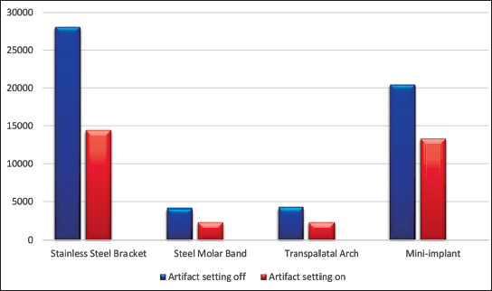
The percentage decrease in artifact area for different components of fixed orthodontic appliance ranged from 39.67% to 48.58% as shown in Table 1. The stainless steel bracket showed the significant artifact area reduction with a mean percentage decrease of 48.58% as compared to other components (Figure 5).
The decrease in artifact area between MARoff image and MARon image along with mean percentage decrease. Data are represented as mean ± SD by using one-way analysis of variance (ANOVA) test.
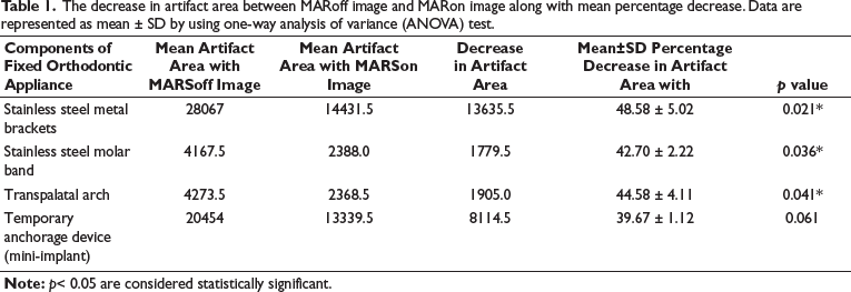
The percentage decrease in artifact area for each component of fixed orthodontic appliance was obtained after applying the MARS system setting of the CBCT unit. The stainless steel bracket showed the significant artifact area reduction with a mean percentage decrease of 48.58% as compared to other components. The significant reduction in artifact area are noted. *P-values are calculated using one-way analysis of variance (ANOVA) correction (p < 0.05).
Discussion
CBCT is a specialized imaging technique that uses a cone-shaped X-ray beam to produce a 3D image of the head and neck region. CBCT imaging is commonly used in dentistry, oral and maxillofacial surgery, and otolaryngology (ENT) to diagnose and plan treatment for a variety of conditions. However, during CBCT imaging various metal artifacts may appear in the form of alternate light and dark areas which can impair the diagnostic quality of CBCT images and render them unusable during diagnosis and treatment planning.4, 5, 7, 8 Some dense metal objects used in patient’s oral cavity can cause over-attenuation of X-rays mainly on the center of the dense metal object as compared to the periphery of the object which results in a cupping artifact which can cause resultant inaccuracy of the object size. 9 Basically, various CBCT units have been equipped with MARS system to reduce those artifacts which are induced by dense metal objects. The software eliminates voxels with too high or too low grey values and reconstruct these areas to nearby voxels. 8 However, streaking artifacts which appear more extensive are removed by MARS system effectively than cupping artifact, although the MARS system can also have a positive influence in reducing the cupping artifact per se.
Various types of dense metal objects with high atomic number, which are to be used as a part of fixed orthodontic treatment can create special issues because they cause generation of extensive artifacts.5, 12 Different components of fixed orthodontic appliances can induce different amounts of artifacts. 5 Previously various research studies that have been carried out analyzed the metal artifacts by insertion of different types of metal rods into dental arch shaped phantoms.16, 17. These studies mainly considered the frequently used materials and their locations and shape in the dental arch so as to simulate the clinical situation. Thus, the present study focused on the metal artifacts in various shapes, size and location of dental materials in the form of different components of fixed orthodontic appliances. In the present study, we fabricated four dental arch shaped phantoms, and each dental arch-shaped phantom similar to patient’s oral conditions was fabricated using resin material. Each phantom was equipped with one of the four components of fixed orthodontic appliances to reflect the clinical setting. Therefore, despite the controlled setting of present experiment, the limitation should be understood of using the differences in size and shape of each component.
The amount of artifact reduction varied according to different components of the fixed orthodontic appliance. The artifacts from the stainless steel metal bracket were more efficiently removed than those from the other components of fixed orthodontic appliances. This might be due to the fact that the brackets are the most important metallic component of fixed orthodontic appliance and is the most frequently used material in orthodontic appliance and, MARS system of CBCT was developed based on this clinical situation. The other components of fixed orthodontic appliance (stainless steel molar band, transpalatal arch and temporary anchorage device (mini-implant) showed lower levels of artifact reduction compared to the stainless steel metal bracket).
Metal artifact is known to be reduced by using high tube voltage in CT.11, 18 This is because high tube voltage induces less beam hardening tendency. In this study, there was no tendency to cause any change in efficiency of MARS system by altering the tube voltage. This is because the amount of tube voltage was not changed enough and was kept at a consistent rate.
Metal artifact reduction on CT images is known to be closely related to voxel size which in turn is determined by matrix size, FOV and slice thickness of CT images.19, 20 The effect of voxel size on MAR during CBCT imaging was controversial. Voxel size did not show significant effect on metal artifact reduction for amalgam and copper–aluminum alloy as reported by some recent studies on CBCT metal artifact. 21
Evaluating the efficacy of the MAR in an objective and quantitative manner during CBCT imaging is quite challenging. Grey-level measurements in the region of interest (ROI) were used for quantification of artifacts. However, constant grey values cannot be found for a single material during CBCT imaging even with the same equipment. 22 Moreover, there are numerous factors, including patient’s position, CBCT unit model, and even room temperature and exposure conditions which are especially sensitive to the ROI according to size or area. 23
Since some of the patients undergoing fixed orthodontic treatment had to undergo CBCT imaging in the head and neck region for diagnosis and treatment planning of various conditions, but there is an apprehension that during CBCT imaging in orthodontic patients, the various metallic components of these appliance may generate various artifacts which makes the interpretation of CBCT images difficult, and the vital diagnostic information may be lagging, this creates a dilemma between orthodontists and oral radiologists, that whether the orthodontic appliance should be debonded or not before subjecting the patients to CBCT imaging in the head and neck region. No research has been carried out in this field till now and very sparse information is available, so our study quantitatively evaluated the performance of MARS system of a CBCT unit in reducing the amount of artifacts generated by various components of fixed orthodontic appliances and, our study supported the fact that innovative aMAR (autoadaptive Metal Artifact Reduction) function is a proprietary algorithm developed by NewTom that can considerably reduce the artifacts generated by amalgam, implants or other metal elements including fixed orthodontic appliances that can impair image quality. This facilitates planning and designing of specialist treatments that require segmentation of anatomical structures without renouncing the original data acquired. Therefore this method can be quite effective in reducing the amount of artifacts generated by fixed orthodontic appliances and patients can be subjected to CBCT imaging without removing the orthodontic appliances.
Conclusion
MARS system of the CBCT machine can effectively reduce the amount of metal artifacts generated by various components of fixed orthodontic appliances during CBCT imaging in the head and neck regions.
It can enhance the diagnostic quality of CBCT images without removing the orthodontic appliances during CBCT imaging, and among the various components, MARS was more efficient for stainless steel bracket in reducing the artifact.
Other factors should be taken into the consideration like voxel size, tube voltage and should be adjusted accordingly to increase the efficiency of MARS system of CBCT unit.
Ethical Approval Statement
This study is purely an in vitro non-clinical phantom-based CBCT study. No patients and animals were involved in this study and subjected to any invasive procedure, and no ethical clearance was needed to carry out this research study, and hence the ethical approval statement does not apply here.
Footnotes
Declaration of Conflicting Interests
The author and co-author have seen and agree with the contents of this manuscript, and there is no financial interest to report, and there is not any conflict of interest and the submission is original work and is not under review at any other publication.
Funding
The authors received no financial support for the research, authorship and/or publication of this article.
Informed Consent
Not applicable.