
Abstract
In orthodontic diagnosis and treatment planning, soft tissue profile assessment is of prime importance. Due to the emerging soft tissue paradigm, greater emphasis has been given to the clinical examination of soft tissue function and esthetics. Various cephalometric and photographic methods were introduced in the past to assess and measure profile angle and other facial angles. Our new device, that is, profilometer, helps to measure profile angle clinically.
Introduction
The success of the orthodontic treatment is frequently determined by improvement gained in the patient’s facial appearance, which includes the soft tissue profile. 1 Legan and Burstone described the angle of convexity or profile angle which is formed using soft tissue glabella, subnasale, and soft tissue pogonion. 2 According to Bergman, a class I subject has a profile angle in the range of 165 to 175 degrees which decreases in class II and increases in class III. 3 However, traditional cephalometric measurements do not provide all the answers to the aesthetic considerations of the face and dentition, particularly in relation to the soft tissues. 1 The present article describes a new device, that is, profilometer, which will help to measure profile angle clinically.
Design and Fabrication
To fabricate the profilometer, a protractor, which is often available while manual cephalometric tracing by the orthodontists, is used.
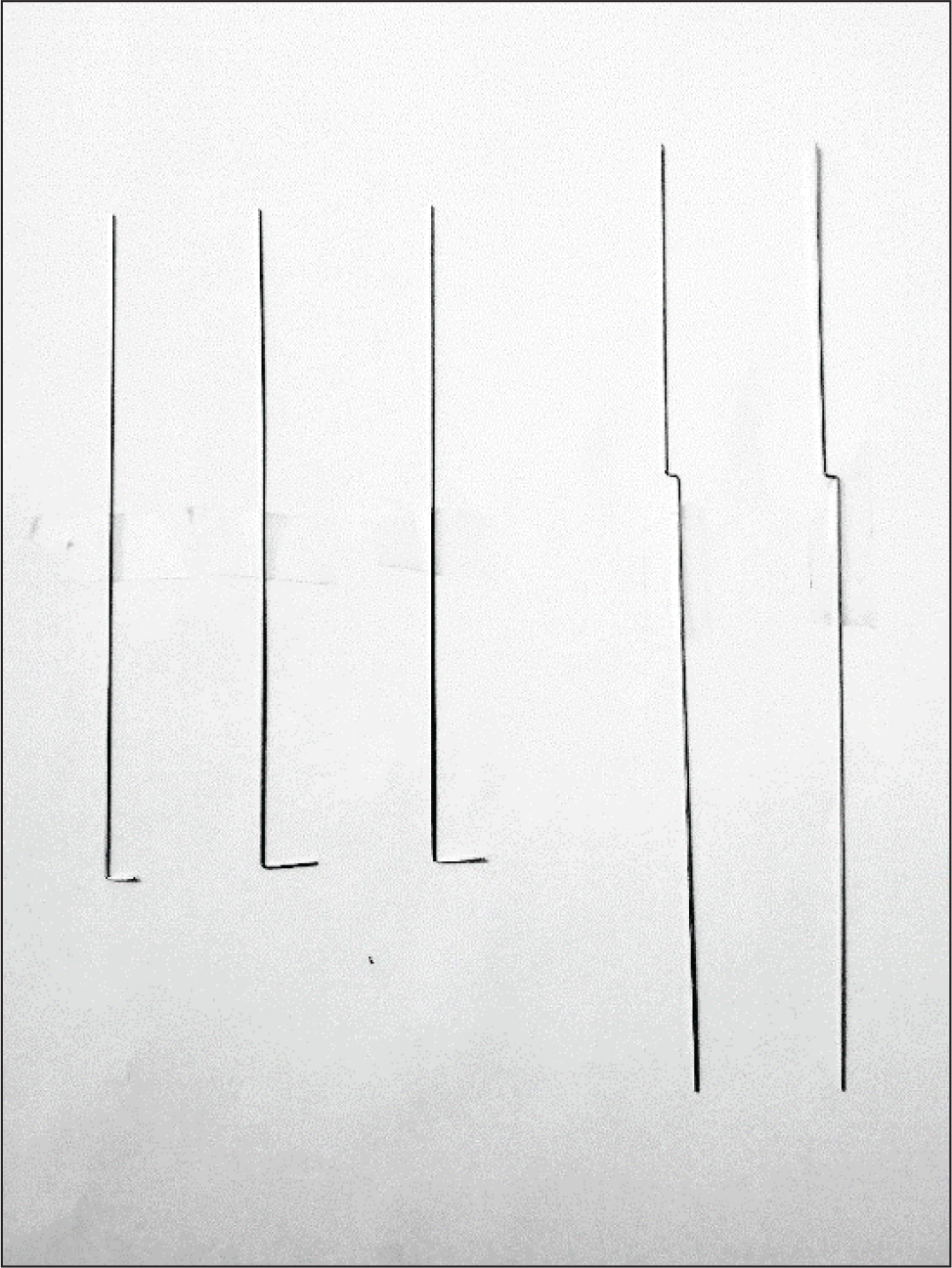
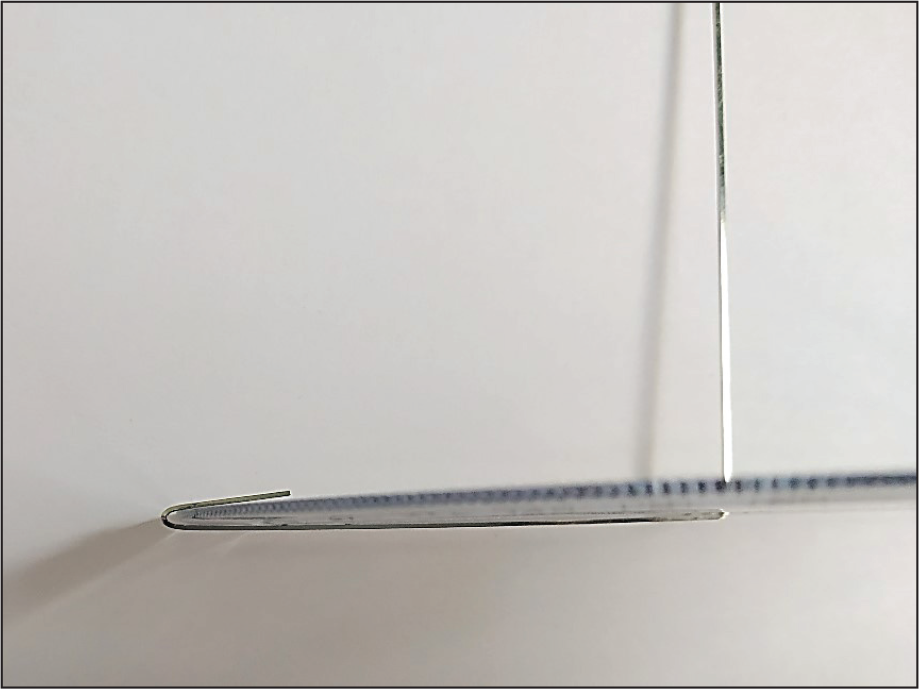
A 0.019 × 0.025 straight stainless steel wire is cut into 3 pieces each of 8 cm each and 2 pieces of 15 cm each (Figure 1).
Wires of 8 cm are bent at their ends at a distance of 1 cm by 90 degrees. A 90-degree step bend of 2 to 2.5 mm is made at 6 cm at one end of the 15-cm wire (Figure1). At the point of step bend, the wires are slightly trimmed and made rounded in shape.
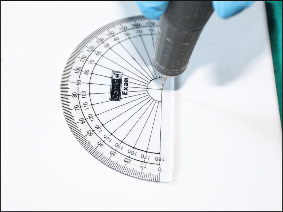
A hole of 2 to 2.5 mm in diameter is drilled in the protractor at the center of its circumference using straight cylindrical bur (Figure 2).
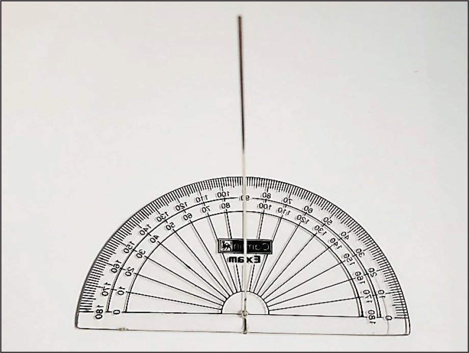
One 8-cm wire is inserted in the hole and glued permanently perpendicular to the rear surface of the protractor. It forms the middle horizontal arm of the device which is fixed (Figure 3).
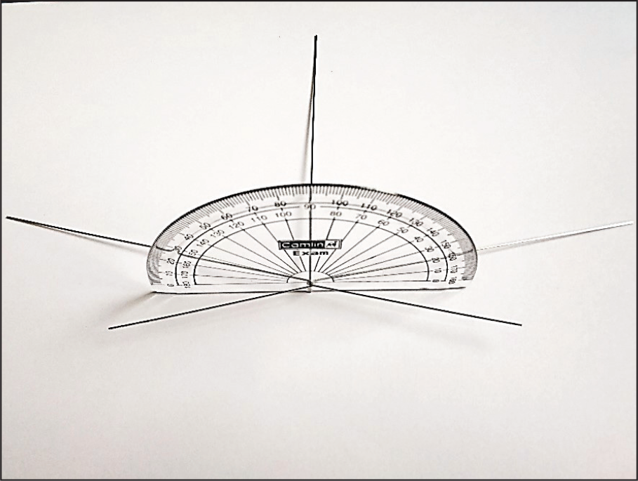
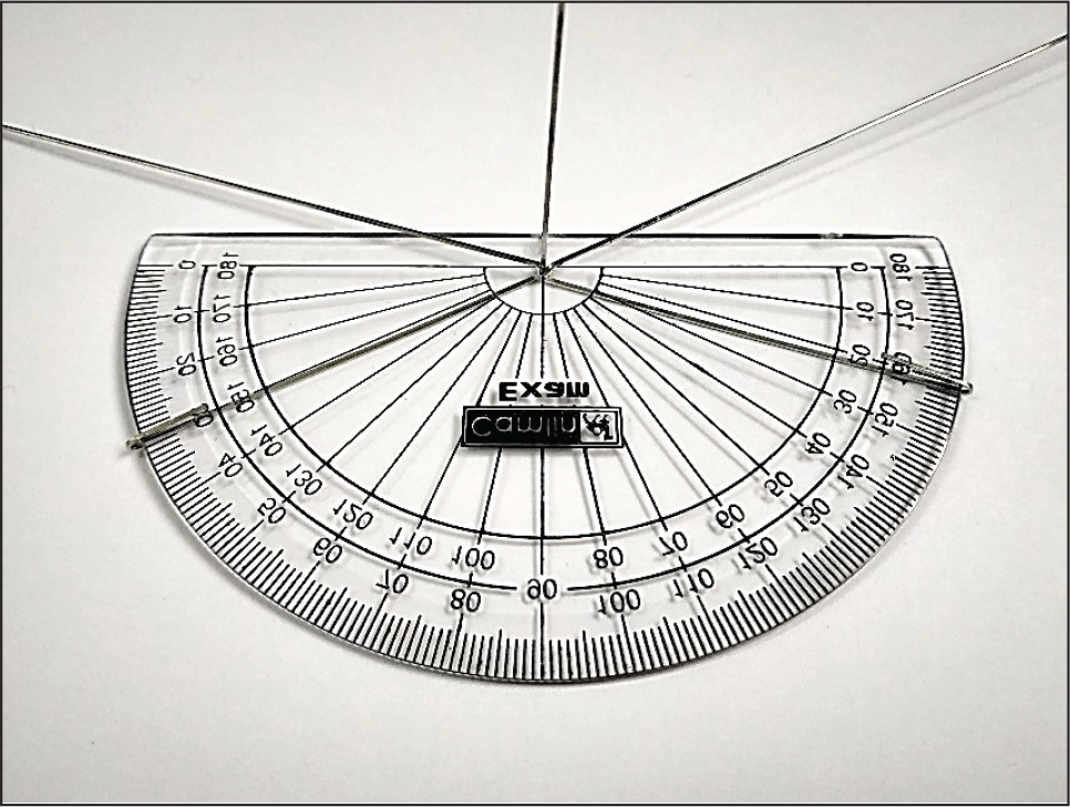
Both the 15-cm wires are inserted in the hole up to step bend in a crisscross manner (Figure 4). The smaller end of the wire is bent and encircled around the border of the protractor by 180 degrees (Figures 5 and 6). Its longer end serves as the vertical arms of the device, that is, upper and lower vertical arms.
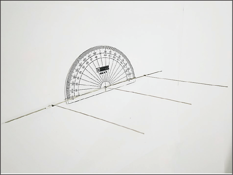
A wire of 8 cm is ligated perpendicular to the longer end of the 15-cm wire using a folded elastic module (e-module) such that it can glide on it. It forms the upper and lower movable horizontal arms of the device. The upper and lower horizontal arms can be glided on the vertical arm of the device (Figure 7).
All the three horizontal arms (upper, middle, and lower) should be in one plane parallel to each other and perpendicular to the vertical arms (upper and lower) of the device (Figure 8).
Three Horizontal and 2 Vertical Arms Are Made Using 0.019 × 0.025 Straight Stainless Steel Wire.
A Hole of 2 to 2.5 mm in Diameter Is Drilled in the Protractor at the Center of Its Circumference Using Straight Cylindrical Bur.
Middle Horizontal Arm Is Inserted in the Hole and Glued Permanently Perpendicular to the Rear Surface of the Protractor.
Both the Vertical Arms Are Inserted in the Hole up to Step Bend in a Crisscross Manner.
The Smaller End of the Vertical Arms Wire Is Bent and Encircled Around the Border of the Protractor by 180 Degrees.
Folded Smaller Ends of the Vertical Arms.
Upper and Lower Horizontal Arms Are Ligated Perpendicular to Vertical Arms Using Folded E-Module.
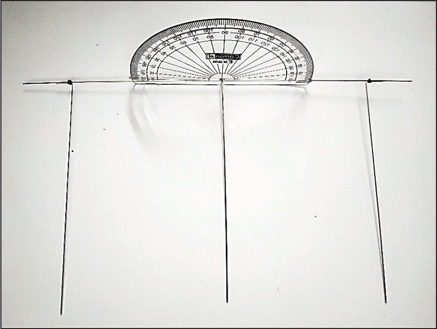
All the Three Horizontal Arms Are Parallel to Each Other and Perpendicular to the Vertical Arms and Protractor.
Clinical Use
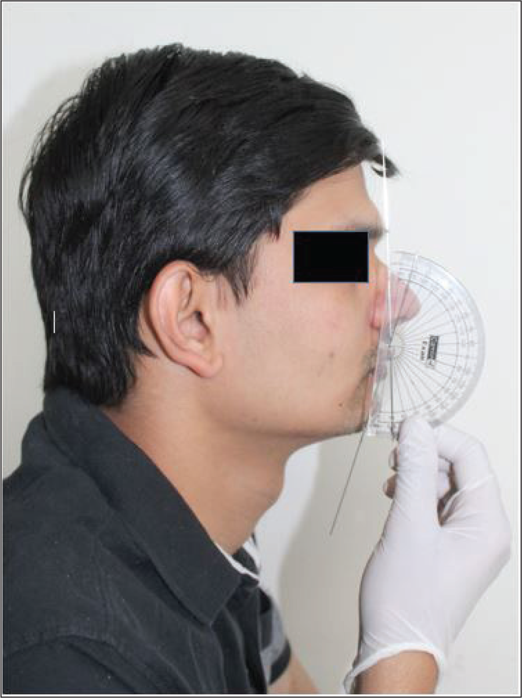
The profilometer can be used to measure the profile angle: first, the protractor is placed in a plane such that its base is posterior and convex surface is in the anterior direction of the patient’s face (Figure 9).
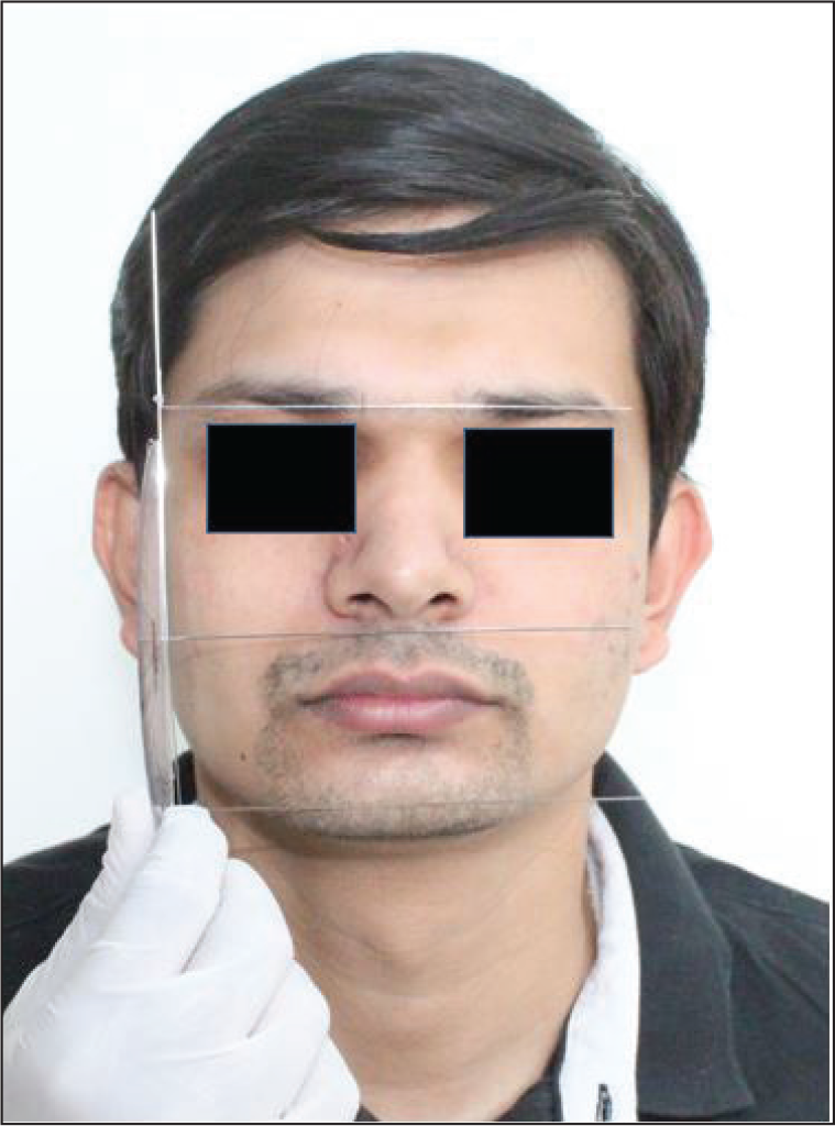
The middle horizontal arm is first placed parallel to the soft tissue subnasale point, and later, upper and lower vertical and horizontal arms are rotated and glided and brought parallel at the soft tissue glabella and pogonion point, respectively. The horizontal arms should passively touch the landmark points (Figure 10).
The angle formed between the smaller vertical arm on the protractor is measured as the profile angle for patients who have a convex and straight profile (Figure 9).
For severe concave profile patients who have a profile angle more than 180 degrees, the protractor is inverted 180 degrees such that the base is anterior and convex surface faces in the posterior direction of the patient’s face, and the profile angle is calculated by deducting the angle formed between smaller vertical arms from 360 degrees.
Measurement of Profile Angle.
Horizontal Arms Touching the Landmarks Passively.
Advantages
The pre-treatment and post-treatment profile angles of the profilometer can be measured and compared clinically.
It can be used in all patients without any design modification.
It is easy to use.
It is easy to fabricate in very little time.
It is inexpensive and cost-effective.
It does not require any exposure to X-rays.
Limitations
Any wire if bent or fractured will have to be replaced.
Free ends of the wire can cause trauma.
Not easy to transport.
A number of modifications can be made to the profilometer as follows:
Tip of the wires can be covered with plastic sleeves or can be made blunt to avoid tissue trauma during use. A 360-degree protractor can be used to measure angle more conveniently in patients who have a profile angle more than 180 degrees.
Conclusion
While using the profilometer, the clinician should be aware of the importance of other methods of measuring profile angle. It can be incorporated in routine orthodontic record taking, diagnosis, and treatment planning.
Footnotes
Statement of Informed Consent
Written informed consent was obtained from the subject for the use of photographs for publication.
Declaration of Conflicting Interests
The authors declared no potential conflicts of interest with respect to the research, authorship, and/or publication of this article.
Funding
The authors received no financial support for the research, authorship, and/or publication of this article.