
Abstract
Introduction:
The upper lateral incisor is the most commonly missing tooth in the anterior segment. It leads to esthetic and functional imbalance for the patients. The ideal solution is the one that is most conservative and which fulfills the functional and esthetic needs of the concerned individual. Canine substitution is evolving to be the treatment of choice in most of the cases, because of its various advantages. These are special cases that need more time and effort from the clinicians due to space discrepancy in the upper and lower arches, along with the presentation of individual malocclusion.
Aims and Objectives:
Malocclusion occurring due to missing laterals is more complex, needing more time and effort from the clinicians because of space discrepancy, esthetic compromise, and individual presentation of the malocclusion. An attempt has been made in this article to review, evaluate, and tabulate the important factors for the convenience of clinicians.
Method:
All articles related to canine substitution were searched in the electronic database PubMed, and the important factors influencing the decision were reviewed. After careful evaluation, the checklist was evolved.
Result:
The malocclusions in which canine substitution is the treatment of choice are indicated in the tabular form for the convenience of clinicians. Specific treatment-planning considerations and biomechanics that can lead to an efficient and long-lasting result are also discussed.
Conclusion:
The need of the hour is an evidence-based approach, along with a well-designed prospective randomized control trial to understand the importance of each factor influencing these cases. Until that time, giving the available information in a simplified way can be a quality approach to these cases.
Keywords
Hypodontia is a common dental anomaly and involves one or more teeth of the individual. The overall prevalence of hypodontia is 6.4%, with the highest number of cases seen in Africa (13.4%). The region is followed by Europe (7%), Asia (6.3), and Australia (6.3%), with lowest prevalence in North America (5%) and Latin America (4.4%). Females are more commonly affected than males. The commonly affected teeth are the maxillary lateral incisors, mandibular second premolars, and the maxillary second premolars.1-3 Etiological factors in missing maxillary laterals include familial tendency, with one or more point mutations in a closely linked polygenetic system, most often transmitted in a autosomal dominant pattern with incomplete penetrance and variable expressibility.4, 5
Missing maxillary lateral incisors, a type of true partial anodontia, is also associated with other dental anomalies, such as agenesis of other permanent teeth, increased occurrence of microdontia of the maxillary lateral incisor of the contralateral side, palatal displacement of canines, and distal angulation of mandibular second premolars. 4 Being part of anterior dentition, missing maxillary laterals lead to functional imbalance and esthetic disability, which can have a negative psychological impact.5-7 Options in treating such patients include either the possibility of orthodontic space closure or a combination of orthodontic space opening and replacement of the missing tooth.8-10 Treatment should be planned as conservatively as possible while fulfilling the patient’s esthetic and functional needs. The choice between the two has been a controversial topic of discussion.11, 12 However, because of the superiority of periodontal health, lack of long-term biological and technical considerations, and possibility of early completion of definitive treatment, canine substitution can be the treatment of choice whenever possible.6, 13-16 With the number of variables involved, it may be beneficial to have a simplified approach for selecting this treatment modality.
All the publications related to canine substitution were searched in the electronic search engine “PubMed” using various keywords related to the topic. The relevant articles were reviewed to understand the factors affecting the decision of substituting the canine for the missing maxillary lateral incisor. An attempt is made in this article to evolve a straightforward checklist of all the key factors that help clinicians in the selection of space closure as a treatment modality. The specific mechanics involved during the orthodontic treatment of these cases are also discussed for the same reason.
Diagnosis of a missing lateral incisor needs to be confirmed with the interpretation of various diagnostic aids, which include case history, clinical examination, photographs, intra-oral peri-apical (IOPA) X-rays, study models, and orthopantomographs (OPG) along with the addition of supplementary aids, like cone-beam computed tomography (CBCT). The patient could present with either of the following cases:
Over-retained deciduous lateral incisor and missing permanent lateral incisor: In this case, extraction of the deciduous tooth should be planned to address the patient’s esthetic and psychological needs for the time being, to guide the eruption of the canine adjacent to the central incisor and to maintain the alveolar ridge thickness.
13
Missing both the deciduous and permanent upper lateral incisors.
The decision to either open the space or close the space for canine substitution also depends on the various factors such as facial profile, type of skeletal and dental malocclusion, age, cost affordability, treatment time considerations, canine position, color balance, and its anatomy. The following are the guidelines that can be helpful in the decision-making process.
Anteroposterior discrepancy: Angle’s class I with crowding, dentoalveolar protrusion in the lower arch and Angle’s class II malocclusion are the types of cases in which stable occlusion can be achieved at the end of the treatment with canine substitution. Angle’s class III with dentoalveolar discrepancy less than 5 mm can also be considered for canine substitution.9, 14, 19 Vertical and transverse discrepancy: In patients with maxillary vertical excess, canine substitution is possible, along with added considerations to correct the gingival levels of the substituted tooth because of increased visibility upon smiling.
9
In cases where there is a large difference in color or size, reduction in the inciso-gingival and mesiodistal dimensions, flattening of the labial surface, steepening of the lingual convexity, use of bleaching, composite bonding, or veneering to mimic the replaced tooth can be considered. Similarly, the dimensions of the premolar can be increased mesiodistally for it to replace the canine.9, 13, 20
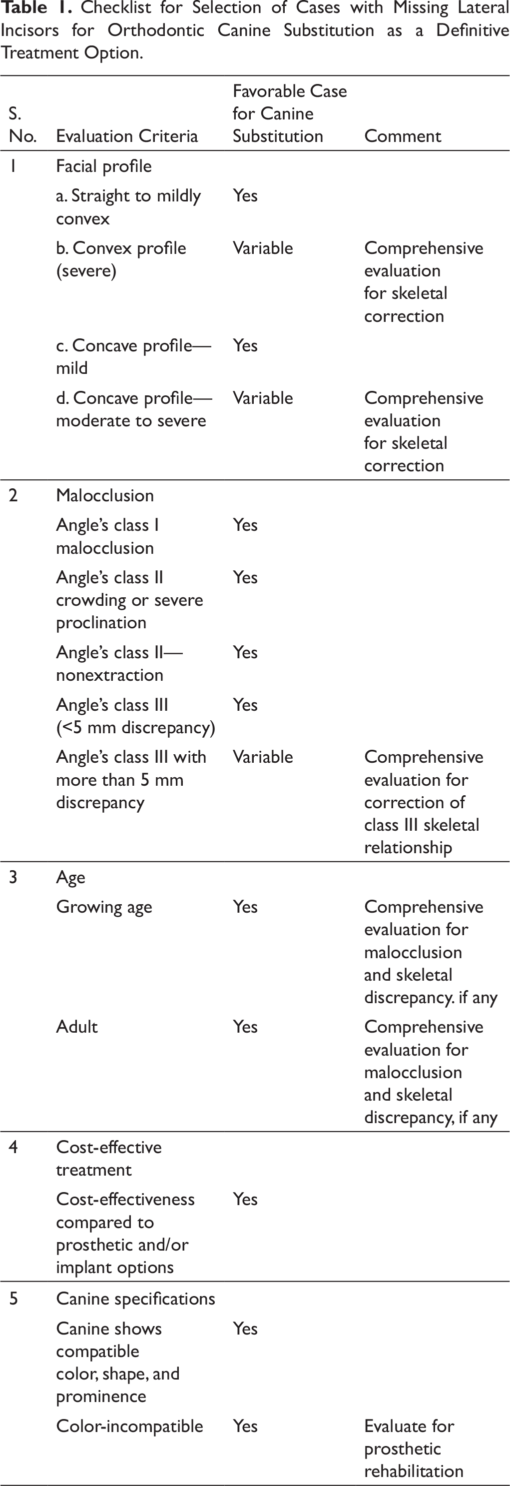
Checklist for Selection of Cases with Missing Lateral Incisors for Orthodontic Canine Substitution as a Definitive Treatment Option.
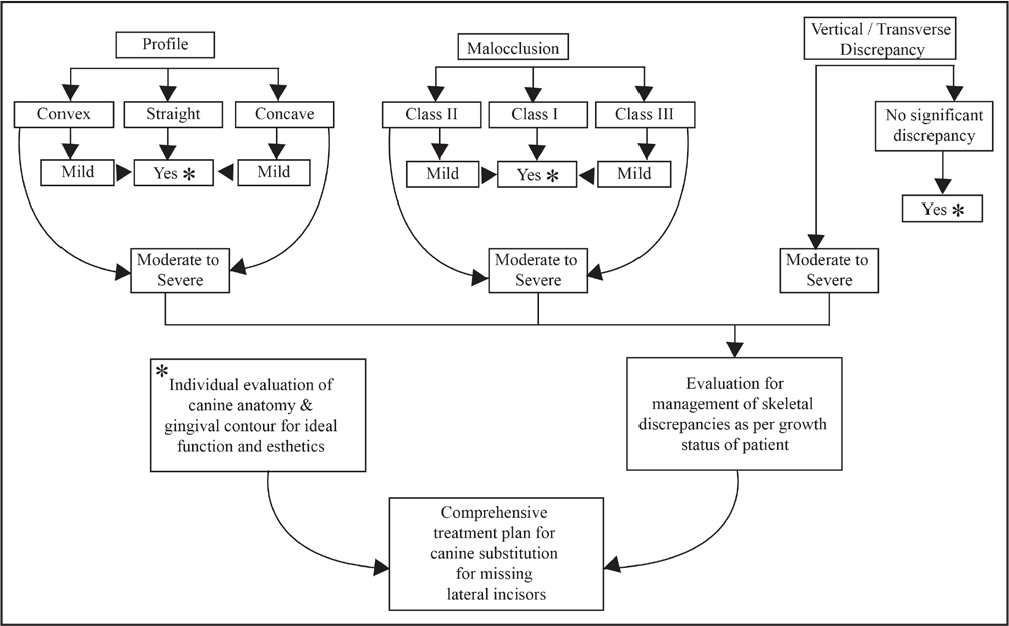
Flow Chart Showing Evaluation of Facial Characteristics and Malocclusion to Determine Comprehensive Treatment Planning for Canine Substitution in Patinets With Missing Lateral Incisors.
Orthodontic Considerations
The orthodontist plays a key role in managing the treatment of missing laterals. Clinical considerations other than those discussed earlier are divided into two parts: treatment planning and mechanics.
Usage of the contralateral lateral incisor as a guide, if present, and assessing whether it is without any shape anomaly.
Every case is unique in its own way in orthodontic practice. In patients with missing lateral incisors, the selection of a suitable treatment modality has always been a challenge. These cases need information which differs from routine orthodontic treatment protocol. The advantage of making a ready-to-use checklist is the gathering of all case-specific information under one roof and in turn reduction of the time needed for the clinician to decide on a treatment plan. The mechanics specifically used for replacing a lateral incisor with a canine and a canine with a premolar are also discussed for the same reason. The need of the hour is an evidence-based approach, along with a well-designed prospective randomized control trial, for understanding the importance of each factor discussed in this article. Treatment of missing laterals needs a thorough discussion with the patient and the family members to understand their expectations, so that the clinician can guide them correctly as per the factors influencing the situation. Above all, an interdisciplinary approach is the best way and should always be considered during the treatment planning stage in missing-lateral cases.
Footnotes
Declaration of Conflicting Interests
Funding
The author received no financial support for the research, authorship, and/or publication of this article.