
Abstract
Abstract
Objective: A vivo study was conducted to evaluate the surface roughness produced by two different methods: hand-held mechanical and air-rotor stripping and also by HORICO and Ortho-Organizer strips (Bengaluru, India), before and after polishing with 3M Sof-Lex Finishing Strips under Atomic Force Microscope.
Methodology: Study included 44 proximal surfaces of extracted premolars divided into a control group and 3 experimental groups with 12 surfaces in each. Hand-held mechanical stripping was done by 40 passages of 6 cm long abrasive strips and air-rotor stripping using high-speed air-rotor turbine hand piece. Polishing was done using 3M Sof-Lex finishing strips. Reduced teeth samples were viewed under Atomic Force Microscope and the proximal strips under Confocal microscope for surface roughness.
Results: Air-rotor stripping produced statistically significant more surface roughness compared to the mechanical reduction technique (P = .01). There was no significant difference between the roughnesses produced by 2 different proximal strips. Tooth surface after IPR with polishing had less roughness compared to unpolished surface. There was no mean difference between the wear of proximal strips.
Conclusion: The mechanical reduction technique of interproximal surface produces less surface roughness compared to air-rotor stripping. Polishing with 3M Sof-Lex strips after reduction irrespective of the technique and material used gives smoother surface than even normal enamel.
Keywords
Introduction
Interproximal reduction (IPR) technique involves the reduction, anatomic recontouring, and protection of proximal enamel surfaces of teeth. 1 It is an important adjunct for the extraction of teeth in border line cases to gain space for relieving crowding. It is also used to correct Bolton index discrepancies and prevent relapse.
The procedure creates roughness on the proximal enamel surface which increases plaque retention and may lead to increased caries risk. The procedure may also lead to gingival inflammation, periodontal tissue breakdown, gingival recession, or increased sensitivity. But studies conducted in the recent years proposed that proximal reduction, if carried out systematically, will not cause iatrogenic dental caries or periodontal pathology.2-5
There are various IPR techniques including mechanical, air-rotor, or chemical stripping methods. 6 Mechanical stripping uses abrasive diamond coated or perforated strips. Air-rotor stripping (ARS) is done using abrasive burs or discs. 7 Since various techniques and materials were available, studies were conducted to evaluate the most efficient IPR procedure.8, 9 Scanning Electron Microscopic studies were undertaken to compare the surface roughness produced by different reduction techniques and polishing procedures.10–14 They concluded that mechanical reduction followed by polishing provided smoother enamel surface compared to other techniques. Use of fluoride varnishes or other remineralizing agents after reduction did not provide significant results in caries protection.10, 15 The Scanning Electron Microscopic studies provided a qualitative measure of the tooth enamel surface, while surface roughness of human dental enamel in microscopic scale is a quantitative measurement. 16 There is very little evidence in literature on quantitative measurement of enamel surface roughness.12, 16 Hence, the present study was designed to measure the enamel surface roughness after IPR under Atomic Force Microscope.
The objectives of this study were to study the enamel surface roughness after IPR by hand-held stripping and ARS techniques using Ortho Organizers and HORICO proximal strips and HORICO burs. It also aimed at studying the enamel surface roughness after proximal stripping followed by polishing with 3M Sof-Lex finishing strips.
Materials and Methods
A total of 22 extracted premolars of patients indicated with extraction treatment plan for fixed orthodontic mechanotherapy were collected. The inclusion criteria were (a) extracted teeth with intact enamel from orthodontic patients indicated for extraction, (b) noncarious proximal surfaces, and (c) teeth with sufficient enamel thickness measured by radiographs. And the exclusion criteria were (a) teeth with abraded proximal surface, (b) premolars with proximal caries, (c) proximal surfaces with white spots and enamel cracks, (d) teeth with proximal surface restorations, and (e) teeth with any developmental anomalies.
The extracted teeth were cleaned with hydrogen peroxide and normal saline and stored in 0.99% thymol solution in an air tight container. At the time of conducting the study, the selected teeth were washed and dried to eliminate the existing film and mounted on orthodontic stone (class III) such that the roots were covered. Digital radiovisiographs were taken to measure the enamel thickness using paralleling cone-beam technique. The selected samples were randomly distributed to 4 groups according to the IPR procedure and color coded for easy identification:
Group 1 (black): Control group with 8 sound nontreated surfaces. Group 2 (yellow): Hand-held proximal reduction with strips from HORICO
R
Group 3 (orange): Hand-held proximal reduction with strips from Ortho Organizer. Group 4 (green): ARS with burs from HORICO
R
company, number FG 166 010.
Each group except for the control group was further divided into 2 subgroups containing 6 surfaces each: subgroup I (without polishing) coded as red and subgroup II (with polishing) coded as blue.
The mesiodistal width of each tooth sample was measured using a digital caliper and the proximal strips were analyzed for surface roughness under confocal microscope. Hand-held proximal stripping was carried out using a stripper handle with 40 passages of 6 cm long abrasive strips, and finishing strips were used. 10 The enamel was reduced by 0.5 mm on each proximal surface. ARS was carried out as per the guidelines given by Sheridan 17 who recommended the use of a high-speed air-rotor turbine handpiece with a light, wiping stroke. The mesiodistal width of each tooth was measured using a digital caliper. The proximal surfaces coded blue in each group were reduced and polished. The reduced crown samples were cut longitudinally in labiolingual direction. The sectioned samples were then mounted on acrylic such that the reduced surfaces face upward. The prepared samples were then analyzed under Atomic Force Microscope for surface roughness. The proximal strips after reduction were analyzed for surface roughness under a confocal microscope. Proximal strips were not cleaned, after stripping, to avoid disturbance to the surface after procedure.
Results
All the teeth samples subjected to reduction were viewed under an Atomic Force Microscope (VEECO DIMENSION V) in nanometers with the image Ra values. The proximal strips were viewed under a confocal microscope (OLYMPUS LEXT OLS4000) for surface roughness in micrometers with the image Sa value. The mean surface roughness of the proximal surfaces for 2 different company proximal strips, 2 different proximal reduction techniques, effect of polishing, and wear of the proximal strips of 2 different companies before and after reduction were compared using independent t test. The change in morphology of the tooth crown was compared with dependent t test.
Tooth surface after mechanical IPR with abrasive strips without polishing (96.52 nm) had 57.83 nm less roughness compared to stripping with an air-rotor, unpolished (154.35 nm) which was statistically highly significant (P = .01).
Tooth surface after IPR with Ortho Organizers strips, unpolished (96.52 nm), had 21.96 nm less roughness compared to HORICO strips, unpolished (118.48 nm), but this difference was not statistically significant (P = .29).
Tooth surface after IPR with polishing (37.12 nm) had 86 nm less roughness compared to unpolished surface (123.11 nm). This difference was statistically significant (P < .001). Tooth surface roughnesses from all 3 procedures after polishing were statistically significantly lower than normal tooth irrespective of the IPR technique. Mean tooth surface roughness of different groups are depicted in Graph 1.
The mean reduction after procedure, in the surface roughness of the Ortho Organizers strip was 5.9 µm (4.62-7.28 µm, 95% CI) and that of HORICO strip was 5.4 µm (3.88-7.04 µm, 95% CI). So, the reduction in roughness was 0.5 µm less for the HORICO strips, but this difference between the 2 strips was statistically not significant (P = .0.56).
Mean Tooth Roughness of different groups

Mean mesiodistal width before the proximal stripping was 7.50 mm and after the procedure was 6.48 mm with mean reduction in mesiodistal width of 1.02 mm after the procedure. This difference was statistically highly significant (P < .001).
Discussion
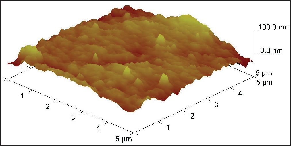
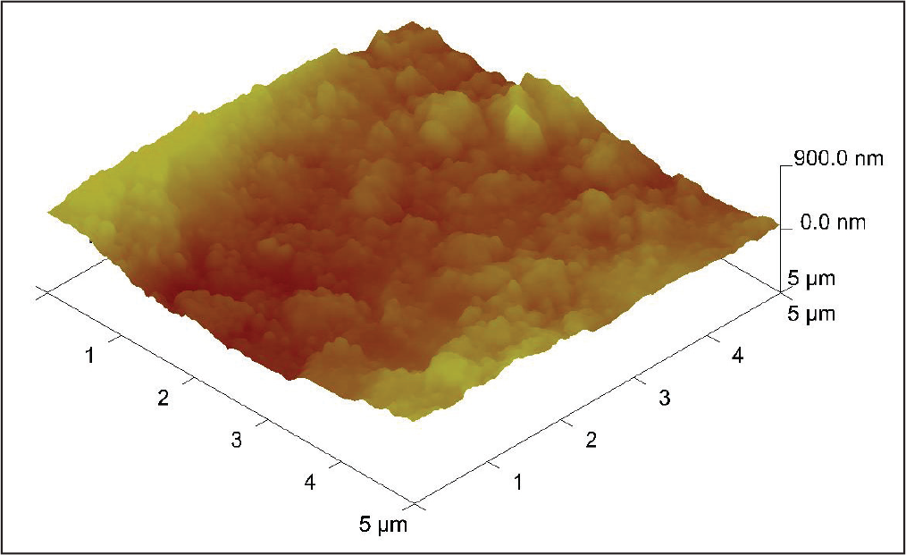
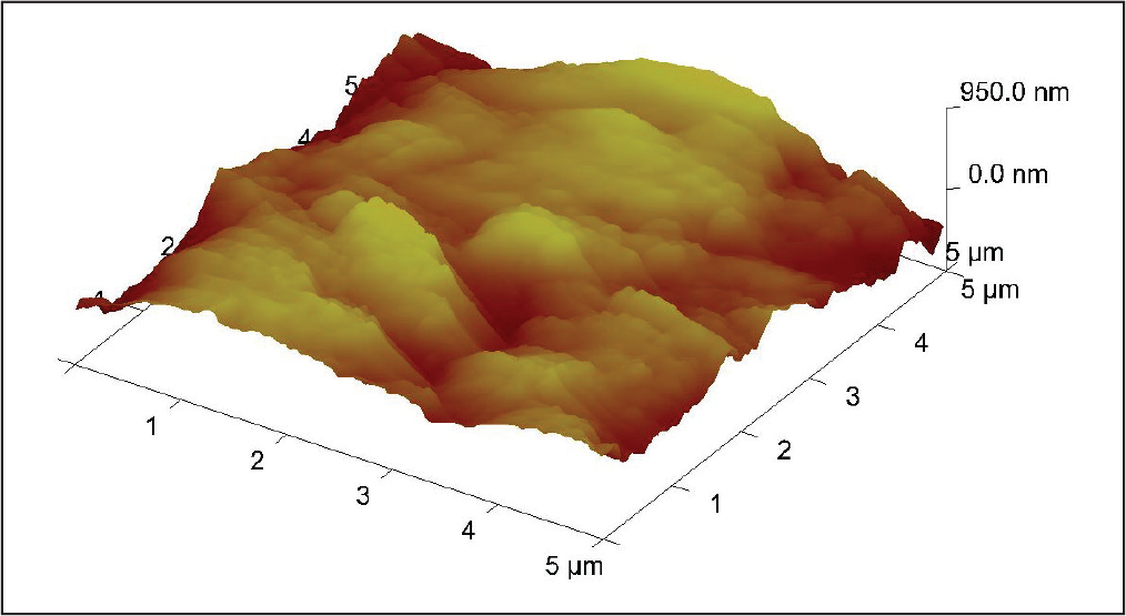
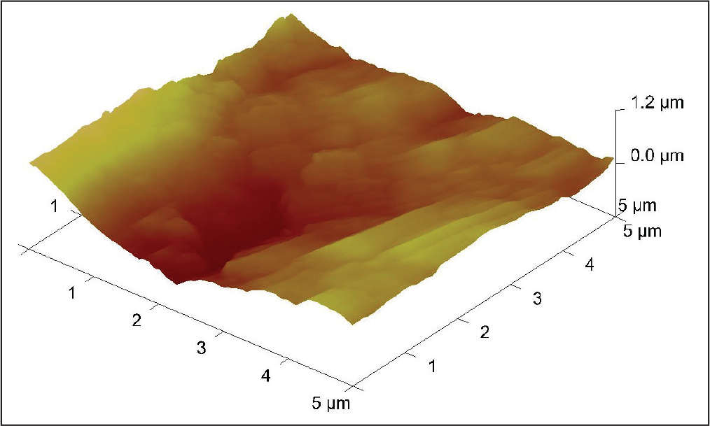
The surface roughness created by mechanical IPR using abrasive strips was less compared to that of the ARS using burs. The mean surface roughness produced by the ARS procedure using HORICO number FG 166010 burs was found to be 154.35 nm. This was 35.87 nm greater than the mean roughness produced by IPR with HORICO strips (118.4 nm) and 57.83 nm to that of Ortho Organizer strips (96.52 nm). This was statistically significant. Hence the current study shows that hand-held mechanical reduction procedure is better than ARS in terms of the surface roughness. This result matches with other studies10, 16 which proved that hand-held mechanical reduction procedures are better and safe compared to the motor-driven air-rotor reduction technique. The surface roughness appearance of normal tooth surface and after IPR as viewed under Atomic Force Microscope are shown in Figures 1 to 4.
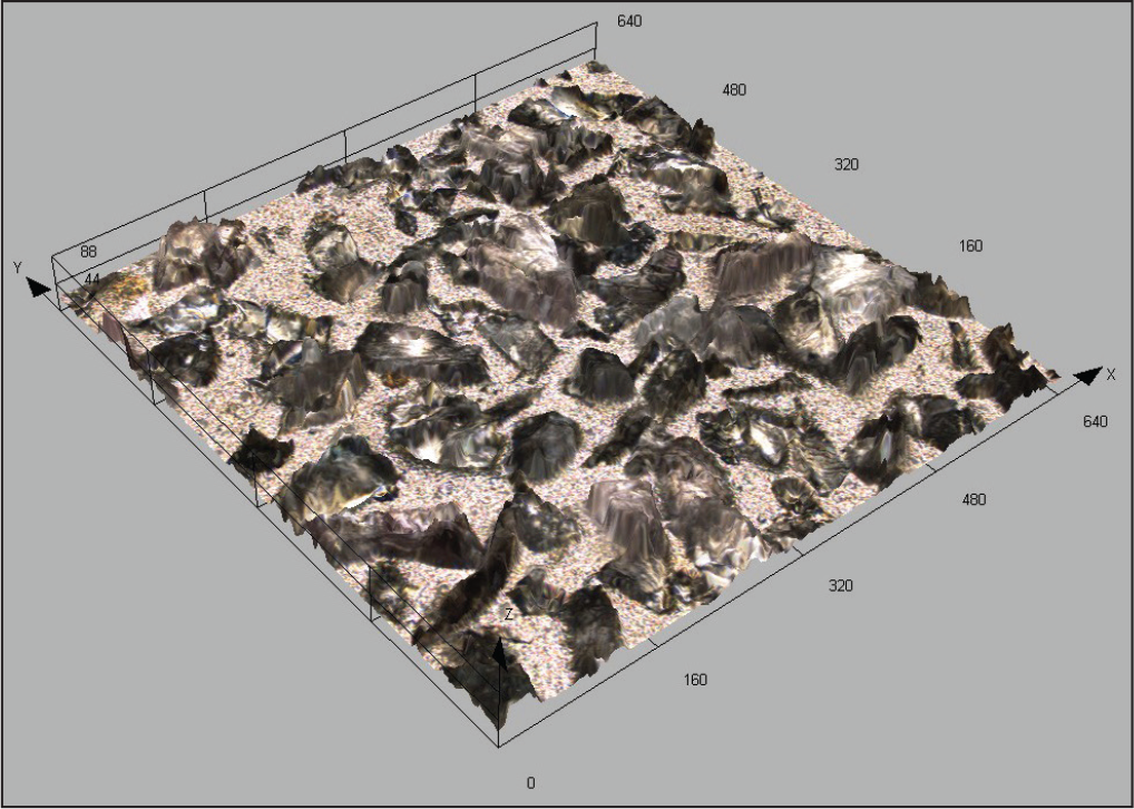
3D Image of Surface Roughness Appearance of Normal Tooth Sample Under AFM.
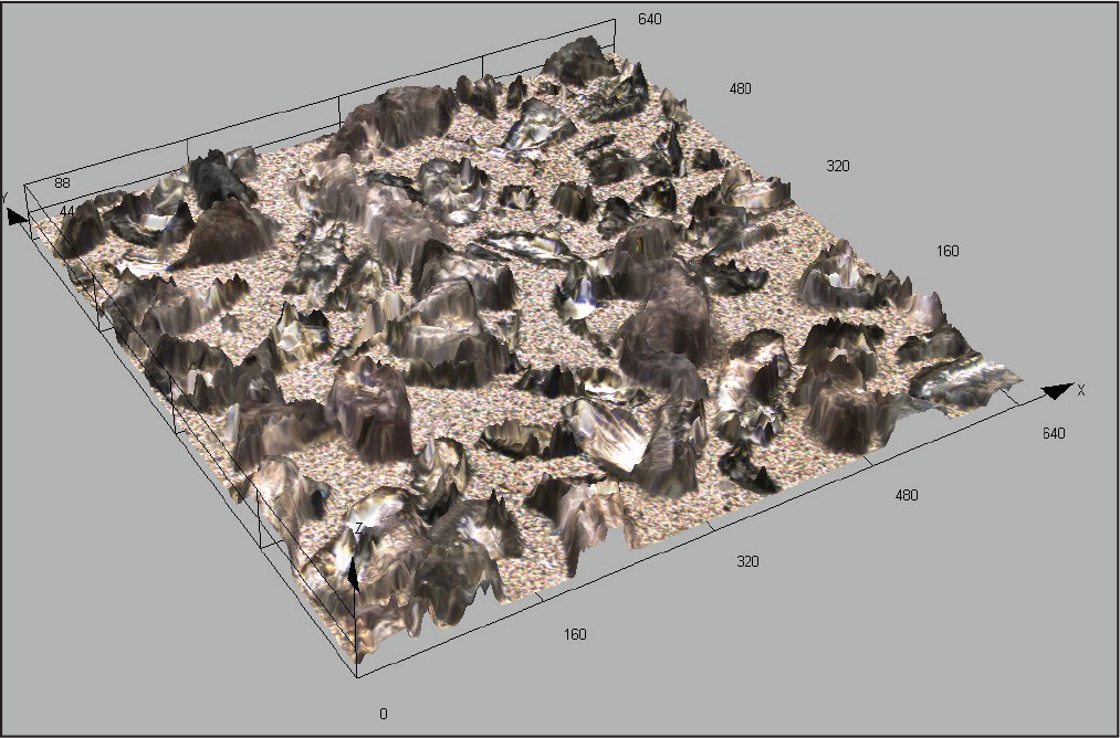
Surface Roughness Appearance of Enamel After IPR with HORICOR Strips without Polishing.
Surface Roughness Appearance of Enamel After IPR with Ortho Organizer Strips without Polishing.
Surface Roughness Appearance of Enamel After IPR with HORICOR Abrasive Burs without Polishing.
The values of surface roughness produced by 2 commercially available abrasive strips were compared in this study. The proximal strips considered were medium grit, single sided, diamond strips from HORICO and Ortho Organizer. The mean surface roughness produced by Ortho Organizer strips was 21.96 nm less compared to that of HORICO strips after IPR without polishing. But this difference was statistically not significant. Hence, strips from both the companies can be preferred for interproximal enamel reduction.
The present study also checked the wear of diamond-coated abrasive strips of HORICO and Ortho Organizer before and after the IPR procedure under a confocal microscope (OLYMPUS LEXT OLS4000). The mean reduction in surface roughness of the HORICO strip was 5.4 µm which was 0.5 µm less than that of Ortho Organizers strip with 5.9 µm. The 3D images of the surface appearance before and after the procedure are shown in Figures 5 to 8. A study conducted by Lione et al 17 concluded that the strips lost their abrasive property by 60% after 5 min and it was more rapid in vivo as greater force was exerted by the clinician. Though there was no statistical significance between the roughness, HORICO strips abraded more than Ortho Organizer strips, and care must be taken to frequently change the strips during procedure.
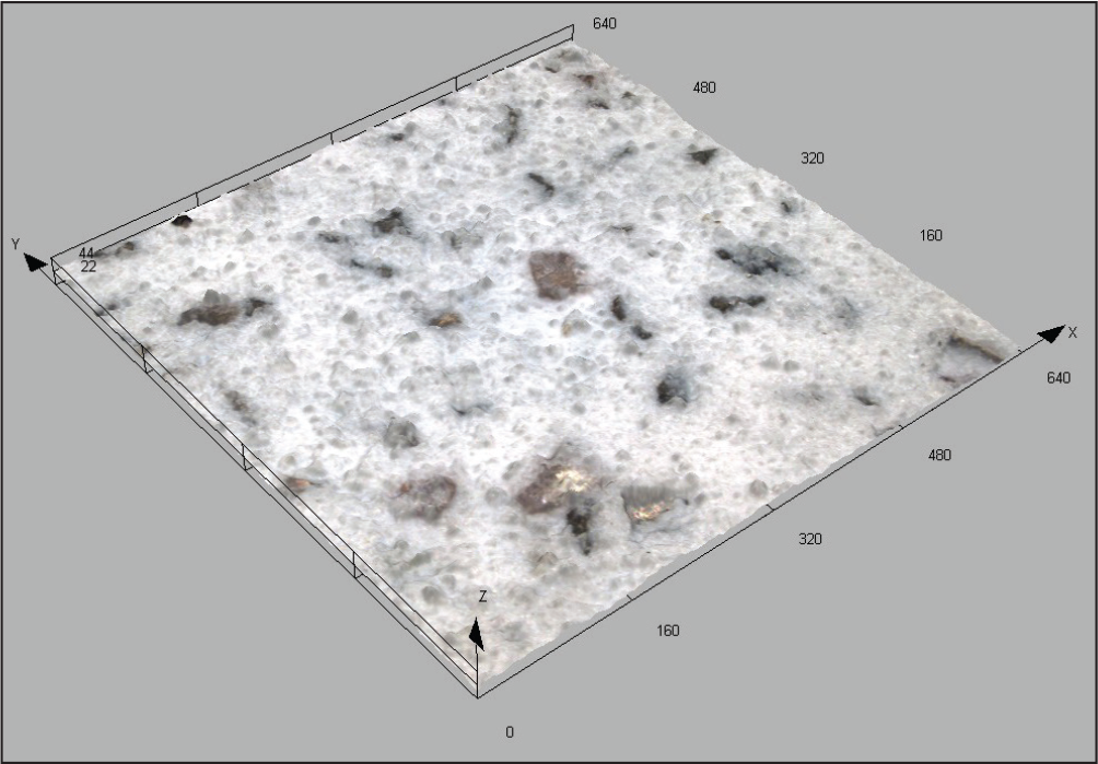
3D Colour Image of Ortho Organizer Proximal Strips Before Reduction Under Confocal Microscope.
3D Colour Image of HORICOR Proximal Strips Before Reduction Under Confocal Microscope.
3D Colour Image of Ortho Organizer Proximal Strips After Reduction Under Confocal Microscope.
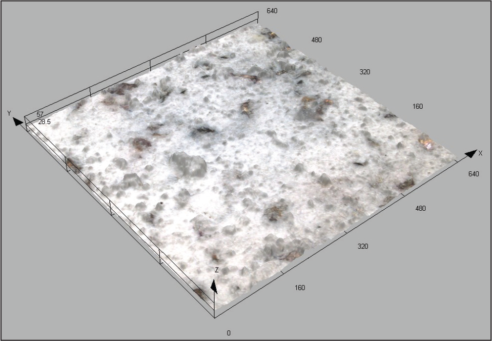
3D Colour Image of HORICOR Proximal Strips After Reduction Under Confocal Microscope.
Morphological changes in the tooth crown before and after IPR without polishing were studied. The mesiodistal widths of the crown were measured before and after the procedure using a digital caliper. The mean reduction in mesiodistal width after IPR without polishing (7.50 mm) was 1.02 mm less than that before procedure (6.48 mm). This was statistically highly significant and hence proved that there is change in the tooth morphology after interproximal reduction. This change in morphology could affect accurate bracket positioning. 18 Hence, care must be taken while bonding, and repositioning of bracket must be considered after the IPR procedure.
The major limitation was the artificial setting of the study. In the oral cavity, the surface roughness produced by IPR depends on various factors such as saliva, blood, force applied by the clinician, duration, and contact points which were standardized during the study. Also, the direction of insertion and strokes of abrasive strips and burs will vary in the oral cavity. Study can be conducted with a larger sample size. Chemical reduction techniques can be considered. In vivo studies must be conducted on interproximal reduction.
Conclusion
Based on the above findings it is concluded that
hand-held mechanical reduction procedure produces less surface roughness compared to ARS technique; the surface roughness produced by HORICO
R
and Ortho Organizers abrasive strips is not statistically significant; the surface roughness of a polished enamel surface with 3M Sof-Lex finishing strips after IPR is smoother than the normal enamel surface; there is no significant difference between the wear of HORICO
R
and Ortho Organizers proximal strips; significant morphological changes are seen in the mesiodistal widths of teeth after IPR.
The polishing of tooth interproximal surfaces with 3M Sof-Lex finishing strips following various methods of IPR is the best way of obtaining an enamel surface more similar to the untreated teeth.
Footnotes
Declaration of Conflicting Interests
The authors declared no potential conflicts of interest with respect to the research, authorship, and/or publication of this article.
Funding
The authors received no financial support for the research, authorship, and/or publication of this article.