Abstract

Image 1
B. Leporipoxvirus.
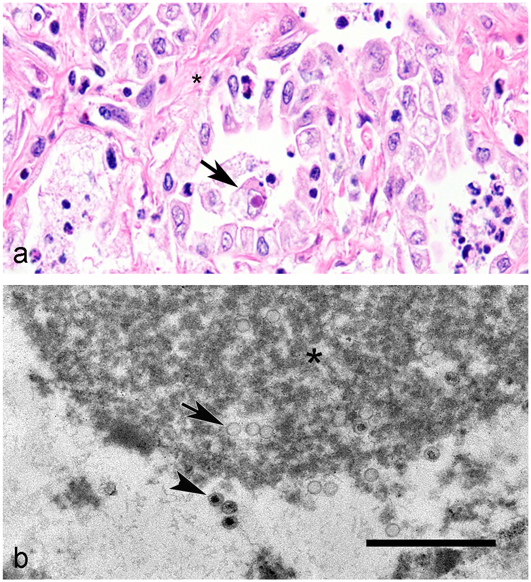
The myxoma virus belongs to the genus Leporipoxvirus and causes myxomatosis in rabbits. Facial and anogenital edema, conjunctivitis, and cutaneous “myxomas” occur. Severe cutaneous epithelial hyperplasia (Image 1a, asterisk) with intracytoplasmic inclusion bodies (Image 1a, arrows) and “stellate mesenchymal cells” (Image 1a, arrowhead) embedded in a mucinous matrix are microscopic hallmarks. Ultrastructurally, poxviruses are 250–450 nm in length by 140–260 nm in width with brick-shaped morphology, an envelope, lateral bodies, and a dumb-bell–shaped core (Image 1b, bar = 200 nm). Viral replication sites contain crescent membranes, and mature and immature virions surrounded by electron-dense and electron-lucent viroplasm. Herpesviruses are smaller (120–140 nm in diameter). Chlamydia has elementary bodies with a double membrane and an eccentric nucleoid as well as reticular bodies with diffuse fibrillary nucleic acid.
Additional Reading: Barthold SW, Griffey SM, Percy DH. Pathology of Laboratory Rodents and Rabbits. 4th ed. Wiley-Blackwell; 2016.
Contributor: Omar Gonzales-Viera, California Animal Health and Food Safety, Davis Branch, School of Veterinary Medicine, UC Davis.
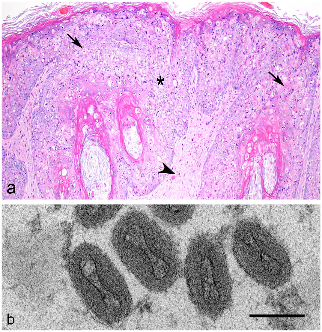
Image 2
B. Adenovirus
Photo Credit: Anibal G. Armien, California Animal Health and Food Safety, Davis Branch, School of Veterinary Medicine, UC Davis. Fowl adenoviruses (FAdVs) cause inclusion body hepatitis, hepatitis–hydropericardium syndrome, and gizzard erosion in chickens. Fowl adenoviruses D and E are the most common species detected in outbreaks. Microscopically, the hepatocytes have basophilic or amphophilic intranuclear inclusion bodies (Image 2, arrows) accompanied by hepatocellular degeneration and necrosis (Image 2, asterisk). The adenoviral virions are nonenveloped and 70–90 nm in diameter, with an icosahedral capsid consisting of 240 nonvertex capsomers (hexons), 12 vertex capsomers, and fibers protruding from the virion surface. Herpesviral icosahedral cores are 120–140 nm in diameter and 160–300 nm in diameter with the envelope. Reoviruses are nonenveloped, 60–70 nm in diameter and nonicosahedral (smooth surface).
Additional Reading: Mete A, Armien AG, Rejmanek D, Mott M, Crossley BM. Emergence of fowl aviadenovirus C-4 in a backyard chicken flock in California. J Vet Diagn Invest. 2021;33(4): 806–809.
Contributor: Omar Gonzales-Viera, California Animal Health and Food Safety, Davis Branch, School of Veterinary Medicine, UC Davis.
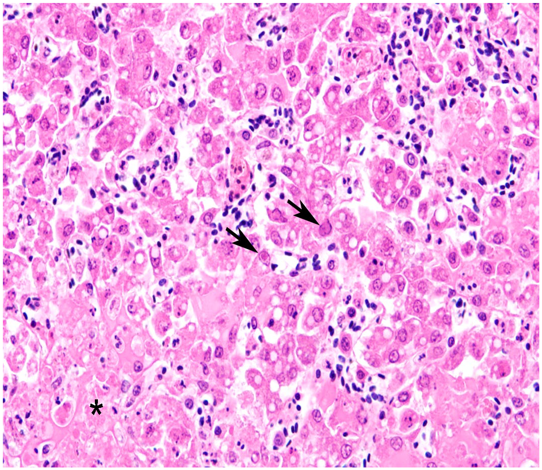
Image 3
C. Amyloid
Photo Credit: Anibal G. Armien, California Animal Health and Food Safety, Davis Branch, School of Veterinary Medicine, UC Davis. Chronic staphylococcal pododermatitis resulting from exposure of the footpad to poor-quality bedding can progress to systemic amyloidosis as seen in this case. AA-amyloid is deposited in the interstitium of organs like the kidney (Image 3), liver, and spleen. In tissue sections stained with hematoxylin and eosin, amyloid deposits are smooth and eosinophilic (Image 3a, asterisk) and, with Congo red stain, they are brick red with yellow to green birefringence under polarized light (Image 3b). Ultrastructurally, 6–20 nm diameter, nonbranching amyloid fibrils are haphazardly organized within the extracellular matrix. Collagen fibrils are larger, at 30–100 nm, with periodic striations. Reticulin fibers are <30 nm and are organized in a delicate network instead of being haphazardly arranged like the amyloid fibrils.
Additional Reading: Williams BH. Non-infectious diseases. In: Suckow MA, Stevens KA, Wilson RP, eds. The Laboratory Rabbit, Guinea Pig, Hamster, and Other Rodents. Academic Press; 2012.
Contributor: Omar Gonzales-Viera, California Animal Health and Food Safety, Davis Branch, School of Veterinary Medicine, UC Davis.
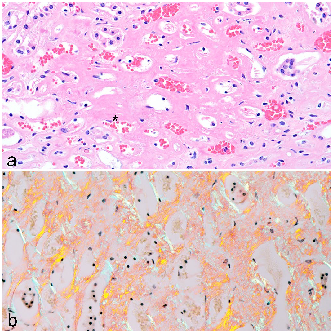
Image 4
B. Herpesvirus
Photo Credit: Anibal G. Armien, California Animal Health and Food Safety, Davis Branch, School of Veterinary Medicine, UC Davis. Equine herpesvirus 5 (EHV-5) is the cause of equine multinodular pulmonary fibrosis, which is characterized by multifocal, white-tan, sharply delineated nodules consisting of interstitial fibrosis (Image 4a, asterisk), cuboidal to columnar pneumocytes, and macrophages containing rare intranuclear eosinophilic inclusion bodies (Image 4a, arrow). Ultrastructurally, nuclear inclusions consist of viral replication and nucleocapsid assembly sites containing electron-dense viroplasm (asterisk), empty capsids (arrow), and DNA containing 120–140 nm diameter icosahedral nucleocapsids (arrowhead; Image 4b, bar = 2 µm). The virions acquire an envelope before being released from the cells. Adenovirus nucleocapsids mature within the nucleus, forming paracrystalline arrays. Paramyxovirus particles have pleomorphic morphology.
Additional Reading: Williams KJ, Robinson NE, Lim A, et al. Experimental induction of pulmonary fibrosis in horses with the gammaherpesvirus equine herpesvirus 5. PLOS ONE. 2013;8(10): e77754.
Contributor: Omar Gonzales-Viera, California Animal Health and Food Safety, Davis Branch, School of Veterinary Medicine, UC Davis.
Photo Credit: Anibal G. Armien, California Animal Health and Food Safety, Davis Branch, School of Veterinary Medicine, UC Davis.
Veterinary Pathology invites submission of exceptional gross or microscopic images for consideration as an Image Challenge, along with a multiple-choice question and answer. For details, see the Instructions to Authors on the journal website.