
Abstract
Yellow fever is an important zoonotic viral disease that can be fatal for both human and nonhuman primates. We evaluated histopathologic changes in free-ranging neotropical primates naturally infected with yellow fever virus (YFV) compared with uninfected cohorts. The most frequent lesions in primates infected with YFV were hepatic changes characterized by midzonal necrosis with lipidosis and mild inflammation including lymphocytes, macrophages, plasma cells, and infrequently neutrophils. Importantly, severe necrotizing hepatic lesions were often observed in Alouatta sp. (howler monkeys), whereas Callithrix sp. (common marmosets) had nearly no hepatic changes. Moderate to severe hepatic necrosis was present in 21/23 (91%) of the YFV-positive Alouatta sp. compared with 10/29 (34%) of the YFV-positive Callithrix sp. (P < .0001; odds ratio = 20). Similarly, hepatitis was more intense in Alouatta sp. compared with Callithrix sp. Furthermore, the frequency of YFV infection was significantly higher in Alouatta sp. compared with Callithrix sp. or Sapajus sp. (capuchin monkeys). Therefore, these data support the notion that Alouatta sp. is highly susceptible to infection and YFV-induced lesions, whereas Callithrix sp. is susceptible to infection but has a lower frequency of YFV-induced lesions.
Yellow fever is a mosquito-borne viral hemorrhagic disease of both human and nonhuman primates caused by a flavivirus. 12 Yellow fever occurs within sylvatic and urban environments, where it is transmitted to a susceptible host by Haemagogus sp. and Sabethes sp., or Aedes aegypti, respectively. 12 In Brazil, the Amazon region is considered endemically affected, but outbreaks often occur in nonendemic areas. The most recent outbreak took place mainly in the southeastern region of Brazil from 2016 to 2018, resulting in more than 1000 confirmed human cases, with a case fatality rate of 35.1%. 4,8
Neotropical primates are divided into 5 large families: Callithrichidae, Cebidae, Aotidae, Pithecidae, and Atelidae, totaling 110 species and 205 subspecies. At least 36 of these species are endemic in Brazil, and all of them are included in the Convention on International Trade in Endangered Species of Wild Fauna and Flora (CITES), indicating some degree of vulnerability. 17
Studies published decades ago demonstrated the susceptibility of neotropical primates to yellow fever virus (YFV), 5,6 and since then yellow fever became a significant concern for conservation of free-ranging populations. 2,7,21 During the sylvatic cycle of the disease, death of these animals usually precedes human cases. 22 Therefore, monitoring the occurrence of yellow fever in nonhuman primates serves as an early warning to viral circulation in a given area, which should lead to preventive vaccination of the potentially exposed human populations, as vaccination is the main preventive measure. 3,15
In Brazil, yellow fever is monitored by the Epizootics Surveillance Program of the Brazilian Ministry of Health. Each suspected case in humans or nonhuman primates must be reported immediately. In cases of death of nonhuman primates, samples of the liver, spleen, kidney, heart, lungs, and brain are sent to reference laboratories for official diagnosis. The diagnosis is based on histopathology and/or a positive result by immunohistochemistry and/or real-time reverse transcriptase-polymerase chain reaction (RT-PCR), allowing differentiation from other flaviviruses. 18
Although histopathologic changes associated with yellow fever in humans have been extensively described, there is only limited information about pathologic findings in nonhuman primate species, both in natural and experimental infections. 9,10,15 Similarly, although there is evidence for differences in susceptibility among neotropical primate species, such differences have not been previously associated with the pattern of pathologic changes in these animals. Briefly, Sapajus sp. (capuchin monkeys) are resistant, whereas Alouatta sp. (howler monkeys) and Callithrix sp. (common marmosets) are considered to be susceptible to yellow fever. 1,12,15,22 A recent study demonstrated lower viral loads in Callithrix sp. compared with other genera. 4
The goal of this study was to assess histopathologic changes in free-ranging neotropical primates of the Brazilian Atlantic Forest naturally infected with YFV, and to compare the intensity and nature of lesions in different primate species, particularly Alouatta sp. and Callithrix sp.
Materials and Methods
From 2016 to 2018, the Pathology Department of Instituto Municipal de Medicina Veterinária Jorge Vaistman (Rio de Janeiro, Brazil) received 1304 free-ranging neotropical primates that were found dead in all regions of the State of Rio de Janeiro, Brazil. Species, sex, and estimated age were recorded prior to necropsy. All 57 of the 1304 primates that tested positive by the official diagnostic laboratory were included in the YFV-positive group. Real-time RT-PCR is the definitive diagnostic test employed by the official laboratory as previously described. 13 Thus, 2 groups were defined: one composed of 57 neotropical primates positive for YFV and another group of 51 animals negative for YFV and with no gross lesions suggestive of yellow fever or any other infectious disease. Negative control animals were selected based on species, sex, and approximate age to match the animals included in the YFV-infected group. Samples of the liver, spleen, heart, and brain were collected and submitted to the official yellow fever diagnostic laboratory at Fiocruz (Rio de Janeiro, Brazil). Samples of these same organs were fixed in 10% neutral-buffered formalin and routinely processed for histopathologic evaluation. Sections (3-4 µm thick) were stained with hematoxylin and eosin, periodic acid–Schiff (PAS), and Perl’s Prussian blue.
Histologic slides were blindly evaluated by 2 veterinary pathologists (DOS and ARO) and a histopathologic score for specific lesions in each organ were established (as described in Supplemental Table S1). Frequencies of lesions were compared between groups using the χ2 test. Histopathologic scores were compared by the Mann-Whitney nonparametrical test and the Spearman correlation test. Statistical analyses were performed using Graphpad Prism software version 7.0.
Results
Frequencies of YFV-Positive Neotropical Primates
Among the 1304 free-ranging neotropical primates found dead in the State of Rio de Janeiro and necropsied from 2016 to 2018, the most common species was Callithrix sp. (n = 1219), followed by Alouatta sp. (n = 48), Sapajus sp. (n = 26), Leontopithecus sp. (golden lion tamarins; n = 7), and Callicebus sp. (titis; n = 4). A total of 57 primates were considered positive by the official diagnostic laboratory. All species included in this study had at least one positive individual; 19/48 (35%) of the Alouatta sp. and 31/1219 (2.5%) of Callithrix sp. tested positive for YFV (Fig. 1). There were no statistically significant differences in frequencies of positivity between different sex and age groups as assessed by the χ2 test. Considering the entire population of primates included in this study, frequencies of positivity were 31/346 (5.7%) and 19/477 (4.0%) in males and females, respectively (P > .05); the sex of 281 was not identified or recorded. Frequencies of positivity in adult and young primates were 48/958 (5.0%) and 6/244 (2.7%), respectively (P > .05). Age information was not available for 121 primates.
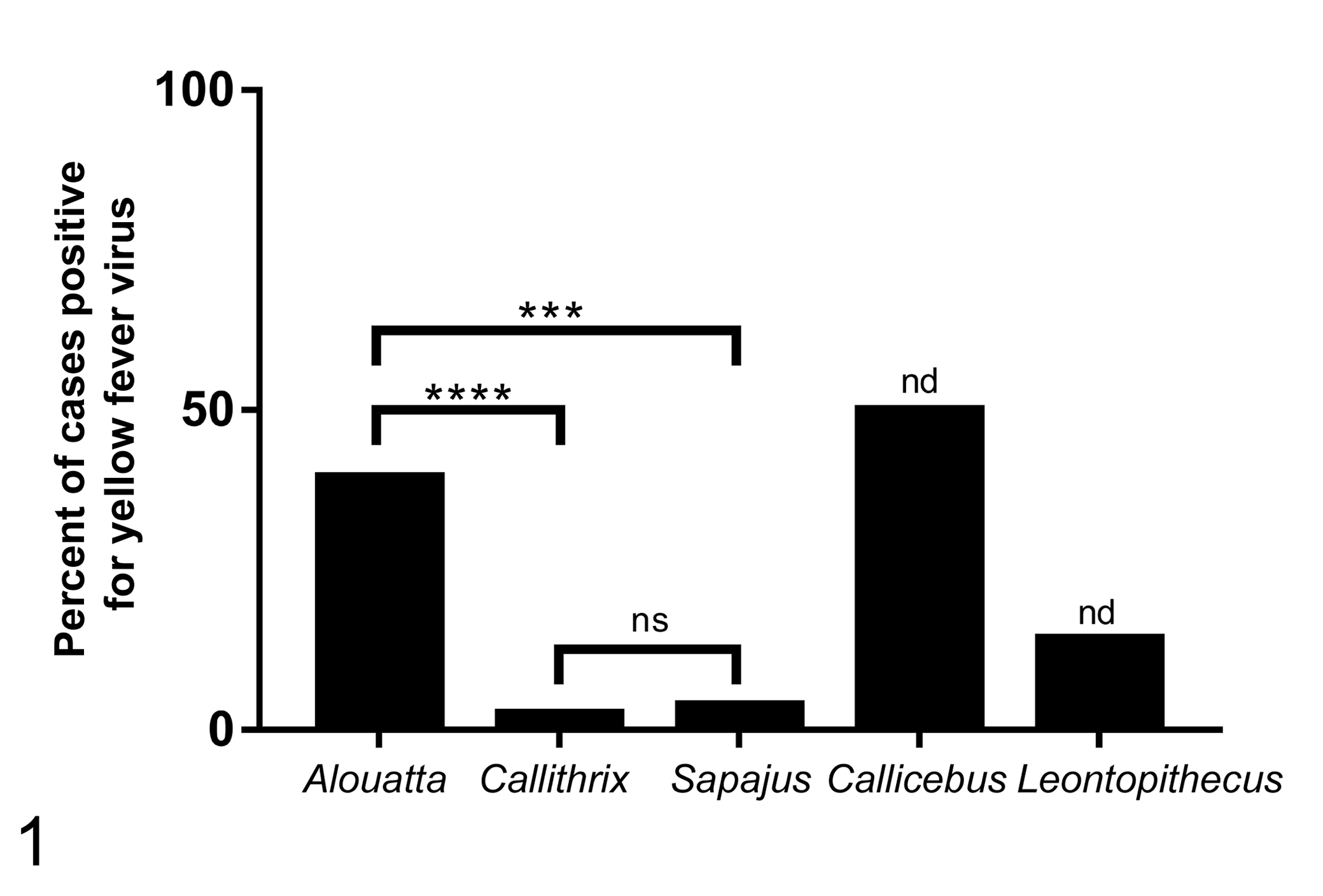
Frequency of yellow fever virus (YFV) positivity in free-ranging neotropical primates: Alouatta sp. (19/48), Callithrix sp. (31/1219), Sapajus sp. (1/26); Callicebus sp. (2/4), and Leontopithecus sp. (1/7). ***P < .001, ****P < .0001 (χ2 test). Sample numbers from Callicebus sp. and Leontopithecus sp. were insufficient for statistical analysis. ns. not significant; nd, not determined.
Histopathology
To evaluate lesion intensity, a histopathologic score was established (Supplemental Table S1). Liver, kidney, spleen, heart, lungs, and brain were included in the histopathologic analysis, but only hepatic lesions resulted in statistically significant differences in histopathologic scores between YFV-infected primates and controls (Supplemental Table S2). Importantly, occasional lesions observed in other organs had scores that were statistically similar between both groups. Therefore, these lesions were not considered to be associated with YFV infection and therefore they were not included in this study. The data for each individual animal is available in Supplementary Table S3.
Severe hepatic necrosis (Fig. 2) was the most common and marked lesion associated with YFV natural infection, affecting 40/56 (71%; P < .0001; odds ratio = 61.25; χ2 test) of infected neotropical primates. This lesion often extended to all zones of the hepatic lobule (Fig. 3). Hepatitis was observed in both YFV-positive and YFV-negative groups, but it was significantly more intense in YFV-positive animals. The inflammatory cells were mostly lymphocytes, plasma cells, and histiocytes, but there were also neutrophils in some cases (Fig. 4). Inflammatory infiltrates were most often in the portal region, but were occasionally randomly distributed in other regions of the hepatic lobule. Lipidosis was more frequent in YFV-positive primates than in controls (36/56 [64%] vs 10/51 [19%], respectively; P < .0001; odds ratio = 7.38; χ2 test; Fig. 2). Glycogenosis was less frequent in YFV-positive primates than in controls (20/56 [35%] vs 34/51 [66%], respectively; P = .0014; odds ratio = 0.28; χ2 test; Fig. 5). Hemorrhage was found in 2 YFV-positive animals that had multifocal to coalescent areas of moderate to severe hemorrhage in the midzonal region of the hepatic lobule.
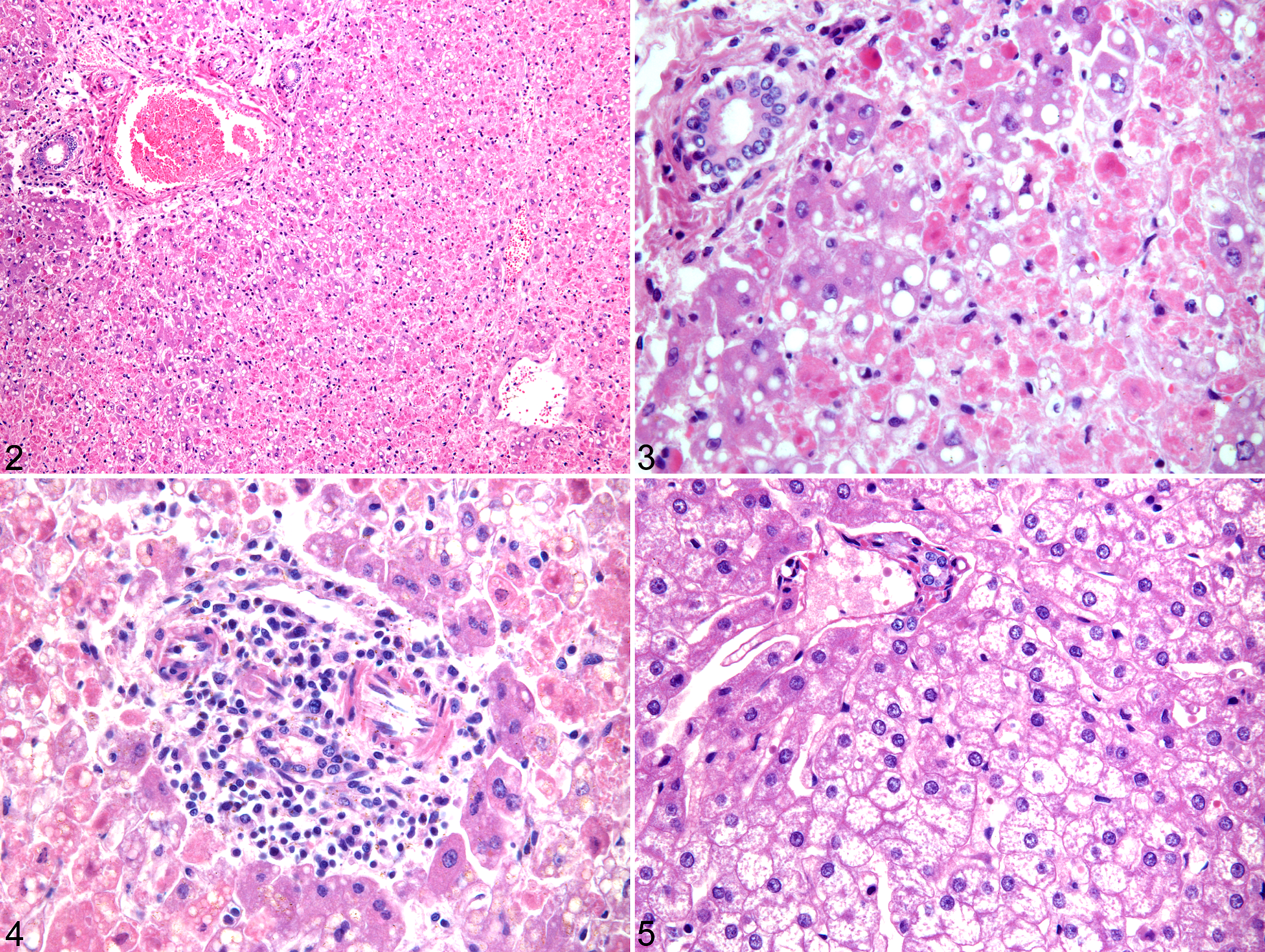
Yellow fever virus (YFV) infection, liver.
In order to evaluate whether there were differences in lesion intensity among different primate species, scores were compared between YFV-positive and negative Alouatta sp. and Callithrix sp. (Table 1). Based on this analysis, Alouatta sp. had significantly higher scores of necrosis, hepatitis, and lipidosis when compared with Callithrix sp. Indeed, 21/23 (91%) of the YFV-positive Alouatta sp. had high scores of hepatic necrosis (from 2 to 4) as compared with only 10/29 (34%) of the YFV-positive Callithrix sp. (P < .0001; odds ratio = 19.95; χ2 test). Similarly, hepatitis was more severe in Alouatta sp. compared with Callithrix sp.
Histopathologic Scores in the Liver of YFV-Positive and YFV-Negative Alouatta sp. (n = 40) and Callithrix sp. (n = 61).
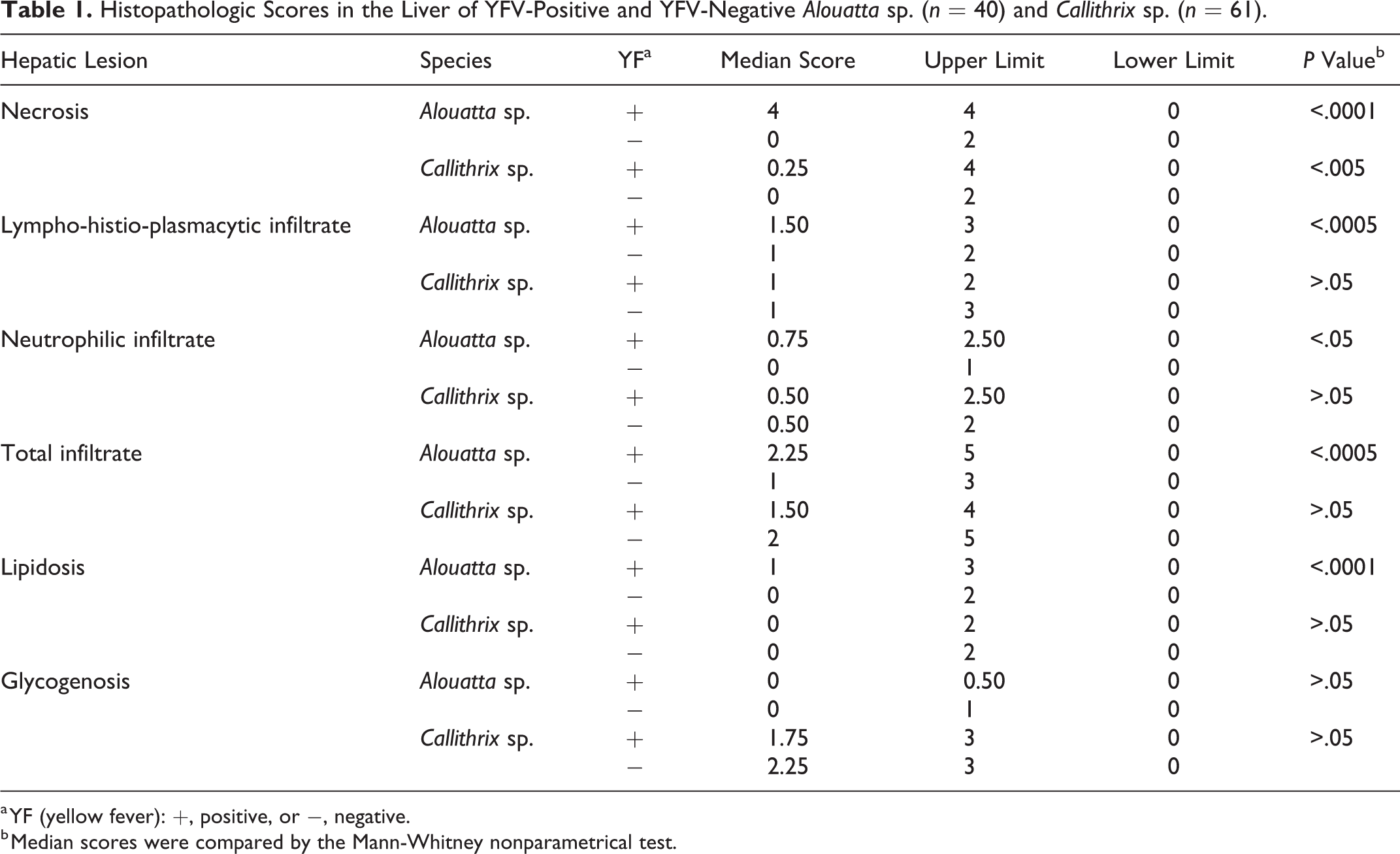
a YF (yellow fever): +, positive, or −, negative.
b Median scores were compared by the Mann-Whitney nonparametrical test.
There was some degree of lipidosis in 22/23 of YFV-positive Alouatta sp. individuals (95%; P < .0001; odds ratio = 36; χ2 test), and lipidosis scores tended to decrease as the necrosis scores increased (Spearman correlation test; Fig. 6). In Callithrix sp., lipidosis was not as frequent as in Alouatta sp. (11/29 [37%]; P < .0001; odds ratio = 36; χ2 test), but the correlation with necrosis was the opposite, that is, animals with higher scores of necrosis also had higher scores of lipidosis (Fig. 7). Glycogenosis, as confirmed by PAS staining (Fig. 8), was found in both positive and negative Callithrix sp. (18/29 [62%] and 26/32 [81%], respectively; P = .0952; odds ratio = 0.38; χ2 test). YFV-infected Alouatta sp. did not have hepatic glycogenosis.
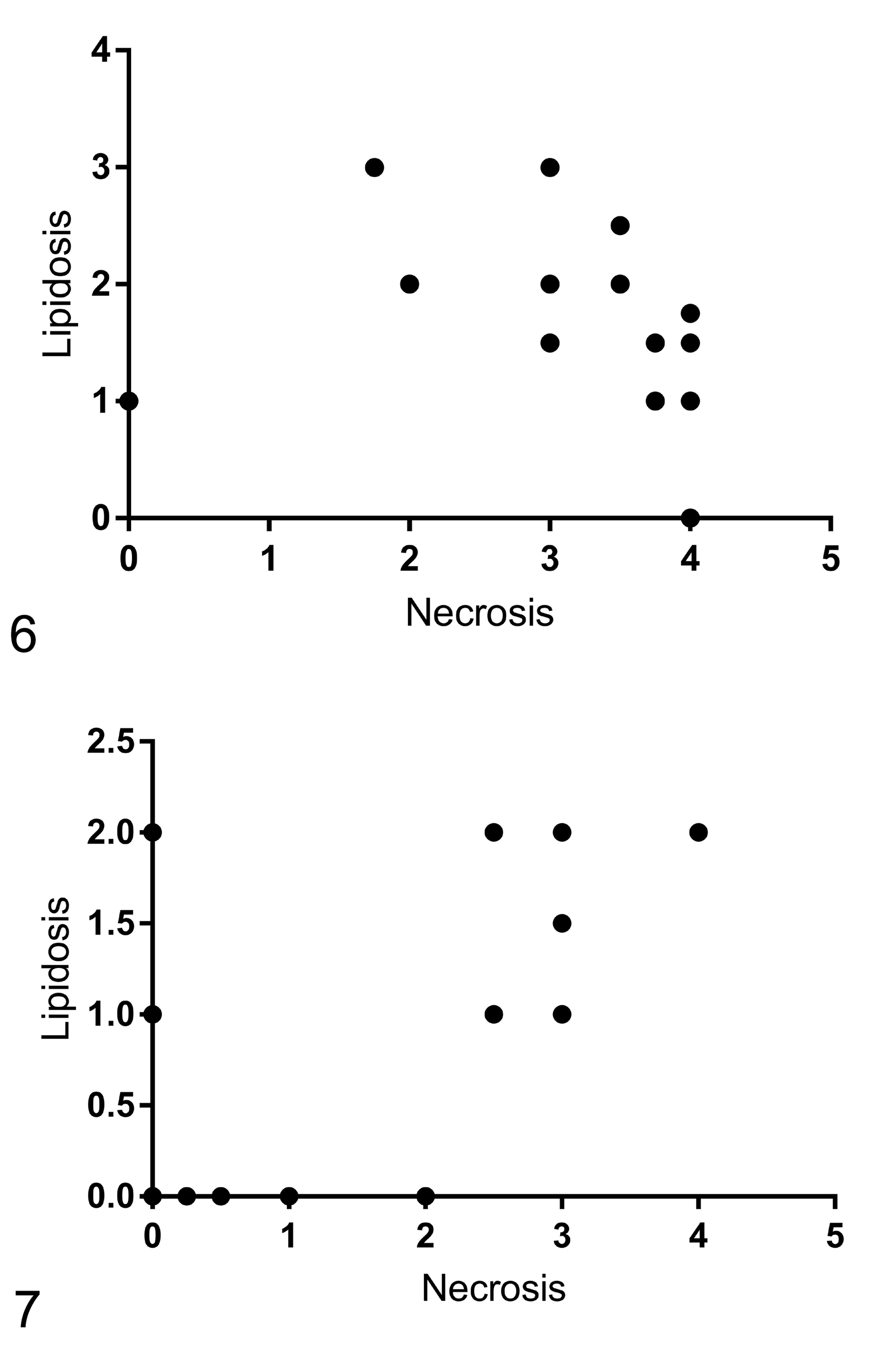
Correlation between hepatic necrosis and lipidosis in Alouatta sp. Spearman correlation test (r = −0.6823; ***P = .0003).
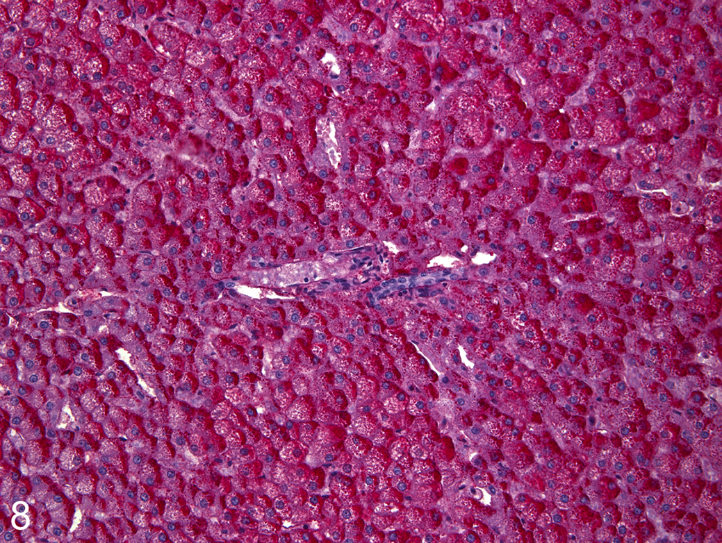
Yellow fever virus infection, liver, Callithrix sp. Severe glycogenosis characterized by abundant accumulation of intracytoplasmic material that is periodic acid–Schiff-positive.
Intracellular pigment was observed in hepatocytes of both YFV-positive and negative primates (17/56 [31%] and 15/51 [29%], respectively; P = .92; odds ratio = 1.046; χ2 test), but Perl’s Prussian blue staining was positive in only a fraction of those cases (3/17 [17%] and 4/15 [26%] for YFV-positive and negative animals, respectively; P = .5380; odds ratio = 0.59; χ2 test). Therefore, hemosiderosis was not a relevant finding in these animals.
Discussion
We evaluated several species of neotropical primates naturally infected with YFV. Alouatta sp. and Callithrix sp. were most commonly represented in our population, including 23 and 30 YFV-positive individuals, respectively, whereas the other species, namely, Callicebus sp., Sapajus sp., and Leontopithecus rosalia included only 2, 1, and 1 YFV-positive individuals, respectively. Our results clearly demonstrated differences in the pattern of liver injury of YFV-infected among different species of neotropical primates. The susceptibility of several nonhuman primate species to YFV infection has been reported, and presumably there are differences in susceptibility among those species. 1,12,15,22 Alouatta sp. and Callithrix sp., which comprised most of this study population, are considered susceptible to yellow fever (as is L. rosalia), while the other species are considered resistant. 1,12,15,22 However, a study of the recent yellow fever epidemics in Brazil suggested that Callithrix sp. may have milder disease compared with other neotropical primates. 4 Comparing histopathologic changes among Alouatta sp. and Callithrix sp. indicated 2 clear distinct patterns of lesions: (1) severe hepatic necrosis with mild to moderate inflammatory infiltrate and occasional lipidosis, indicating severe disease, which was the most common presentation in Alouatta sp. (with the exception of one single individual) and (2) absence of necrosis with mild inflammatory infiltrate and occasional non-specific glycogenosis, which was the pattern observed in Callithrix sp. These findings support the notion that Alouatta sp. are highly susceptible to acute and severe YFV-induced hepatic disease, whereas Callithrix sp. are resistant to developing these lesions. In addition, the 2 Callicebus sp. and the Leontopithecus rosalia that were YFV-positive developed lesions comparable to Alouatta sp., whereas the only YFV-positive Sapajus sp. had diffuse glycogenosis with mild inflammatory infiltrate with an absence of necrosis similar to Callithrix sp. These findings are in agreement with previous reports that Sapajus sp. is a resistant species. 1,12,15,22
There were no significant differences in the frequencies of YFV positivity when comparing different sex and age groups. In humans, adult males are more often infected, not due to susceptibility but because this group is the most exposed to sylvatic environments, 12 a behavioral pattern that obviously does not affect risk in the case of nonhuman primates.
Hepatic necrosis/apoptosis was the most severe lesion in the liver of YFV-positive primates, as for humans. 16 Necrosis was usually severe and extended from the midzonal to the central and portal hepatocytes, frequently with only a few unaffected hepatocytes. This is the classic pattern of lesions previously described in humans 16 and in experimentally YFV-infected rhesus monkeys. 20 This pattern is associated with virus tropism for hepatocytes. 14
Lipidosis was another hepatic lesion that was often observed in this study. Lipidosis has been diagnosed in human livers from YFV-infected patients. 8,16,19,23 Interestingly, Alouatta sp. had lower scores of lipidosis when compared with necrosis, which is likely due to the loss of most viable hepatocytes associated with the severe disease in this species.
The inflammatory response observed in the liver in this study was mild and mainly composed of lymphocytes and macrophages. Inflammatory processes are usually not found in YF cases, but when present are usually mild and composed of the same cell types observed in this study. 15,16,22 Impairment of an inflammatory response during YFV infection is likely due to the action of transforming growth factor-β (TGF-β), which induces apoptosis, while acting as an anti-inflammatory cytokine. 15
Outbreaks of yellow fever happen at intervals of 5 to 10 years, when human cases are preceded by a rise in nonhuman primate cases, which are due to an increased susceptibility of the nonhuman primate population. 12 Between 2016 and 2019, which coincides with the period of this study, a major yellow fever outbreak occurred in Brazil, considered to be the most severe in the past 70 years. 18 In this context, highly susceptible nonhuman primate species such as Alouatta sp. likely play an important role in amplifying the virus and therefore infecting large numbers of invertebrate vectors. In contrast, animals that develop a mild YFV infection and develop long-lasting immunity, including resistant neotropical primates such as Sapajus sp. as well as Callithrix sp. as demonstrated in this study, probably prevent circulation of YFV during outbreak intervals, with an epidemiologic effect of broad vaccination coverage in human populations. However, the ability of these primates to infect invertebrate hosts is unknown and warrant further study of their role in YFV transmission.
In human yellow fever patients, renal failure due to acute tubular necrosis is a major complication in fatal cases. 15 However, in this study renal tubular necrosis was not observed in any YFV-positive animal, which is in agreement with Monath, 14 who demonstrated that renal function is not altered in primates. However, our findings contrast with previous reports of mild tubular degeneration and necrosis in rhesus monkeys experimentally infected with YFV. 11,20
In conclusion, this study described the histopathologic pattern in cases of natural infections with YFV as well as variable responses in different host species. Alouatta sp. often developed severe YFV-induced hepatic injury, while Callithrix sp. was considered as a susceptible species but had only mild hepatic lesions associated with YFV natural infection.
Supplemental Material
Supplemental Material, Combined_supplemental_materials-Oliveira_dos_Santos_et_al - Histopathologic Patterns and Susceptibility of Neotropical Primates Naturally Infected With Yellow Fever Virus
Supplemental Material, Combined_supplemental_materials-Oliveira_dos_Santos_et_al for Histopathologic Patterns and Susceptibility of Neotropical Primates Naturally Infected With Yellow Fever Virus by Daniel Oliveira dos Santos, Ayisa Rodrigues de Oliveira, Fabiana Pizzolato de Lucena, Sara Aquino de Mattos, Thaynara Parente de Carvalho, Fabíola Barroso Costa, Larissa Giannini Alves Moreira, Tatiane Alves da Paixão and Renato Lima Santos in Veterinary Pathology
Supplemental Material
Supplemental Material, Table_S3 - Histopathologic Patterns and Susceptibility of Neotropical Primates Naturally Infected With Yellow Fever Virus
Supplemental Material, Table_S3 for Histopathologic Patterns and Susceptibility of Neotropical Primates Naturally Infected With Yellow Fever Virus by Daniel Oliveira dos Santos, Ayisa Rodrigues de Oliveira, Fabiana Pizzolato de Lucena, Sara Aquino de Mattos, Thaynara Parente de Carvalho, Fabíola Barroso Costa, Larissa Giannini Alves Moreira, Tatiane Alves da Paixão and Renato Lima Santos in Veterinary Pathology
Footnotes
Declaration of Conflicting Interests
The author(s) declared no potential conflict of interest with respect to the research, authorship, and/or publication of this article.
Funding
The author(s) disclosed receipt of the following financial support for the research, authorship, and/or publication of this article: Work in RLS lab is supported by CNPq (Conselho Nacional de Desenvolvimento Científico e Tecnológico, Brazil), FAPEMIG (Fundação de Amparo à Pesquisa do Estado de Minas Gerais, Brazil), and CAPES (Coordenação de Aperfeiçoamento de Pessoal de Nível Superior, Brazil). TAP and RLS have fellowships from CNPq (Brazil).
Supplemental Material
Supplemental material for this article is available online.
References
Supplementary Material
Please find the following supplemental material available below.
For Open Access articles published under a Creative Commons License, all supplemental material carries the same license as the article it is associated with.
For non-Open Access articles published, all supplemental material carries a non-exclusive license, and permission requests for re-use of supplemental material or any part of supplemental material shall be sent directly to the copyright owner as specified in the copyright notice associated with the article.