
Abstract
Cutaneous mucinosis is a cutaneous disorder described in humans, dogs, and rarely cats but never reported in birds. Twenty-six brown egg–laying chickens between ages 43 and 46 weeks had a history of feather loss, scaly, dry skin, weight loss, and decreased egg production. Microscopic findings in the skin included fragmentation of collagen bundles and interstitial, periadnexal, and perivascular dermal accumulation of wispy, mildly basophilic material that was also occasionally observed within the follicular epithelium. A moderate lymphoplasmacytic and heterophilic perivascular dermatitis was also observed. The wispy to granular material was diffusely Alcian blue positive and periodic acid–Schiff negative (consistent with mucin), suggesting a diagnosis of primary or secondary cutaneous mucinosis. The cause of this condition could not be determined.
Cutaneous mucinosis is a syndrome characterized by mucin deposition in the dermis. 10 Mucin, an acid mucopolysaccharide produced by fibroblasts, is composed of hyaluronic acid (HA) bound to heparin and chondroitin sulfate B. 34 HA is a straight-chain glycosaminoglycan (GAG) polymer of the extracellular matrix with remarkable hydrodynamic characteristics, especially in terms of its viscosity and ability to retain water. 33 Several cell types, including fibroblasts, keratinocytes, and endothelial cells, synthesize HA by means of a specific hyaluronan synthases. 15,21,32 HA is a normal component of the dermis, and its production decreases with age. 13 Mucin is also found in lesser quantities within the superficial and follicular epidermis as well as in other adnexal structures. 16,28
In humans, cutaneous mucinoses are heterogeneous groups of disorders, classified as focal, follicular, and diffuse. 10 In dogs, mucinosis may occur as a primary condition, due to heredity or acquired metabolic or endocrine defects, or as a subclinical condition in association with other diseases (secondary mucinosis). 30 Primary mucinosis can be divided into hereditary mucinosis (generalized or multifocal), myxedema, papular mucinosis, and nodular mucinosis. 8 Among animals, a generalized cutaneous mucinosis, probably of genetic origin, occurs primarily in the Chinese Shar-Pei dogs. 18 Shar-Peis have more dermal mucin than do other breeds 7 and an association between the Shar-Pei breed and genetic variation (single-nucleotide polymorphism) in hyaluronic acid synthase 2 (HAS2) has been demonstrated. 1 The term myxedema is used specifically to denote primary mucinosis observed with hypothyroidism. 3,17 Focal cutaneous mucinosis presenting as asymptomatic nodules, papules, or plaques on the skin has been described in 7 dogs of different breeds. 2 Since dermal mucin is closely bound to collagen fibers, collagen degeneration can cause mucin deposition. 9 In humans, secondary mucin deposition has been observed in connective tissue injury or diseases of many types, including lupus erythematosus, hypertrophic scarring, dermatomyositis, scleroderma, and actinic elastosis. 6,12,27 In dogs, secondary mucinosis has been observed in pyoderma, allergy, eosinophilic skin disease, lupus erythematosus, acromegaly, dermatomyositis, and mast cell tumor. 8,29,30 An unusual case of generalized cutaneous mucinosis characterized by multiple dermal swellings on the head, back, elbows, hocks, and digital pads occurred in association with thyroid carcinoma in a dog. 11
Here we describe for the first time the gross and histopathologic findings of diffuse cutaneous mucinosis in 26 brown egg–laying chickens, a condition rarely described in other species of animals 3 but never in birds.
Materials and Methods
Chickens
Twenty-six live brown egg–laying chickens aged between 43 and 46 weeks from a flock of 41 000 were submitted for necropsy to the California Animal Health and Food Safety Laboratory System (CAHFS)–San Bernardino branch. The brown egg–laying chicken is a strain of chicken originating from a cross between male Rhode Island Red (RIR) white variety and female RIR brown variety. The birds were humanely euthanized by use of carbon dioxide. The flock had a history of 20% egg production drop over a 10-week period starting at 33 weeks of age (Fig. 1), decreased feed consumption, weight loss, and feather loss on the head, neck, keel, and thigh. Other chickens of the same strain but different ages on the same premises but in different houses were not affected. No other clinical signs were noted in the flock. The birds were raised indoors and housed in cages (7 birds per cage; cage size: 7.31 × 6.09 m), with adequate ventilation and standard vaccination for layers. Feed and water were provided ad libitum. There was no history of pesticide application on the birds or in the house, and no mites or lice were found on the birds submitted for necropsy. For treatment, the birds were supplemented with vitamin A, biotin, and electrolytes, with no improvement.
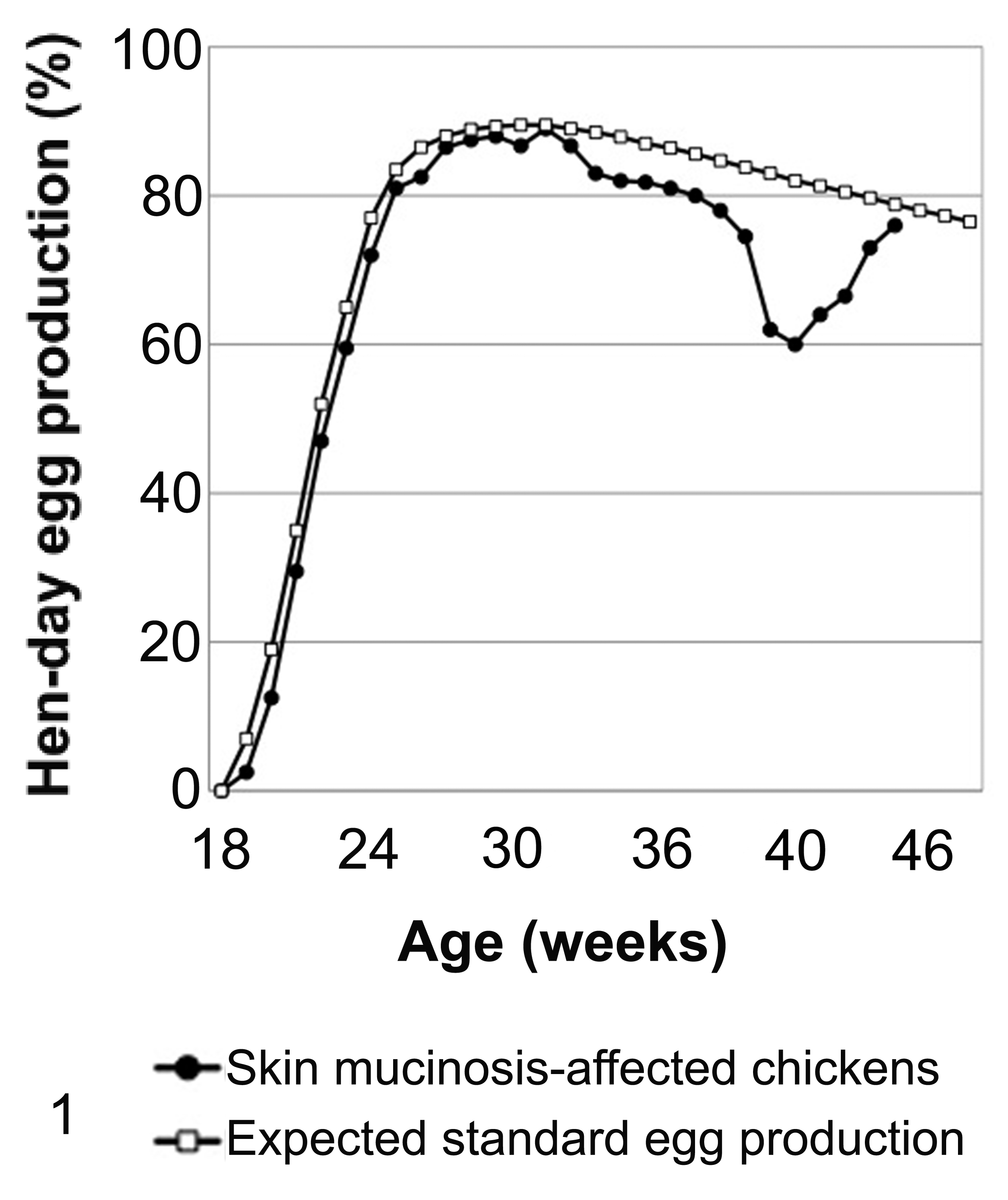
Hen-day egg production in skin mucinosis–affected chickens compared with the expected standard of egg production in age-matched normal brown egg–laying chickens. Note the egg production drop starting at 33 weeks of age in the affected group. The hen production is the number of eggs produced per day/number of birds in the flock that day, expressed as a percentage.
Pathology
A complete necropsy was performed on all the submitted chickens. Representative tissue samples, including multiple samples of skin from affected and normal skin, including comb and wattles, were collected for histologic examination. The samples were fixed in 10% neutral buffered formalin, processed, and embedded in paraffin. Tissue sections were stained with hematoxylin and eosin (HE) and examined by light microscopy. Special stains, such as Alcian blue (pH 2.5), periodic acid–Schiff (PAS), and toluidine blue were applied in selected cases. Skin samples from 4 normal 16-week-old and 46-week-old brown egg–laying chickens (age-matched controls) unrelated to the chickens in the present study were sampled, processed, and examined for comparison. In addition, skin samples from two 46-week-old brown egg layers with mild dermatitis were also stained with Alcian blue.
Additional Diagnostic Tests
Sera from all live chickens were tested for Newcastle disease (ND), avian influenza (AI), avian encephalomyelitis (AE), infectious bronchitis (IB), Mycoplasma gallisepticum (MG), and Mycoplasma synoviae (MS). Routine bacteriology, mycology, and virology were performed on the skin samples. Livers from 10 chickens were tested for heavy metals, while selenium and vitamins A and E were tested in 4 chickens.
Results
Gross Lesions
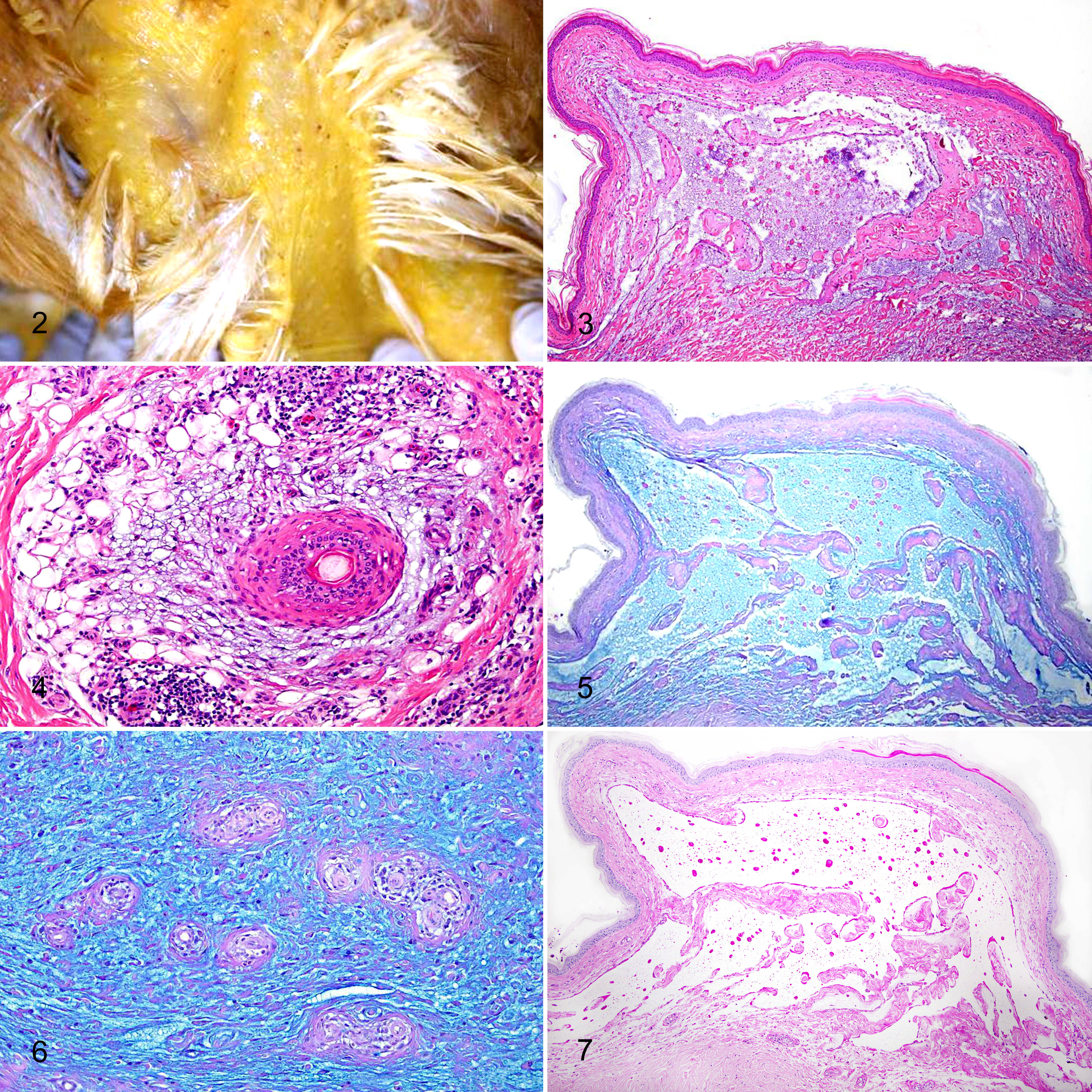
Postmortem examination revealed loss of feathers over 60% to 80% of the body and a scaly, dry skin with a slight green tinge and occasional petechiae scattered throughout the body but most prominent on the keel, crop, and anterior stifle. The dermis was mildly thickened (Fig. 2). The skin around the eyes, comb, and wattles was not affected. Most of the birds were out of production, as evidenced by inactive ovaries. No other gross lesions were observed.
Histopathology
Throughout the dermis, collagen bundles were fragmented, distorted, and widely separated by abundant, granular to wispy, lightly basophilic material and clear spaces suggestive of mucin and edema fluid, respectively. Mucin accumulation was more evident in the superficial dermis, forming extensive lakes or dermal vesicles (Fig. 3). Occasionally, similar material was observed expanding the perifollicular and perivascular spaces, as well as the follicular epithelium (Fig. 4). Dilated lymphatics were observed in the superficial dermis. A moderate number of lymphocytes and plasma cells infiltrated the perivascular spaces in the superficial, mid, and deep dermis, while a few to a moderate number of heterophils were randomly scattered within the interstitium. Rare aggregates of heterophils were observed within the epidermis. There were also multifocal epidermal spongiosis and intercellular edema and mild, diffuse orthokeratotic hyperkeratosis. The wispy to granular material was diffusely and strongly Alcian blue-positive (Figs. 5, 6) while mostly PAS-negative (Fig. 7). Alcian blue stain revealed positive staining dispersed throughout the dermis, characterized by irregular strands of alcianophilic material, or accumulating in lakes or vesicles in the superficial dermis. Occasionally, small amounts of wispy to granular Alcian blue–positive material was observed in the superficial dermis of normal control skin and in the dermis and perivascular spaces of 2 chickens with dermatitis but never in the epidermis. Low numbers of metachromatic mast cells were observed in the superficial and deep dermis, similar to the cell density of normal control skin.
Additional Diagnostic Tests
Routine bacteriology, mycology, and virology of the skin detected no isolates. Serology for ND, AI, AE, and IB reflected vaccination titers. Eight of 26 and 26 of 26 sera were positive for MG and MS, respectively. Heavy metal, selenium, and vitamin A and E levels in the liver were within normal limits.
Discussion
The histologic and histochemical results demonstrated unequivocally that a dermal mucinosis similar to that described in dogs might occur in brown egg–laying chickens. To our knowledge, this study represents the first report of this condition in an avian species. It is often difficult to visualize dermal mucin on HE-stained histologic slides, and only in severe cases of mucinosis are collagen bundles dissociated by a granular or wispy basophilic material. 25 In the affected chickens, the alcianophilic reaction confirmed acid GAGs as the main components of the dermal deposits. Interestingly, the combs and wattles, which generally have large amounts of mucin in the dermis, were not affected in the chickens in this study. The reason for this is not known. The positive titers for MG and MS were considered not significant as other chickens on the same premises without skin lesions were also positive for MG and MS. In addition, MG and MS have not been known to cause any skin or feather conditions in chickens.
Although the precise cause of cutaneous mucinosis in these chickens cannot be determined, 2 possible mechanisms could be hypothesized: a primary hereditary mucinosis, in which mucin is the main histologic feature, or a secondary mucinosis due to inflammation.
The predominant occurrence of this condition in the submitted strain of chickens might suggest a genetic basis. Other birds on the premises, raised under similar conditions of management, environment, and diet but from different genetic sources, were not affected. Brown egg–laying chickens are bred under similar management conditions to other strains of chickens, therefore suggesting that management has no influence on the occurrence of cutaneous mucinosis in the brown egg–laying chickens. Furthermore, it is well known that the strain of brown egg layer chickens in this study has a genetic tendency to develop other diseases, such as amyloid arthropathy 19 and intestinal dilatation syndrome (IDS). 36 However, potential genetic analyses are hindered in our cases by the lack of available tissue samples as well as readily available technology. Further genetic studies are necessary to confirm the genetic basis of skin mucinosis in the brown egg layer–type chickens and to ascertain the alterations responsible for this dysfunction.
Regarding inflammation as a putative trigger of dermal mucinosis, it is well known that cytokines may play an important role in the induction of GAG synthesis, specifically tumor necrosis factor α and β, interleukin (IL)–1, transforming growth factor (TGF) β, IL-6, and epidermal growth factor. 4,5,20,22 Inflammatory cytokines released by lymphocytes, plasma cells, and heterophils in the submitted birds may be responsible for stimulating mucin production from dermal fibroblasts, producing a secondary dermal mucinosis. It is interesting that skin from 2 dermatitis-affected brown egg–laying chickens with mild to moderate accumulation of mucin confirms the association between inflammation and mucin production. Skin samples of the affected chickens in this study had similar mast cell density to the control skin, ruling out the pathophysiologic role of mast cells in the development of mucinosis, as demonstrated in humans. 14,26
Interestingly, in the submitted birds, the severe and diffuse dermal mucinosis was associated with a moderate multifocal follicular mucinosis. Follicular mucinosis is an epithelial mucinosis, in which mucin infiltrates the outer root sheath of the follicles and sebaceous glands. 34 Occasionally, the mucin leaks back into the dermis. 2 Usually, this condition is associated with cutaneous epitheliotropic lymphoma, 31,35 or otherwise a lymphocytic infiltrate is constantly present around the affected follicles and in the surrounding dermis. 8 Reed 24 has speculated that T lymphocytes stimulate follicular keratinocytes to produce mucin. In the submitted chickens, a lymphocytic inflammatory infiltrate was noted around the affected epithelium. Based on this finding, we propose that the feather loss was due to a primary dermatitis with secondary dermal and follicular mucinosis.
One differential diagnosis for dermal mucin accumulation includes mucopolysaccharidoses, which are characterized by accumulation of glycosaminoglycans, such as dermatan and heparan sulfate, but these substances have different staining characteristics from hyaluronic acid. 23
In conclusion, we have described a distinctive histopathologic form of dermal mucinosis in brown egg–laying type chickens. As further cases are reported, a clearer picture of cutaneous mucinosis in avian species may emerge. A dermal mucinosis by itself is only a consequence of an altered metabolism or catabolism of dermal GAGs, whatever its cause. Further studies are needed to elucidate the origin and pathogenesis of this unique dermatosis in birds.
Footnotes
Acknowledgements
We thank all the technical staff at CAHFS, Tulare and San Bernardino and histotechnologists at VMTH, Davis for their help.
Declaration of Conflicting Interests
The author(s) declared no potential conflicts of interest with respect to the research, authorship, and/or publication of this article.
Funding
The author(s) received no financial support for the research, authorship, and/or publication of this article.