
Abstract
A 14-year-old neutered male cat presenting with chronic vomiting had 2 masses within the submucosa of the stomach that were excised. They presented histologically as circumscribed, submucosal masses consisting of diffusely arranged medium-sized round cells with a moderate amount of cytoplasm and interspersed eosinophils, separated by trabecular fibroblastic stroma. The overlying mucosa was diffusely infiltrated by the same round cells, and marked epitheliotropism was present. Neoplastic cells labelled positive for CD3 and negative for CD79a and CD117. Giemsa staining and silver staining (SNOBA) were also negative. A T-cell lymphoma with reactive fibroplasia was diagnosed, and differential diagnoses including mast cell tumor and feline gastrointestinal eosinophilic sclerosing fibroplasia could be excluded.
A 14-year-old neutered male European shorthair cat was presented to the Clinic for Small Animal Internal Medicine of the University of Zurich for chronic vomiting, anorexia, fever, and apathy. Hematologic abnormalities included mild, hypochromic, microcytic, nonregenerative anemia (hematocrit 29%, reference interval 33%–45%; MCH 12 pg, reference interval 14–17 pg; MCV 36 fl, reference interval 41–49 fl; slight anisocytosis and poikilocytosis of erythrocytes) and mild hypoeosinophilia (0.05 × 10³/μl, reference interval 0.10–0.60 × 10³/μl). Ultrasonographic changes included 2 submucosal, hypoechogenous oval masses (1.5 × 1.0 × 1.0 and 0.8 × 0.6 × 0.6 cm) in the fundus between the small and large curvature of the stomach. Accordingly exploratory laparotomy and gastrotomy revealed 2 well-circumscribed solid masses with a light-gray cut surface. One mass near the small curvature was completely removed, whereas the second, located in the fundus, was only partially removed by surgery. Both masses were submitted for histological examination.
Differential Diagnoses
Based on the ultrasonographic and macroscopic findings, differential diagnoses included primarily neoplastic lesions, most likely mast cell tumor or lymphoma, and inflammatory lesions, most likely feline gastrointestinal eosinophilic sclerosing fibroplasia. Formation of parasitic nodules induced by Gnathostoma sp or Cylicospirura sp were considered less likely, as they are not reported in Switzerland.
Microscopic Findings
The submucosal masses were well circumscribed, nonencapsulated, and moderately to highly cellular and consisted of variably sized round cells with nuclei measuring 2.5 to 3.5 times a red blood cell diameter, forming islands that were separated by broad, hyalinized, fibroblastic, branching trabeculae (Fig.1). The round cells had a moderate amount of pale eosinophilic cytoplasm. Nuclei were round to irregular and often vesicular with finely stippled chromatin and 1 distinctive nucleolus (Fig. 2). There were moderate to high numbers of eosinophils between the round cells. The mucosa overlying the masses was multifocally eroded, and loss of glands was evident. Diffuse infiltration with the same cell population as detected within the masses was present in the lamina propria mucosae, and there was marked epitheliotropism (Fig. 3). Anisocytosis and anisokaryosis were mild, and mitotic activity was low. No metachromatic intracytoplasmic granules were discernible when stained with Giemsa, and silver staining (SNOBA) was negative. The nodular neoplastic part within the submucosa of both masses was completely excised; however, the neoplastic cells within the mucosa reached the margin in both of them.
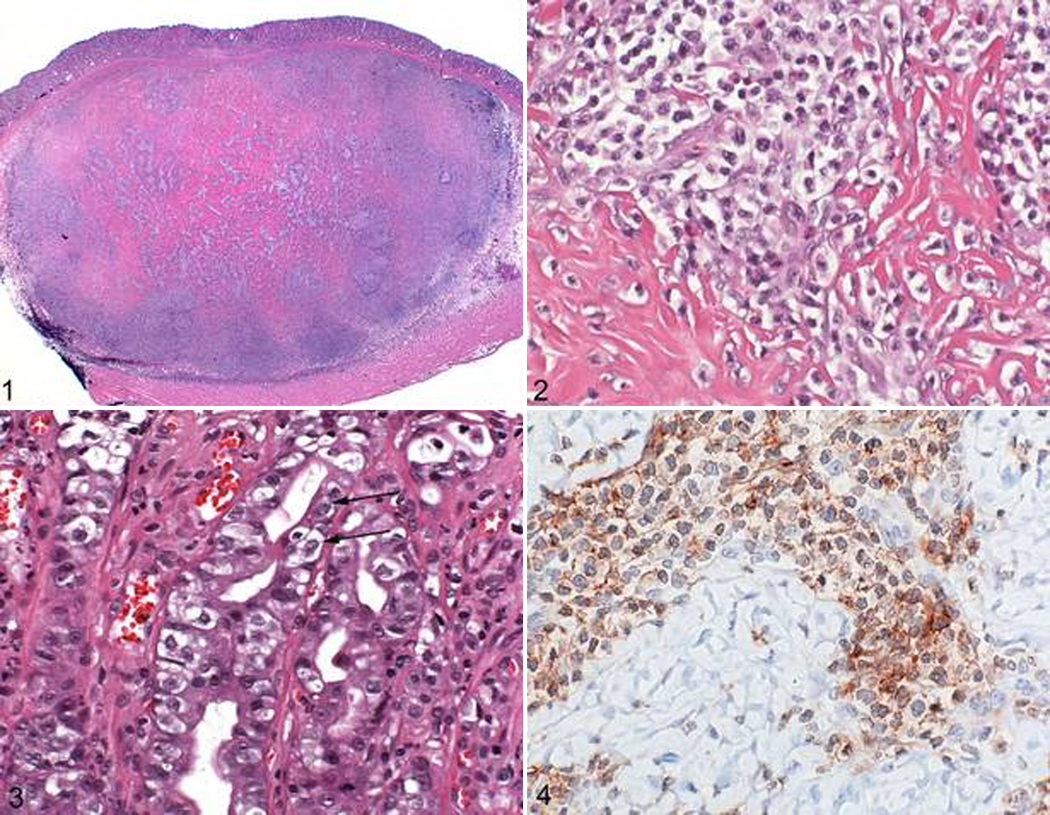
Stomach; cat. Submucosal well-circumscribed nodular mass composed of medium-sized round cells forming islands that were separated by broad, hyalinized, fibroblastic, branching trabecules. HE.
Immunohistochemistry
Unstained paraffin-embedded tissue was sectioned at 3.5 μm and placed on positively charged glass slides. Immunohistochemical staining was performed using an automated staining device (Dako Autostainer, Dako, Carpinteria, CA) with AEC (Dako) as the chromogen and a Meyer’s hematoxylin counterstain. Antibodies used were CD3 (mouse/monoclonal, 1:50, DAKO, M725401), CD79a (mouse/monoclonal, 1:250, DAKO, M705101), and CD117 (rabbit/polyclonal, 1:400, DAKO (A4502). High-temperature antigen retrieval was employed for all 3 antibodies. All neoplastic round cells were labeled intracytoplasmatically for CD3 (Fig. 4). Immunohistochemistry for CD79 and CD117 was negative.
Diagnosis
Based on histomorphology and the immunohistochemical findings, the final diagnosis was T-cell lymphoma with reactive fibroplasia. Four months postsurgically, the cat was free of clinical signs, suggesting curative surgical excision without regrowth of the mass, but was lost for follow-up.
Discussion
Gastrointestinal masses with marked eosinophilic infiltration and severe reactive fibroplasia represent a peculiar feline lesion evoking some controversy in the veterinary literature.4,10 They may represent reactive inflammatory lesions, such as feline gastrointestinal eosinophilic sclerosing fibroplasia. Craig et al described these lesions as intramural masses that usually were located at the pylorus, the ileocecocolic junction or colon, and less frequently in the small intestine of affected cats. 2 Lesions were transmural or restricted to the inner layers of the gastrointestinal wall. They consisted of branching trabeculae of dense collagen and contained variable numbers of predominantly eosionophils and fewer mast cells, neutrophils, plasma cells, and lymphocytes. In more than 50% of the cases, intralesional bacteria were demonstrated. Gross and histological differential diagnoses are neoplastic lesions, such as mast cell tumors reported to occur as sclerosing variant or lymphoma, which both can be associated with fibroplasia and reactive eosinophilic inflammation.1,4,6 Special stainings such as Giemsa, Toluidine blue, and Gram to demonstrate metachromatic granules and bacteria as well as immunohistochemical stainings to characterize the cell of origin are needed to make a final diagnosis. The presence of high numbers of large blastlike cells in the case presented here was indicative of a neoplastic process; however, the cell of origin could not be determined solely on histomorphological features. Giemsa staining and CD117 were needed to exclude an intestinal mast cell tumor, including undifferentiated ones that might not have been diagnosed with the Giemsa staining but should have been labeled by CD117. The intense labeling with CD3 and the absence of positivity for CD79 identified the lesion as T-cell lymphoma with extensive reactive fibroplasia.
Lymphoma is the most common hematopoietic tumor in the cat, and the gastrointestinal tract is a frequent location. An increasing prevalence of alimentary lymphoma is reported in recent years. 12 Cats affected by the alimentary form of lymphoma tend to be older than cats affected by multicentric, central nervous system and mediastinal lymphoma or leukemia.8,11 Depending on the extension of the lymphoma, affected animals may suffer from anorexia, weight loss, vomiting, and diarrhea. Hematological abnormalities may include nonregenerative anemia due to chronic disease, bone marrow depression due to lymphomatous infiltration, FeLV infection, or blood loss into the gastrointestinal tract. Up to 50% of the cats have a mild hypoalbuminemia that is problably caused by protein loss or malassimilation in the gastrointestinal tract.8,12 Gastrointestinal lymphomas may be classified as low grade (lymphocytic or small cell lymphoma), intermediate, or high grade (immunoblastic, lymphoblastic, or large cell) with the predominant form being a low-grade lymphoma.8,11,12 Another class consists of granular lymphocytes that most likely originate from intraepithelial lymphocytes, primarily affects the intestine, and is usually characterized by an aggressive clinical course. 9 Neoplastic lymphatic infiltrations usually affect the mucosa and progress to the submucosa and muscularis, causing localized or multifocal masses within the gastrointestinal tract or circumferential diffuse thickening of the intestine and occur more often in the intestine than in the stomach.3,8,11,12
The lymphoma we present here has an unusual submucosal localization, and the presentation as circumscribed mass with severe reactive fibroplasia and eosinophilia is unusual. Infiltration of eosinophils within a lymphoma is reported to be related to the synthesis of IL-5 by neoplastic lymphocytes of both lineages; however, it is more common in T-cell lymphomas. 1 The eosinophils on their part may be responsible for the fibroplasia within the lesion by producing major basic protein, TGF-beta, and IL-1beta, which induce proliferation of fibroblasts and deposition of extracellular matrix.2,5 The hematologic abnormalities of this cat, consisting of a mild, hypochromic, microcytic, nonregenerative anemia, have not been further clarified. An underlying iron deficiency would be a possible etiology. The primary differential diagnoses for a lymphoma with numerous eosinophils and fibroplasia is a mast cell tumor and feline gastrointestinal eosinophilic sclerosing fibroplasia. The latter is hypothesized to be a unique nonneoplastic fibroblastic reaction of felines to an eosinophilic inflammation and is possibly related in some cases to a bacterial infection. 2 Immunohistochemistry is the method of choice to differentiate between lymphoma and mast cell tumor. However, the epitheliotropism of the neoplastic cells that, to the authors’ knowledge, is not described in intestinal mast cell tumors of cats may be another important feature to distinguish lymphomas and mast cell tumors as reported in dogs. 7
Footnotes
Acknowledgements
We thank the laboratory technicians of the Institute of Veterinary Pathology, Vetsuisse Faculty, University of Zurich, Switzerland, for technical assistance.
Declaration of Conflicting Interests
The author(s) declared no potential conflicts of interest with respect to the research, authorship, and/or publication of this article.
Funding
The author(s) received no financial support for the research, authorship, and/or publication of this article.