
Abstract
Hemophagocytic syndrome (HPS) is a macrophage hyperactivation disorder triggered by disrupted T–cell macrophage cytokine interaction. HPS has been reported in humans, dogs, cats, and cattle, and it is infrequent and poorly characterized in animals. A 16-year-old male rhesus macaque was euthanized because of severe pancytopenia, including nonregenerative anemia (hematocrit = 5.5%), neutropenia (0.29 K/μl), and thrombocytopenia (21 K/μl). Bone marrow was hypocellular with normal maturation, myeloid hypoplasia, and few megakaryocytes. There were numerous morphologically normal macrophages (12% of nucleated cells), with 6% of nucleated cells being hemophagocytic macrophages in the bone marrow. Serology was negative, but polymerase chain reaction and immunohistochemistry were positive for simian retrovirus type 2. Blood and bone marrow findings were consistent with HPS. Cytopenias are common in simian retrovirus–infected macaques, but HPS has not been reported. An association between simian retrovirus infection and HPS is undetermined, but retrovirus-associated HPS has been observed in humans.
Hemophagocytic syndrome (HPS) is a potentially life-threatening, nonneoplastic proliferative disorder of phagocytically active macrophages. It manifests as cytopenias of multiple cell lines in bone marrow and other tissues. 24 In human medicine, symptoms such as fever and splenomegaly are often observed in conjunction with abnormal laboratory findings. 7 HPS is considered rare and life threatening in any species, but it has been best described in human medicine as a genetic disorder (primary or familial HPS) or as an acquired condition (secondary or reactive HPS).7,19 The acquired form has been associated with a variety of infectious or immunologic underlying causes, including viral infections. 17 HPS-associated viruses include Epstein-Barr virus, human herpesvirus 6, cytomegalovirus, parvovirus B19, hepatitis virus, and human immunodeficiency virus (HIV).17,21 Regardless of the underlying cause, the downstream event is T-cell activation with resultant overproduction of macrophage-activating cytokines and induction of a hyperinflammatory state. In contrast to human medicine, information on HPS in veterinary medicine is limited.23,24 Twenty-eight cases in dogs and a single case of a cat have been reported and were associated with underlying infections (ehrlichiosis, Lyme disease, parvovirus, and blastomycosis), neoplasia, or immune-mediated diseases.23,24
Simian retrovirus (SRV) is a betaretrovirus predominantly found in Asian nonhuman primates of the genus Macaca (macaques). It is well established that the virus is able to cause latent infections in macaques, with the potential for reactivation and serious associated disease. 9 Five distinct SRV serotypes have been recognized, with a species predilection for each serotype. Although all macaque species are potentially susceptible to infection by each serotype, 9 the prevalence of SRV infection in captive macaque populations significantly varies, from minimal (3–5%) to majority (> 50%). 9 Clinical manifestations of SRV range from subclinical carriers to severe immunosuppression and death.4,9 Opportunistic infections and hematologic abnormalities are commonly associated with active or recrudescent SRV infections. Hematologic abnormalities include anemia, leukopenia (neutropenia and lymphopenia), and thrombocytopenia with bone marrow hyperplasia of the granulocytic and erythroid cell lines. Severe pancytopenia and bone marrow depletion may occur during the late-stage SRV infections.5,10 The cause for SRV-related pancytopenia is not well understood, but there is some evidence of direct viral cytotoxicity for hematologic precursors 9 (S. T. Yoshioka, “Effects of Simian Type D Retrovirus Serotype 1 on In Vitro Differentiation of Bone Marrow Derived Progenitor Cells of the Rhesus Macaque (Macaca mulatta),” unpublished master’s thesis, University of California, Davis, 2000).
Case History
A 16-year-old male rhesus macaque was presented for pallor to the veterinary staff at an academic institution accredited by the Association for Assessment and Accreditation of Laboratory Animal Care. The animal was individually housed in a stainless-steel nonhuman primate cage, fed a commercial diet (Lab Diet 5045, PMI Nutrition International, Brentwood, Missouri), and provided filtered city water ad libitum by an automated water system. It was maintained at the institution for 10 years without significant medical conditions. It was also undergoing behavioral training for a pharmacologic study (approved by an Institutional Animal Care and Use Committee), and it had not undergone invasive experimentation for 3 years before presentation.
There were no abnormalities on visual examination. Food and water consumption was normal. Physical examination under ketamine sedation (10 mg/kg; Ketaset, Fort Dodge, Iowa) was unremarkable apart from pallor and poor body condition (body condition score, 2 out of 5). There had been weight loss of 0.8 kg (6.5%) since examination 3 months prior. Femoral venipuncture was performed for complete blood count and serum chemistry, and a small hematoma formed at the venipuncture site. The serum chemistry was unremarkable except for mild hypoproteinemia (5.6 g/dl, normal: 5.9–7.6). Hematologic alterations consisted of severe pancytopenia—specifically, normocytic normochromic anemia, neutropenia, lymphopenia, and thrombocytopenia (Table 1). Although reticulocytes were present, the absolute reticulocyte count was not significant with respect to the degree of anemia; therefore, the anemia was classified as nonregenerative.
Hematologic Findings
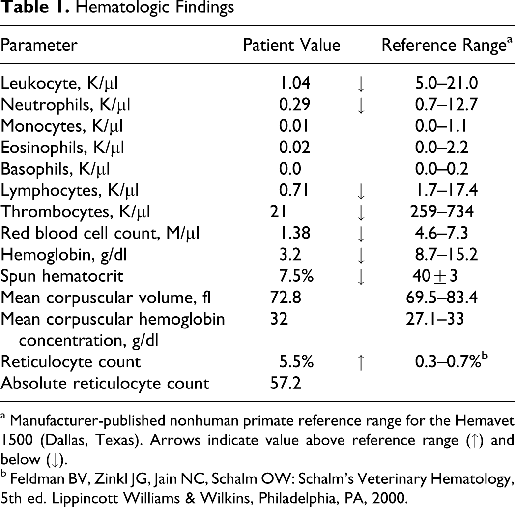
a Manufacturer-published nonhuman primate reference range for the Hemavet 1500 (Dallas, Texas). Arrows indicate value above reference range (↑) and below (↓).
b Feldman BV, Zinkl JG, Jain NC, Schalm OW: Schalm’s Veterinary Hematology, 5th ed. Lippincott Williams & Wilkins, Philadelphia, PA, 2000.
Hematologic parameters were confirmed by microscopic blood smear evaluation. The white blood cell population was severely decreased. The majority of remaining leukocytes were lymphocytes. The red blood cell morphology was normal with the exception of hypochromasia. In addition, platelets were severely decreased.
Due to the severe hematologic abnormalities and the poor prognosis for return to experimental function, the animal was euthanized and submitted for necropsy.
Pathologic Findings
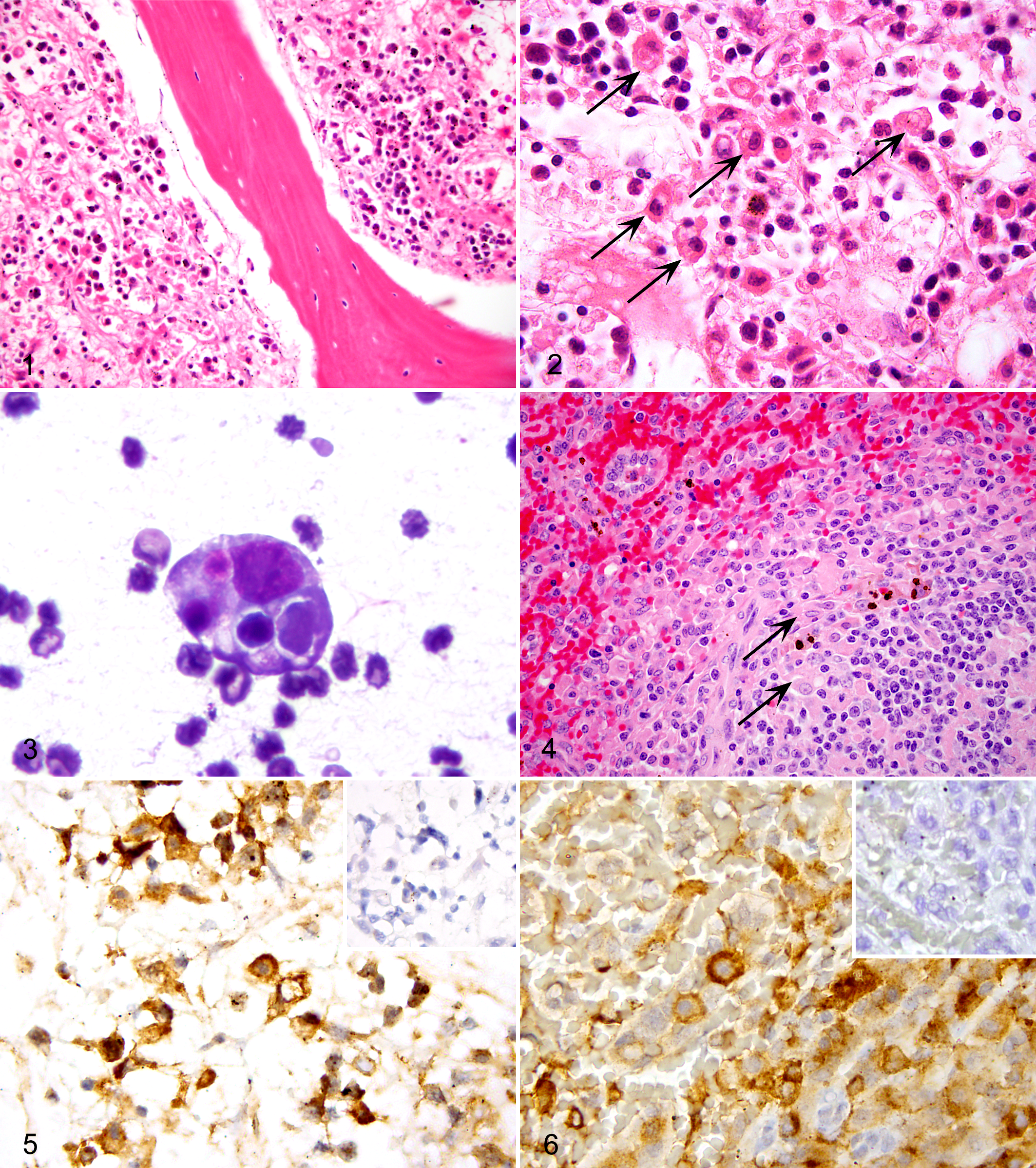
Gross findings included pallor, mild cardiomegaly, and a focal pulmonary abscess in the right middle lung lobe. Histologically, a postmortem bone marrow core biopsy taken from the proximal humerus was hypocellular with minimal iron stores and decreased megakaryocytes (Fig. 1). Although overall cellularity was reduced, there was a relative increase in erythroid precursors, and the myeloid lineage decreased. Numerous morphologically normal macrophages were present, many of which contained mature erythrocytes or degenerate nucleated cells within the cytoplasm, consistent with hemophagocytosis (Fig. 2). Overt hemorrhage or necrosis was not evident. In a bone marrow aspirate from the humerus, there was an inversion of the myeloid:erythroid ratio (1.0:1.8, normal = 1.36:1.00). 20 Additionally, 12% of total nucleated cells in the aspirate consisted of macrophages (normal rhesus values = 0.3–0.9%) 20 with 6% of the nucleated cells consisting of hemophagocytic macrophages (Fig. 3). In humans, hemophagocytic macrophages should normally comprise < 1% of the nucleated cells in bone marrow. 15 In veterinary medicine, information is available only for dogs, where bone marrow hemophagocytic macrophages are normally < 2% of nucleated cells. 24 Additional histologic findings included numerous hemophagocytic macrophages within splenic red and white pulp, splenic lymphoid depletion with numerous macrophages in marginal zone (Fig. 4), and mild multifocal myocardial degeneration. Extramedullary hematopoiesis was not observed in any tissue. The right middle lung lobe contained a well-demarcated abscess with numerous bacterial rods and cocci. Moderate Klebsiella pneumoniae and α hemolytic streptococcus were isolated. Aerobic blood cultures were negative.
Ancillary Diagnostics
Viral serology by indirect ELISA, performed at a commercial laboratory (VRL laboratories, San Antonio, Texas), was positive for Macacine herpesvirus 1 (herpes B virus) and seronegative for measles, SRV, simian immunodeficiency virus, and simian T-lymphotropic virus 1.
Polymerase chain reaction (PCR) and immunohistochemistry detection of SRV were performed at the Pathogen Detection Laboratory at the California Primate Research Center (Davis, California). Real-time PCR for SRV was performed on spleen (frozen) and bone marrow (formalin fixed) using primers and probe targeting the env gene. 25 The animal was positive by PCR for SRV in both tissues. Further PCR established the serotype as SRV type 2. Immunohistochemistry was performed with a laboratory-generated mouse monoclonal antibody against the gp20 transmembrane protein of SRV. Numerous cell lines had positive cytoplasmic reactivity in bone marrow and spleen (Figs. 5, 6 ), with strongest immunoreactivity in macrophages. Megakaryocytes and mature red blood cells were negative. Additional PCR for simian parvovirus was negative for spleen and bone marrow.
PCR for herpes B virus and rhesus lymphocryptovirus, performed on formalin-fixed spleen and bone marrow, was negative (Zoologix, Chatsworth, California).
Discussion
This case involved severe chronic pancytopenia in an adult male rhesus macaque. Although the macaque was seronegative for SRV, it was PCR and immunohistochemistry positive, which is common with active SRV infection. 9 SRV has been associated with severe cytopenias in macaques. 10 The pathogenesis is poorly understood, and bone marrow findings are not extensively characterized. In previous descriptions of SRV-related cytopenias, bone marrow findings ranged from erythroid hyperplasia and a myeloid left shift to profound depletion of all hematopoietic elements.5,12 Immune-mediated hemolysis or hemophagocytosis was not reported with these findings. In vitro, evidence indicated that SRV alters the bone marrow microenvironment, disrupting progenitor cell maturation and inhibiting myeloid differentiation.9,11 Large numbers of phagocytically active macrophages, in the absence of hemorrhage or necrosis, are not typical of bone marrow that is depleted because of maturation deficits.
In this case, the bone marrow contained an expanded population of morphologically normal macrophages with hemophagocytosis of multiple cell lines. Differentials for this finding consist of a variety of neoplastic histiocytic disorders, including malignant histiocytosis, disseminated histiocytic sarcoma, and hemophagocytic histiocytic sarcoma.3,12 Hemophagocytosis by macrophages with normal morphology is inconsistent with a histiocytic neoplasm.3,12,16 Other conditions associated with hemophagocytosis (necrosis, hemorrhage) were also absent.
This case is most consistent with a diagnosis of HPS, which is a clinical condition with multiple potential causes culminating in aberrant phagocytic activation of morphologically normal macrophages. Diagnostic criteria have been established for humans and dogs (Table 2). In this case, the human diagnostic criteria of cytopenia and > 1% hemophagocytically active macrophages in bone marrow was met, but the additional criteria of fever, splenomegaly, and various serum abnormalities were difficult to apply to a nonhuman primate. 15 For example, body temperature can decrease with sedation, and a mildly elevated temperature may not be appreciated in physical examination of a macaque. Splenomegaly was not noted at necropsy, and it is not possible to determine if this finding was present in an earlier clinical stage. Additionally, the diagnosis in this case was reached postmortem, and samples were not available for determination of serum abnormalities, such as increased ferritin, triglycerides, and CD25 (soluble interleukin-2 receptor).
Published Diagnostic Criteria6 for Human or Veterinary Cases of Hemophagocytic Syndrome
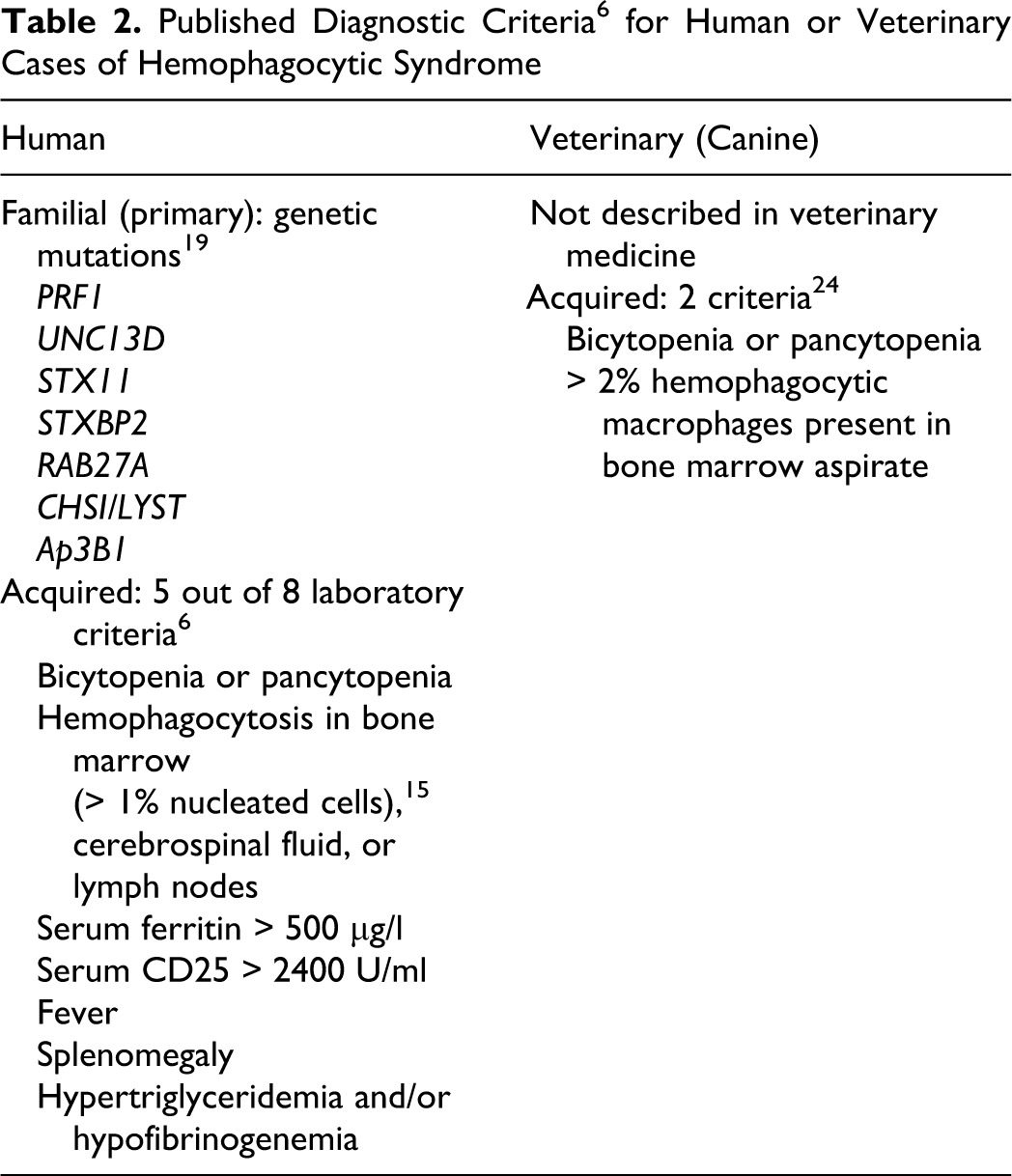
In veterinary medicine, HPS has been described in dogs, cattle, and a cat.13,23,24 In contrast to the more extensive criteria used in human medicine, the diagnostic criteria used for veterinary patients have consisted of bicytopenia or pancytopenia with concurrent hemophagocytic macrophages > 2% of nucleated cells in the bone marrow aspirate (Table 2).23,24 In this macaque, 6% of bone marrow nucleated cells were hemophagocytic macrophages, and there was severe pancytopenia.
The pathogenesis of HPS remains poorly understood. Most current understanding of the condition is derived from the genetic forms of the disease in human medicine (familial hemophagocytic lymphohistiocytoses). A consistent finding is impaired cytotoxic function of natural killer cells and CD8+ T cells, which leads to excessive activation of lymphocytes and macrophages with subsequent cytokine overproduction, including interferon γ, interleukin-6, interleukin-18, and tumor necrosis factor α.19,22 The generalized proinflammatory state induced by these cytokines causes macrophage hyperstimulation, leading to organ dysfunction and aberrant phagocytosis. 22 A similar pathogenesis of cytokine overproduction has been suggested in reactive (secondary) forms of the condition. 2
Acquired HPS in humans has been associated with a variety of underlying viral infections, including HIV, which is a lentivirus in the family Retroviridae. HPS has been reported to occur in all stages of HIV infection. 1 HPS is often associated with other opportunistic viral, bacterial, or protozoal infections in HIV-infected patients; however, HIV has been posited as the primary cause in some cases. 18 Studies suggest that the incidence of HPS in HIV-infected patients could be higher than previously reported. 1 In a study of 56 HIV-positive patients, histopathologic evidence of HPS was reported in 20% of those examined on postmortem. 14
In macaques, concurrent viral infections are common in SRV-infected animals. Simian parvovirus has been associated with anemia, but it was not PCR-detected in this case. Herpes B was serologically identified but not detected by PCR in bone marrow or spleen. In contrast, SRV was PCR detected in both bone marrow and spleen and was immunologically demonstrated within the depleted bone marrow. SRV exhibits broad cellular tropism and could alter cytokine profiles and cytotoxic T-cell function. 9 Although work is limited in this area, enhanced phagocytosis was demonstrated in vitro in 3 of 17 macaques with clinically advanced SRV-related disease, lending support to the idea that hemophagocytosis may be involved in causing cytopenia in a subset of SRV-infected animals. 8
In summary, primary causality cannot be definitively established in this case. Nevertheless, this case fulfilled previously established veterinary criteria and partially fulfilled human diagnostic criteria for HPS. Given the well-established occurrence of virus-associated HPSs in the human literature, the possibility that this condition plays a role in a subset of SRV-associated cytopenias in macaques warrants consideration. Serum testing of triglyceride levels, ferritin, and, possibly, CD-25 in suspect macaque cases would be valuable to establish further diagnostic criteria and facilitate comparison to HPS in humans.
Footnotes
Acknowledgment
We would like to thank Jessica White and Ann Rosenthal at the Pathogen Detection Laboratory at the California National Primate Research Center, University of California, Davis, for their generous assistance in virologic and immunohistochemical procedures. We also thank Paula Arrowsmith at the University of Michigan Pathology Cores for Animal Research for histology services.
The authors declared that they had no conflicts of interest with respect to their authorship or the publication of this article.
The authors declared that they received no financial support for their research and/or authorship of this article.