
Abstract
Angiomatoid lesions in a lymph node associated with a thyroid carcinoma of a dog were restricted to the subcapsular and medullary sinuses. Lymphoid atrophy was present, but nodal architecture was not distorted and normal structures were not invaded. Immunohistochemical staining indicated that the vascular spaces formed by spindloid cells were lined by endothelium with a low mitotic index. The spindloid cells were positive for smooth muscle actin, vimentin, and desmin and thus were likely to be fibroblasts, myofibroblasts, smooth muscle cells, and/or pericytes. These features are comparable to vascular transformation of lymph node sinuses in humans (nodal angiomatosis), a nonneoplastic condition often associated with mechanical or functional blockage of efferent lymphatics and veins.
Tissue from a neck swelling of an 11.5-year-old, neutered male, fox terrier was submitted for histologic evaluation to the Veterinary Diagnostic Laboratory at Oregon State University. The owner had noticed the mass a month previously and it had been growing rapidly. Hematoxylin and eosin, Masson’s trichrome, and Prussian blue–stained 4- to 5-μm-thick sections of formalin fixed tissues were prepared using standard methods. The largest portion of the mass was of thyroid gland origin. It was composed of broad sheets, cords, and lobules of uniform, cuboidal cells separated by a delicate fibrovascular stroma. Occasional acini contained inspissated to mineralized protein. The cells had scant amounts of rarified, eosinophilic cytoplasm and central round euchromatic nuclei. Mitotic activity was low, and the mass was punctuated by numerous vascular sinuses. Large areas of hemorrhage and necrosis were present, and the neoplastic cells invaded the capsule of the thyroid gland. A small lymph node (15 mm × 7 mm × 4 mm) was unknowingly included in the formalin-fixed materials.
Differential Diagnoses
Differential diagnoses for a lymphadenopathy associated with a neoplasm include metastatic foci within the subcapsular and medullary sinuses, siderophagocytosis and edema, infarction from tumor emboli, lymphoid hyperplasia or depletion, lymphadenitis from necrotic and inflamed neoplastic tissue, an unrelated primary neoplasm, and vascular transformation of lymph node sinuses (nodal angiomatosis).
Microscopic Findings
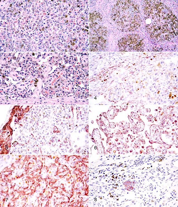
The microscopic lesions of note in the lymph node were effacement of the subcapsular and medullary sinuses by broad sheets and nodules of densely packed, polyhedral to spindloid cells (Fig. 1 ). These cells had abundant eosinophilic cytoplasm and indistinct borders. Nuclei were somewhat pleomorphic, euchromatic to hyperchromatic with undulating membranes. Chromatin was dispersed and nucleoli were inconspicuous. Scattered multinucleate cells were present along with 0–1 mitotic figures per ten 400× microscope fields. Many of these cells contained Prussian blue–positive cytoplasmic granules (hemosiderin). Cleft-like spaces often containing erythrocytes were present throughout the spindle and polyhedral cell populations. Erythrophages and siderophages were present within lymphoid follicles. These follicles had marked lymphoid depletion (Fig. 2 ). Of note, spindloid cells forming myriads of vascular channels separated by mature collagen were present, especially in subcapsular sinuses (Fig. 3 ). These channels were lined by elongate cells with flattened nuclei. Extramedullary hematopoiesis and moderate sinusoidal sclerosis were present.
Immunohistochemical Staining
Unstained, paraffin-embedded lymph node was sectioned at 3–5 μm and placed on positively charged glass slides. Immunohistochemical staining was performed using an automated staining device (DakoCytomation Autostainer, Carpinteria, CA) with Nova Red (Vector Labs, Burlingame, CA) as the chromogen and a hematoxylin counterstain. Antibodies tested were monoclonal mouse anti-swine desmin (1:50), monoclonal mouse anti-human BLA36 (1:10), monoclonal mouse anti-human CK (AE1-AE3; 1:200), polyclonal rabbit anti-bovine CK (wide spectrum screening; 1:500), monoclonal mouse anti-human MAC387 (1:200), monoclonal mouse anti-human smooth muscle actin (1:300), monoclonal mouse anti-bovine vimentin (1:100), and rabbit polyclonal anti-human von Willebrand factor (1:100; all from DakoCytomation). High-temperature antigen retrieval 1 was used for BLA36, desmin, vimentin, and von Willebrand factor, whereas proteinase K retrieval (5 minutes) was an added step for both cytokeratins, smooth muscle actin, and Mac387. Cells of the test slide (internal control) and appropriate normal canine tissues were used as positive controls. The negative control for all tissues and antibodies was the universal negative control provided by Dako.
The spindloid cells within the lymph node that outlined the neovascular channels were immunohistochemically positive for desmin (Fig. 4 ), smooth muscle actin (Fig. 5 ), and vimentin (Fig. 6 ). Endothelial cells were also positive for vimentin. The cells directly lining the channels were positive for von Willebrand factor (Fig. 7 ). BLA36-positive dendritic cells were present in all locations within the node including germinal centers. Mac387-positive cells were most numerous in germinal centers. A raft of AE1–AE3 cytokeratin–positive cells was present adjacent to the subcapsular sinus and presumed to be metastatic thyroid carcinoma (Fig. 8 ).
Diagnosis
The animal was diagnosed as having vascular transformation of lymph node sinuses (nodal angiomatosis).
Discussion
Vascular transformation of lymph node sinuses, sometimes referred to as nodal angiomatosis, is a pressure-induced, nonneoplastic conversion of nodal sinuses into complex, anastomosing vascular channels. 6 Nodules of sinusoidal spindloid pericytes and smooth muscle cells are often present. 10 Speculation exists that angiogenic factors may also be involved. 5,7 There are often sclerosis, erythro- and siderophagocytosis, and lymphoid atrophy. 4,6 In many but not all cases of vascular transformation of lymph nodes, a causative obstructive process is identified. 4,18
In people, vascular transformation of lymph nodes is believed to be secondary to obstruction of the efferent hilar lymphatics. The lesion was reproduced experimentally by ligation of the efferent lymphatics or partial ligation of the nodal veins and lymphatics of rabbits. 19 Venous ligation by itself does not produce vascular transformation. 19 In humans, vascular transformation of lymphoid sinuses has been reported in association with various neoplasms (lymphoma, 15 mammary carcinoma, 17 bronchogenic carcinoma, 15 renal carcinoma, 4 ovarian carcinoma, 4 and lingual carcinoma 10 ), venous obstruction, 16 hepatic cirrhosis, 20 pulmonary veno-occlusive disease and/or hypertension, 7,14 myeloproliferative disease, 11 congestive heart failure, 3 constrictive pericarditis, 3 and cat scratch fever. 2 The latter condition is associated with an abundance of neutrophils, a lack of spindle cell fascicles, and plump endothelial cells.
The florid erythrophagocytosis and numerous siderophages in our case and in the human literature are postulated to be due to either trauma at the primary site being drained by the affected lymph node or venolymphatic anastomoses that occur in vascular transformation of lymph nodes. 7
Vascular transformation of lymph node sinuses is easily confused with Kaposi’s sarcoma in people. Unlike the case in Kaposi’s sarcoma, however, mitotic activity and periodic acid–Schiff-positive, hyaline globules are absent in cases of vascular transformation, cellular pleomorphism is minimal, 3,4,16 and capsular involvement does not occur. 8
Vascular transformation of lymphoid sinuses is described as occurring in 4 patterns that may present in various combinations in particular lymph nodes. 3 The plexiform pattern is the least common, in which there is a lack of spindle cell fascicles but there are interconnected, mature vascular channels and noninvolvement of capsule, a flat endothelial cell lining, and low mitotic activity. Sinusal is characterized as having cleft-like spaces and is the most common type seen. Solid or nodular is composed of haphazardly arranged spindloid to plump cells among vascular spaces. In the round vascular type, the channels are usually empty or contain amorphous material presumed to be lymph. These types are believed to be on a continuum related to the duration and degree of venous/lymphatic obstruction.
Plexiform vascularization of lymph nodes was reported from 9 cats in 1987. 12 The authors of that article discussed what was known about vascular transformation of lymph node sinuses in humans at that time and concluded that the lesion in cats was different. However, 3 of the cats in that cohort had space-occupying lesions (2 presumptive mammary carcinomas and 1 presumptive salivary cyst), and postoperative edema was suggestive of vasoconstriction or lymphatic blockage. Cardiomyopathies, asthma, pulmonary hypertension, and other conditions associated with vascular transformation of human lymph nodes were not mentioned and presumably not tested for in the feline cohort.
With immunohistochemical staining, the spindle cells in vascular transformation of lymphoid sinuses of humans are positive for smooth muscle actin, vimentin, and CD34 and negative for factor VIII (von Willebrand factor)–related antigen, keratin, and desmin. Similar results were obtained in the case reported here. However, desmin was detected in the spindloid cells in this case, contributing to the supposition that some of the spindloid cells are pericytes. 13
Differential diagnoses for vascular transformation of lymph node sinuses in domestic animals include vascular hamartomas, hemangiomas, and lymphangioma. In a report of primary vascular neoplasms in aged dogs, 8 hemangiomas and 1 lymphangioma were reported. 9 Nodal angiomas occur principally in children and affect the medulla in humans, whereas the reported nodal angiomas in the dogs affected aged animals and were cortical. Because vascular transformation of lymph nodes begins peripherally, it may be that these presumptive angiomas of aged dogs were actually vascular transformations of the round vascular type. In addition, these putative neoplasms were nonexpansile and lined by flattened endothelium. It appears untoward that more than 5% of 175 control beagles on a life span study had these vascular tumors when they had not been previously described in any domestic species. At least 1 of the masses in this study was associated with obstructed drainage. The histologic distinction between angioma and vascular transformation can be difficult, particularly if the investigator is not familiar with the latter condition.
In conclusion, vascular transformation of lymph node sinuses, a nonneoplastic reaction to blocked efferent lymphatics and veins, is a recognizable condition in domestic animals that is easily confused with nodal hemangioma and lymphangioma, which are exceedingly rare conditions of humans and domestic animals. The salient features of vascular transformation of lymph node sinuses are polyhedral to spindle cell aggregates in nodal sinuses with angiomatous transformation beginning in subcapsular regions. These masses are noninvasive, have a low mitotic rate, and are frequently associated with lymphoid atrophy, erythrophagocytosis/hemosiderosis, and variable fibrosis. In humans and in the dog described herein, the lesions are often associated with malignant neoplasia of the nodal-associated tissues, but other mechanisms of vascular blockage are causative. In many cases the blockage may be physiologic (vasoconstriction) without histologic lesions (idiopathic). Anecdotal evidence suggests that vascular transformation of lymph node sinuses is a not uncommon lesion in domestic animals as it is in humans.
Footnotes
Acknowledgements
We sincerely thank Kay Fischer and the personnel of the histopathology section of the Veterinary Diagnostic Laboratory for their excellent technical assistance. We thank Jill Bartlett for her assistance with the photomicrographs.
The authors declared that they had no conflicts of interest with respect to their authorship or the publication of this article.
The authors declared that they received no financial support for their research and/or authorship of this article.