
Abstract
Objective
Inflammation plays a crucial part in osteoarthritis (OA) development. This work aimed to explore loganin’s role and molecular mechanism in inflammation and clarify its anti-inflammatory effects in OA treatment.
Methods
Chondrocytes were stimulated using interleukin (IL)-1β and loganin at two concentrations (1 μM and 10 μM). Nitric oxide (NO) and prostaglandin E2 (PGE2) expression was assessed. Real-time polymerase chain reaction was used to evaluate inducible NO synthase (iNOS), cyclooxygenase (COX)-2, IL-6, and tumor necrosis factor (TNF)-α mRNA levels. Western blot was used to investigate TLR4, MyD88, p-p65, and IκB-α expression. p65 nuclear translocation, synovial inflammatory response, and cartilage degeneration were also assessed.
Results
Loganin significantly reduced IL-1β-mediated PGE2, NO, iNOS, and COX-2 expression compared with that of the IL-1β stimulation group. The TLR4/MyD88/NF-κB pathway was suppressed by loganin, which decreased inflammatory cytokine (TNF-α and IL-6) levels compared with those of the IL-1β stimulation group. Loganin inhibited IL-1β-mediated NF-κB p65 nuclear translocation compared with that of the IL-1β stimulation group. Loganin partially suppressed cartilage degeneration and the synovial inflammatory response in vivo.
Conclusion
This work demonstrated that loganin inhibited IL-1β-mediated inflammation in rat chondrocytes through TLR4/MyD88/NF-κB pathway regulation, thereby reducing rat cartilage degeneration and the synovial inflammatory response.
Keywords
Introduction
Osteoarthritis (OA) is a chronic joint disease that affects and disables 1 × 10⁸ people around the world and contributes to a large societal burden. 1 Several studies have revealed the importance of chronic inflammation in OA development. Various inflammatory factors, including tumor necrosis factor (TNF)-α and interleukin (IL)-1, -8, -6, -15, and -33, increase in the cartilage in OA patients’ joints, and these factors mediate cartilage degeneration.2–4 IL-1β has been widely studied in OA pathophysiology to stimulate inflammatory cytokine release and chondrocyte apoptosis. 5 Matrix metalloproteases (MMPs) and a disintegrin and metalloproteinase with thrombospondin motifs (ADAMTS) are degradation enzymes that can be upregulated by several transcription factors, especially nuclear factor kappa B (NF-κB). 6 The microenvironment of inflammatory cytokines combined with older age and biomechanical stress increase the oxidative stress, contributing to up-regulation of reactive oxygen species (ROS) including nitric oxide (NO), prostaglandins (PGs), and superoxide anion and down-regulation of anti-oxidative enzymes. 7 These changes contribute to joint cartilage destruction and bone metabolism, increasing the generation of chondrocytes, osteoblasts, and their precursors. 7 Therefore, regulation of the inflammatory microenvironment homeostasis contributes to OA development.
Loganin is an iridoid glycoside that is isolated from herbs such as Flos lonicerae and Cornus mas L. It mediates immune function and has anti-inflammatory and anti-oxidative effects through inhibiting NF-κB signaling to decrease the release of IL-6, TNF-α, and other inflammatory factors. Loganin attenuates the intestinal inflammatory reaction and oxidative stress through TLR4/NF-κB modulation. 8 It also had an obvious anti-oxidative stress and anti-inflammatory effect on diabetes mellitus-induced reproductive damage through restoration of glutathione levels and superoxide dismutase activity and reduction of reactive oxygen species. 9 Our previous research showed that loganin caused reduced extracellular matrix (ECM) catabolism and IL-1β-induced apoptosis in rat chondrocytes through the regulation of PI3K/Akt signaling. This suggests that loganin is a potential alternative in OA treatment. 10 To further explore loganin’s protective mechanisms, the present research aimed to clarify whether loganin had anti-inflammatory effects in OA treatment.
Materials and methods
Reagents
Loganin was obtained from Sigma-Aldrich (St. Louis, MO, USA, ≥97.0%, Figure 1a). IL-1β was obtained from R&D Systems (St. Paul, MN, USA). Antibodies were purchased from Cell Signaling Technology (Beverly, MA, USA) and Santa Cruz Biotechnology Inc. (Santa Cruz, CA, USA). All other reagents were obtained from Sigma-Aldrich.
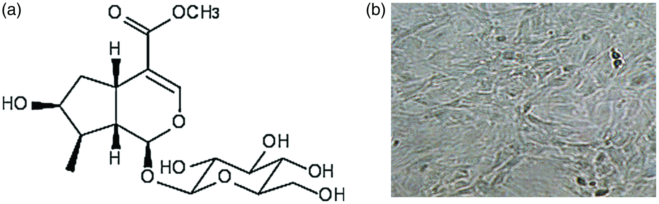
(a) Loganin chemical structure and (b) Primary chondrocyte morphology.
Chondrocyte culture and drug administration
Chondrocytes were isolated from the knee joint of normal Sprague–Dawley rats. 11 Cartilage was removed, minced, and incubated with collagenase II (2 mg/mL) in a cell incubator for 3 to 6 hours.12,13 The samples were then filtered via a filtration system (70 μm) and the solution was centrifuged (300 × g, 5 minutes, 37°C), washed, and incubated with DMEM/F12 medium containing 10% fetal bovine serum. We evaluated the cell morphology (isogenous and elliptic) from primary chondrocytes (Figure 1b). The medium was changed every 2 days, and chondrocytes were passaged after reaching 70% to 80% confluence. Loganin or IL-1β at 10 ng/mL was added to chondrocytes at P2 for 2 hours. The cells were then collected for subsequent testing. All animal experiments were approved by the Animal Care and Use Committee of Hainan Medical College (2016-35).
Chondrocytes were divided into the following four groups: control, IL-1β, 1 μM loganin, and 10 μM loganin. Among these groups, COX-2, iNOS, IL-6, and TNF-α mRNA expression and TLR4, MyD88, p-p65, and IκB-α were evaluated.
Quantitative real time polymerase chain reaction
COX-2, iNOS, IL-6, and TNF-α mRNA expression was assessed. mRNA was isolated using the TRIzol method. The specimen concentration was measured spectrophotometrically at 260 nm, and cDNA synthesis was performed with 1 μg of RNA using the cDNA Synthesis Kit (Thermo Scientific, Waltham, MA, USA). Quantitative real time polymerase chain reaction (PCR) was conducted using SYBR Premix Ex Taq™ under the following conditions: 10 minutes at 95°C and 40 cycles of 15 s at 95°C and 1 minute at 60°C. iNOS, COX-2, TNF-α, and IL-6 mRNA levels were compared with β-actin.
Western blot
Chondrocytes were lysed via a protease- and phosphatase-containing RIPA buffer. The protein concentration was calculated using the Bio-Rad Laboratories protein reagent (Bio-Rad, Hercules, CA, USA). Cellular lysates (20 ng) were separated using polyacrylamide gel electrophoresis. The protein was then transferred onto poly(vinylidene fluoride) membranes for antibody blotting. Specific antibodies were used (e.g., anti-TLR4, anti-MyD88, anti-p-p65, anti-IκB-α, and anti-β-actin) overnight followed by the appropriate secondary antibodies. An enhanced chemiluminescence system (Amersham Biosciences, Piscataway, NJ, USA) was used to visualize the immunoreactive bands.
NO measurement and enzyme-linked immunosorbent assay
NO production was tested by measuring the levels of the stable NO metabolite nitrite using sodium nitrite resuspended in distilled H2O as a standard. 14 The NO concentration was quantitatively measured using the Griess reaction. 15 NO expression was tested by evaluating the expression of the stable metabolite nitrite of NO. After incubation with loganin for 2 hours, cells were simulated with IL-1β (10 ng/mL) for 1 day. The supernatant was incubated with the Griess solution in a 96-well plate for 10 minutes. The specimen was tested using a spectrophotometer at 540 nm. PGE2, IL-1β, IL-6, and TNF-α expression within the medium and rat synovial fluid were measured using ELISA kits (R&D Systems, Minneapolis, MN, USA) in accordance with the manufacturer’s protocol.
Rat osteoarthritis model
Sprague–Dawley rats (200–250 g) were divided into the following three groups: sham, OA, and OA + loganin. Rats in the OA and OA + loganin groups underwent anterior cruciate ligament (ACL) transection and medial meniscus resection in the right knee. The sham rats underwent the same surgery without ACL or meniscus intervention. After surgery, the rats in the OA + loganin group were administered loganin (20 mg/kg/day subcutaneously) for 8 weeks until they were sacrificed using CO2 inhalation (40% vol/minute for 5 minutes). 16
Hematoxylin and eosin staining and Mankin’s score
Eight weeks after surgery, the tissues were harvested and incubated with 4% (v/v) paraformaldehyde for 1 day followed by de-calcification and paraffin embedding. The specimens were cut into slices (5 μm thick), which were stained with hematoxylin and eosin and observed using a light microscope. The degree of cartilage degeneration was assessed using Mankin’s score (0 to 13), which indicates the degradation level of articular cartilage, with a higher score representing more cartilage degeneration. 17
Immunofluorescence assay
Paraffin sections (5 μm) were deparaffinized and rehydrated, followed by 3% (v/v) H2O2 for 10 minutes. Cells were incubated in 4% (v/v) paraformaldehyde for 30 minutes. The specimens were incubated with 5% (w/v) BSA for 60 minutes and then with primary antibodies (anti-CD68 and anti-P65) for 1 hour with Alexa Fluor 488-conjugated anti-IgG secondary antibody. This was followed by incubation with diamidinophenyl indole for 7 minutes. The images were captured under a Nikon Eclipse Ti microscope (Nikon, Tokyo, Japan).
Statistical analysis
Results are presented as the mean ± standard deviation and analyzed using SPSS version 17.0 software (SPSS Inc., Chicago, IL, USA). A one-way analysis of variance was used to compare three or more groups. Values with P < 0.05 were considered to be statistically significant.
Results
Effect of loganin in IL-1β-mediated inflammatory reactions in chondrocytes
Chondrocytes that were isolated from normal rats and cultured as described above were assessed. The Griess reagent was used to determine the NO concentration in the supernatant after centrifuging the chondrocytes, while an ELISA was used to measure the PGE2 production in the culture medium. As shown in Figure 2a and b, NO and PGE2 secretion was significantly increased after IL-1β exposure compared with that in the control group (P < 0.001), while loganin significantly inhibited the IL-1β-mediated increase in PGE2 and NO compared with that in the IL-1β group (P < 0.05). As shown in Figure 2c and d, IL-1β significantly increased the COX-2 and iNOS expression compared with that in the control group (P < 0.001). Loganin also inhibited IL-1β-mediated increase in COX-2 and iNOS mRNA expression in a dose-dependent manner (P < 0.05). TNF-α and IL-6 levels in the culture medium were quantitatively measured using an ELISA. Figure 2e and f shows that compared with that in the control group, IL-6 and TNF-α mRNA expression was significantly up-regulated by IL-1β in chondrocytes (P < 0.001). However, loganin down-regulated the IL-1β-induced increase in IL-6 and TNF-α mRNA expression in a dose-dependent manner (P < 0.05). ELISA was also used to evaluate the effect of loganin on IL-1β-mediated IL-6 and TNF-α protein production. Figure 2g and h shows that TNF-α and IL-6 protein expression was significantly increased by IL-1β compared with that in the control group (P < 0.001). However, TNF-α and IL-6 expression were significantly decreased after loganin pretreatment compared with that in the IL-1β group (P < 0.05).

Role of loganin in IL-1β-mediated inflammatory reaction in chondrocytes. (a) PGE2 levels were measured by ELISA. (b) NO production was assayed by measuring the levels of the stable NO metabolite nitrite using sodium nitrite resuspended in distilled H2O as a standard. The Griess reaction was performed to evaluate the nitrite contents within the medium. (c–d) (c) COX-2 and (d) iNOS mRNA expression was detected using PCR. (e–f) (e) TNF-α and (f) IL-6 expression was detected using PCR. (g–h) TNF-α and IL-6 levels were calculated using ELISA. Data are presented as the mean ± SD. *P < 0.05, **P < 0.01, ***P < 0.001 compared with controls.
The role of loganin in IL-1β-mediated activation of TLR4/MyD88/NF-κB pathway in chondrocytes
To investigate loganin’s anti-inflammatory mechanism, its role in activating NF-κB by IL-1β was evaluated using western blot. Figure 3a–d shows that IL-1β significantly increased TLR4 and MyD88 expression and NF-κB p65 phosphorylation of chondrocytes compared with that of the control group (P < 0.01). IL-1β also caused significant IκB-α degradation in chondrocytes compared with that in the control group (P < 0.01) (Figure 3a and e). Loganin dose-independently decreased IL-1β-induced TLR4 and MyD88 expression and NF-κB p65 phosphorylation compared with those of the IL-1β group (P < 0.05), and it also reduced the IL-1β-induced IκB-α degradation in chondrocytes in a dose-dependent manner compared with that in the IL-1β group (P < 0.05) (Figure 3). Chondrocyte immunofluorescence in response to IL-1β-mediated NF-κB activation was measured to explore the role of loganin in NF-κB p65 nuclear translocation. Clear and enhanced nuclear p65 staining was observed in chondrocytes upon IL-1β stimulation, indicating that nuclear NF-κB p65 translocation occurred in these cells. However, loganin reversed NF-κB p65 subunit translocation compared with that in the IL-1β stimulation group (Figure 3f). These findings may indicate that loganin exerts protective effects on IL-1β-mediated chondrocytes via the TLR4/MyD88/NF-κB signaling pathway.

Roles of loganin in IL-1β-mediated expression of TLR4/NF-κB signaling in chondrocytes. (a–e) Western blotting analysis of TLR4, MyD88, p-p65, and IκB-α in each group. Data are shown as the mean ± SD. *P < 0.05, **P < 0.01 compared with controls and (f) Effects of loganin on IL-1β-activated nuclear translocation of p65 in chondrocytes. Fluorescence immunostaining of p65 (green) and nucleus (blue).
Loganin inhibits synovial inflammation in OA rats
To further clarify the anti-inflammatory role of loganin in OA, synovial fluid was isolated from joints in OA model rats, and its response to an inflammatory stimulus and loganin was measured using an ELISA. Figure 4a–c shows that surgery increased IL-1β, IL-6, and TNF-α expression in knee synovial fluid compared with that in the sham group (P < 0.01). However, loganin administration reduced these increases compared with those in the OA group (P < 0.05). Additionally, an immunofluorescent assay showed that loganin decreased the number of CD68-labeled inflammatory cells compared with that in the OA group (Figure 4d). All data suggested that loganin attenuated synovial inflammation in OA rats compared with that caused by OA alone.

Roles of loganin in the synovial inflammatory response in OA rats. (a–c) Loganin inhibited synovial inflammatory factor expression in OA rats. IL-1β (a), IL-6 (b), and TNF-α (c) protein expression was tested using an ELISA. (d) Representative synovial fluid stained with CD68. Data are presented as the mean ± SD. *P < 0.05, **P < 0.01 compared with the OA group.
Effects of loganin on OA cartilage histopathology
Hematoxylin and eosin staining showed that compared with the sham group, OA rats had severe articular cartilage destruction. Loganin treatment increased knee articular cartilage thickness and seems to have mitigated cartilage decomposition (Figure 5a). Moreover, the Mankin’s score in the OA group was significantly increased compared with that in the sham group (P < 0.05), and this increase was attenuated by loganin treatment (P < 0.01) (Figure 5b). Thus, loganin showed protective effects and enhanced the recovery of OA cartilage in this rat OA model.

Articular cartilage histology was measured using hematoxylin and eosin staining (×40), and cartilage damage was assessed using the Mankin’s score in rats. Data are presented as the mean ± SD. ***P < 0.05 compared with the sham group; **P < 0.01 compared with the OA group.
Discussion
An increasing amount of evidence indicates the importance of IL-1β in cartilage degradation caused by OA.18–20 Chondrocytes stimulated with IL-1β showed increased iNOS and COX-2 levels, which contributed to NO and PGE2 expression. Both NO and PGE2 are catabolic factors that are stimulated by MMP production and ECM synthesis inhibition in chondrocytes from OA rats.21–23 NO produced by iNOS is an inflammatory mediator that can trigger MMP secretion and activation, which can also compromise proteoglycan and collagen-II production in OA pathophysiology. 24 PGE2 is a main mediator of inflammation and pain in OA joints, such as enhancing ECM degradation and suppressing chondrocyte proliferation during OA pathogenesis. 25 Previous work demonstrated that suppressing inflammatory factor secretion including NO and PGE2 attenuates cartilage degeneration. 26 Synovial fibroblasts in OA were shown to be sensitive to local NO donors, which contributed to the increase in oxidative stress. 27 Oxidative stress in OA fibroblasts activates mitogen-activated protein kinase and NF-κB signaling and up-regulates COX-2 and PGE2 levels, leading to degenerative changes. 27
Our previous research showed that loganin administration compromised ECM catabolism and IL-1β-mediated apoptotic activity in chondrocytes via activation of PI3K/Akt signaling, which suggests that loganin is a potential therapeutic drug for treating OA. The significant inhibitory role of loganin in IL-1β-stimulated COX-2 and iNOS expression was revealed by the subsequent PGE2 and NO generation. The results of this study were consistent with previously published studies. Loganin decreased the TNF-α mRNA level and the iNOS and COX-2 levels in β-amyloid protein (Aβ)-treated PC12 cells. 28 Additionally, loganin treatment prevented TNF-α, IL-6, MCP-1, NO, and PGE2 over-production and COX-2 and iNOS expression in the Aβ-stimulated BV-2 cells. 29 These findings show loganin’s anti-inflammatory effects that protect cartilage through suppressing IL-1β-mediated COX-2 and iNOS expression with subsequent PGE2 and NO generation in the OA pathogenic mechanism.
TLR4 was shown to be overexpressed in cartilage in OA, and it was essential in the pathological process of cartilage degeneration. 30 The combination of TLR4 and the relevant ligand MyD88 results in NF‐κB activation and phosphorylated NF‐κB translocation to the nucleus, contributing to inflammatory‐related gene transcription. 31 TLR signaling contributes to NF-κB signaling activation, thereby enhancing inflammatory factor and degradative enzyme production. 31 Lipopolysaccharide (LPS) enhances ECM protein degradation through binding with TLR4 in rheumatoid arthritis. 32 LPS, which is a specific TLR ligand, enhances proteoglycan and aggrecan degradation and collagen II expression via TLR4 signaling activation in murine and human articular chondrocytes. 33 This research revealed the significant role of IL-1β in increasing the TLR4 signaling level in chondrocyte cells. We also showed a significant inhibitory role for loganin in IL-1β-induced TLR4 expression.
TLR4 is a main receptor in the inflammatory response. TLR4 ligation activates NF-κB, a downstream transcription factor and main catabolic signal related to OA progression. TLR4 ligation coupling to NF-κB activation is achieved by the adaptor protein MyD88, which is recruited to activate IκB kinase followed by removal of cytosolic p65 subunit sequestration that triggers nuclear translocation of these proteins. Our results showed that loganin markedly attenuated NF-κB p65 phosphorylation and IκB-α degradation in IL-1β-stimulated chondrocytes, causing a decrease in inflammatory cytokines including IL-6 and TNF-α. The NF-κB pathway is a main catabolic signaling pathway engaged in OA pathogenesis, and it exerts significant effects on regulating OA-related inflammatory mediators and the ROS level. 34 Inactivated NF-κB interacts with the inhibitory protein IκB-α and appears within the cytoplasm in the non-activated state. Stimulation by inflammatory mediators and oxidative stress triggered nuclear translocation of activated NF-κB. This translocation up-regulated multiple inflammation-related genes including iNOS, COX-2, NO, PGE2, MMPs, and ADAMTS, thereby facilitating catabolic factor synthesis, cartilage inflammation, and OA chondrocyte apoptosis. 14 Thus, targeted suppression of NF-κB signaling may be helpful in OA treatment. Previous studies revealed that NF-κB suppression inhibited COX-2, MMP-9, and iNOS levels in chondrocytes exposed to IL-1β chondrocytes. 35 Moreover, NF-κB inhibition decreased TNF-α-triggered chondrocyte expression of MMP-3 and MMP-13. 36
Loganin was reported to prevent chronic neuropathic pain triggered by constriction injury through inhibition of TNF-α/IL-1β-mediated NF-κB activation. 37 Loganin reduces Aβ-induced inflammation in BV-2 microglial cells by regulating the TLR4/TRAF6/NF-κB pathway. 29 Loganin seems to prevent M1 macrophage-mediated inflammation through modulating the Sirt1/NF-κB axis and lowering TNF-α, IL-6, and IL-1β levels to ameliorate dextran sulfate sodium-induced ulcerative colitis. 38 Our work and other previous research indicated that loganin prevented IL-1β-triggered inflammation by suppressing TLR4/MyD88/NF-κB activation. The current research revealed the underlying mechanism through which loganin blocks IL-1β-induced TLR4/MyD88/NF-κB activation in chondrocytes. Additionally, our in vivo experiments showed that loganin partially suppressed the synovial inflammatory response. However, more studies are required to illustrate the mechanism by which loganin regulates chondrocyte inflammation.
In summary, this study shows the anti-inflammatory effects of loganin in IL-1β-treated chondrocytes. The results indicated that loganin suppressed IL-1β-induced levels of NO, PGE2, iNOS, COX-2, IL-6, and TNF-α through suppressing the TLR4/MyD88/NF-κB pathway in IL-1β-activated chondrocytes. Our in vivo experiment revealed that loganin partially suppressed cartilage degeneration and the synovial inflammatory response, indicating the potential role of loganin in OA treatment.
Footnotes
Declaration of conflicting interest
The authors declare that there is no conflict of interest.
Funding
The authors disclosed receipt of the following financial support for the research, authorship, and/or publication of this article: The present study was supported by the scientific research projects in the health and family planning industry in Hainan Province (Grant No. 20A200096).