
Abstract
The mechanisms by which Y chromosome microdeletions cause infertility have been well described; however, the therapeutic targets remain a challenge. Here, we used whole-genome sequencing to explore the mechanism of Y chromosome deletion and potential therapeutic targets in a patient with infertility. There were no abnormalities in the patient’s medical history. Routine semen analysis showed immotile sperm and only two motile spermatozoa were occasionally see after centrifugation, indicating that the direct cause of infertility was an abnormal sperm count and motility. A Y chromosome microdeletion test revealed partial deletion of the AZFc region, including AZFc1, AZFc2, AZFc3, and AZFc4. Whole-genome sequencing showed that the patient had seven harmful mutations, with only one significant epigenetic mutation, SH3KBP1. Gene Ontology analysis of these meaningful mutations indicated involvement of cAMP signaling pathways. The patient’s wife became pregnant following in vitro fertilization, and no significant abnormalities were found during prenatal examination. This case suggests that Y chromosome microdeletion and gene mutation may affect the cAMP signaling pathway, leading to reduced sperm quality and male infertility.
Keywords
Introduction
The human reproductive system affects the health of the offspring, and is susceptible to environmental exposure. Recent increases in workplace stress and environmental pollution have severely affected human reproductive health, with male infertility becoming a global health problem.1,2 The main causes of male infertility are reduced sperm quality and impaired sperm function. 1 Evolutionarily, spermatozoa are the most divergent cell type, possibly as a consequence of their need to perform their function outside the male body, in the female reproductive tract. 3 Spermatogenesis occurs in the testes, and spermatozoa are morphologically intact but functionally immature when they leave the testes. To achieve their goal, spermatozoa thus need to undergo maturation and capacitation in the epididymis, followed by the acrosome reaction in the female reproductive tract. These are interdependent processes, and any errors could lead to abnormal morphology or sperm dysfunction. 4 Although spermatogenesis and sperm maturation are strictly regulated through complex signal transduction pathways, an in-depth understanding of these is currently lacking. 5 Spermatogenesis is controlled by azoospermia factor (AZF) genes on the long arm of the Y chromosome, and deletion or mutation of these genes may cause spermatogenesis disorders and consequent male sterility. Previous studies suggested that AZF deletions were the second most common genetic cause of azoospermia and oligospermia, accounting for about 3% to 29% of patients. 6 We report the case of a patient with Y chromosome microdeletion. Based on his clinical symptoms and genetic alterations, we aimed to analyze the mechanisms underlying sperm dysfunction and provide clues for the diagnosis and treatment of similar cases.
Case report
A 28-year-old male patient was admitted to our hospital with a 3-year history of reproductive failure. He had normal development, and the couple had regular sexual intercourse with endovaginal ejaculation and no contraception. The patient had no history of mumps or other specific viral infections, trauma or surgery, chronic diseases (such as hypertension and diabetes), and no family history of genetic diseases. His partner had no obvious abnormalities. Further examination showed normal development of the external genitalia, small bilateral testes, no obvious masses, and no obvious abnormality of the bilateral epididymides or spermatic cords.
B-scan ultrasonography of the prostate, seminal vesicles, testes, epididymides, and spermatic cords was carried out using a Siemens Acuson X150 ultrasound device (Siemens Healthcare, Erlangen, Germany). The patient was placed in a supine position with the lower abdomen fully exposed and the probe surface was modified with a coupling agent and placed on the symphysis. Color Doppler ultrasound examination indicated slightly smaller testes on both sides, no obvious abnormalities in the bilateral epididymides, slightly enlarged inner diameter of the left spermatic cord, no abnormalities of the seminal vesicles, a slightly enlarged prostate, and normal blood flow signals in the prostate (Figures 1 and 2).

B-scan ultrasonography of the testes.

B-scan ultrasonography of the seminal vesicles.
We also evaluated sex hormone levels, semen quality, presence of anti-sperm antibody, viral infections, blood karyotype, and Y chromosome microdeletion.
Semen was harvested after 3 days of ejaculatory abstinence and liquefied at room temperature for 30 minutes. The color (yellowish-white), viscosity (moderate), pH (pH = 7), and volume (5 mL) were normal. Routine semen-quality analysis (Huijia, Beijing, China) detected no sperm. Immotile sperm and two motile spermatozoa were occasionally seen after centrifugation.
Based on the abnormal semen analysis, we examined the patient’s hormones levels to further investigate the cause of his azoospermia. Peripheral blood was harvested at 8:00 am and testosterone, estradiol, progesterone, prolactin, luteinizing hormone, follicle-stimulating hormone, sex hormone binding globulin, and dehydroepiandrosterone sulfate levels were assessed using a Cobas e601 analyzer (Roche, Switzerland). The results were within normal ranges (Table 1).
Sex hormone determination.

We also collected peripheral blood and tested for the presence of anti-sperm IgG, IgA, and IgM antibodies using Kanghua ELISA kits (Kanghua, Shandong, China) and for viral immunological response by detecting IgM and IgG antibodies against rubella virus, cytomegalovirus, Toxoplasma, and herpes simplex virus type 2 using Aikang ELISA kits (ACON, Hangzhou, China). The results were all normal (Table 2).
Immunological analysis.

We collected peripheral blood and extracted DNA for karyotype analysis and to evaluate Y chromosome microdeletion. Karyotype analysis showed 46, XY, and no obvious abnormalities were observed by G-banding (Figure 3). Detection of Y chromosome microdeletion confirmed that the male sex-determining gene SRY was not affected. DNA samples were submitted for analysis on a MassARRAY nucleic acid mass spectrometer (Agena Bioscience, CA, USA) to detect sequence tagged sites (STS) in the AZFa (sY84/sY86), AZFb (sY121/sY127/sY134), and AZFc (sY160/sY252/sY255/sY1191/sY1291) regions of the Y chromosome. Partial deletions (AZFc1-4) in the AZFc region were detected, with no deletion of AZFc5 (Table 3 and Figure 4).

Karyotype analysis of the peripheral blood.
Y chromosome microdeletion detection.

STS, sequence tagged site.

Male Y chromosome microdeletion report and explanatory chart.
Library construction and genome sequencing were performed by Beijing Novogene Co., Ltd. The integrity and purity of the DNA samples were determined by agarose gel electrophoresis. DNA concentration was determined with a Qubit 3.0 fluorometer (Thermo Fisher Scientific, MA, USA), and samples containing more than 0.5 µg DNA were used for library construction. A DNA library was constructed by disruption of the genomic DNA into ∼350-bp fragments using a Covaris crusher (Covaris, MA, USA), end-repair, and poly(A) tailing, followed by adapter ligation at both ends. The quality of the library was evaluated using a Qubit 2.0 fluorometer, and the insert size was detected with NGS3K/Caliper (Novogene Co., Ltd., Beijing, China). Finally, quantitative polymerase chain reaction was performed to quantify the effective concentration of the library (3 nM).
After library quality assessment, DNA sequencing was performed using the Illumina platform (Integrated DNA Technologies, Inc., Coralville, IA, USA) according to the effective concentration and data output requirements. Sequencing data quality was assessed in terms of sequencing error rate, data volume, and comparison rate. If sequencing data met the standards, subsequent analysis was performed; otherwise, the library was rebuilt and tested. Evaluation of the base distribution showed no separation of AT and GC, and the sequencing data met the experimental requirements. The sequencing depth was 30×, and the chromosome coverage was 100%, except for the Y chromosome (Figure 5).

Coverage depth (left coordinate) and coverage (right coordinate) for each chromosome.
Raw sequenced reads were then processed and variations were detected by aligning high-quality sequences to the human reference genome GRCh37/hg19, followed by statistics and annotation. However, such tests generate massive data and relevant disease-related mutations are limited. The mutation detection results were thus used to screen for pathogenic mutation sites/genes and predict disease correlations.7,8
First, potentially pathogenic or splicing-affecting single nucleotide polymorphism/InDel mutations were screened and classified according to the American College of Medical Genetics and Genomics 2015 classification criteria (Table 4). Five pathogenic mutations (KDM2A, ACOX1, NEB, PREX1, and RNF168) were identified (Table 5). Given that the noncoding region plays critical roles in gene regulation, variations in the noncoding regions were screened (Table 6). Genomiser software screening identified 574,029 non-coding region variants related to disease or phenotype. The conserved sites were identified by Combined Annotation Dependent Depletion and Genome Wide Annotation of VAriants, and 3714 candidate sites were obtained. Finally, we used the GTEx, Roadmap, and Encode databases to screen for variants that might affect gene expression in specific tissues, and annotated them as DNA functional elements. After screening, the important noncoding gene, SH3KBP1, was identified and annotated (Table 7).
Screening of single nucleotide polymorphism/InDel pathogenic mutations.
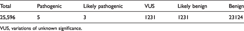
VUS, variations of unknown significance.
Annotation of pathogenic mutations.

ACMG, evidence level of American College of Medical Genetics and Genomics; Position, absolute position of variation sites in the chromosome; ID, dbSNP annotation ID; Ref, reference; ALT, sample genome base; Qual, variation score, positively correlated with quality; H, high.
Variations in the noncoding region.

Genomiser, variation counts in the noncoding region using Genomiser; Conservation, conserved variation counts by Combined Annotation Dependent Depletion and Genome Wide Annotation of VAriants screening; Epigenome, variation sites affecting gene expression with DNA functional elements screened by GTEx, Roadmap, and Encode databases.
Identification and annotation of variations in the noncoding region.

HR, chromosome; ID, dbSNP annotation ID; Ref, reference; ALT, sample genome base; Qual, variation score, positively correlated with quality.
In living organisms, genes exert their biological functions cooperatively, and phenotypic differences in complex diseases may be caused by mutations in multiple genes. The major metabolic and signaling pathways affected by genetic mutations can be determined through functional enrichment analysis. We therefore carried out Gene Ontology (GO) enrichment analysis to identify the mutations involved in cell component, biological pathway, and molecular function. The results are shown in Figure 6. We also carried out Kyoto Encyclopedia of Genes and Genomes analysis. A scatter plot of KEGG pathways indicated that the variant genes were mainly involved in the cAMP signaling pathway (Figure 7). P values were calculated using hypergeometric distribution and bioinformatics software (R; http://www.R-project.org/).

Gene Ontology (GO) functional enrichment scatter plots. (a) Cell component, (b) biological pathway, and (c) molecular function. X-axis indicates the ratio of genes mapped to that pathway to total genes; y-axis indicates enriched GO terms; dot size indicates number of genes mapped to the GO term; and color indicates P-value.

Scatter plot of Kyoto Encyclopedia of Genes and Genomes (KEGG) pathway analysis. X-axis represents ratio of genes mapped to that pathway to total genes; y-axis represents name of enriched KEGG pathway; dot size indicates number of genes mapped to the pathway; and color indicates P-value.
The final diagnosis in the present case was male infertility due to Y chromosome microdeletion and genetic mutations. Considering that the patient had few active sperm, we recommended in vitro fertilization. The couple underwent pre-implantation genetic diagnosis to select normal embryos for implantation with the aim of improving the clinical pregnancy rate. The patient’s wife subsequently became pregnant and the fetus was growing well, with no obvious abnormalities during prenatal examination.
The patient provided consent for the treatment and for the publication of the case report and accompanying images. We have de-identified all patient details.
Discussion
Infertility can be caused by abnormalities in sperm development, morphology, and motility, which in turn depend on metabolic and signaling pathways. Defects in genes involved in these metabolic and signal transduction pathways may thus lead to sperm dysfunction and infertility.
The current patient was admitted with infertility; however, no congenital abnormalities of the reproductive system and no significant alterations in laboratory tests, such as androgen levels, were identified. Nevertheless, no spermatozoa were detected by routine semen analysis, and only two motile sperm were seen occasionally after centrifugation, indicating that the direct cause of infertility was abnormal sperm count and motility. Chromosome karyotyping detected the Y chromosome, and the AZFc region contained microdeletions of AZFc1, AZFc2, AZFc3, and AZFc4. Y chromosome microdeletions are the second most frequent genetic cause of male infertility. However, there are also individual differences in terms of the effects of these microdeletions, and the exact mechanism of their action in relation to male infertility remains unknown. The role of AZFc microdeletion in infertility is particularly unclear.
Genomic sequencing in the current patient identified seven harmful mutations, including KDM2A, ACOX1, NEB, PREX1, and RNF168, as well as a sense epigenetic mutation in SH3KBP1. GO analysis of these sense mutations mapped to the cAMP signal pathway, which is widely recognized as a regulator of mammalian sperm cell motility. Activation of the CatSper channel is required for calcium entry into sperm. HCO3– and Ca2+ regulate atypical sperm adenylyl cyclase, leading to production of cAMP and activation of protein kinase A (PKA), which in turn induces phosphorylation of axonemal dynein, leading to depletion of ATP and increased intracellular pH. PKA activates serine threonine tyrosine kinase, triggering a phosphorylation cascade of proteins related to sperm motility. 9 Moreover, sperm capacitation is mainly regulated through the cAMP-dependent PKA-mediated protein phosphorylation cascade. 10 Binding of PKA regulatory subunits to the A-kinase anchoring protein family promotes tyrosine phosphorylation of sperm proteins by indirectly activating tyrosine kinases. Protein tyrosine phosphorylation of AKAP proteins (AKAP3 and FSP95) in human spermatozoa is critical for sperm capacitation. Tyrosine kinase receptors are transmembrane proteins with an extracellular ligand-binding domain and an intracellular tyrosine kinase domain. Upon extracellular ligand binding, these receptors activate and undergo autophosphorylation or phosphorylate other proteins. Tyrosine phosphorylation of sperm flagellin results in human sperm capacitation. Elucidation of the specific pathogenic mechanism could enable the development of targeted therapies, such as specific agents to improve sperm motility.11,12 As such, altering cAMP metabolism using phosphodiesterase inhibitors was reported to increase sperm motility in human spermatozoa. 13 In vitro fertilization represents a good option for men with severe sperm loss, 14 while additional pre-implantation genetic diagnosis can avoid inherited problems in the next generation.
A reduced sperm count and function could thus be attributed to Y chromosome microdeletions and genetic mutations, eventually leading to infertility. However, this was based on a single case and individual differences cannot be ruled out, and the results thus need to be validated by further screening in large samples. Furthermore, the relationship between the Y chromosome microdeletion and the genetic mutations is unknown, and further studies are needed to identify the specific mechanism.
Conclusion
We applied whole-genome sequencing to investigate Y chromosome deletions and explore related gene mutations in a patient with male infertility. Several gene mutations were detected, and GO analysis found that these mutations were mainly mapped to the cAMP signal pathway, indicating that abnormalities in this signaling pathway may affect sperm production and capacitation, resulting in male infertility. However, further research is needed to clarify the mechanisms involved, including the relationship between Y chromosome microdeletions and related gene mutations, to help clinicians provide targeted treatment.
Footnotes
Declaration of conflicting interest
The authors declare that there is no conflict of interest.
Funding
The authors disclosed receipt of the following financial support for the research, authorship, and/or publication of this article: This work was supported by the Project of “the team of clinical medicine experts” of Suzhou [SZYJTD201705], Opening Subject of State Key Laboratory of Radiation Medicine and Radiation Protection of Soochow University [GZK1201901], Project of Cultivating Gusu Health Personnel [GSWS2019042], and The Project of Nuclear Industry (Key Cells and Molecules in Spermatogenic Disturbances Induced by Ionizing Radiation and Corresponding Interventions).