
Abstract
Objective
Expression of microRNA-22 (miR-22) and ezrin protein (a membrane–cytoskeleton linking protein) in hepatocellular carcinoma (HCC) was investigated.
Methods
Specimens of HCC and paracancerous tissue (control; ∼5 cm away from tumour tissue) were collected from 192 patients. miR-22 expression was detected by real-time polymerase chain reaction; ezrin protein expression in tumour tissue was assessed immunohistochemically. Associations between miR-22 expression and clinicopathological features of HCC and ezrin expression were analysed.
Results
miR-22 expression was lower in HCC tissue than in paracancerous tissue samples (median relative expression 0.676 versus 1.000 for control tissue). Expression of miR-22 was significantly associated with histological differentiation (relative expression 0.431 for lower grades of differentiation versus 0.918 for higher grades), and was associated with lymphatic metastasis (relative expression 0.518 if metastasis was present, 0.919 if absent). Survival time was shorter in patients with low miR-22 expression than in those with high expression (31.0 ± 2.6 versus 52.2 ± 5.1 months). There was a significant negative correlation between the expression of miR-22 and that of ezrin.
Conclusions
miR-22 is downregulated in HCC and its expression is associated with the differentiation, metastasis and prognosis of the carcinoma. Ezrin is a potential regulatory protein of miR-22.
Introduction
MicroRNA (miRNA) is a type of endogenous noncoding small RNA (19–24 nucleotides) that negatively regulates target gene expression at the post-transcriptional level.1–4 The aberrant expression of miRNA was found to be closely associated with tumour occurrence, development and prognosis.5–8 It has been reported that ezrin is one of the targets of microRNA183.9,10 So far, however, no studies on the relationship between microRNA-22 (miR-22) and ezrin are available. In this study we explored miR-22 and ezrin protein expression in HCC tissue, and analysed the relationships between miR-22 expression and the pathological characteristics and prognosis of HCC.
Materials and methods
The study was approved by the Ethics Committee of Central South University. All patients gave written informed consent to participate in the study.
Clinical material
Samples of HCC tissue and paracancerous tissues (control; ∼5 cm from the tumour margin) were collected from 192 patients (139 males and 53 females), diagnosed with HCC at The Xiangya Hospital of Central South University (Changsha, China) between January 2004 and December 2004. Tissue samples were embedded in paraffin wax for molecular genetic and immunohistochemical studies. Patients ranged between 28 and 81 years old (mean age, 60 years) and were diagnosed with HCC pathologically. The tumour–node–metastasis classification system was used to stage each patient’s cancer. 11
Tumour histological differentiation was graded using the Edmondson HCC grading method;12–14 there were 40 cases of grade I (lowest degree of differentiation), 53 of grade II, 59 of grade III and 40 of grade IV (highest degree of differentiation) tumours. Tumour diameter was ≤2.0 cm in 62 cases, 2.0–5.0 cm in 84 cases and ≥5.0 cm in 46 cases. No patients received chemotherapy or radiation therapy before surgery, and all were diagnosed pathologically after surgery. Of the 192 paracancerous tissue specimens presented, 143 had different degrees of hepatocirrhosis. The follow-up rate was 95.3% (183/192) and the follow-up period was 4–88 months. The 5-year survival rate after surgery was 44.6%.
Reagents
The RecoverAll™ Total Nucleic Acid Isolation Kit and the miRCURY LNA™ Universal RTmicroRNA PCR kit were purchased from Applied Biosystems (ABI, Foster City, CA, USA) and Exiqon (Vedbaek, Denmark), respectively. Primers for miR-22 and U6 small nuclear RNA (snRNA) were synthesized by Exiqon. Mouse antihuman ezrin antibody and the streptavidin–peroxidase (immunochemistry assay) kit were obtained from Santa Cruz (Santa Cruz, CA, USA) and ZSGB-Bio (Beijing, China), respectively.
Total RNA extraction
Paraffin wax-embedded HCC and paracancerous tissue samples were sectioned at a thickness of 20 µm. Four sections were dewaxed, and total RNA was extracted using TRIzol® Reagent (Invitrogen, Carlsbad, USA) according to the manufacturer’s instructions. Concentration and absorbance were determined using an enzyme-linked immunosorbent assay reader. The A260/A280 (ratio of absorbance at 260 to that at 280 nm) of the RNA was between 1.8 and 2.1, and the RNA was further used to synthesize cDNA, as described below.
Fluorescence qPCR
For the fluorescence quantitative (q)polymerase chain reaction (PCR), first, 100 ng/µl of RNA was used to synthesize cDNA. The reaction system contained 4 µl of 5 × reaction buffer (pH 7.0), 2 µl of enzyme mix, 10 µl of nuclease-free water and 4 µl of template total RNA. The mixture was incubated at 42℃ for 60 min then at 95℃ for 5 min. Expression of miR-22 was detected using an ABI 7500 PCR instrument (ABI), and U6 snRNA was used as an internal control. The reaction system consisted of 10 µl of SYBR® Green Master Mix (ABI), 2 µl of PCR primer mix and 8 µl of diluted cDNA template. The PCR programme was as follows: one cycle of 50℃ for 2 min and 95℃ for 10 min, and 40 cycles of 95℃ for 15 s, 60℃ for 1 min and 72℃ for 30 s. The melting curve was obtained using the following conditions: 95℃ for 15 s, 60℃ for 1 min and 95℃ for 15 s. Three repeated wells were set. The 2−ΔΔ C T method was used to determine the fold difference in target gene expression in tumour tissue compared with that in normal (paracancerous) tissue: ΔΔCT = tumour (CTmiR-22 − CTU6) − normal (CTmiR-22 − CTU6).
Immunohistochemistry assay
Mouse antihuman ezrin monoclonal antibody was used at a ratio of 1 : 100 and antigen was retrieved with citrate buffer (pH 6.0). Known positive tissue was used as positive control, and the primary antibody in the negative control was replaced with phosphate-buffered saline (pH 7.2). Colour was developed with diaminobenzidine. Staining results were scored using the percentage of positive tumour cells and the intensity of staining. The percentage of positive tumour cells was scored as follows: <5%, score 0; 6–25%, score 1; 26–50%, score 2; >50%, score 3. Staining intensity was scored as follows: no staining, 0; weak yellow staining, 1; yellow staining, 2; brown staining, 3. The product of the scores for percentage of positive cells and staining strength was calculated, and the samples were categorized as showing low ezrin expression if the product was ≤3, and showing high ezrin expression if the product was >3.
Statistical analyses
Expression of miR-22 was represented as median and quartiles. Data were analysed using the nonparametric Wilcoxon rank test, the Mann–Whitney U-test (two groups) and the Kruskal–Wallis H-test (multigroup comparisons) with SPSS® software, version 18.0 (SPSS Inc, Chicago, IL, USA). Spearman’s rank correlation was used to evaluate the relationship between miR-22 and ezrin expression. The Kaplan–Meier method was used to analyse the relationship between miR-22 expression and prognosis. P-values <0.05 denoted statistical significance.
Results
miR-22 expression
Fluorescence qPCR data indicated that miR-22 expression in HCC tissue was significantly lower than that in paracancerous tissue (P = 0.016; paired Wilcoxon rank test). The relative expression of miR-22 in HCC tissue was 0.676 (0.330–1.253) versus 1.000 for control (paracancerous) tissue (Figure 1).
Relative expression of miR-22 in hepatocellular carcinoma and paracancerous tissue samples from 192 patients. Columns show the mean and vertical lines show the SD.
Correlation between miR-22 expression and pathological characteristics of HCC
Expression of miR-22 in relation to patient characteristics and pathological features of hepatocellular carcinoma.

Expression data are median (interquartile range) expression relative to control (paracancerous) tissue (= 1).
2-test used for statistical analysis.
NS, difference not statistically significant; TNM, tumour–node–metastasis cancer staging system. 11
Correlation between ezrin and miR-22 expression in HCC
Expression of ezrin on the plasma membrane of paracancerous tissue samples was low or absent (Figure 2a). A limited amount of brown granular ezrin staining was observed in the cytoplasm, but ezrin was not observed in nuclei (Figure 2b). Ezrin expression was significantly greater in HCC tissue samples than in paracancerous tissue samples (P < 0.05). Among 192 cases of HCC, ezrin expression was high in 123 (64.1%) and low in in 69 (35.9%) cases, while ezrin expression was low in 54 (28.1%) and absent in 138 (71.9%) of the paracancerous tissue samples. The median relative expression of miR-22 in the high-ezrin group (0.551, 0.241–1.069) was lower than that in the low-ezrin group (1.013, 0.478–1.387). Spearman’s rank correlation showed that miR-22 expression was negatively correlated with ezrin expression (r = −0.254, P = 0.023).
Expression of ezrin in hepatocellular carcinoma (original magnification × 100) in tissue samples from a representative patient with the disease. (a) Paracancerous tissue (∼5 cm from tumour). (b) HCC tissue. Diaminobenzidine staining; black arrows indicate particles staining positively for ezrin. The colour version of this figure is available at: http://imr.sagepub.com.
miR-22 expression and HCC prognosis
Tissue samples (HCC and paracancerous)were divided into a group with low expression (96 cases) and a group with high expression (96 cases), using a median miR-22 relative expression value of 0.676 as a boundary. The survival periods (mean ± SD) in these two groups were 31.0 ± 2.6 and 52.2 ± 5.1 months, respectively (P = 0.046; Figure 3).
Kaplan–Meier survival curves for 96 patients with hepatocellular carcinoma with high expression, and 96 patients with low expression, of miRNA-22 in tumour tissue. The boundary between the two groups was a median relative expression level of 0.676, with respect to control (paracancerous) tissue.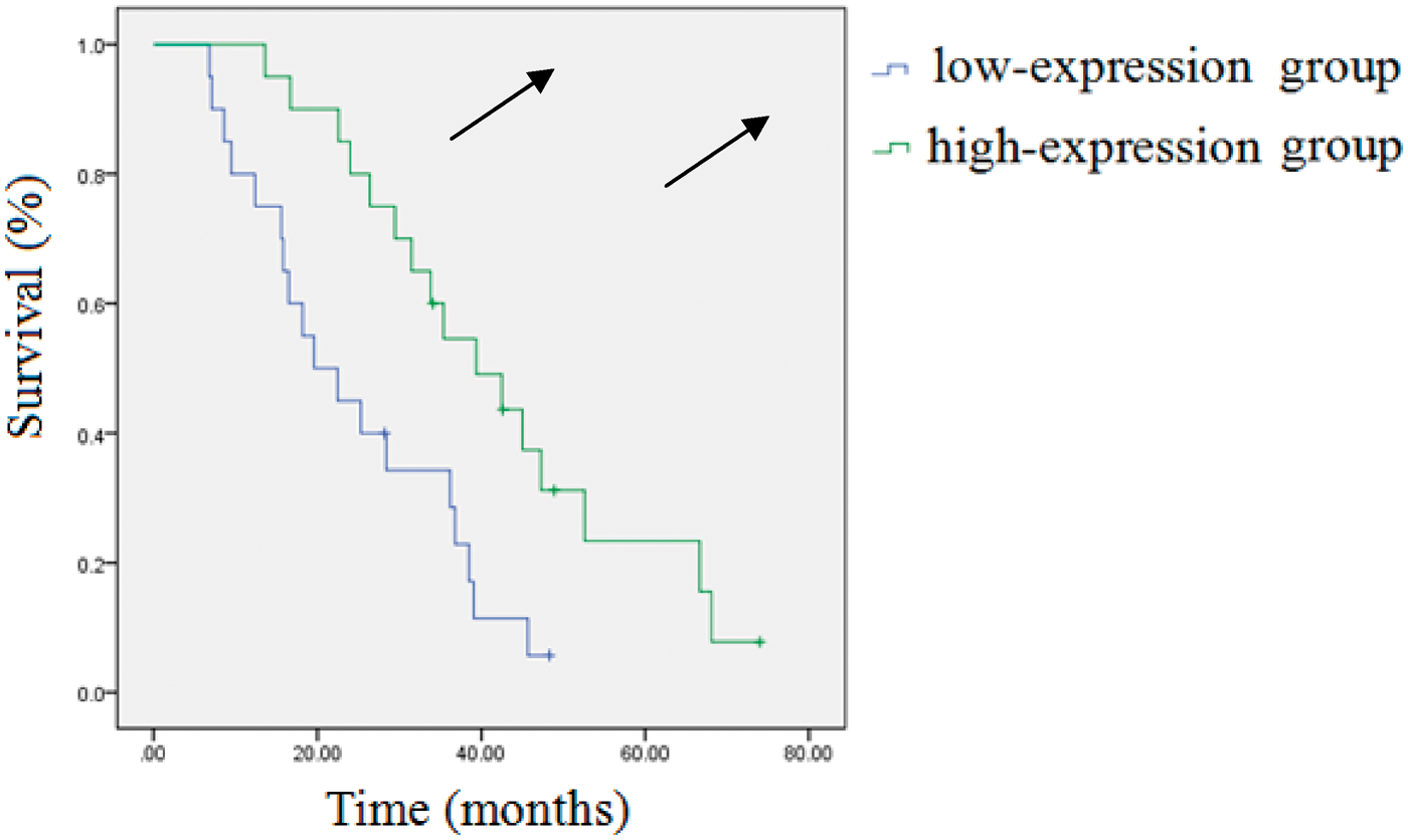
Discussion
MicroRNA is a type of endogenous noncoding small RNA that binds completely or partially to the mRNA of the target gene. miRNA degrades the target mRNA or inhibits translation, and regulates gene expression post-transcriptionally.15–17 Ji et al. 18 studied miRNA expression patterns in HCC tissues from 43 HCC cases, and found that 35 tissue samples presented differential miRNA expression when compared with paracancerous tissue samples. These findings suggest that miRNAs may play a role in promoting or inhibiting tumour development.19–22 Tang et al. 23 investigated miRNA expression in adipose stromal cells and found that miR-22 played an important role in regulating the development of these cells. miR-22 was aberrantly expressed in many tumour tissues, including breast cancer, colorectal cancer and cervical carcinoma.24–27 These findings suggest that miR-22 plays a specific role in different tumours; however, hitherto the effect of miR-22 has not been investigated in HCC.
In the present study, miR-22 expression in 192 HCC tissue samples was detected using qPCR and was found to be significantly downregulated in these samples compared with control (paracancerous) tissue samples. Additionally, miR-22 expression in the high tumour differentiation group was higher than that in the group with low differentiation. Expression was lower in the group with lymphatic metastasis than in the group with nonlymphatic metastasis, suggesting that miR-22 may play a role in regulating HCC infiltration and metastasis.
Furthermore, miRanda, mirBase, TargetScan and PicTar softwares have been used to perform bioinformatic analysis, and it has been postulated that ezrin may be one of the targets of miR-22 in HCC tissue (Y Zhang J Zhao and Y Pan, unpublished results). Ezrin is a member of the ezrin–radixin–moesin (ERM) family that is mainly involved in the link between the cytoskeleton and the plasma membrane, suggesting that ezrin is associated with HCC infiltration and metastasis.28–30 Nowak et al. 31 detected ezrin expression in four human colon adenocarcinoma cell lines that were characterized by different metastatic abilities, and suggested that the highest ezrin expression occurs in cells with the strongest invasive ability. Li et al. 32 analysed the correlation between ezrin expression and pathological characteristics of gastric cancer. They suggested that ezrin expression was associated with age, tumour size, depth of infiltration, tumour stage, lymphatic metastasis and 5-year survival rate, and that ezrin expression can be used for the prediction of gastric cancer prognosis. Wang et al. 10 reported that miR-183-transfected lung cancer cells showed downregulation of ezrin expression, and the luciferase assay demonstrated that ezrin was the target gene of miR-183. In the present study, high ezrin expression was detected in HCC tissue. Furthermore, miR-22 expression in HCC tissue that expressed ezrin at a high level was significantly lower than that in HCC tissue with low ezrin expression, indicating that ezrin expression is negatively related to miR-22 expression. These findings indicate that miR-22 in HCC may regulate ezrin expression.
Studies have used miRNA as a molecular marker for tumour diagnosis and prognosis. Li et al. 32 established an early warning model that included seven miRNAs (miR-10 b, miR-21, miR-223, miR-338, let-7 a, miR-30 a-5 p and miR-126); this model can be used to predict the total survival rate and recurrence-free survival rate of gastric cancer patients. Patel et al. 33 found that aberrant expression of miR-22 in breast cancer and serum was associated with prognosis. The present study analysed the relationship between miR-22 expression and 5-year survival rate, and found that the prognosis of the high miR-22 expression group was better than that of the low miR-22 expression group. This suggests that miR-22 may be used as a biomarker for HCC prognosis.
In conclusion, we found that miR-22 expression in HCC tissue was significantly higher than that in paracancerous tissue, and was associated with the degree of differentiation of the tumour, lymphatic metastasis and prognosis. Ezrin may be one of targets of miR-22. Further exploration of the biological effects and molecular mechanisms of miR-22 might lead to the identification of a novel therapeutic target for HCC.
Footnotes
Declaration of conflicting interest
The authors declare that there are no conflicts of interest.
Funding
This research received no specific grant from any funding agency in the public, commercial or not-for-profit sectors.