
Abstract
Objectives
To investigate pigment epithelium-derived factor (PEDF) mRNA and protein levels in condyloma acuminatum, and their relationship with angiogenesis and keratinocyte proliferation.
Methods
Lesions from male patients with condyloma acuminatum and skin from healthy male (control) subjects were collected. Levels of PEDF protein and its corresponding mRNA (SERPINF1) were determined via Western blotting and reverse transcription–polymerase chain reaction, respectively. Immunohistochemical staining for Ki-67 and CD34 was performed to calculate keratinocyte proliferation index (PI) and microvessel density (MVD), respectively.
Results
Levels of both PEDF protein and SERPINF1 mRNA were significantly lower in lesions from patients with condyloma acuminatum (n = 30) than in skin from healthy control subjects (n = 30). There were significant negative correlations between PEDF levels and both PI and MVD.
Conclusions
The reduction in PEDF levels in condyloma acuminatum was associated with an increase in angiogenesis and cell proliferation. PEDF may be involved in the pathogenesis of condyloma acuminatum.
Introduction
Condyloma acuminatum, also known as genital warts, is a benign hyperplasia of the skin and mucous membrane caused by human papilloma virus, and is one of the most common sexually transmitted diseases. 1 It is characterized by several histopathological features including abnormally thickened epithelium, hyperplasia of cells in the spinous and basal layers, and proliferation and congestive expansion of dermal capillaries. 1
Pigment epithelium-derived factor (PEDF) is a 50-kDa endogenous soluble secretory protein of the serine proteinase inhibitor family, encoded by the SERPINF1 gene. 2 It is ubiquitous in humans and is functionally involved in biological processes such as antiangiogenesis, endothelial cell migration and inhibition of differentiation of tumour and neural cells.3–6 There are few studies regarding PEDF expression in the skin,7,8 and none in condyloma acuminatum. The aim of the present study, therefore, was to investigate SERPINF1 mRNA and PEGF protein levels in condyloma acuminatum and in healthy skin, using reverse transcription–polymerase chain reaction (RT–PCR) and Western blotting, respectively. In addition, the relationship between PEDF levels, proliferation index (PI) and microvessel density was examined.
Materials and methods
Study population
The study included male patients with condyloma acuminatum attending the Department of Dermatology, Qilu Hospital, Shandong University, Jinan, China, between September 2009 and September 2010. Inclusion criteria were: (i) first occurrence of histologically confirmed condyloma acuminatum; (ii) no previous treatment; (iii) absence of systemic disease. Healthy age-matched male volunteers undergoing circumcision at the Department of Urology, Qilu Hospital were recruited as controls.
The study was approved by the Ethics Committee of Qilu Hospital, Shandong University, Jinan, China, and conformed to the Declaration of Helsinki. All study participants provided written informed consent.
Tissue samples
Samples were obtained from condyloma acuminatum lesions in patients, and a similar anatomical site in control subjects. Skin was disinfected and 2% lidocaine was used for topical anaesthesia, then a sample of skin measuring 0.5 cm × 0.5 cm was removed using scissors. Samples for Western blotting and RT–PCR were frozen at −80°C until use. Tissue for immunohistochemical staining was fixed in 10% formaldehyde, embedded in paraffin wax and cut into 3-µm serial sections.
Western blotting
Tissue samples (20 mg) were homogenized with 100 µl of lysis buffer (Beyotime, Shanghai, China), centrifuged at 3000
RT–PCR
Sequences of primers used for reverse transcription–polymerase chain reaction analysis of SERPINF1 (pigment epithelium-derived factor) and ACTB (β-actin.

Immunohistochemistry
Following an antigen retrieval step (Tris–EDTA buffer, ethylenediamine tetra-acetic acid, pH 9.0; ZSGB-Bio, Beijing, China) at 96°C for 15 min, tissue sections were incubated with mouse antihuman CD34 or mouse antihuman Ki-67 (both ready to use; ZSGB-Bio) for 1 h at 37°C. Sections were washed three times at room temperature (2 min each wash) with 0.01 M phosphate buffered saline (PBS; pH 7.2), incubated with horseradish peroxidase-conjugated goat antimouse IgG for 15 min at 37°C and washed again, as before. Immunoreactive staining was visualized directly using an EnVision® + System-HRP (DAB) kit (Dako, Glostrup, Denmark) according to the manufacturer's instructions, then examined via light microscopy.
The number of Ki-67-positive cells from 10 randomly selected views under a × 200 objective was recorded, and the PI was calculated as the number of stained cells/100 cells × 100%. 9 CD34-positive vascular endothelial cells were counted in three randomly selected fields (×200 magnification); microvessel density (MVD) was defined as the mean number of microvascular cells per mm 2 . 10
Statistical analyses
Data were expressed as mean ± SD; between-group comparisons were made using Student's t-test. Correlations between PEDF levels and PI or MVD were analysed using Spearman's rank correlation coefficient. Statistical analyses were performed with SPSS® software, version 16.0 (SPSS Inc, Chicago, IL, USA) for Windows®. A P-value of <0.05 was considered statistically significant.
Results
The study included skin samples from 30 male patients with condyloma acuminatum (mean age 29.7 ± 10.1 years, range 19–47 years; disease course 1 week–2 months) and thirty age-matched male control subjects (mean age 27.9 ± 4.6 years, range 17–40 years).
Levels of pigment epithelium-derived factor (PEDF) protein and its corresponding mRNA (SERPINF1), proliferation index (PI) and microvessel density (MVD) in lesions from male patients with condyloma acuminatum and skin samples from matched healthy male control subject.
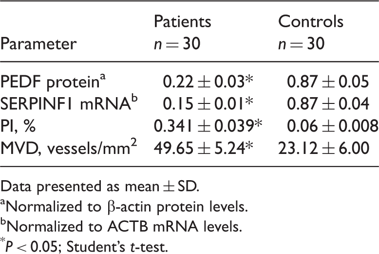
Data presented as mean ± SD.
Normalized to β-actin protein levels.
Normalized to ACTB mRNA levels.
P < 0.05; Student's t-test.
Discussion
Pigment derived-epithelial factor was originally isolated from retinal pigment epithelial cell cultures and was considered to have a potentially differentiative ability in human retinal cells. 11 PEDF is widely expressed and possesses multiple biological functions. Several evolutionarily conserved PEDF-specific protein sequences may be responsible for the antiangiogenic effects of PEDF, 12 supported by the finding that PEDF is a more potent inhibitor of angiogenesis than angiostatin and endostatin. 13 Studies have reported absent or abnormally low PEDF levels in prostate, pancreatic, nonsmall cell lung, Wilms' tumour, malignant melanoma and ovarian cancer cells and cell lines, and a potential correlation with increased invasion and poor prognosis.14–19 Taken together, these findings suggest that PEDF is able to delay the process of cell proliferation and tumour development via an antiangiogenic effect.
In situ hybridization and tissue immunofluorescence have been used to demonstrate the expression of PEDF in normal skin. 20 In addition, PEDF was expressed in two cell lines derived from malignant melanoma, and inhibited the development of melanoma by inducing apoptosis of vascular endothelial cells. 21 Others have shown that PEDF expression was lost during malignant progression of human melanoma. 22 Studies regarding PEDF in inflammatory skin diseases have so far been limited to psoriasis.7,8 Both PEDF mRNA and protein were found to be present at lower levels in condyloma acuminatum lesions than in normal controls, in the present study.
The process of angiogenesis is under the control of both stimulating and inhibiting factors, with vascular epidermal growth factor (VEGF) and PEDF known to be the most potent stimulator and inhibitor of angiogenesis, respectively. 23 The negative correlation between MVD and PEDF levels in the present study further confirms the antiangiogenic role of PEDF. It is thought that PEDF may interrupt the protein kinase B (Akt)/nuclear factor κB (NFκB) signalling pathway by preventing the phosphorylation of the VEGF receptor Akt, thereby inhibiting angiogenesis. 24 PEDF levels have been shown to increase with apoptosis in vascular epidermal cells, 25 and PEDF promoted apoptosis and inhibited VEGF expression in osteosarcoma-derived MG63 cells. 26 It is possible that abnormally low levels of PEDF in epidermal basal layers and echinoderm layer cells may attenuate its antiangiogenic ability, and inhibit abnormal cell proliferation and vascular proliferation in condyloma acuminatum.
The present study revealed a negative correlation between PEDF levels and PI, indicating that PEDF can inhibit cell proliferation and migration. PEDF may inhibit cell proliferation in epidermal basal layers and echinoderm layers by blocking the S to G2/M cell-cycle transition and by inducing apoptosis through the Fas/Fas1 pathway. 27 The level of PEDF was decreased in condyloma acuminatum lesions in the present study, directly or indirectly attenuating its ability to inhibit cell proliferation, and resulting in the abnormal proliferation and thickening of epidermal basal layers and echinoderm layers which are characteristic of condyloma acuminatum.
The authors acknowledge that this study had some limitations. Further research needs to be undertaken with a larger sample size. In addition, further experiments involving the cell lines, and studies undertaken in vivo, are also required.
In conclusion, the present study shows that levels of PEDF protein and mRNA are significantly lower in condyloma acuminatum lesions than in normal control tissue, in men. This reduction in PEDF levels was associated with an increase in angiogenesis and cell proliferation, indicating that PEDF may be involved in the pathogenesis of condyloma acuminatum.
Footnotes
Funding
This study was funded by the Natural Science Foundation of Shandong Province (ZR2010HM018).
Declaration of conflicting interest
The Authors declare that there are no conflicts of interest.