
Abstract
Objectives
Lysophosphatidic acid (LPA) is a bioactive lipid mediator involved in tumour progression, cell invasion and metastasis. The mechanism of action of LPA in the invasive and metastatic capacity of human hepatocellular carcinoma (HCC) is not fully understood. This study investigated the effects of LPA on HCC cell invasion and induction of matrix metalloproteinase (MMP) −2 and −9.
Methods
LPA receptor levels in HCC cell lines were detected by Western blot analysis; HCC cell invasion was analysed by the Transwell® system. The LPA receptor blocker Ki16425 was used to determine whether HCC cell invasion was LPA dependent. Expression of the MMP2 and MMP9 genes in HCC cells was determined by real-time quantitative reverse transcription–polymerase chain reaction (qPCR).
Results
LPA increased HCC cell invasion, which was LPA-receptor dependent. Real-time RT–qPCR showed that LPA increased MMP9, but not MMP2, expression in HCC cells. Pharmacological inhibition of LPA receptors with Ki16452 significantly attenuated LPA-induced HCC cell invasion.
Conclusions
HCC invasiveness is facilitated by LPA and may be relevant to tumour progression. Thus, LPA receptors may be a potential therapeutic target for HCC.
Keywords
Introduction
Hepatocellular carcinoma (HCC) is one of the most common cancers in the world, 1 with a higher incidence in China than other countries. 1 The early invasion of blood vessels by HCC cells, together with intrahepatic extension and later extrahepatic metastasis, results in poor rates of survival. 2 Tumour invasion is a complex biological process that involves the loss of cell–cell contact, followed by detachment from the primary tumour, active cell migration and invasion, and infiltration the surrounding tissue.3,4 Thus, understanding the processes involved in the development of HCC invasion and metastasis is important to improve future treatment strategies and prognosis.
Lysophosphatidic acid (LPA) is a product of phospholipid metabolism. 5 LPA plays an important role in de novo lipid synthesis and acts as a bioactive lipid mediator in diverse cellular processes that include smooth-muscle contraction6,7 and cytoskeletal reorganization.8,9 LPA also increases the proliferation, migration and invasion of cancer cells by activating cell surface G protein-coupled receptors (GPCRs),10–12 and can act as an important autocrine or paracrine mediator. Exogenous LPA mediates downstream biological signalling through binding to cell-surface GPCRs called LPA receptors (LAPRs).13,14 Six subtypes of LPARs (LPAR1–6) have been identified and there is evidence for three additional LPARs.15,16 The regulation of LPAR genes and the role of LAPRs in HCC progression has yet to be established.
Data have shown that LPA enhances the invasion potential of human HCC, 17 colon cancer 18 and ovarian cancer 19 cells. In HCC, cancer-cell invasion is associated with extensive remodelling of the extracellular matrix, angiogenesis and hepatocyte injury. 20 Matrix metalloproteinases (MMPs), a large family of structurally related zinc-endopeptidases that degrade most of the components of extracellular matrix, are key mediators of these processes. 21 Among MMPs, MMP-9 (gelatinase B) has been postulated to have a critical role in HCC cell invasion and metastasis. 22
A study reported that LPA enhances the migration, invasion and adhesion of HCC cells, as well as upregulating MMP-9; 17 however, the relevant mechanism involved remains unclear. On this basis, and using previously research 17 as a methodological model, the present study investigated whether the effects of LPA on HCC cell invasion and MMP2 or MMP9 gene expression in HCC cells were LPAR dependent, using the LPAR blocker, Ki16425 (3-(4-[4-([1-(2-chlorophenyl) ethoxy] carbonylamino)-3-methyl-5-isoxazolyl] benzylsulphanyl) propanoic acid) – a type of synthesized isoxazole derivative that antagonizes LPA binding, particularly to LPAR1 and LPAR3 (rank order of antagonizing affinity of Ki16425, LPA1 ≥ LPA3 ≫ LPA2). 23
Materials and methods
Culture of HCC Cell Lines
The HCC SMMC-7721 and HepG2 cell lines were obtained from the American Type Tissue Collection (Manassas, VA, USA) and cultured in Dulbecco’s modified Eagle’s medium (DMEM; Invitrogen, Carlsbad, CA, USA) supplemented with 10% fetal bovine serum (FBS, GIBCO® Cell Culture; subsidiary of Invitrogen), 100 IU/ml penicillin and 100 µg/ml streptomycin. Cells were incubated at 37°C with humidified air containing 5% CO2.
Western Blotting Analysis
Expression of endogenous LPAR genes in HCC cells (1 × 10⁶ cells/ml) was determined by Western blotting analysis. SMMC-7721 and HepG2 cell lysates were prepared using cell lysis buffer: 20 mM Tris-Cl (pH 7.5), 150 mM NaCl, 1 mM ethylenediaminetetra-acetic acid, 1 mM ethylene glycol tetra-acetic acid, 1% Triton X™-100 (Sigma-Aldrich, St Louis, MO, USA), 2.5 mM sodium pyrophosphate, 1 mM β–glycerolphosphate, 1 mM Na3VO4 and 1 µg/ml leupeptin) with 1 mM phenylmethylsulphonyl fluoride. The concentration of the extracted protein was determined using the Pierce BCA Protein Assay Kit (Thermo Fisher Scientific, Rockford, IL, USA). Samples were separated using a 10% sodium dodecyl sulphate–polyacrylamide gel electrophoresis (SDS–PAGE) gel; proteins were then transferred to nitrocellulose membranes. Membranes were initially blocked with 5% nonfat dry milk in 0.01 mM Tris-buffered saline (TBS, pH 7.4) for 2 h at room temperature, then incubated with a 1 : 1000 dilution of primary antibodies (anti-LPAR1, anti-LPAR2 and anti-LPAR3 [Cayman Chemical, Ann Arbor, MI, USA]) overnight at 4°C. The antibody for glyceraldehyde 3-phosphate dehydrogenase (GAPDH; Santa Cruz Biotechnology, Santa Cruz, CA, USA) was used as the internal control. Membranes were then washed three times with 0.01 mM TBS in 0.1% Tween-20 (TBST) and incubated with a 1 : 200 dilution of horseradish peroxidase-labelled secondary antibody (Cell Signaling Technology®, Danvers, CA, USA) for 1 h at room temperature. The membranes were washed a further three times with TBST and immunoreactive bands were detected by enhanced chemiluminescence detection reagents (Amersham Biosciences, Piscataway, NJ), according to the manufacturer’s instructions.
Cell Invasion Assay
Cell invasion was detected using Transwell® chambers (Corning Costar Corporation, Cambridge, MA, USA). Cultured SMMC7721 cells, at a density of 3 × 10⁴ cells/400 µl of serum-free DMEM, were placed in the 1 : 10 diluted Matrigel®-coated (8-µm pore) upper chamber (BD Biosciences, San Jose, CA, USA). Cells were serum starved for 24 h before the lower chamber was filled with 1 ml of DMEM containing 5 µM LPA or PBS (Sigma-Aldrich), then cultured for 24 h at 37°C. To determine the role of LPARs in cell invasion, cells were serum starved for 24 h, pretreated with 10 µM of the LPAR blocker Ki16425 (Cayman Chemical) for 1 h at 37°C, then stimulated with 5 µM LPA for 24 h at 37°C. A cotton swab was used to remove the cells on the upper surface of the filters, which were then fixed and stained with 0.5% crystal violet solution (Sangon, Shanghai, China). The degree of cell invasion was visualized by light microscopy at magnification of ×200; three to five fields on each slide were randomly selected under a light microscope and cells adhering to the under surface of the filter were counted. Three independent experiments were conducted.
Real-time qRT–PCR Analysis
Total RNA was isolated from 1 × 10⁶ cells per sample, treated as described above, using a homogenizer and TRIzolTM reagent (Invitrogen), according to the manufacturer’s instructions. RNA extraction and reverse transcription (RT) for cDNA synthesis were carried out, as described previously. 24
Specific products of human MMP9 and MMP2 gene expression were amplified by real-time quantitative polymerase chain reaction (qPCR) using the SYBR® Green Master Mix kit (Applied Biosystems, Foster City, CA, USA) and the ABI Prism® 7000 Real-Time PCR System (Applied Biosystems), according to the manufacturer’s instructions. Specific primers for the MMP9 and MMP2 genes were as follows: MMP9, 5′-GGACGGCAATGCTGATG-3′ (forward), and 5′-CAGGGCGAGGACCATAGA-3′ (reverse); MMP2 5′-TCA CATACA GGATCATTG GCTAC-3′ (forward) and 5′ -GCC AGG AGT CCG TCC TTA-3′ (reverse); GAPDH, 5′-TAAGTATGACTCCACCCACG-3′ (forward) and 5′-CTAGCACCTTCCCAACTA-3′ (reverse) was used as an internal control. The reaction conditions were: preliminary denaturation at 95°C for 10 min; followed by 40 cycles of denaturation at 95°C for 15 s, annealing at 56°C for 30 s, elongation at 72°C for 30 s followed by a final elongation step at 72°C for 5 min. PCR products were separated on 12% agarose gels and visualized using ethidium bromide staining and ultraviolet light.
The threshold cycle numbers (CT) were transformed using the Δ C T comparative method. Each sample was performed in triplicate. Gene-specific expression values were normalized to expression values of GAPDH (internal control) within each sample. The amount of target, normalized to an endogenous control and relative to a calibrator, was determined by the comparative CT method (ΔΔ C T ).
Statistical analyses
Statistical analyses were performed using the SPSS® software package, version 13.0 (SPSS Inc., Chicago, IL, USA) for Windows®. Quantitative data were presented as mean ± SD. Comparisons between two groups were conducted by Student’s t-test, where appropriate; multiple-group comparisons were conducted by one-way analysis of variance. A P value <0.05 was considered statistically significant.
Results
All three LPA receptor subtypes (LPAR1, LPAR2 and LPAR3) were present in SMMC-7721 cell lines; however, no detectable protein bands were observed in the HepG2 cell lines (Figure 1). Thus, the SMMC-7721 cell line was used for the remaining experiments.
Representative protein bands from Western blot analysis of lysophosphatidic acid receptor (LPA)1, LPA2 and LPA3 protein levels in lysates from the human hepatocellular carcinoma cell lines, SMMC-7721 and HepG2. All three subtypes were present in the SMMC-7721 cell line. No obvious protein bands were observed in the HepG2 cell line. GAPDH, glyceraldehyde 3-phosphate dehydrogenase (internal control).
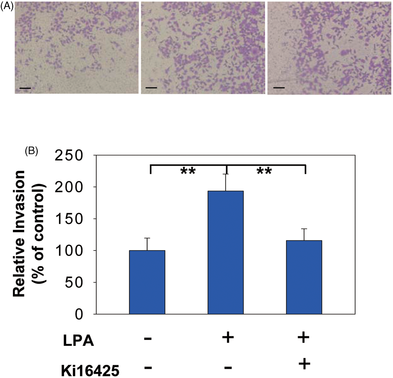
The invasion capacity of SMMC-7721 cells was significantly increased in response to treatment with 5 µM LPA compared with the control group (P < 0.01) (Figure 2). This effect was completely and significantly inhibited by the pharmacological inhibitor of LPARs Ki16425 (P < 0.05; Figure 3). LPA also significantly induced expression of the MMP9 gene (P < 0.05), but not the MMP2 gene, in SMMC-7721 cells (Figure 4). LPA-induced MMP9 expression was dramatically inhibited by Ki16425 (P < 0.05) (Figure 4).
Effect of lysophosphatidic acid (LPA; 5 µM) on invasion potential of the human hepatocellular carcinoma cell line SMMC-7721, detected using a Corning® Transwell® chambers system. Cells were serum starved for 24 h before being treated with 5 µM LPA or phosphate-buffered saline (0 µM; control) for 24 h at 37°C. Relative percentage of invasive cells was expressed as no. of LPA-treated invasive cells/no. of control-treated invasive cells × 100. Results presented as mean ± SD of three independent experiments with similar results. **P < 0.01 versus control; one-way analysis of variance. Effect of pretreatment with lysophosphatidic acid (LPA) receptor blocker (Ki16452) on invasion potential of the human hepatocellular carcinoma cell line SMMC-7721, detected using a Corning® Transwell® chambers system. Cells were serum starved for 24 h before being treated with 5 µM LPA or phosphate-buffered saline (0 µM; control) for 24 h at 37°C, or were pretreated with 10 µM Ki16425 for 1 h at 37°C then stimulated with 5 µM LPA for 24 h at 37°C. (A) Representative photomicrographs (scale bars, 10µM) of invading SMMC-7721 cells: control-treated cells, left panel; stimulated with LPA 5 µM, middle panel; pretreated with Ki16452 LPA stimulation, right panel. (B) Quantification of relative invasion of SMMC-7721 cells treated as described above. Results presented as mean ± SD of three independent experiments, carried out in triplicate. **P < 0.01 for LPA-treated group versus controls and Ki16452-pretreated group; one-way analysis of variance. Effect of lysophosphatidic acid (LPA) on expression of matrix metalloproteinase −2 and −9 (MMP2 and MMP9) genes, detected by quantitative real-time polymerase chain reaction (PCR), in the human hepatocellular carcinoma cell line, SMMC-7721. SMMC-7721 cells were serum starved for 24 h before being treated with 5 µM LPA or phosphate-buffered saline (PBS; 0 µM; control) for 24 h at 37°C, or were pretreated with 10 µM Ki16425 for 1 h at 37°C then stimulated with 5 µM LPA for 24 h at 37°C. (A) Expression of MMP2 and MMP9 genes in SMMC-7721 cells relative to glyceraldehyde 3-phosphate dehydrogenase (GAPDH) housekeeping gene; (B) Representative 12% agarose gels showing MMP-2 and MMP-9 mRNA levels. Results are mean ± SD of three independent experiments, carried out in triplicate. *P < 0.05 for LPA-treated group versus control and Ki16452-pretreated groups; one-way analysis of variance.

Discussion
Metastasis is an important feature of malignant tumours and is a direct cause of mortality. Metastasis is a multistep process that includes detachment of cancer cells from the primary tumour, migration, adhesion and invasion of cancer cells into the blood or lymphatic vessels, extravasation and, finally, interactions with the target tissue and the formation of metastastic foci in distant organs.3,25 Although encouraging progress has been made in the treatment of primary tumours, metastases still affect prognosis for patients with malignancies, with an even poorer treatment effect seen in those with advanced disease. 26
Aberrant expression of LPA and LPARs has been shown in ovarian cancer, with high levels found predominantly in malignant ascites and plasma from patients. 27 However, few studies have investigated the effects of LPA in HCC. The present study showed that LPA significantly induced HCC cell invasion, which is consistent with reports that LPA promotes the invasion of human ovarian 19 and colon 18 cancers. This effect was completely attenuated by the pharmacological inhibition of LPARs with Ki16452, suggesting that LPARs are involved in LPA-induced cell invasion.
Elevated MMP-9 levels in HCC correlate with increased tumour recurrence or metastasis after resection. 28 LPA was previously shown to upregulate MMP-9 in HCC cells, 17 which is supported by our study. Similarly, increased MMP9 gene expression was inhibited by the LPAR antagonist, Ki16452, whereas MMP2 gene expression was not significantly changed after LPA treatment. Upregulation of MMP-9 in response to LPA might be more sensitive than upregulation of MMP-2 in SMMC-7221 cells, suggesting that MMP-9 might be predominant in LPA-induced HCC cell invasion.
Our study provided preliminarily insights into the mechanisms by which LPA might augment HCC cell invasion. LPA activates multiple intracellular signalling pathways including the mitogen-activated protein (MAP) kinase (MAPK) and phosphatidylinositol 3-kinase (PI3K)/Akt pathways, promoting growth factors and protease production, and induction of tumour cell invasion. 29 LPAR-mediated downstream molecular signalling pathways have yet to be fully investigated, however. LPA upregulates the hypoxia-inducible factor-1α subunit by activating PI3K and MAPK signalling in SK-Hep1 HCC cells, 28 and MMP9 expression is induced through activation of extracellular signal-regulated kinase (ERK) and PI-3K/Akt pathways. 30 Simon et al. 31 demonstrated that p38 MAPK is more critical than ERK for heregulin-β1-induced MMP-9 induction in breast cancer cells, which is consistent with the present study, where LPA induced MMP-9 production in HCC cells.
Because of the important biological activities of Ki16425, LPA antagonists have attracted considerable attention and numerous small molecules having LPA antagonistic activity have been reported. Ki16425 is a small-molecule LPA antagonist with oral activity and high selectivity towards LPA1–3, 23 and has been used as a standard compound for evaluating the potency of LPA antagonists. The present study showed that Ki16425 blocked LPA-induced invasiveness of HCC, suggesting that Ki16425 (as a LPAR antagonist) may have potential as a therapeutic agent for HCC patients to prevent tumour metastasis. Preclinical experiments with Ki16425 drugs are ongoing.
The present study showed that HCC invasiveness is facilitated by LPA and inhibited by the LPAR blocker Ki16452. LPARs may, therefore, be a potential therapeutic target in HCC. Accordingly, analysis of the mechanism of cancer cell invasion might lead to new strategies to prevent HCC progression. Although cell invasion promoted by LPA was shown to play a critical role in HCC cell progression, further investigations into the mechanisms of the LPAR-mediated downstream molecular signalling pathway need to be conducted.
Footnotes
Declaration of Conflicting Interest
The authors declare that there is no conflict of interest.
Funding
This research received no specific grant from any funding agency in the public, commercial, or not-for-profit sectors.