
Abstract

PB01-B01
The effects of an iron chelator, deferasirox, on the hemorrhagic cellular damage
1Mol. Pharmacol., Dept. of Biofunct. Eval., Gifu Pharmaceutical University, Gifu, Japan
Abstract
Objectives
After hemorrhagic stroke, leakage blood has toxicity to normal tissue contains blood brain-barrier and neuronal cells as secondary brain injury. Especially, hemoglobin and iron which are major factors in blood components play essential roles in cellular damage mechanism via inducing oxidative stress (1). Previous studies showed that deferoxamine, an iron chelator which is approved in chronic iron overload at transfusion had protective effect on intracranial hemorrhage rat model (2). On the other hands, deferasirox which is orally administratable drug had protective effects on ischemic stroke mouse model (3). However, there is no report to investigate whether deferasirox has any effects on hemorrhagic stroke. In this study, we investigated the effects of deferasirox on hemorrhagic stroke model by using both neuronal and endothelial cells.
Methods
Human brain microvascular endothelial cells (HBMVECs) were cultured to reach confluent, and hemorrhagic injury model was established by incubating with hemin (50 µM) which is metabolized materials of hemoglobin. Deferasirox (0.1– 100 µM) was co-incubated with hemin for 1 h or 24 h. After then, we evaluated several parameters that intracellular bivalent iron accumulation, cell viability rate, cell death rate, reactive oxygen species (ROS) production and expression of an apoptosis marker. Moreover, we also used human neuroblastoma cells (SHSY-5Y) with hemin (10 µM) and evaluated the cell death rate for each several hour.
Results
After incubation with hemin for 1 h, intracellular bivalent iron accumulation level in HBMVECs was increased. In addition, hemin exposure for 24 h induced reduction of cell viability, cell death, ROS over-production and increased cleaved caspase-3 up-regulation. Interestingly, these reactions were suppressed by co-treatment with deferasirox at 10 µM. Moreover, deferasirox co-treatment ameliorated the cell death and ROS over-production in neuronal injury model.
Conclusions
The secondary cellular damage caused hematoma after hemorrhagic stroke may be depended on oxidative stress via iron accumulation. These findings indicate that deferasirox has protective effects on both brain endothelial and neuronal cells against hemorrhagic injury through suppressing the iron overload. Therefore, deferasirox orally administration may improve the patient outcome after hemorrhagic stroke.
References
PB01-B02
Unraveling mechanisms of axonal degeneration and endothelial cell damage in intracerebral hemorrhage
1Institute for Experimental and Clinical Pharmacology and Toxicology, University of Luebeck, Germany
2Fraunhofer Research Institution for Marine Biotechnology and Cell Technology, Germany
Abstract
Objectives
It is established that adverse outcomes after intracerebral hemorrhage (ICH) result from irreversible damage to neurons resulting from primary and secondary injury. Secondary injury has been attributed to hemoglobin and its oxidized product hemin from lysed red blood cells. However, our advances in understanding neuronal demise after ICH have not translated into effective therapeutic approaches. There are many possible explanations for the lack of success of current therapeutics at the bedside. One reason may be that they primarily focus on neurons and, more specifically, on neuronal cell bodies. We here hypothesize that the molecular mechanisms underlying cell death and degeneration may be different in different cell types as well as compartments of the cells, such as the axon in comparison to the soma.
Methods
We investigated cell death mechanisms in cultured primary neurons, isolated axons, and primary endothelial cells exposed to hemin. We systematically screened pharmacological inhibitors of different known cell death pathways (apoptosis, necroptosis, ferroptosis, parthanatos) to identify the underlying molecular signaling pathways involved in the different cell types or compartments.
Results
We developed a quantitative method to analyze axonal degeneration in vitro based on deep learning/convolutional networks. We show that different cell death pathways are activated in primary endothelial cells versus primary neurons and that the mechanisms are different between neuronal cell body demise and axonal degeneration.
Conclusions
Our results indicate that different therapeutic approaches addressing the numerous types of brain cells are needed to effectively treat patients with ICH and, potentially, other neurological diseases.
PB01-B03
Nogo-A/PIR-B/TrkB signaling pathway activation inhibits neuronal survival and axonal regeneration after experimental intracerebral hemorrhage in rats
1Department of Neurosurgery & Brain and Nerve Research Laboratory, The First Affiliated Hospital of Soochow University, China
Abstract
Objectives
Intracerebral hemorrhage (ICH) leads to widespread pathological lesions in the brain, especially impacting neuronal survival and axonal regeneration. This study aimed to elucidate whether the Nogo-A (a myelin-related protein)/PIR-B (paired immunoglobulin-like receptor B)/TrkB (tropomyosin receptor kinase B) pathway could exert a regulatory effect in ICH.
Methods
An ICH model was first established in Sprague Dawley rats, followed by different administrations of vehicle, k252a, or NSC 87877. The Morris water maze test was performed to observe ICH-induced cognitive dysfunction in rats.
Results
Rats in the ICH+NSC 87877 group showed better cognitive performance compared with those injected with vehicle or k252a. Neurobehavioral scores were identical. By harvesting brain tissues at different time points after ICH, we detected the expression levels of Nogo-A and PirB with western blot and immunofluorescence and found they were markedly upregulated at 48 h after ICH. TUNEL and Fluoro-Jade B staining showed that NSC 87877 treatment attenuated ICH-induced apoptosis and neuronal death, whereas k252a treatment aggravated these pathological changes. The expression levels of growth-associated protein 43 (GAP43) and neurofilament 200 (NF200) were higher in the ICH+NSC 87877 group compared with the ICH+vehicle group, but were lower in the ICH+k252a group. Finally, we confirmed the protective role of p-TrkB/TrkB in ICH by western blot.
Conclusions
To sum up, our study identified the inhibitory role of the Nogo-A/PirB/TrkB pathway in ICH; however, p-TrkB/TrkB may serve as a potential target for secondary brain injury post-ICH.
PB01-B04
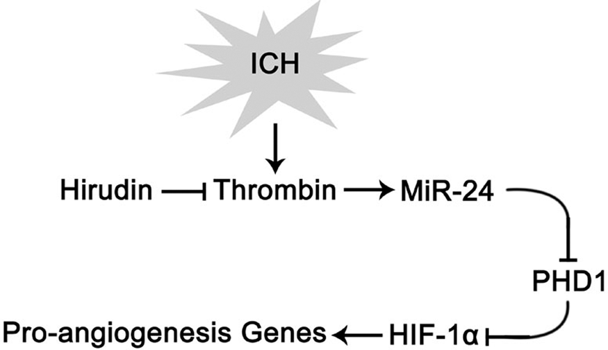
Thrombin-induced miRNA-24-1-5p upregulation promotes angiogenesis by targeting PHD1 in intracerebral hemorrhagic rats
1Institute of Integrative Medicine, Xiangya Hospital, Central South University
Abstract
Objectives
Thrombin is a unique factor to trigger post-intracerebral hemorrhage (ICH) angiogenesis by increasing hypoxia inducible factor-1α (HIF-1α) in protein level. However, HIF-1α mRNA remains no change, which arouses our interest. MicroRNAs (miRNAs) mediate post-transcriptional gene regulation by suppressing protein translation in mammal. Present study aimed to determine miRNAs which might involve in thrombin-induced angiogenesis after ICH by targeting HIF-1α or its upstream molecules—HIF prolyl hydroxylase domain (PHDs).
Methods
In part 1, miRNA array combined with miRNA target prediction was used to identify candidate miRNAs and their target gene in thrombin-infused basal ganglia (BG) compared with the opposite. In the experiment 1 of part 2, rats were randomly divided into sham group, the ICH group, and the ICH+hirudin-treated (thrombin inhibitor) group. In experiment 2, the rats were randomly divided into the sham group, ICH group, ICH+antagomir group, ICH+antagomir-Co group and ICH+vehicle group. MiR-24 (short for miR-24-1-5p) level was determined by qRT-PCR, the protein expression of PHD1 and HIF-1α were detected by western blot. The angiogenesis was evaluated by double-labeling immunofluorescence. Neurological function was evaluated by body weight, modified Neurological Severity Scores as well as corner turn and foot-fault tests.
Results
Part 1 showed that miR-24 significantly increased after thrombin infusion and interplayed with PHD1. Part 2 found that hirudin treated rats showed downregulated miR-24 level, hindered angiogenesis, higher PHD1 expression and lower HIF-1α expression after ICH. Inhibition of miR-24 impeded angiogenesis and neurological recovery after ICH.
Conclusion
The present study suggested that thrombin reduces HIF-1α degradation and initiates angiogenesis by increasing miR-24 targeting PHD1 after ICH.
PB01-B05
Rbfox-1 contributes to CaMK IIα expression and intracerebral hemorrhage-induced secondary brain injury via blocking the binding of microRNA-124 to CaMK IIα mRNA
1Department of Neurosurgery & Brain and Nerve Research Laboratory, The First Affiliated Hospital of Soochow University, China
Abstract
Objectives
Rbfox-1, an RNA-binding protein in neurons, was thought to be associated with many neurological diseases. In this study, we mainly aimed to explore the role of Rbfox-1 in intracerebral hemorrhage (ICH)-induced secondary brain injury and its underlying mechanisms.
Methods
ICH models were established by injecting autologous blood into the brains of adult Sprague Dawley rats, and cultured primary neurons were exposed to oxyhemoglobin to mimic ICH in vitro.
Results
After ICH, the expression of Rbfox-1 in neurons was significantly increased, accompanied by increases in the binding of Rbfox-1 to CaMK IIα mRNA and the protein level of CaMK IIα. In addition, when exposed to exogenous upregulation or downregulation the Rbfox-1, the protein level of CaMK IIα showed a same change trend in the brain tissue, which further suggested CaMK IIα as a downstream target protein of Rbfox-1. And, downregulating Rbfox-1 improved ICH-induced neuronal apoptosis and necrosis. Furthermore, we found that Rbfox-1 promoted the expression of CaMK IIα via blocking the binding of microRNA-124 to CaMK IIα mRNA.
Conclusions
ICH-induced increase in Rbfox-1 blocked the binding of microRNA-124 to CaMK IIα mRNA, which subsequently promoted the expression of CaMK IIα and finally neuronal death and secondary brain injury.
PB01-B06
Siponimod reduces perihemorrhagic edema and improves neurological outcome in experimental intracerebral hemorrhage
1Department of Neurology, University of Erlangen-Nuremberg, Germany
2Department of Radiology, University of Erlangen-Nuremberg, Germany
3Department of Neuroradiology, University of Erlangen-Nuremberg, Germany
Abstract
Objectives
Perihemorrhagic edema (PHE) is a risk factor for poor outcome after intracerebral haemorrhage (PHE). Recent studies suggest that treatment with sphingosine-1-phosphate receptor (S1PR) modulators improve outcome in ICH. In this study we investigate the impact of Siponimod on perihemorrhagic edema and neurological outcome in a mouse model of ICH.
Methods
ICH was induced by bacterial collagenase in C57Bl/6 mice. Animals were assigned to different treatment groups. Siponimod was administered intraperitoneally as a single shot 30 minutes after ICH induction or for three consecutive days. Evolution of PHE, neurological defects and survival were assessed after ICH.
Results
Siponimod significantly reduced PHE 72 hours after ICH, measured by MR-Imaging (p = 0.0212) as well as wet-dry method (p < 0.001). Consecutive treatments with Siponimod for three days significantly attenuated neurological deficits measured by Garcia-Score. Survival at day 10 was significantly improved in mice treated with multiple dosages of Siponimod (p = 0.037).
Conclusions
S1RP modulation via Siponimod provides protection for secondary brain injury after ICH. Siponimod reduced ICH-induced PHE and demonstrated improved neurological outcome after ICH in our model of experimental ICH. Our results suggest that S1PR1 modulation is a viable approach for treatment of PHE in ICH.
PB01-B07
Early rehabilitation inhibits inflammation of the sensorimotor cortex and promotes motor function recovery in intracerebral hemorrhage rats
1Department of Physical Therapy, Niigata University of Health and Welfare
Abstract
The present study examined the effects of early exercise on brain damage and recovery of motor function following intracerebral hemorrhage (ICH) in rats. Under deep anesthesia, animals were placed inside a stereotaxic apparatus and ICH was induced by injection of collagenase in 0.9% saline into the left striatum. Animals were randomly assigned to four groups: no training after ICH (ICH), no training after sham surgery (SHAM), early treadmill exercise after ICH (ICH + ET), or late treadmill exercise after ICH (ICH + LT). The ICH + ET and ICH + LT groups were trained for seven consecutive days starting on day 2 or day 9 after surgery, respectively. Sensorimotor function was assessed by forelimb placing and the horizontal ladder test. At day 16 post-surgery, brains were removed and lesion volume, cortical thickness, neuronal number, dendritic length, and dendritic complexity were analyzed. Expression levels of IL-1b, TGF-b1, and IGF-1 mRNAs in ipsilateral sensorimotor cortex were measured by RT-PCR. The ICH + ET group showed significantly improved sensorimotor function compared to the ICH and ICH + LT groups. In addition, cortical thickness and neuronal number were significantly higher in the ICH + ET group than the ICH and ICH + LT groups. The length and complexity of dendrites were also significantly greater in the ICH + ET group compared to the ICH and ICH + LT groups. Expression of IL-1b mRNA was significantly lower in the ICH + ET group than that in the ICH group. Taken together, these results suggest that early treadmill exercise after ICH promotes recovery of sensorimotor function by preventing neuronal death and subsequent cortical atrophy as well as by preserving dendritic structure. Early exercise may prevent neurodegeneration and functional loss by inhibiting neuroinflammation. This work was supported by a Grant-in-Aid for Scientific Research from Niigata University of Health and Welfare (H30B18) and the Japan Society for the Promotion of Science, Grant-in-Aid for Scientific Research (16K16445). We have no financial relationships to disclose.
PB01-B08
Activation of NLRP3 inflammasome is associated with down-regulation of estrogen receptor alpha in ovariectomized rat intracranial aneurysm model
1Dept. of Neurosurgery, Tokushima University, Japan
Abstract
Background
The rupture of intracranial aneurysms is cause of major subarachnoid hemorrhage. Its prevention requires understanding of its pathogenesis in detail. In the arteries prone to rupture of our newly establishedintracranial aneurysm model subjected to estrogen-deficiency, hemodynamic change and hypertension (HT), estrogen receptors (ERs) were down-regulated, matrix metallopoteinase-9 and the tissue inhibitor of metalloproteinase-2 (MMP9/TIMP2) were imbalanced, and interleukin-1β (IL-1β) was increased. As NLRP3 inflammasome activates the production of caspase1-dependent IL-1β, we focused on its role in the elicitation and rupture of intracranial aneurysms. We hypothesized that NLPR3 is mediated by ERs and associated with vascular vulnerability.
Methods
Ten-week-old female Sprague-Dawley rats were randomized into two groups of 19 rats each. Group 1 was ovariectomized (HT-OVX+), group 2 was intact (HT-OVX−). They were subjected to hemodynamic changes and HT. Sham-operated rats were the control. Human brain microvascular endothelial cells were treated with estradiol,ER-α- or ER-β agonistunder the condition of estrogen deficient.
Results
During 12-week observation, aneurysms ruptured in 47% of the HT-OVX+and 16% of the HT-OVX−rats (p < 0.05). Immunohistochemically, the expression of ERα but not of ERβ was decreased, while the expression of the inflammasome components NLRP3, caspase1, IL-1β, of MMP-9, and the mRNA level of NLRP3, IL-1β and MMP-9 was increased in HT-OVX+rats. In human brain microvascular endothelial cells, the expression of NLRP3 was increased by estrogen-deficiency and abrogated by the ER-α agonist (PPT) but not the ER-β agonist(DPN). These findings suggest that in intracranial aneurysms prone to rupture, the activation of NLRP3, mediated by a decrease in ERα, plays a part.
Conclusion
For a better understanding of the relationship between the rupture of intracranial aneurysms and the activation of inflammasomes under estrogen-deficient conditions, additional studies are underway.
PB01-B09
Study on effectiveness of prothrombin complex concentrate on emergency correction of anticoagulant effect
1Department of Cerebrovascular Medicine and Neurology, National Hospital Organization Kyushu Medical Center, Japan
Abstract
Background and purpose
Prothrombin complex concentrate (Kcentra®: PCC-KC) has been available in the clinical field in Japan since September 2017, but post marketing experience of use has not been reported. Then, in our hospital we investigated the actual aspect of the PCC-KC administration, including undelaying disease, dose, change in PT-INR, hemostatic effect, and incidence of thromboembolism in 17 patients who received the PCC-KC.
Results
There were 17 subjects (12 men, 5 females), aged between 64–95 years old (mean 77 years old). Thirteen patients had severe hemorrhagic complications during warfarin therapy (brain hemorrhage in 10 patients, subdural hemorrhage in 3), 3 patients needed urgent surgery or procedure (femoral neck fracture surgery in 2 patients, thoracic puncture in one) during warfarin therapy, and one had brain hemorrhage during apixaban therapy. In all 16 cases of warfarin therapy, vitamin K (median 10 mg, range 10 −20 mg) was simultaneously administered. PT-INR before administration of PPC-KC was median 2.51 (1.06 to 4.72) and an average of 26.1 IU / kg (10.5 to 35.9 IU / kg) of PCC-KC were administered, and the PT-INR after administration was 1.17 (1.03 to 1.47) (p < 0.01, paired t test). In 4 cases of PT-INR 4 or more and less than 6 before administration (median 4.41, range 4.00 to 4.72) (4-6 group), 35 IU / kg was administered in 3 cases and 25 IU / kg was administered in the other one and the PT-INR decreased to a value of 1.12 (1.07 to 1.26). In the 9 patients with PT-INR 2 or more and less than 4 (median 2.51, range 2.05 to 3.80) (INR 2–4 group), 25 IU / kg was administered in all cases, and the PT-INR decreased to median 1.22 (range 1.09 to 1.47). In three patients, PT-INR before administration was less than PT-INR 2 (1.59, 1.85, and 1.93) (less than INR 2 group), PCC-KC of 19 IU / kg, 15 IU / kg, and 11 IU / kg was administered and they were reduced to 1.17, 1.16, and 1.42, respectively. Expansion of hematoma was seen in a patient with brain hemorrhage in the INR 2–4 group. There was no intraoperative major bleeding in the 3 patients with urgent surgery or procedure. In one patient with brain hemorrhage during apixaban therapy, PCC-KC of 27 IU / kg was administered and the PT-INR before and after administration was 1.06 and 1.03. There was no thromboembolic events.
Conclusions
In patients with PT-INR 2 or higher, PCC-KC administration was performed according to the dosage regimen in most cases, and sufficient reduction of PT-INR was obtained. In patients with IPT-INR less than 2.0, administration of 15 to 19 IU / kg is effective, but administration of 11 IU / kg may be insufficient. It seems difficult to evaluate usefulness of PCC-KC administration in patients treated with apixaban due to small number.
PB01-B10
Leakage sign for intracerebral hemorrhage in relation to the site of hemorrhage
1Dept. of Neurosurgery, Kurume University, Japan
Abstract
Background and Purpose
Recent studies of intracerebral hemorrhage (ICH) treatments have highlighted the need to identify reliable predictors of hematoma expansion. Several studies have suggested that the “spot sign” on computed tomography angiography (CTA) is a sensitive radiological predictor of hematoma expansion in the acute phase. In this study, we evaluated the usefulness of a novel predictive method, called the “leakage sign.”
Methods
We performed CTA for 228 consecutive patients presenting with spontaneous ICH. Two scans were completed: CTA phase and delayed phase (5 min after the CTA phase). By comparing the CTA phase images, we set a region of interest (ROI) with a 10-mm diameter and calculated the Hounsfield units (HU). We defined a positive leakage sign as a >10% increase in HU in the ROI. Additionally, hematoma expansion was determined on plain CT at 24 h in patients who did not undergo emergent surgery.
Results
M:F = 124:104 (mean age 67.6). Positive leakage signs were present in 89 (39%) patients. The most common site of ICH was the putamen (103 patients, 45.1%), followed by the thalamus (50, 21.9%), lobar areas (31, 13.5%), brainstem (20, 8.7%), cerebellum (24, 10.5%). The leakage sign had higher sensitivity (69.2%) and specificity (92.8%) for hematoma expansion in putamen than the other site. By contrast, in cerebellar hemorrhage, the positive rate of leakage sign was low and it was difficult to predict the expansion hematoma. The leakage sign was different in features depending on the site of hemorrhage.
Conclusions
The results indicate that the leakage sign is a useful and sensitive method to predict hematoma expansion. Furthermore, it is necessary to analyze the leakage sign for each site of ICH.
PB01-B11
Perihemorrhagic edema revisiting hematoma volume, location and surface
1Department of Neurology, University of Erlangen-Nuremberg, Germany
2Department of Neuroradiology, University of Erlangen-Nuremberg, Germany
Abstract
Objectives
To determine the influence of intracerebral hemorrhage (ICH) location, volume and hematoma surface on perihemorrhagic edema evolution.
Methods
ICH patients of the prospective UKER-ICH cohort study (NCT03183167) between 2010 and 2013 were analyzed. Hematoma and edema volume during hospital stay were volumetrically assessed, and time course of edema evolution and peak edema correlated to hematoma volume, location and surface to verify strength of parameters on edema evolution.
Results
Overall 300 patients with supratentorial ICH were analyzed. Peak edema showed high correlation with hematoma surface (R2 = 0.864, p < 0.001) rather than with hematoma volumes, irrespective of hematoma location. Smaller hematomas with higher ratio of hematoma-surface-to-volume showed exponentially higher relative edema (R2 = 0.755, p < 0.001). Multivariable logistic regression analysis revealed a cut-off ICH-volume 30 ml beyond which an increase of total mass lesion volume (combined volume of hematoma and edema) was not associated with worse functional outcome; specifically, peak edema was associated with worse functional outcome in ICH<30 ml (OR 2.59[1.55–4.31]; p < 0.001), contrary to ICH ≥30 ml (OR 1.20[0.82–1.75]; p = 0.339). There were no significant differences between patients with lobar versus deep ICH after adjustment for hematoma volumes.
Conclusions
Peak perihemorrhagic edema, though influencing mortality, is not associated with worse functional outcomes in ICH volumes above 30 ml. Although hematoma volume correlates with peak edema extent, hematoma surface is the major parameter for edema evolution. The effect of edema on functional outcome is therefore more pronounced in smaller and irregularly shaped hematomas and these patients may benefit more significantly from edema-modifying therapies.
PB01-B12
Crossed cerebellar tracer uptake on acute-stage123I-iomazenil SPECT imagingpredicts 3-month functional outcome in patients with non-fatal hypertensive putaminal or thalamic hemorrhage
1Department of Neurosurgery, Iwate Medical University, Japan
2Cyclotron Research Center, Iwate Medical University, Japan
Abstract
Objective
To determine whether crossed cerebellar tracer uptake on acute-stage123I-iomazenil (IMZ) single photon emission computed tomography (SPECT) image predicts 3-month functional outcome in patients with non-fatal hypertensive putaminal or thalamic hemorrhage.
Methods
We comprised forty-six patients with non-fatal hypertensive putaminal or thalamic hemorrhage. The SPECT image was scanned from 2 to 7 days after the onset and the images obtained 30 and 180 min after the administration of123I-IMZ were defined as early and late images, respectively. On the SPECT images standardized by statistical parametric mapping software, an anatomical ROI template covering the whole brain was automatically placed. The affected/contralateral cerebral hemisphere ratio (ARcrb) and the contralateral/affected cerebellar hemisphere ratio (ARcbl) based on the side determined by the cerebral lesion. Each patient’s physical function was estimated using the modified Rankin scale (mRS) score 3 months after the onset.
Results
ARcbl on early (ρ = −0.511; p = 0.0003) and late (ρ = −0.714; p < 0.0001) images correlated with the mRS score, although ARcrb on both images did not. Multivariate analysis showed that the only ARcbl in late image was significantly associated with a functional outcome (3-month mRS score ≥3) (p = 0.0212). ARcbl in late images was a good indicator to predict a poor functional outcome (sensitivity, 77%; specificity, 100%; positive- and negative-predictive values, 100% and 83%).
Conclusion
Crossed cerebellar tracer uptake on acute-stage123I-IMZ SPECT images predicts 3-month functional outcome in patients with non-fatal hypertensive putaminal or thalamic hemorrhage.
Referemces
PB01-B13
Hypo-intensity of the drainage vessels in Susceptibility-weighted MR image may relate to lower risk of the intracerebral hemorrhage in patients with intracranial arteriovenous shunt(s)
1Dept. of Neurosurgery, Nagasaki Harbor Medical Center, Nagasaki, Japan
2Dept. of Neurosurgery, Nagasaki University, Nagasaki, Japan Withdrawn
PB01-B14
Role of complement system in SAH-induced hippocampal alterations
1Dept of Neurosurgery, Houston Methodist Hospital
Abstract
Objectives
Overwhelming majority of subarachnoid hemorrhage (SAH) survivors (up to 95%) experience various long-term memory and cognitive abnormalities1 associated with atrophy of the temporomesial area2,3. Comparable changes also observed in mice model of SAH4. However, the mechanisms of these impairments remain unclear. We hypothesize that complement system5,6 may be involved in SAH-induced long term hippocampal damage.
Methods
We explored transcriptome, morphological and functional changes in hippocampus (Hpc) following the SAH induced by perforation of the circle of Willis in mice (male C56BL/6J). Animals without perforation constituted sham group. Four days following SAH animals were anesthetized, transcardially perfused with saline followed by 4% formaldehyde for immunohistochemistry, or non-perfused hippocampi were extracted (for RNA sequencing). In a separate groups of animals long term potentiation (LTP) of population spike (PS) by stimulation of perforant pathway (PP) in dentate gyrus (DG) in vivo and behavioral changes 4 and 30 days after SAH were explored.
Results
Immunohistochemistry revealed significant activation of astro- and microglial cells in CA1 and DG areas (p = 0.035). Hpc neuroinflammation was accompanied by loss of dendritic spines of the DG and CA1 neurons (p < 0.001).
To identify leading processes in the hippocampus following SAH we used RNA next-generation sequencing 4-days after SAH. Functional analysis of the differentially expressed genes between SAH and Sham groups showed upregulation of 11 complement system-related genes. RtqPCR confirmed significant increase in C3, C4 and CfB expression. Quantitative immunofluorescence revealed increase in stratum lacunosum moleculare (SLM), terminal field of PP, levels of C3 by 21% (p = 0.02, n = 6, confirmed by WB) and C4 by 28% (p = 0.04, n = 4).
The immunofluorescent intensity of C3 and C4 staining varied significantly between Hpc layers (p < 0.001) suggesting diverse roles of complement in different hippocampal areas. In sham animals C3 was colocalized with astrocytes. Following SAH, confocal microscopy revealed colocalization of GFAP-positive cells and PSD95 suggesting engulfment of postsynaptic elements by astroglial cells. However, Iba1-positive microglial cells demonstrated colocalization with presynaptic marker synaptophysin after SAH. Observations suggest that mechanisms of dendritic spines pruning may differ from elimination of axonal terminals.
LTP in DG in response to PP stimulation was suppressed 4 days after SAH. These changes were accompanied by behavioral abnormalities: appearance of the anxiety and impairment in working memory.
To assess participation of the complement system in the observed hippocampal alterations we blocked the central element of complement system C3 by intracerebroventricular injection of compstatin during 4 days following the SAH. In animals, which received compstatin loss of dendritic spines was prevented, suppression of LTP and SAH-induced behavioral changes were reversed.
Conclusions
Hippocampal neuroinflammation following the SAH is accompanied by activation of the complement system, which may participate in loss of dendritic spines, suppression of LTP and behavioral disturbances.
References
PB01-B15
Subarachnoid hemorrhage leads to hippocampal atrophy and suppresses oligodendrocyte genes expression
1Dept. of Neurosurgery, Houston Methodist Hospital
2Translational Imaging Center, Houston Methodist Hospital Research Institute
3Center for Biostatistics, Institute for Academic Medicine, Houston Methodist Research Institute
Abstract
Objectives
Forty five percent of subarachnoid hemorrhage (SAH) survivors are unable to continue with their professional activities due to permanent cognitive or emotional disabilities. The SAH is often followed by atrophy of temporomesial area correlating with decreased neurocognitive scores1-3. However, the mechanism of post-SAH hippocampal atrophy remains unclear. We explored changes in hippocampal myelination, volume and expression of oligodendrocyte-related genes expression at 4 days following the SAH in mice.
Methods
We used SAH model of filament perforation of the circle of Willis in anesthetized male C56BL/6J mice. To identify leading biological processes in the hippocampus following SAH we used RNA next-generation sequencing to explore the changes in gene expression at the transcriptional level 4-days after SAH. Under deep anesthesia animals were decapitated, brain removed, and hippocampi extracted. RNA of the whole hippocampus of SAH (n = 4), sham (no perforation, n = 3) group was extracted and differential gene expression analysis was performed (−1.5<fold change>1.5; p < 0.05).
For imaging and immunohistochemistry, deeply anesthetized animals were intracardially perfused with saline followed by 4% PFA. Brains were extracted and postfixed. For MRI brains washed in PBS for 24 hours were placed in tubes filled with 3% PBS agar. The tubes were placed in a bore-mounted coil (Micro MRI Bruker). T2 weighted images were acquired using a TurboRARE_3D sequence. Hippocampal volume was determined by manual segmentation using ITK-SNAP4.
Brain cryo-slices were processed for immunohistochemistry. Quantitative fluorescent immunohistochemistry was used to evaluate intensity of fluorescent staining.
Results
To explore the possible mechanisms of hippocampal atrophy we evaluated cell loss in hippocampal formation. Using Nissl staining we counted number of cells in Hpc CA1 area. Number of cells was comparable in sham and SAH animals (n = 4/group, p>0.05). In agreement with others5, we did not observe fluoro-jade C or activated caspase 3 positive cells in CA1 or DG.
Gene set enrichment analysis (GSEA, Broad Institute) of all genes sequenced revealed a significant (FDR <0.15) depletion in CAHOY_Oligodendrocyte’s gene set with 32 genes out of 100 significantly reduced, suggesting a decrease in oligodendrocyte phenotype. Decrease of immunofluorescent intensity of myelin oligodendrocyte glycoprotein staining (by 33%) and its level (by WB) in hippocampi (by 30%, p < 0.05, n = 3/group) in the animals with SAH is in line with the reduction of oligodendrocyte genes expression.
Hippocampal volume as measured by MRI in mice 4 days after SAH (22.7 ± 1.2 mm3) was significantly (n = 3/group, p < 0.001) diminished compared to sham group (26.3 ± 0.47 mm3) suggesting SAH-induced atrophy.
Conclusions
Our data demonstrate that in agreement with others6 SAH induces hippocampal atrophy, which is reminiscent to such observed in humans. Our data suggest that decrease in hippocampal volume occur prevalently due to decrease in oligodendrocyte gene expression and hence decrease in myelination.
References
PB01-B16
RIP3 mediates early brain injury by inducing necroptosis and promoting inflammation after subarachnoid hemorrhage in rats
1Department of Neurosurgery & Brain and Nerve Research Laboratory, The First Affiliated Hospital of Soochow University, China
Abstract
Objectives
Necroptosis is a regulated form of necrosis that is mediated by a variety of proteins including tumor necrosis factor-α (TNF-α) and receptor-interacting proteins (RIPs). TNF-α, a critical inflammatory molecule, is one of the initiating signals in the necroptosis pathway, and RIP3 acts as a switch that commits the cell to necroptosis. Subarachnoid hemorrhage (SAH) is a common type of hemorrhagic stroke with high mortality and disability rates. RIP3 has been studied in many central nervous system (CNS) diseases, but its role in SAH has not been investigated in depth.
Methods
We used an autologous-blood injection model to study the role of RIP3 in brain injury induced by SAH in rats. Several indexes such as brain edema, loss of blood-brain barrier (BBB) integrity, and behavioral tests of neurological function were used to evaluate brain damage in SAH-injured rats.
Results
We found that the expression of RIP3 was increased in the rat brain after SAH, reaching the highest point 24 h post-injury. We also showed that genetic or pharmacological inhibition of RIP3 or TNF-α reduced the brain damage induced by SAH, whereas overexpression of RIP3 aggravated brain injury and neurological damage. Additionally, we verified the presence of RIP3-mediated necroptosis in an in vitro SAH model of primary cultured neurons treated with conditioned medium from primary microglia activated by oxygen hemoglobin (OxyHb).
Conclusions
Collectively, our findings indicated that RIP3 contributed to brain damage after SAH by inducing necroptosis.
PB01-B17
Relationship between blood cholesterol level and cerebral aneurysm
1Dept. Medical Education, Hamamatsu University School of Medicine, Japan
2Dept. of Pharmacology, Hamamatsu University School of Medicine, Japan
3Dept. of Anesthesiology, Hamamatsu University School of Medicine, Japan
4Dept. of Neurosurgery, Hamamatsu University School of Medicine, Japan
Abstract
Introduction
Subarachnoid hemorrhage (SAH) is a life-threatening type of stroke and can be frequently caused by a ruptured aneurysm of cerebrovascular blood vessels. Although one third of patients could survive with good recovery; one-third will survive with a disability; and one-third will die.
It is well accepted that lowering blood cholesterol level is mandatory in prevention of cerebral circulatory disorder. However, the relationship between cholesterol and cerebral aneurysm is still controversial.
In this study, we elucidate the above relationship by monitoring aneurysm and SAH in 1) aneurysm model of LDL receptor/ Apobec 1 double knock out (LA-/-) mice and that of control mice. 2) Reducing cholesterol intake by administering Cholestyramine, cholesterol lowering cationic resin, to LA -/- aneurysm model mice together with daily food.
Method
Experiments were conducted in accordance with the guidelines of the Institutional Animal Care and Use Committee of Hamamatsu University School of Medicine, Hamamatsu, Japan.
Hashimoto model of animal cerebral aneurysms was performed.
Briefly, left kidney was excised one week before the experiment. Elastase was administered to the subarachnoid space to damage cerebral artery and sustained-release deoxycorticosterone was placed subcutaneously. Drinking water was substituted with 1% salt solution. Three weeks later, the brain tissue was harvested for evaluation of cerebral aneurysm and subarachnoid hemorrhage.
Results
1) lesser amount of cerebral aneurysm and SAH were detected in aneurysm model of LA-/- mice compared to control mice.
2) Increasing trend of SAH was observed in LA-/- mice with cholestyramine administered group.
Conclusion
In this study, we identified the relationship between cholesterol level and subarachnoid hemorrhage. As the next step, whether blood cholesterol is directly related to rupture cerebral artery or influences through enhancement of coagulation system, is still needed to clarify further.
PB01-C01
Osteoprotegerin prevents the growth of intracranial aneurysms promoting collagen biosynthesis and vascular smooth muscle cell proliferation via TGFβ1
1Dept. of Neurosurgery, Kyoto University, Japan
2Dept. of Cinical Innovative Medicine Insitute, Kyoto University, Japan
Abstract
Objectives
The pathogenesis of intracranial aneurysms (IAs) are mainly characterized by reduced extracellular matrix and decreased number of vascular smooth muscle cells (VSMCs) in cerebral vascular walls as response to chronic stimuli of inflammatory cytokines. It is previously reported that osteoprotegerin (OPG) promotes cell proliferation [1] and increases collagen content of VSMCs via TGF-β1 [2]. Here we investigated whether to prevent the development of IAs through the effect of OPG on collagen expression and VSMCs proliferation in experimentally induced IAs in rats.
Methods
IAs were surgically induced in 7-week-old male Sprague Dawley rats. 1 week after the operation, mouse recombinant OPG at 0.125 g/L (37 ng/h) or vehicle (Phosphate buffered salts containing 2% bovine serum albumin) was continuously infused into the lateral ventricle via an osmotic pump from day 0 to day 28. Rats were sacrificed 5 weeks after the 1st operation. Incidence, size, media thickness of induced IAs were measured in the both groups after aneurysm induction. The effect of OPG on collagen (Col1a2 and Col3a1) expression and pSmad2/3 in aneurysmal walls were examined by immunohistochemistry. Expression of Col1a2, Col3a1, TGFβ1 and inflammation-related genes (MMP-2, MMP-9, MCP-1 and ICAM-1) was analyzed by reverse transcription polymerase chain reaction (RT-PCR). The total RNA was extracted from the whole Willis ring. To examine whether OPG treatment (1, 10 or 100ng/mL) upregulates the expression of collagen and promotes the proliferation of VSMCs via TGF-β1, primary cell culture of mouse aorta VSMCs (Passage 3 to 6) were analyzed by RT-PCR and MTS assay with or without SB 431542 (TGFβ-R1 inhibitor, 2 or 20μM).
Results
There were no difference in the incidence rate of IAs between both groups, however, in the OPG treatment group the mean diameter of IAs and the media wall thickness were significantly smaller (42 ± 6.5µm vs 66µm ± 6.1µm, p < 0.05, n = 10, each) and thicker (0.55 ± 0.07 vs 0.34 ± 0.03, p < 0.05, n = 10, each) than in the control group. In the immunohistochemistry, collagen type 1 and 3, and pSmad2/3 were upregulated in the aneurysmal wall in the OPG treatment group. In the RT-PCR study, OPG treatment significantly upregulated the expression of Col1a2, Col3a1 and TGFβ1 genes in the intracranial arteries (p < 0.05, n = 15, each), however, did not alter the expression of MMP-2, −9, MCP-1 and ICAM-1 genes. In vitro study, MTS assay revealed the OPG treatment significantly promoted cellular proliferation of VSMCs and this effect was canceled by TGFβR1 inhibitor.
Conclusions
Our results indicate that OPG has a suppressive effect on IAs development through the activation of collagen biosynthesis and VSMC proliferation via TGFβ1 in aneurysmal walls without altering the expression of inflammation-related genes. OPG may represent a novel therapeutic target of the medical treatment for IAs.
References
PB01-C02
A pilot study to detect intracranial aneurysm rupture using a video tracking system on iPad application
1Department of Anesthesiology, Hamamatsu University School of Medicine
2Department of Pharmacology, Hamamatsu University School of Medicine
3Department of Neurosurgery, Hamamatsu University School of Medicine
Abstract
Objectives
Intracranial aneurysm rupture is a life-threatening event. To research intracranial aneurysm rupture, Hashimoto created a new mouse model. Hashimoto induced intracranial aneurysm using combination of elastase injection into basal cistern and experimental systemic hypertension. Those mice develop intracranial aneurysm almost in one week. The aneurysm growths and ruptures spontaneously in around one to three weeks. It is difficult to detect exact timing of spontaneous aneurysm rapture, so we had to do daily neurological observation.
The purpose of this study is to detect the aneurysm rupture for preventing sudden death in the mice model. We used our developed video tracking system on iPad.
Methods
Experiments were conducted in accordance with the guidelines of the Institutional Animal Care and Use Committee of Hamamatsu University School of Medicine, Hamamatsu, Japan.
Mouse model of intracranial aneurysm rupture: Intracranial aneurysms were induced in C57BL/6J male mice (9 wkold; Japan SLC, Inc., Hamamatsu, Japan). To induce intracranial aneurysm in mice, we combined systemic hypertension with a single elastase injection into the cerebrospinal fluid. Deoxycorticosterone acetate-salt hypertension was used to induce systemic hypertension. A single dose of elastase (35 mU) was injected into the cerebrospinal fluid at the right basal cistern. The definition of aneurysm in this study was as follows: a localized outward bulging of the vascular wall with a diameter greater than the parental artery diameter.
Video tracking system
We developed a video tracking system on iPad application. We put a color ball marker on mouse skin. The application recognizes a color of the ball marker. The marker was painted with fluorescent paint. In the night, the marker illuminated by ultra violet. Video tracking application can recognize the marker although in the night dark surroundings. The application provides mouse movement distance, mouse traces, mouse pictures to us.
Result
We could detect a normal mouse; without intracranial aneurysm induction, movement throughout the day including night time. Mouse moved well in night time compare to day time. Mouse moved periodically.
On the mouse with intracranial aneurysm, its movement reduction observed at the night of day 3 after aneurysm induction (Fig. 1 left). At that day mouse movement in night time was less than daytime. Especially mouse movement decreased, and mouse movement rhythm was lost from around 0:10 of day 4. The mouse was euthanized. Its brain sample showed the occurrence of intracranial aneurysm rupture (Fig. 1 right). Intracranial aneurysm rupture and related deconditioning might happen around the time.

Conclusion
In the pilot study, we could detect the approximate time of spontaneous aneurysm rupture with using video tracking system. Further study is required.
PB01-C03
Cerebral blood flow after bypass with parent artery occlusion for ruptured blister aneurysms of the internal carotid artery
1Department of Neurosurgery, Tohoku University Hospital, Japan
2Department of Neurosurgery, Kohnan Hospital, Japan
3Department of Neurosurgery, Akita University, Japan
4Department of Neurosurgery, Sendai Medical Center, Japan
Abstract
Background
Blister aneurysms of the internal carotid artery (ICA) arise at a nonbranching site of the dorsal wall of the supraclinoid portion. Surgical management for this type of aneurysms is challenging due to their fragile wall with its high rate of intraoperative rupture. In this report, we reviewed patients with ruptured blister aneurysm treated by bypass and trapping to investigate the hemodynamic changes and safety of this surgical strategy.
Methods
We retrospectively reviewed 45 patients with ruptured blister aneurysms. We principally perform high flow bypass using saphenous vein graft as first-line treatment. STA-MCA bypass was selected for the patients with good collateral circulation. After completion of the anastomosis, we performed trapping of the aneurysm. We assessed pre- and postoperative radiological findings and clinical course of these patients.
Results
Forty-three patients were surgically treated, but 2 patients cannot be treated because of clinical deterioration due to preoperative rebleeding. Twenty-six patients were treated immediately after the diagnosis. Elective surgery was scheduled in 17 patients because of delayed diagnosis. Among these 17 cases, 9 cases developed rebleeding while waiting for the surgery, and treated by emergent bypass surgery. We created high flow bypass for 25 patients and STA-MCA bypass for 18 patients, respectively. Among 18 cases treated by STA-MCA bypass, we selected STA-MCA bypass for 2 patients with severe brain swelling, instead of high flow bypass. Five patients manifested symptomatic vasospasm (high flow: 4 patients, STA-MCA: 2 patients), which did not affect final outcomes. Postoperative cerebral blood flow (CBF) was assessed by IMP-SPECT in 17 patients. CBF was transiently decreased in the first postoperative week, which recovered in the second postoperative week. This transient CBF decrease was significant compared with the patients treated by clipping of the saccular ICA aneurysms. Transient CBF decrease did not result in symptomatic cerebral ischemia. Postoperative bleeding did not occur in any of the patients. Postoperative angiography showed good bypass patency in 42 patients (98%). Final clinical outcomes were favorable in 34 patients (76%).
Conclusions
Bypass with parent artery occlusion is safe and effective treatment method for ruptured blister aneurysms, though transient CBF decrease was observed within one week after surgery.
PB01-C04
Possible role of matricellular protein tenascin-C after subarachnoid hemorrhage: clinical and experimental studies
1Dept. of Neurosurgery, NHO Mie Chuo Medical Center, Japan
2Dept. of Neurosurgery, Suzuka Kaisei Hospital, Japan
3Dept. of Neurosurgery, Mie University Graduate School of Medicine, Japan
Abstract
Objectives
Subarachnoid hemorrhage (SAH) is a devastating disease and many pathophysiologic mechanisms have been suggested to contribute to poor outcomes after SAH. Our previous studies indicated that tenascin-C (TNC), one of matricellular proteins, increased in cerebrospinal fluid in SAH patients1, was involved in vasospasm of major cerebral arteries and related with blood-brain barrier permeability in experimental animal models2, 3. However, correlation between TNC expression levels and vasospasm-unrelated intracranial events, called early brain injury (EBI), in SAH patients was not fully investigated. The data as to localization of TNC expression in brain are also limited. In this study, we examined relationship between plasma TNC levels and post-SAH events in clinical settings and localization of TNC in brain in mice SAH models.
Methods
Between 2013 and 2015, SAH patients with pre-onset modified Rankin Scale (mRS) 0–2 and no previous history of inflammatory diseases or malignant tumors underwent aneurysmal clipping within 48 hours of onset, and their peripheral blood was collected at days 1–3, 4–6, 7–9, and 10–12. Plasma TNC concentration was measured using enzyme-linked immunosorbent assay method, and compared between admission modified Fisher (mFisher) grades 1–3 and 4, with and without angiographic vasospasm, delayed cerebral ischemia (DCI) or cerebral infarction, and 3-month good (mRS 0–2) and poor outcomes. Experimental SAH models were made by endovascular perforation in C57BL/6 mice. At 24 or 72 hours after surgery, mice were sacrificed after neurological score assessment, and the brain was used for immunohistochemistry.
Results
Ninety-one SAH patients were registered for the study. Plasma TNC levels were significantly elevated in patients with mFisher grade 4 compared with grades 1–3. However, there were no differences in TNC levels between the presence and absence of angiographic vasospasm, DCI, or cerebral infarction over the 4 terms, although poor outcome patients had significantly higher TNC levels than good outcome patients from an acute phase of SAH. In experimental animal studies, sham animals had little expression of TNC, but expression of TNC was upregulated in the endothelial and adventitial cell layers of cerebral arteries, brain capillary endothelial cells, microglia, and sometimes neurons in SAH mice. Upregulated TNC expressions were observed at both 24 and 72 hours after SAH.
Conclusions
From clinical and experimental studies, TNC expression levels were suggested to reflect the severity of SAH at onset, which also may reflect the severity of EBI. This study showed no relationships of TNC levels with occurrence of cerebral vasospasm, DCI and cerebral infarction; this may be because some therapeutic interventions influenced TNC levels during the so-called vasospasm period. Acute-phase TNC upregulation may cause miserable outcomes through EBI, and therapies performed in this study may have failed to inhibit EBI-related TNC upregulation because of limited time window.
References
PB01-C05
Altered expression of microRNAs in body fluids in after aneurysmal subarachnoid hemorrhage
1Dept. of Neurosurgery, Keio University, Japan
2Dept. of Neurosurgery, Tokyo Dental College Ichikawa General Hospital, Japan
3Dept. of Neurosurgery, Mihara Memorial Hospital, Japan
Abstract
Background
Cerebral vasospasm (CVS) is a major determinant of prognosis in patients with subarachnoid hemorrhage (SAH). Alteration in the vascular phenotype contributes to development of CVS. However, little is known about the role of microRNAs (miRNAs) in the phenotypic alteration after SAH. We investigated the expression profile of miRNAs in the expression of microRNA-451-a(miR-451a) and microRNA-15a (miR-15a) in the plasma of patients with SAH.
Methods
Peripheral blood were collected from 13 patients with aneurysmal SAH. Samples obtained from 7 patients without SAH were used as controls in the analysis. Exosomal miRNAs were isolated and subjected to microarray analysis with the three-dimensional-gene miRNA microarray kit. The expression of miRNAs ware analyzed using quantitative real- time polymerase chain reaction.
Results
Microarray analysis showed miR-451a was upregulated in plasma after SAH. There was no trend in group analysis.
Conclusions
Our results suggest that an increase in miR-451a may contribute to the altered vascular phenotype.
PB01-C06
Characteristics of nonconvulsive status epilepticus in patients with aneurysmal subarachnoid hemorrhage
1Department of neurosurgery, Tokyo women's university, Japan
Abstract
Objectives
Characteristics of nonconvulsive status epilepticus (NCSE) after aneurysmal subarachnoid hemorrhage (aSAH) are unclear. To determine NCSE incidence among patients with aSAH and clinical features of NCSE that differentiate it from aSAH without NCSE.
Methods
We enrolled 66 patients with aSAH treated in our center from April 2013 to March 2015. All patients clinically suspected with NCSE underwent routine electroencephalography (EEG) or continuous EEG monitoring (cEEG). Patients diagnosed with NCSE were aggressively treated with antiepileptic drugs including fosphenytoin and levetiracetam. We calculated NCSE incidence among all patients with aSAH and analyzed their presentation characteristics.
Results
NCSE incidence was 15.1% (10/66) in patients with aSAH. One patient had convulsive status epilepticus. All patients with NCSE underwent craniotomy. Severe neurological conditions (p = 0.021) and postoperative hydrocephalus (p = 0.044) were associated with NCSE occurrence. Advanced age, Fisher grade, and location of the aneurysm were not associated with NCSE. In multivariate analysis, independent risk factor of NCSE were severity of SAH were associated with occurrence of NCSE (p = 0.03 OR 6.27). In addition, NCSE, DCI, and ventriculoperitoneal shunt (VP shunt) significantly correlated with extended hospitalization. However, NCSE, DCI, and VP shunt were not significantly associated with mRS after 3 months.
Conclusions
Poor-grade aSAH was an independent risk factor for NCSE. Hydrocephalus and craniotomy tend to be correlated with NCSE. The use of EEG for patients with aSAH who are at high risk for NCSE enables earlier diagnosis and therapeutic intervention of NCSE, which may prevent secondary brain injury and epileptogenesis.
References
PB01-C07
Factors that predicts poor outcome in patients with subarachnoid hemorrhage
1Department of Neurosurgery, Keio University, School of Medicine, Japan
Abstract
Background
The incidence of delayed cerebral ischemia (DCI) due to major cerebral artery stenosis in patients with subarachnoid hemorrhage (SAH) decreases along with the development of modern treatment strategies. On the contrary, poor clinical outcome in patients with SAH due to early brain injury (EBI) has been noticed recently. In the present study, we evaluated the impact of EBI on outcome of SAH patients.
Methods
Data of 39 patients with SAH due to rupture of saccular aneurysm treated at our institution during the periods of 3.5 years from January 2015 was retrospectively analyzed. Baseline characteristics were compared using χ2 test. Multivariate logistic regression analyses were performed to account for patients’ characteristics and clinical parameters.
Results
In univariate analyses, older age, LOC at ictus, initial WFNS poor grade, radiographic vasospasm, and DCI were associated with poor outcome. Multivariate logistic regression analyses revealed older age (p < 0.0001) and LOC at ictus (p = 0.0018) were associated with poor outcome.
Conclusions
The influence of EBI on outcome in patients with SAH emerges along with the development of modern treatment strategies those prevent vasospasm. Finding out the pathologic clarification of EBI as well as developing new therapeutic strategies to prevent EBI seems to be important in the future.
PB01-D01
Mapping the laminar activity and connectivity of the newborn pig cortex affected by hypercapnia and NMDA stimulation
1Department of Physiology, University of Szeged, Hungary
Abstract
Objectives
The newborn pig cerebral cortex has been an important model to study the microvascular reactivity of the neonatal brain to hypercapnia and NMDA, however, cortical neuronal activity induced by these stimuli has been virtually unstudied. Topical application of NMDA evokes spreading depolarization (SD) in the cortex of mature brains but not in neonates. Therefore, our major aims were to describe the laminar activity of the newborn cortex in anesthetized piglets by exploring the Local Field Potential (LFP) and the spiking activity at baseline conditions, during hypercapnia, and NMDA stimulation.
Methods
Anesthetized, artificially ventilated, male newborn pigs (<24 h, 1,5-2 kg, n = 7) were equipped with open cranial windows over the parietal cortex for studying the LFP and unit activity. The broadband signals were sampled at 20 kHz (down-sampled for LFP analysis to 1250 Hz) using a 16-channel acute laminar Neuronexus© probe from 0,1 to 1,6 mm cortical depths. Graded hypercapnia was elicited with 5–10% CO2 ventilation, followed by topical application of 0,1-1 mM NMDA (7–7 min/ stimulus). Spike sorting and clustering were performed off-line with the Klusta package. The interneurons and pyramidal cells were putatively identified by their waveform characteristics and autocorrelograms (ACGs). LFP and current source density (CSD) analyses were performed in MATLAB environment.
Results
Graded hypercapnia and NMDA increase spiking activity mainly in the II/III. and IV. layers down to 900 µm. From the recorded 149 cells (total spike counts: 152.089) all neurons fire with a low frequency (hypercapnia: 0,34 -> 0,69 Hz; NMDA: 0,78 -> 2,48 Hz; baseline -> stimulus). Increased firing allowed the more precise identification of interneuronal connections, from the total 164 connections 71% have been associated with layer II/III. Inhibitory and excitatory connections between pyramidal cells, interneurons and between a pyramidal cell and an interneuron could also be clearly distinguished.
Hypercapnia first increases then reduces LFP power in the Theta range (maximal θ change: 184,4 ± 1,4%**), the changes originating in deeper layers with gradually shifting upward that return to the baseline activity after restoration of normocapnia. Furthermore, 1 mM of NMDA highly increases LFP power in the Delta range (maximal δ change: 305,0 ± 7,7%**) and evokes Delta oscillation (∼2,5 Hz) down to 600 µm. After NMDA, suppression of both the LFP and the single unit activity changes evoked by hypercapnia was observed lasting at least 1 hour.
Conclusions
Our findings show the layer-specific and concentration-dependent effects of hypercapnia and NMDA on both the LFP and unit activity as well. The developing brain cannot sustain SD waves, however, the observed delta oscillations may be the sign of depolarization events triggered by NMDA in this model raising the question how the response would change in the early postnatal period suggesting further investigations in older animals. Our studies may help the better understanding the maturation of neurovascular events in this important large animal model of the term neonate.
Support
Hungarian Brain Research Program 2.0 (2017–2.1 NKP 2017 00002), the EU-funded Hungarian grant EFOP-3.6.1-16-2016-00008 and the GINOP 2.3.2 15 2016 00034. V.K. is supported by OTKA-PD128464 from the NRDI.
PB01-D02
Live imaging of cerebrovascular remodeling and barrier properties in the postnatal brain using in vivo two-photon microscopy
1Center for Developmental Biology and Regenerative Medicine, Seattle Children’s Research Institute, Seattle, Washington, USA
2Department of Pediatrics, University of Washington, Seattle, Washington, USA
Abstract
Introduction and Objectives
The blood-brain barrier (BBB) is a highly selective vascular interface that regulates brain access and homeostasis, formed by endothelial cells of cerebral blood vessels in cooperation with pericytes and astrocytes. The intimate contact between neurons, microglia, astrocytes, pericytes and blood vessels, and the functional interactions and signaling between them form a dynamic functional unit, known as the neurogliovascular
Methods and Results
Here, we report a neonatal reinforced thin-skull preparation for time-lapse in vivo imaging microvasculature in mice using two-photon laser scanning microscopy. We demonstrate the use of iterative shaving with scalpel blades to thin the delicate calvarium of early postnatal pups (P0 to P9) to 15 µm average thickness, permitting imaging to 100–250 µm below the cortical surface without breaching the intracranial cavity. We further show that the thickness of the skull can be evaluated during the surgery process using second harmonic fluorescence signals generated by the bone. The restraining head caps for head-fixation are light-weight and do not hinder the movement of pups between imaging sessions, providing a system that is conducive to repeated imaging over multiple days. As a proof of principle for the ability to visualize cells in the developing BBB, we tracked tdTomato-positive capillary pericytes in the vasculature of transgenic mice. We observed a marked shift in the pericyte morphology and endothelial coverage between P2 to P8, pericytes of the neonatal brain have greater coverage of the vessel lumen and more inconspicuous cell bodies compared to pericyte in the mature brain. This suggests active remodeling at the pericyte-endothelial interface, and potentially the BBB, over a relatively short period of time. Our ongoing studies will characterize in vivo BBB permeability during this critical period in brain vascular development.
Conclusions
This approach will make it possible to capture brain vasculature development dynamics in unprecedented detail, provide insight into the coordination of neurogliovascular cells of the growing brain.
PB01-D03
Neurochemical evolution of murine embryonic brain, an in vivo 1H MRS study at 14.1T
1Center for Biomedical Imaging (CIBM), Ecole Polytechnique Fédérale de Lausanne (EPFL), Lausanne, Switzerland
2Faculty of Medicine, University of Geneva, Geneva, Switzerland
3Faculty of Science, University of Geneva, Geneva, Switzerland
Abstract
Neurochemical profiling murine embryonic brain in utero may shed insights towards normal fetal brain development and opens possibilities to investigate large number of transgenic mouse models.
We stuided C57/BL6 mouse embryonic brains at E12.5-13 and E17.5-18 in a 14T MR scanner. The pregnant animals were lying on a surface coil with one side. Throughout the entire study, animals were kept anesthetized under 1.5-2% isoflurane mixed with air and oxygen (1:1) through a mask to maintain their respiration rates within the range of 80–100 beats-per-minute. Their body temperatures were monitored and well-maintained at 36–37C. Anatomical images were acquired with fastest spin echo (effective-echo-time/repetition-time = 50/4000 ms, 4 averages, 256×256-data-matrix) with sufficient field-of-view (30×30 mm). Once embryo brain volume was identified, field homogeneity was improved and the resulting water linewidths were no more than 25 Hz. STEAM (TE/TM/TR = 2.8/20/4000 m) and SPECIAL (TE/TR = 2.8/4000 ms) with outer-volume-suppression and water suppression was used for localized1H MR spectroscopy (MRS). When comparing to those spectra from the identical volume using STEAM, both spectra at TE = 2.8 ms in solution were nearly identical and no substantial differences from in vivo studies. Thus, both spectral results with quality were reported. Typical volumes for embryonic brain at E12.3-13 were 5.5-7 µL and at E17.5-E18 were 15–18 µL. To reach satisfactory signal-to-noise ratios (SNRs), sufficient number of scans were acquired, e.g. 240–480 for STEAM and 80–160 for SPECIAL, respectively.
LcModel (4) was applied to analyze spectral data referencing to the endogenous water (90%, 5). All metabolites except macromolecules (Mac) in the basisset of the LCModel were simulated, i.e. alanine (Ala), ascorbate (Asc), aspartate (Asp), creatine (Cr), myo-inositol (Ins), γ-aminobutyric acid (GABA), glucose (Glc),glutamine (Gln), glutamate (Glu), glycine (Gly), glycerophosphocholine (GPC), glutathione (GSH), lactate(Lac), N-acetyl-aspartate (NAA), N-acetyl-aspartylglutamate (NAAG), phosphocholine (PCho),phosphocreatine (PCr), phosphorylethanolamine (PE), scyllo-inositol (scyllo), and taurine (Tau). The sum of slected metabolites, i.e. Glu and Gln (Gln+Glu), NAA and NAAG (NAA+NAAG), total choline (GPC+PCho) and total creatine (PCr+Cr), were reported.
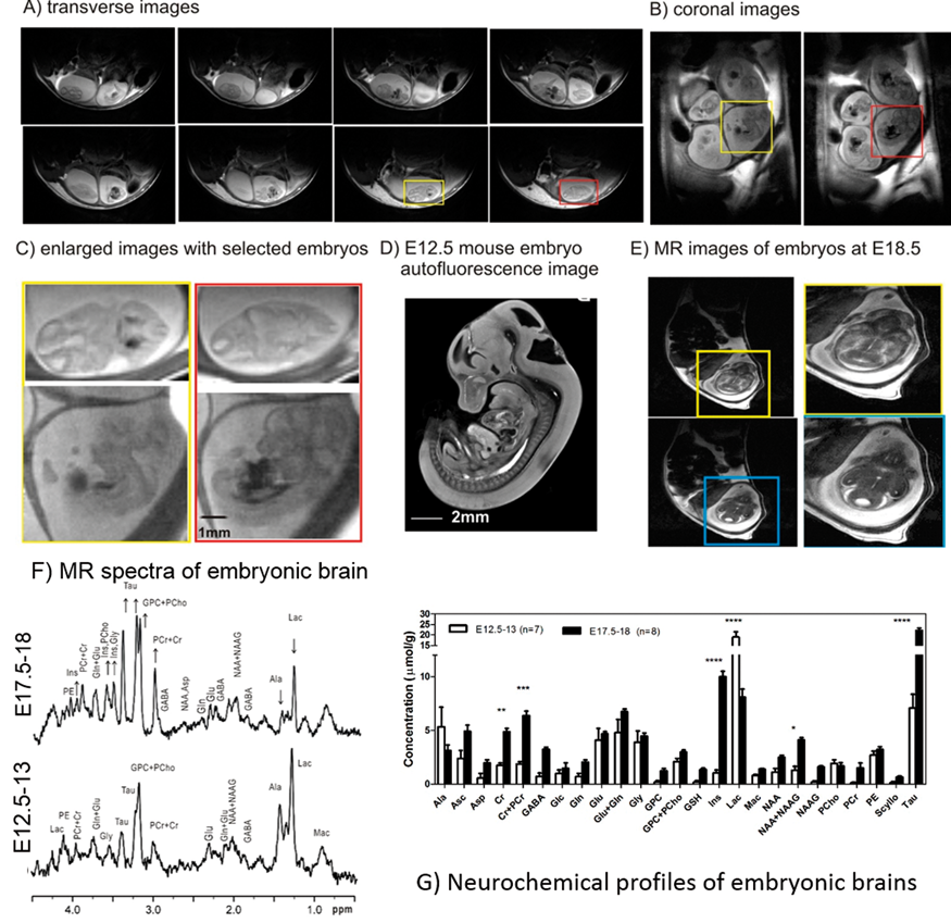
Anatomical images were obtained with sufficient resolution to depict embryonic brain, its structures and development (Figure 1A, B, C and E). Immediately after improvements of field homogeneity, magnetic resonance spectra with sufficient scans and satisfactory water suppression exhibited adequate quality to identify numerous metabolite resonances, as shown in Figure1F. At E17.5-18, increased number of resonances appeared in the spectra (Figure 1F). Such neurochemical profile of embryonic brain at E17.5-18 confirmed spectral differences and was noticeably different from the neurochemical profile of brain at E12.5 (Figure 1G).
In this study, we applied anatomical MRI to locate embryonic brains of mouse in utero as early as E12.5-13. Typical neurochemical profile of embryonic brains is the first to be reported in mice. The non-invasive characteristics of MR techniques allow us following metabolic development longitudinally. Thus, the further 1H MRS assessments of embryonic brains at E17.5-18 revealed substantial neurochemical evolution towards the birth.
PB01-D04
Development of a neonatal neuromonitor for concurrent measurements of cytochrome c oxidase and CMRO2
1Department of Medical Biophysics, Western University, London, Canada
2Imaging Program, Lawson Health Research Institute, London, Canada
Abstract
Objectives
Having very limited energy stores, the brain is susceptible to injury related to impaired cerebral blood flow (CBF). This is particularly evident in preterm infants as the underdeveloped vascular system in the immature brain can lead to poor CBF control. For example, cerebral autoregulation is known to be impaired in this age group [1]. However, the impact of cerebrovascular dysfunction on the coupling of CBF to cerebral energy metabolism in the developing brain is unknown due to a lack of adequate technologies for assessing these measures in such a fragile population. This work outlines the development of a neuromonitor for the neonatal intensive care unit (NICU) to measure CBF and energy metabolism. Metabolic measures were the cerebral metabolic rate of oxygen (CMRO2) and the oxidation state of cytochrome c oxidase (oxCCO) – the final electron acceptor in the electron transport chain and a direct marker of oxidative metabolism [2]. A preliminary demonstration of the system’s ability to track changes in these metabolic markers was conducted in a porcine model.
BFI (by DCS) measurements plotted against concurrent rCBF measurement (by 15O-water PET) on six piglets (coded by symbols), for a total of sixty-two measurements. The black line indicates the results of the Deming regression (slope of 1.15 (1.05, 1.27) (cm2/s)/(ml/100g/min) and intercept of -1.54 (-4.88, 1.47) (cm2/s) with 95% confidence interval in brackets.)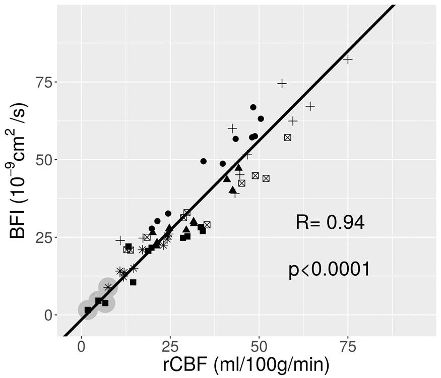
Methods
The neonatal neuromonitor (NNeMo) combines broadband near-infrared spectroscopy (B-NIRS) to measure both cerebral tissue saturation (StO2) and oxCCO with diffuse correlation spectroscopy (DCS) to provide continuous CBF monitoring (Fig 1a). The combination of these systems was achieved using a multiplexing shuttering system capable of continuous quantification of StO2, CBF, oxCCO, and CMRO2 with a temporal resolution of 6 seconds [3].
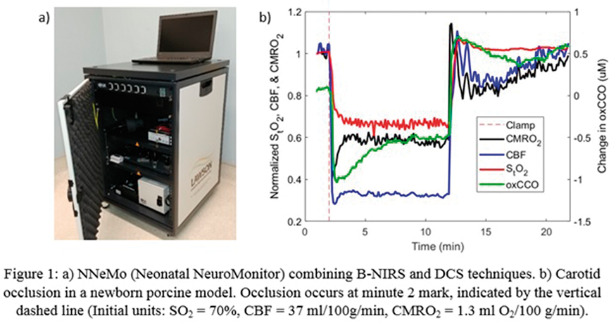
In four newborn piglets, cerebral energy metabolism was altered by (i) injections of an anesthetic (propofol 1.6 mg/kg) and (ii) occluding the common carotid arteries to induce transient ischemia (10-min), leading to CBF-driven changes in metabolism.
Results
Propofol injections resulted in a reduction of all parameters (CBF: 9%, StO2: 5%, CMRO2: 6%, and oxCCO: 0.3μM). Vascular occlusion (Fig 1b) produced larger decreases (CBF: 72%, StO2: 35%, CMRO2: 60%, and oxCCO: 1μM, at their respective nadirs). A temporal delay in oxCCO was observed compared to the CBF and CMRO2 responses.
Conclusions
The combination of B-NIRS with DCS provides a unique monitoring approach to study the coupling of CBF and metabolism in the developing brain. When manipulating metabolism directly (anesthetic) and through vascular occlusion, expected reductions in CBF and metabolic markers were observed; however, the temporal differences in CMRO2 and oxCCO responses requires further investigation. The immediate aim is to translate the developed system to the NICU to assess if flow/metabolic monitoring will provide clinicians with greater sensitivity to changes in cerebral hemodynamics that precede preterm brain injury.
References
PB01-D05
Over-estimation of cerebral oxygen saturation by commercial oximeter during deep hypothermic circulatory arrest
1Department of Anesthesiology and Critical Care, University of Pennsylvania, USA
2Division of Neurology, The Children's Hospital of Philadelphia, USA
3Division of Cardiovascular Surgery, University of Pennsylvania, USA
4Division of Cardiothoracic Anesthesia, The Children's Hospital of Philadelphia, USA
5Division of Cardiothoracic Surgery, The Children's Hospital of Philadelphia, USA
6Department of Physics & Astronomy, University of Pennsylvania, USA
Abstract
Introduction
Circulatory arrest is required for a subset of cardiac surgeries, specifically procedures that include repair to the aortic arch and is accomplished under deep hypothermia (typically 18°C) to decrease metabolic demand and thus the risk for hypoxic-ischemic injury while perfusion is stopped. However, the efficacy of deep hypothermia in providing this protection is still being elucidated. For example, in infants born with hypoplastic left heart syndrome (HLHS), there has been conflicting evidence on the effect of deep hypothermic circulatory arrest (DHCA) on the risk for post-operative hypoxic-ischemic brain injury, with some studies showing that longer time on DHCA increased the risk for this injury. Because of these findings, some surgical centers opt to utilize cerebral oximeters during surgeries requiring DHCA to ensure adequate cerebral oxygenation. Previous work from our group has shown that cerebral tissue oxygen saturation (ScO2), quantified using frequency-domain near-infrared spectroscopy (FD-NIRS), continued to decline during DHCA, despite the body being cooled to 18°C. As part of this larger study on intraoperative cerebral hemodynamics in infants with HLHS, we compared ScO2 measured with FD-NIRS to ScO2 measured with commercially available and clinically used continuous-wave near infrared spectroscopy.
Methods
Term neonates (N=3) with HLHS were recruited as part of a larger study. Frequency domain near-infrared spectroscopy (FD-NIRS, Imagent, ISS Inc.,) and continuous wave near-infrared spectroscopy (CW-NIRS, Nonin SenSmart Model X-100) were simultaneously employed to noninvasively quantify cerebral tissue oxygen saturation (ScO2) during deep hypothermic circulatory arrest.
Results
We observed a decrease in ScO2 during DHCA in all patients and with both modalities. In all patients, ScO2 quantified with FD-NIRS decreased lower than ScO2 measured with CW-NIRS. Furthermore, ScO2 quantified with CW-NIRS plateaued at approximately 50% in all three patients, even in ScO2 measured by FD-NIRS continued to decline
Conclusions
Cerebral oxygen extraction persists even under deep hypothermia, however the extent of this is not accurately reflected using commercial cerebral oximeters. As these commercial, continuous-wave oximeters are currently clinically employed for neuromonitoring during DHCA, understanding the limitations and inaccuracies of these devices is of vital importance.
PB01-D06
Noninvasive optical measurement of microvascular cerebral blood flow in children with sickle cell disease
1Wallace H. Coulter Dept. of Biomedical Engineering, Georgia Institute of Technology/Emory University, USA
2Dept. of Pediatrics, Emory University School of Medicine, USA
3Alfac Cancer and Blood Disorders Center, Children's Healthcare of Atlanta, USA
4Children's Research Scholar, Children's Healthcare of Atlanta, USA
Abstract
Objectives
Sickle cell disease (SCD) is an inherited blood disorder affecting ∼300,000 neonates worldwide each year [1] that can have profound effects on the brain. By early adolescence, ∼40% of patients will have a silent cerebral infarct. These silent infarcts are clinically asymptomatic but are associated with worse neurocognitive outcomes and increased risk of overt stroke [2]. Silent infarcts are thought to arise from anemia-induced microvascular perfusion abnormalities and subsequent reduced cerebrovascular reserve that is insufficient to meet tissue metabolic demands. Thus, measurement of microvascular cerebral blood flow (CBF) holds promise as a biomarker for the risk of silent infarcts. Unfortunately, current CBF measurement tools (e.g. MRI or PET) are expensive, have limited access, and require anesthesia and/or external contrast agents, thus limiting their use for routine assessment. Herein, we demonstrate the feasibility of a low-cost and noninvasive optical technique, known as Diffuse Correlation Spectroscopy (DCS), to quantify microvascular CBF in pediatric patients with sickle cell disease.
Methods
A total of 11 children with SCD and 11 sex-/age-matched healthy controls were enrolled at Children’s Healthcare of Atlanta (Median age of 7 y, 4/7 male/female in each group). A fiber optic-based DCS sensor was briefly secured to the subject’s forehead for assessment of an index of resting-state cerebral blood flow (CBFi). The geometry of this sensor ensures sensitivity to an average superficial cortical CBF in the region directly underneath the probe. A complete blood count was obtained in all SCD patients at the time of DCS measurement. Further, transcranial Doppler ultrasound (TCD) was used to assess large artery blood flow velocity in a subset of SCD patients (n = 7) on the same day with DCS measurements.
Results
DCS-measured CBFi in SCD is significantly higher than sex-/age-matched healthy controls (p < 0.001, Fig. 1 a). Within the SCD group, DCS-measured CBFi is inversely proportional to hematocrit (Hct,%) (p = 0.005, Fig. 1b). These results agree with observations made by the other perfusion imaging modalities [3] that CBF is globally increased in SCD as a compensatory mechanism to maintain adequate cerebral oxygen delivery in the face of chronic anemia. Notably, the DCS-measured CBFi did not significantly correlate with TCD-measured blood flow velocity in the middle cerebral or anterior cerebral arteries (p = 0.12 and 0.30, respectively). Presumably, this lack of correlation is due to the differing vascular sensitivities of the two modalities, i.e. DCS is sensitive to microvasculature while TCD to microvasculature.
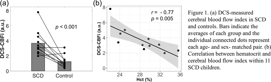
Conclusions
Our study demonstrates that DCS is sensitive to elevations in CBF associated with SCD. Further, the lack of correlation with TCD may suggest an uncoupling of macro and microvascular blood flow and emphasizes the importance of microvascular CBF monitoring in these patients. These results suggest that the technique could provide a low-cost, noninvasive monitor of tissue-level cerebral blood flow in children with sickle cell disease, paving the way for future studies that explore the potential of DCS to detect perfusion abnormalities associated with stroke risk and/or cerebral vasculopathy in these patients.
References
PB01-D07
Validation of diffuse correlation spectroscopy (babylux device) against15O-water PET for regional cerebral blood flow measurement in neonatal piglets
1ICFO-Institut de Ciències Fotòniques, The Barcelona Institute of Science and Technology, Spain
2Department of Neonatology, Copenhagen University Hospital-Rigshospitalet, Denmark
3Department of Clinical Physiology, Nuclear Medicine & PET, Copenhagen University Hospital -Rigshospitalet, Denmark
4Politecnico di Milano-Dipartimento di Fisica, Italy
5Istituto di Fotonica e Nanotecnologie,Consiglio Nazionale delle Ricerche, Italy
6Institució Catalana de Recerca i Estudis Avançats (ICREA), Spain
7HemoPhotonics S.L., Spain
Abstract
Objectives
The BabyLux project aimed at developing a neuro-monitor integrating diffuse correlation spectroscopy (DCS) [1] and time resolved near-infrared spectroscopy (TRS) [2] for neonatal critical care. The BabyLux device allows for non-invasive, continuous, and cot-side monitoring of the regional, microvascular cerebral blood flow (rCBF) by DCS and blood oxygenation by TRS. Our objective was to validate DCS against positron emission tomography (PET) with15O-labelled water [3] on piglets, for the first time.
Methods
Six neonatal piglets were measured during a protocol that consisted of an injection of acetazolamide and hypoxia events. Data was acquired with the BabyLux device to derive a cerebral blood flow index (BFI), a quantity proportional to blood flow and whose units are cm2/s. Concurrently, PET scans were performed and rCBF measured from the cerebral cortex. Correlation and agreement between the two methods were tested using Pearson coefficient (R), Deming regression and Bland-Altman plot, considering repeated measurements. A formula for conversion of DCS data to standard CBF units has hence been derived.
Results
BFI measurement by DCS and rCBF measurement by PET were highly correlated (figure 1). rCBF = 0.89 x10⁹xBFI (ml/100 g/min)/(cm2/s) is the calibration formula for single BFI measurement, as derived from the Bland-Altman analysis.
Conclusions
The good correlation between DCS and PET measurement of rCBF proves the robustness of blood flow measurement by optics in a wide range of rCBF values. The conversion formula can be used to calibrate BFI measurements from groups of piglets and infants.
Acknowledgements & disclosures
BabyLux (620996, EU CIP ICT PSP), Fundació CELLEX Barcelona, the Obra social ”la Caixa” Foundation (LlumMedBcn), Institució CERCA, “Severo Ochoa” Programme (SEV-2015-0522). UW is the CEO, employee and has equity ownership in HemoPhotonics S.L.. His role has been defined by the BabyLux project and was reviewed by the European Commission.
References
PB01-D08
Non-invasive detection of intracranial hypertension with near-infrared light: Pilot results in infant hydrocephalus patients
1Dept. of Neurology, Children's Hospital of Philadelphia, United States
2Dept. of Neurosurgery, Children's Hospital of Philadelphia, United States
3Dept. of Neonatology, Children's Hospital of Philadelphia, United States
Abstract
Objective
This study aims to validate non-invasive intracranial pressure (ICP) assessment with the near-infrared diffuse correlation spectroscopy technique in infants with hydrocephalus.
Introduction
Hydrocephalus affects 1–2 of every 1000 live births, making it the leading cause of brain surgery in US children. A major complication of hydrocephalus is intracranial hypertension (IH), which may compromise cerebral blood flow and cause fatal brain injury.1 Although timely treatment of IH is important for brain tissue viability, its implementation is hindered by the lack of tools for non-invasive IH detection.
Methods
Diffuse correlation spectroscopy (DCS) employs near-infrared light to measure local, microvascular cerebral blood flow (CBF) continuously at the bedside. We recently demonstrated a novel approach for measurement of cerebral critical closing pressure (CrCP) based on DCS measurements of pulsatile CBF in arterioles.2 CrCP, which depends on ICP, defines the arterial blood pressure at which CBF approaches zero. Intraoperative non-invasive CrCP measurements with DCS on the frontal cortex were performed concurrently with invasive ICP measurements in 5 infants with hydrocephalus at the Children’s Hospital of Philadelphia. Invasive ICP was measured during surgical shunt placement.
Results
A strong correlation (r = 0.9) between non-invasive CrCP and invasive ICP measures (ICP range 5–20 mmHg) was observed in 5 infants undergoing shunt placement. CrCP overestimated ICP at lower ICP levels, but was close to ICP at higher ICP levels.

Conclusions
Pilot data shows the potential of using DCS measurement of CrCP for non-invasive ICP measurements in infants. DCS probes are further well-suited for continuous, long-term monitoring. We hypothesize that CrCP overestimation of ICP at lower levels is because of CrCP’s sensitivity to vasomotor tone. Enrollment of more patients is underway.
References
PB01-D09
Cerebral hemodynamic measurements predict treatment response and neurodevelopmental outcome in hydrocephalus infants
1Division of Newborn Medicine, Department of Medicine, Harvard Medical School/Boston Children's Hospital, Boston, Massachusetts, USA
2Department of Psychiatry, Harvard Medical School/Boston Children's Hospital, Boston, Massachusetts, USA
3Department of Neurosurgery, Harvard Medical School/Boston Children's Hospital, Boston, Massachusetts, USA
Abstract
Objectives
Infant hydrocephalus is one of the most prevalent neonatal medical conditions in the United States, afflicting one out of every 500 births1. While hydrocephalus is the most common reason for brain surgery to be performed in children, existing hydrocephalus assessments have low correlation with treatment failure and do not predict the incidence of neurodevelopmental impairment (NDI)2. To address the urgent unmet medical need for diagnostics to guide care, we proposed to develop new approaches for investigating the pathophysiology of brain injury in hydrocephalus with the ultimate goal of providing early, quantitative diagnostics to evaluate treatment effectiveness and predict risk of NDI.
Methods
Infants less than 3 months old with clinical symptoms of progressive hydrocephalus were eligible for the study. We used frequency-domain NIRS (FDNIRS) and diffuse correlation spectroscopy (DCS) to measure regional cerebral hemoglobin concentrations (HbO, HbR), oxygen saturation (SO2), blood volume (CBV), flow (CBF), and metabolic rate of oxygen (CMRO2) non-invasively in the frontal, temporal, and parietal brain regions of each subject. Measurements were performed both pre- and one to three days post- operatively in patients treated by endoscopic third ventriculostomy combined with choroid plexus cauterization (ETV/CPC) surgery. Treatment was categorized to have either “failed” or “not failed” within six months according to clinical assessment. In a subset of patients, Vineland Adaptive Behavior Scales II (VABS-II) were also used to evaluate the neurodevelopmental outcome at six and 12 months.
Results
To date, 29 subjects (GA: 34.48 ± 5.11 wks) with progressive hydrocephalus were enrolled at Boston Children’s Hospital and 23 patients (PMA: 42.62 ± 6.81 wks) underwent ETV/CPC. The success rate of the procedure was 69.5% (16 in the not-failed group and 7 in the failed). Overall, CBV, CBF and CMRO2 increased after surgery while no significant differences occurred in HbO, HbR and SO2. However, the failed and not-failed groups had significantly different changes in HbO, HbR, SO2, and CBF. The not-failed group had significantly greater increases in CBF and CMRO2 with treatment, while the failed group had a significant increase in HbR (Fig. 1). HbO and SO2 increased postoperatively in the not-failed group but decreased in the failed group (Fig. 1). Among all parameters, the change in SO2 showed the strongest power in predicting treatment response (ROC area under curve = 0.74, p = 0.01). In addition, six patients were evaluated with VABS-II at six and 12 months. Post-operative CBF correlated well with socialization (r = 0.83, p < 0.05) and adaptive behavior scores (r = 0.90, p < 0.05) at six months. Post-operative SO2 values were strongly correlated with communication (r = 0.96, p < 0.001) and socialization scores (r = 0.84, p < 0.05) at 12 months. Similarly, strong correlations were observed between post-operative CBV and communication (r = 0.92, p < 0.001), daily living skills and (r = 0.81, p < 0.05) and adaptive behavior scores (r = 0.81, p < 0.05) at 12 months.
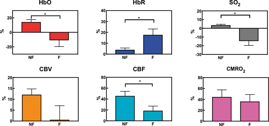
Conclusions
We have demonstrated that FDNIRS-DCS bedside measurements of cerebral hemodynamics is sensitive to the effects of hydrocephalus treatment. Despite the small sample size, our preliminary results show excellent correlation between post-surgical cerebral hemodynamics and long-term neurodevelopmental outcome. Thus, our approach has potential as patient-specific tool for guiding care in infant hydrocephalus.
References
PB01-D10
Is the enlargement of subarachnoid spaces benign? building a pathophysiological frame through the psychomotor development assessment and continuous non invasive optical monitoring
1Neurotraumatology and Neurosurgery Research Unit (UNINN), Vall d'Hebron Research Institute (VHIR), Universitat Autonoma de Barcelona, Barcelona, SPAIN
2ICFO-Insitut de Ciencies Fotoniques, The Barcelona Institute of Science and Technology, Barcelona, SPAIN
3Department of Neurosurgery, Vall d’Hebron University Hospital, Universitat Autònoma de Barcelona, Barcelona, SPAIN
4Institució Catalana de Recerca i Estudis Avançats (ICREA), Barcelona, SPAIN
Abstract
Objectives
Benign enlargement of subarachnoid spaces (BESS) is radiologically characterized by an enlargement of subarachnoid spaces at the frontal and temporo-parietal level. It has been considered a self-limiting, benign cause of macrocephaly in children which does not require any treatment. Even though it has been reported to be associated with an increased risk of subdural hematoma [1] and a delay in the acquisition of cognitive and motor skills [2, 3]. We have hypothesized that the alteration of the cerebrospinal fluid (CSF) dynamics causes alterations in the intracranial pressure (ICP) and cerebral oxygenation thus leading to developmental delays.
Our main objective is to achieve a better knowledge of BESS pathophysiology and sequelae in order to allow an early detection and treatment. We aim to detect the presence of psychomotor delay and highlight the most compromised areas through the Bayley III test and to obtain additional biomarkers about cerebral hemodynamics by introducing a new non-invasive optical method.
Methods
We have recruited 31 children: 24 males and 7 females, median age 15 months (7 to 55 months) and followed up every 6 months. In 13 children, extradural ICP was monitored continuously during 72 hours due to the presence of macrocephaly and/or an acceleration in the growth of the head circumference with associated clinical symptoms or persistent psychomotor delay. Among them, 8 underwent non-invasive monitoring via optical techniques (time-resolved spectroscopy and diffuse correlation spectroscopy) during 2 consecutive nights. To compare the optical parameters (oxy and deoxy-hemoglobin, total hemoglobin concentration, cerebral oxygen saturation and blood flow) with the ICP findings, we have identified ICP pathological patterns: low and high amplitude B waves and plateau waves.
Results
At the baseline assessment, out of 31 children 16 showed a psychomotor delay with the gross motor area affected in all of them. All patients with ICP monitoring showed abnormal findings (elevated ICP values and presence of pathological waves) and received a shunt. We have looked at optical parameters variations in a subset of 4 patients during different ICP patterns and found a linear correlation between ICP and cerebral blood flow (CBF) during B-waves. Other measurements are under analysis. At the 6 months follow up of the 16 patients with psychomotor delay: 3 presented a normal development, 3 shunted presented a delay reduction to one area, 4 still presented a delay. The follow up is still going on.
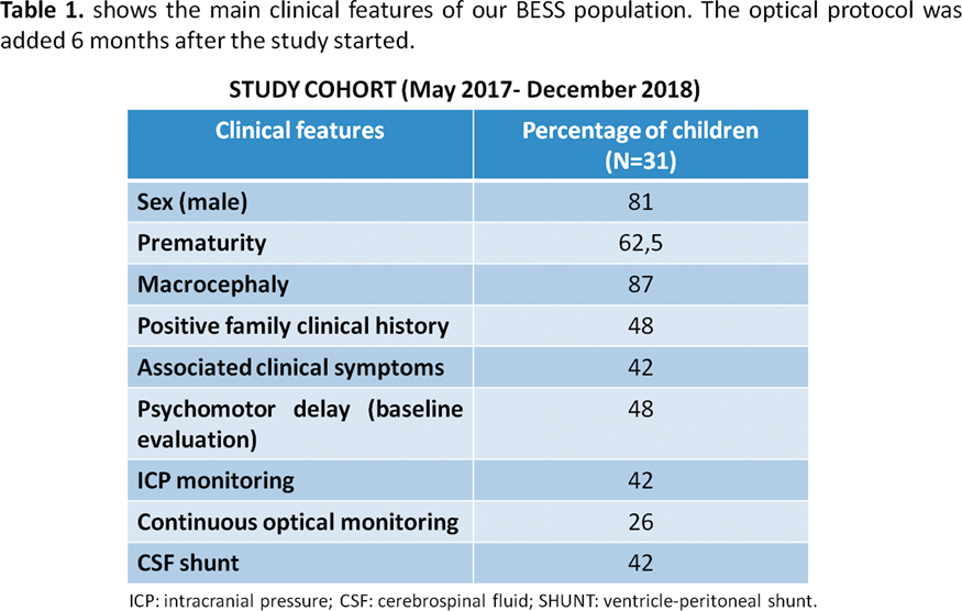
Conclusions
We confirmed the presence of psychomotor delay and ICP alterations in BESS children. We identified the CBF as a potential biomarker to better understand the cerebral hemodynamics in these patients. More children will be recruited to confirm our results.
Acknowledgements
European Union’s Horizon 2020 (Marie Sklodowska-Curie grant 675332), KidsBrainIT-project (ERA-NET NEURON), Instituto de Salud Carlos III grant PI18/00468, European Regional Development, Fundació CELLEX Barcelona, Ministerio de Economía y Competitividad/FEDER (PHOTODEMENTIA, DPI2015-64358-C2-1R), Instituto de Salud Carlos III/FEDER (MEDPHOTAGE, DTS16/0087), “Severo Ochoa” Programme for Centres of Excellence in R&D(SEV-2015-0522), Obra social “la Caixa” Foundation (LlumMedBcn), Institució CERCA, AGAUR-Generalitat (2017-SGR-1380), LASERLAB-EUROPE IV.
References
PB01-E01
Delayed melatonin treatment improves white striatal white matter content after perinatal stroke and promotes oligodendrocyte maturation in vitro
1Dept. of Pediatrics, Division of Neurology, University of Colorado Anschutz Medical Campus
2Dept. of Anesthesiology, University of Colorado Anschutz Medical Campus
3Dept. of Cell and Developmental Biology, University of Colorado Anschutz Medical Campus
4Neuronal Injury and Plasticity Program
Abstract
Introduction
Neonatal stroke is a common cause of lifelong neurologic disability and there are currently no clinically available therapies to improve long term outcome. White matter (WM) injury and repair after full term equivalent neonatal stroke has been relatively under-studied but is a promising therapeutic target given the large window of WM repair after ischemic injury. Melatonin is a commercially available drug that has been shown to protect white matter after neonatal ischemia in humans and rodents.
Objective
To determine the effect of delayed melatonin treatment on striatal white matter and oligodendrocyte progenitor cell (OPC) maturation in a model of neonatal stroke.
Methods
Transient middle cerebral artery occlusion (MCAO) was performed for 45 minutes in p10-11 mice followed by reperfusion. NeuN/TUNEL immunohistochemistry (IHC) was used to assess neuronal cell death at 3 days post-injury. IHC for SMI34 and MBP was used to assess striatal axons and myelin respectively at the same time point. Melatonin (20 mg/kg) was administered intraperitoneally on POD 3–7 and the Black Gold II myelin stain was used to quantify striatal white matter 14 days post-MCAO. Primary OPC cultures were treated with melatonin (0.1 uM) or vehicle (<1% DMSO) for 24 hours in differentiation media. Cells were then fixed and IHC was used to determine the proportion of oligodendrocytes (olig2+) that had a mature phenotype (O4+).
Results
In our model we see robust neuronal cell death and myelin loss with relative preservation of pencil fiber axons in the ipsilateral striatum 3 days post-injury (A in figure). Fourteen days after MCAO there is a significant reduction in pencil fibers in vehicle treated animals (609.2 ± 164.9 pencil fibers, mean ± SD, n = 6), compared to sham surgery animals (812.7 ± 35.9 pencil fibers, n = 6, p = 0.02, one-way ANOVA). In contrast, there was no difference between the number of striatal pencil fibers in animals treated with melatonin after MCAO (739.5 ± 85.72 pencil fibers, n = 4) compared to sham surgery animals (p = 0.59, B and C in figure). In primary OPC culture, 24 hour incubation with melatonin resulted in an increase in the proportion of oligodendrocytes with a mature phenotype (0.31 ± 0.01, mean ± SD, n = 3 coverslips) compared to vehicle treated cultures (0.23 ± 0.03, n = 3 coverslips, p = 0.01, unpaired t-test, D in figure).
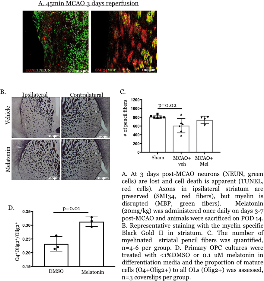
Conclusions
45 minutes of MCAO in neonatal mice produces striatal injury with neuronal death and myelin loss acutely. Melatonin treatment ameliorates striatal white matter loss chronically after p10 MCAO, and melatonin treatment promotes OPC differentiation in culture. Ongoing studies will determine whether melatonin promotes remyelination via enhanced OPC maturation in vivo after neonatal stroke.
PB01-E02
N-SMase2 –dependent vesicle release and injury after neonatal stroke
1Department of Neurology, University of California San Francisco, San Francisco, USA
Abstract
Background
Perinatal arterial stroke, stroke that occurs between the 20th gestation week to the 28th day of life, leads to long-lasting disabilities, including epilepsy, behavioural and learning deficits, and cerebral palsies. Although the inflammatory damage is a major brain injury component in the early postnatal period, microglial cells could limit injury after neonatal stroke (Faustino et al., J Neurosci 2011). These brain resident immune cells are central to the crosstalk between cells in the brain. One largely unexplored mechanism of communication is via release of extracellular vesicles (EV), including Exosomes (Exo) and Microvesicles (MV). Neutral Sphingomyelinase 2 (N-SMase2), a plasma membrane enzyme that belongs to sphingomyelinase family, was demonstrated to control EV release.
Objectives
To understand the role and the underlying mechanisms of N-SMase2 –dependent release and signaling of microglial derived MV and Exo in perinatal stroke.
Methods
To investigate the contribution of N-SMase2 in injury after neonatal stroke, we have chosen to downregulate the gene named Smpd3 (Sphingomyelin phosphodiesterase 3 in mus musculus), which encodes N-SMase2, using a KO-CRISPR/dcas9. Control Vs KO-CRISPR/dcas9 was injected intracerebral at postnatal day 7 (P7) and N-SMase2 enzyme activity examined daily between P9-P12. Transient middle cerebral artery occlusion (tMCAO) was performed at P9. N-SMase2 activity was determined in treated and non-treated pups and cell origin of N-SMase2 expression examined by immunofluorescence. Histological outcomes were determined 72 h after reperfusion. Microglial cells were isolated from injured and uninjured regions (using CD11b-conjugated magnetic beads) and plated. Released MV and Exo were characterized using Nanosight and ImageStreamX. EV protein expression was determined by western blot.
Results
We observed a developmental increase of N-SMase-2 activity in naïve brain from P10 to P12 (p < 0.016) and a significant decrease of N-SMase-2 activity in KO-CRISPR/dcas9-treated mice compared to Control-CRISPR/dcas9 at P12 (16.22 ± 1,79 Vs 7.78 ± 3,54 mUnit/mg of protein, p < 0.0062, respectively). Following tMCAO, N-SMase-2 activity was significantly reduced in injured regions (p < 0.0043) at 72 h post reperfusion. Based on immunofluorescence, N-SMase-2 expression was markedly reduced in neurons within injured regions. Injury volume was significantly reduced 72 h after reperfusion in KO-CRISPR/dcas9-treated mice as compared to Control-treated mice (41.39 ± 3.78 Vs 23.15 ± 2.68, respectively, p < 0.0029). Decreased N-SMase2 activity in Control-CRISPR mice correlated with reduced Glut-1 coverage, an effect in part prevented by KO-CRISPR/dcas9 pre-treatment. Moreover, downregulation of Smpd3 expression correlated with protection against injury. Microglial cells isolated 24 h after tMCAO from ipsilateral hemisphere actively proliferated in culture as compared to cells from contralateral hemisphere or naïves brains. We are characterizing the effects of N-SMase-2 deactivation on the constitution and cargo of microglial MV and Exo.
Conclusion
Downregulation of N-SMase2 protects neonatal brain from stroke, in part due to vasculature preservation. In fine, these results will provide a better understanding of the implication of this novel cell-cell communication on severity of neonatal stroke.
Support by: AHA17IRG33430004, RO1 NS44025, CPA PG0816
PB01-E03
Alteration of brain-derived neurotrophic factor expression in a piglet model of neonatal hypoxic-ischemic encephalopathy
1Department of Physiology, University of Szeged, Hungary
Abstract
Objectives
Hypoxic-ischemic encephalopathy (HIE) is defined as acute/subacute brain injury elicited by asphyxia in the perinatal period associated with adverse neurologic outcomes or death. Neuronal injury is elicited by a complex cascade of processes beginning within minutes of reoxygenation that may evolve for at least 72 hours. Brain-derived neurotrophic factor (BDNF) is a member of the neurotrophic factor family which is highly expressed in the developing brain. Through its anti-apoptotic ability, it plays an important role in promoting neuronal proliferation, differentiation and survival. The overall purpose of the study was to examine the consequence of asphyxia on BDNF, apoptosis-inducing factor (AIF), and caspase-3 mRNA levels in various brain regions in a neonatal piglet HIE model.
Methods
Anesthetized and artificially ventilated newborn (<1 day old) pigs were aseptically instrumented and divided into 3 experimental groups: untreated naïve animals without any interventions (n = 4), normoxic time controls with 48 h of survival (n = 5) and animals exposed to asphyxia with 48 h of survival (n = 6). The animals were intensively monitored: core temperature, MABP, blood gases were tightly controlled, and the EEG was recorded. Asphyxia was induced by ventilation with a gas mixture containing 6% O2 and 20% CO2 for 20 min with simultaneous reduction of the respiratory rate from 30 to 15 /min and suspension of the supportive fluid therapy containing glucose. After asphyxia the animals were reventilated with medical air throughout the 48 h survival period. Then the brain samples obtained from the frontal and occipital cortices as well as from the hippocampus of the euthanized animals were processed for quantitative RT-PCR.
Results
Compared to the untreated naïve group, the relative expression of BNDF mRNA was largely and significantly reduced in all assessed cortical regions in the asphyxia but not in the time control group, despite a similar tendency in the latter. There were no major alterations regarding AIF expression among the groups. However, the relative expression of caspase-3 mRNA in the asphyxia group appeared to be increased in all examined regions compared to the normoxic naïve controls.
Conclusions
Our preliminary results show that BDNF mRNA abundance is largely reduced, however, caspase-3 abundance is moderately increased 48 hours after asphyxia in various cortical areas compared to normoxic controls. Understanding the changing expression patterns during HIE development can contribute to developing rational, more effective therapies that can further reduce mortality and the severity of disabilities in HIE patients.
This work was supported by the Hungarian Brain Research Program 2.0 (2017–2.1 NKP 2017 00002), the EU-funded Hungarian grant EFOP-3.6.1-16-2016-00008 and the GINOP 2.3.2 15 2016 00034. V.K. is supported by OTKA-PD128464 from the NRDI.
PB01-E04
Combination of therapeutic hypothermia and H2 treatment in a piglet model of neonatal hypoxic-ischemic encephalopathy
1Department of Physiology, University of Szeged, Hungary
Abstract
Objectives
Hypoxic-ischemic encephalopathy (HIE) induced by asphyxia remains to be the most common cause of neonatal death and life-long disability affecting annually 0.7 and 0.5 million term infants, respectively. Therapeutic hypothermia (TH) is the only effective neuroprotective therapy in HIE management, however, TH alone often cannot provide full neuroprotection thus supplementary neuroprotective therapies are warranted to improve outcome. We showed previously postasphyxial administration of H2 decreased neurovascular unit dysfunction and neuronal injury in a piglet HIE model (1,2). The aim of this study was to investigate the neuroprotective effects of TH and TH+H2 in the first 48 hours of HIE development following severe experimental asphyxia.
Methods
Newborn (<1 day old), male piglets were anaesthetized, artificially ventilated and continuously monitored (O2 saturation, MABP, core temperature, blood gases and EEG). Animals were divided into 3 groups: 1. asphyxia+normothermia controls (CTRL), 2. asphyxia+TH (TH), 3. asphyxia+TH+H2 (TH+H2). Asphyxia was induced with 4%O2-20%CO2 gas mixture ventilation for 20 min. After asphyxia animals were reventilated with air (CTRL, TH), or air containing 2.1% H2 (TH+H2) for 4 h followed by air. In the CTRL group, the animals were kept normothermic (38,5 ± 0,5°C), whereas in the TH and TH+H2 groups after 2 h normothermia the animals were quickly cooled to 33,5°C in 0.5 h. TH was maintained until the 36 th hour of survival then the animals were gradually warmed up (0,5°C/hour) back to normothermia. At 48 h, the brains were collected then processed for neuropathology. Neuronal injury was assessed in H&E stained sections with cell counting in the hippocampus and the subcortical regions and with a previously published scoring system in the neocortex (2).
Results
Asphyxia resulted in severe hypoxia, hypercapnia, and lactic acidosis, similar in all groups (pH: 6,77 ± 0,03; PaCO2: 167 ± 5 mmHg; lactate: 9,0 ± 0,6 mmol/L). These derangements returned to basal levels within 4 h. Compared to the CTRL, heart rate was reduced in both TH groups, however, there was no significant difference in MABP among the groups. Asphyxia elicited severe cerebrocortical neuronal damage, and although there was a neuroportotective tendency in the TH and TH+H2 groups compared to CTRL but no significant changes could be identified. In a similar fashion, severe neuronal injury was found in the assessed hippocampal regions, basal ganglia and thalamus without significant difference in TH and TH+H2 groups.
Conclusions
Compared to previous studies, we used a more severe hypoxia (4% vs. 6% O2) to generate more uniform injury and longer survival (48 h vs. 24 h) to accommodate TH methodology. Our results suggest that these experimental conditions were not in favor to demonstrate additive/synergistic neuroprotection between TH and H2. Future studies with modifications of the asphyxia protocol and perhaps earlier implementation of TH may be required to satisfactorily address this issue.
Support
Hungarian Brain Research Program 2.0 (2017–2.1 NKP 2017 00002), the EU-funded Hungarian grant EFOP-3.6.1-16-2016-00008 and the GINOP 2.3.2 15 2016 00034. V.K. is supported by OTKA-PD128464 from the NRDI.
References
PB01-E05
Estrogen receptor alpha and tyrosine kinase B signaling in long-term neurological recovery following neonatal hypoxia and ischemia
1Department of Pediatrics, University of Wisconsin-Madison, WI, USA
2Waisman Center, University of Wisconsin, Madison, WI, USA
3Department of Neuroscience, University of Wisconsin, Madison, WI, USA
Abstract
Background
Neonatal hypoxia ischemia (HI) related encephalopathy is one of the major causes of learning disabilities and memory deficits in children. Male brains are two times more vulnerable to the effects of perinatal asphyxia, a phenomenon that is poorly understood. We recently reported that increased hippocampal estrogen receptor alpha (ERa) expression post-HIconfers neuroprotection only in the female neonate hippocampi through crosstalk with the neurotrophin receptor, tyrosine kinase B (TrkB). Activation of the TrkB via its selective agonist, 7,8-dihydroxyflavone (7,8-DHF) decreases apoptosis only in female mice hippocampus in an ERa dependent way. Here, we hypothesize that absence of ERa will ablate the sex differences seen in long-term neurological outcome following 7,8-DHF therapy in neonatal mice following perinatal HI.
Methods
HI was induced in P9 mice by unilateral common carotid artery ligation and exposure to 10% O2for 50 min using Vannucci’s HI model. Cognitive and memory functions were assessed at P60+ by novel object recognition (NOR) and location (NOL) tests, respectively one week apart longitudinally.Thirty seconds of total exploration time (novel+familiar exploration = 30 sec) was recorded. Percent time spent with novel objects or at novel locations were calculated as discrimination ratio. ANOVA was used to compare the discrimination ratios (mean ± SEM) between the groups.
Results
HI decreased the discrimination ratios for both NOR and NOL tests in ERawild type (WT) male (% 28 ± 2 and %23 ± 3) and female (% 25 ± 12 and % 25 ± 4) mice compared to sham male (% 76 ± 4 and % 56 ± 8) and female (% 68 ± 4 and % 73 ± 1) mice (p < 0.001). HI induced decline in recognition and location memories were recovered by 7,8-DHF therapy only in ERa WT females for both NOR and NOL tests, respectively [% 69 ± 6 and %70 ± 3, (p < 0.001)]. 7,8-DHF therapy failed to improve the discrimination ratios for both NOR and NOL tests inERa knockout female and male mice (p < 0.001).
Conclusion
Neurotrophin receptor activation improves the long-term cognitive and memory function only in female WT mice in an ERa dependent way following global brain injury. Understanding the cellular basis of the sex differences seen in ERa dependent neurotrophin mediated neuroprotection is important to impliment sex-specific therapies.
PB01-E06
The HDAC inhibitor, sodium butyrate, stimulates neurogenesis in subventricular zone in a rat model of neonatal hypoxia-ischemia
1NeuroRepair Department, Mossakowski Medical Research Centre Polish Academy of Sciences, 5 Pawinskiego Street, 02–106 Warsaw, Poland
Abstract
Objectives
Neonatal hypoxic-ischemia (HI) brain injury is a leading cause of impaired neurodevelopment, manifested by mental retardation, motor dysfunction, and seizures. The loss of highly vulnerable axons, oligodendrocyte progenitors, and neurons disrupts maturation of the neural network, with a destructive impact on the structure and connectivity in the brain [1]. Despite significant investigations, there is not, yet, any known reliable treatment to reduce brain damage in suffering infants. Our recent studies in an animal model of HI revealed the therapeutic potential of sodium butyrate, a histone deacetylase inhibitor (HDACi). The neuroprotective properties of this agent were connected with the anti-inflammatory action [2]. The present investigation was designed to assess whether SB will stimulate neurogenesis in the subventricular zone (SVZ) in a neonatal rat model of HI. In an effort to explore the potential underlying mechanism which regulates the neuroprotective/neurogenic response to SB-induced HDAC inhibition, we sought to determine the expression of chosen neurotrophins – BDNF and NGF and its receptors.
Methods
Seven-day-old rat pups were subjected to unilateral carotid artery ligation followed by 60 minutes of hypoxia (7.6% O2). SB (300 mg/kg) was administered in a 5- day regime with the first injection given immediately after hypoxic exposure. To determine the phenotype of newborn cells, animals received BrdU injections twice daily (12 h apart) for 3 consecutive days starting 4 days after the onset of hypoxia-ischemia. Double immunohistochemical staining BrdU/ DCX and NeuN/BrdU was used to analyze if BrdU-positive cells represent newly generated neuroblasts and mature neurons. Effects of SB on neurotrophins were assessed by Elisa method. Expressions and activation/phosphorylation of neurotrophins receptors: TrkB and p75 were determined by Western blot.
Results
Sodium butyrate treatment stimulated neurogenesis in SVZ region of damaged ipsilateral side, based on increased number of DCX (+) neuroblasts and NeuN (+) neuronal cells. The neurogenic effect of SB was associated with increased level of neurotrophins – BDNF and NGF in ipsilateral hemisphere in animals 7 days after HI. In addition, the administration of SB increased the activation of TrkB receptor, and reduced the level of HI-induced p75NTR.
Conclusion
Obtained results suggest that BDNF-TrkB signaling plays an important role in SB-induced neurogenesis after HI. These findings provide the basis for clinical approaches targeted at protecting the newborn brain damage, which may prove beneficial for treating neonatal hypoxia-ischemia.
This work was supported by National Science Centre, Poland grant no 2015/17/N/NZ7/00969 and 2017/27/B/NZ3/00582.
References
PB01-E07
Novel mechanism of rescuing synaptic dysfunction following juvenile global ischemia using AMPAkines
1Dept of Pediatrics, University of Colorado
2Dept of Anesthesia, University of Colorado
Abstract
Background
Global ischemia in the developing brain often leads to poor neurologic outcomes, including learning and memory deficits. Using a novel murine model of juvenile cardiac arrest (CA), we investigate the mechanism of ischemia-induced cognitive deficits and recovery. Brain-derived neurotrophic factor (BDNF)-tyrosine kinase (Trk) B signaling is a requisite pathway for memory formation and hippocampal synaptic plasticity and we recently demonstrated that enhanced BDNF-TrkB singaling contributes to recovery of cognitive function after global ischemia. AMPAkines have been shown to augment BDNF levels and therefore may be a novel therapeutic avenue following CA.
Objective
Investigate the effect of LY404187, an AMPAkine reported to increase BDNF levels, on hippocampal function after CA in the developing brain.
Methods
Male juvenile mice (postnatal day 20–25; equivalent to 2–3 year old human) were subjected to 8 min CA and resuscitated. Memory function was measured using contextual fear conditioning, a hippocampal-dependent memory task. Hippocampal CA1 long-term potentiation (LTP), a well-accepted cellular model for learning and memory, was measured in acute brain slices following a theta-burst stimulation (TBS, 40 pulses 100Hz). Increase in field excitatory post-synaptic potential (fEPSP) slope 60 min after TBS was analyzed as a measurement of LTP. Results reported as mean ± SEM.
Results
Memory is impaired 7 days after CA/CPR (52 ± 5% sham, n = 8, vs 24 ± 6% CA, n = 9, p < 0.05) and recovers 30 days after CA (52 ± 5%, n = 7). Hippocampal LTP correlates with this neurobehavioral recovery. In control mice, LTP was 153 ± 5% (n = 8) of baseline (100%). In contrast, 7 days after CA, LTP was impaired (114%, n = 6, p < 0.05 compared to sham control mice), followed by recovery at 30d (155 ± 4%, n = 6, p < 0.05 compared to 7 day but similar to sham). We have previously reported that delayed activation of TrkB receptor signaling by 7,8 dihydroxyflavone (DHF) reversed CA-induced LTP deficits and memory impairments. In paired experiments 7d after CA, 7,8 DHF (250 nM) reversed synaptic impairment (105 ± 5% [n = 6] vehicle vs. 143 ± 10% 7,8 DHF [n = 6], p < 0.05, paired t-test). Using a similar strategy exposing the BDNF inducing AMPAkine LY404187 (25μM) to hippocampal slices 7 days after CA reverses impaired LTP (112 ± 8 [n = 3] vs. 162 ± 2 [n = 3], p < 0.05, paired t-test).
Conclusion
These data provide unique new data that BDNF enhancing drugs are capable of enhancing recovery from global cerebral ischemia in the developing during critical school ages.
PB01-E08
Experimental pediatric stroke shows age-specific recovery of cognition and role of hippocampal Nogo-A receptor signaling
1Department of Anesthesiology, University of Colorado School of Medicine
2Department of Pediatrics, University of Colorado School of Medicine
3Department of Pharmacology, University of Colorado School of Medicine
4Neuronal Injury Program, University of Colorado School of Medicine, Aurora
Abstract
Objective
Ischemic stroke is a leading cause of death worldwide and clinical data suggests that children may recover from stroke better than adults (Anderson, et al., 2011). However, supporting experimental data is lacking. We used our novel mouse model of experimental juvenile ischemic stroke (MCAO) to characterize age-specific cognitive dysfunction following ischemia. This study investigates the hypothesis that juvenile mice exhibit greater recovery of hippocampal synaptic function following experimental stroke compared to adult mice.
Methods
Extracellular field recordings of CA1 neurons were performed to asses hippocampal synaptic plasticity changes 7 or 30 days after recovery from a 45 minute middle cerebral artery occlusion (MCAO) in juvenile (p21-25) and adult mice (p60-90). A contextual fear-conditioning paradigm was done to evaluate memory in both juvenile and adult mice 30 days after MCAO. Biochemistry was used to analyze Nogo-A expression.
Results
In adult mice following MCAO, hippocampal long-term potentiation (LTP) remained impaired for at least 30 days in the ipsilateral and contralateral, non-injured hemisphere (Orfila et al., 2017). However, in juvenile mice following MCAO, LTP was only impaired in the ipsilateral side 7 days after MCAO and showed full recovery of synaptic function at 30 days. Behavioral data confirmed this observation, showing that only adult mice displayed memory deficits 30 days after MCAO. Western blot analysis showed that MCAO caused an increase in Nogo-A expression in the ipsilateral hippocampus 7 days after MCAO compared to contralateral and sham hippocampus. Further, inhibition of Nogo-A receptors reversed MCAO-induced impairment of LTP in acute slices of juvenile mice at 7days after MCAO. However, adult MCAO-induced impairment of LTP was not associated with increased Nogo-A expression. This suggests that Nogo-A receptor mechanisms may account for the impairments seen in juvenile mice during development.
Conclusion
The present study demonstrates that transient focal ischemia causes functional impairment in the hippocampus and that recovery of behavioral and synaptic function is more robust in the young brain. In addition, Nogo-A receptor activity may account for the impairments seen following ischemic injury in the juvenile. Therefore, targeting Nogo-A receptor activity may provide a new therapeutic approach to improve functional recovery after pediatric stroke.
References
PB01-E09
Enhanced stem cell therapy provides more long-term histological and behavioral improvement for neonatal stroke than stem cells alone
1Department of Pediatrics; University of California, San Francisco
Abstract
Background
Stroke in the newborn period is a significant cause of mortality and life-long morbidity. In recent years, cell-based therapies have emerged as a potential treatment for different various CNS diseases. These improved outcomes occur even in the absence of survival of engrafted cells, suggesting transplanted cells may induce repair by stimulating secretion of growth and differentiation factors that provide an environment to enhance repair. It is possible that growth factors that have specific effects on stem cell function, including their role in the development of the neurovascular unit and cell fate, may provide additional benefit.
Objective
To compare the efficacy of single-dose mesenchymal stem cells (MSC) pretreated with erythropoietin [EPO] (MSC-EPO) into the rat CNS versus MSC alone following transient neonatal focal ischemia-reperfusion on long-term histological and behavioral outcomes.
Design/Methods
Focal cerebral ischemia-reperfusion was performed in P10 Sprague-Dawley rats by transient right middle cerebral artery occlusion (MCAO) for 3 hours, or sham surgery under isoflurane anesthesia. This age is considered equivalent to the full-term human newborn. Either vehicle, MSC (1x10⁶ cells) or MSC pre-treated with EPO (1 IU/mL x 24 hours) were administered intranasally 72 hours following reperfusion. N = 4-6 per group. Animals underwent sensorimotor (cylinder rearing) and cognitive (novel object recognition) testing at 8 weeks of age, after which brains were harvested for histological analysis.
Results
MSC alone and MSC-EPO groups had a significant improvement in calculated hemispheric brain volumes (ipsi/contra ratio) compared to untreated MCAO rats at 2 months of age (MCAO: 64.5%, MSC:79.5%, MSC-EPO:85.8%; p < 0.05). While both treatment groups had improved sensorimotor function and cognitive memory retention testing [Fig.1] compared to vehicle-treated MCAO animals, MSC-EPO animals had a significant improvement in both functional measures compared to MSC alone animals.
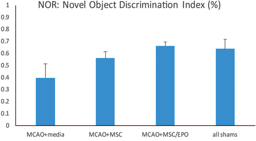
Conclusions
A delayed, single-dose combination strategy to enhance cell type-specific effects using MSC and EPO provides the most long-term benefit following early stroke, lasting until young adulthood. The specific effects of this strategy on vasculogenesis, cell proliferation and fate remains to be determined.
PB01-E10
Selective brain cooling with transnasal flow of ambient air is neuroprotective in a model of pediatric cardiac arrest
1Dept. of Anesthesiology and Critical Care Medicine, Johns Hopkins University, USA
2Dept. of Medicine, Johns Hopkins University, USA
Abstract
Objectives
An out-of-hospital pediatric cardiac arrest trial reported a small, non-significant favorable outcome with hypothermic treatment,1 possibly because of the median 5.9-hour delay in inducing hypothermia. To minimize this delay, we developed a simple yet innovative technique of transnasal cooling. Air at ambient temperature is passed through standard nasal cannula and out of the mouth to produce evaporative cooling of the nasal passages and a countercurrent heat exchange between cooled venous blood draining the nasal turbinates with cephalic arterial blood. Here, we tested the hypothesis that high transnasal airflow is not inferior to standard surface cooling in providing neuroprotection when initiated at either 10 or 120 min after resuscitation in an infant swine model of asphyxic cardiac arrest.
Methods
Under fentanyl-nitrous oxide anesthesia, we induced 45 min of hypoxia (O2 saturation ∼35%) followed by 7 min of airway occlusion to produce asphyxic cardiac arrest in 2-week-old piglets (4 kg). Viable neuronal counts were assessed at 4 days of recovery in 6 groups: 1) sham surgery, 2) normothermic recovery, 3) surface cooling to decrease rectal temperature from 38.5 to 34C between 10–120 min 4) transnasal cooling with airflow of 32 L/min from 10–120 min, 5) surface cooling to decrease rectal temperature from 38.5 to 34C between 120–240 min, and 6) transnasal cooling from 120–240 min. In all 4 cooling groups, hypothermia was sustained at 34C with surface cooling until 20 h followed by 6–8 h of rewarming.
Results
In a separate group of piglets without cardiac arrest, nasal airflow of 32 L/min decreased brain temperature from 38.3 ± 0.3°C to 33.8 ± 0.6 within 60 min without spatial temperature gradients. In the 4-day survival piglets, surface cooling and transnasal airflow rescued the number of viable neurons in putamen from 40 ± 22% (% of sham viable neurons) in the normothermic group (n = 7) to 65 ± 37% (n = 5) and 75 ± 36% (n = 6), respectively, when initiated at 10 after resuscitation, and to 71 ± 30% (n = 5) and 60 ± 25% (n = 7), respectively, when initiated at 120 min. In sensorimotor cortex, surface cooling and transnasal airflow rescued neurons from 56 ± 38% in the normothermic group to 86 ± 41% and 88 ± 29%, respectively, when initiated at 10 after resuscitation, and to 83 ± 19% and 80 ± 27%, respectively, when initiated at 120 min.
Conclusions
These results demonstrate that the use of a high transnasal airflow is as effective as standard surface cooling when initiated at 10 or 120 min after resuscitation in protecting vulnerable putamen and sensorimotor cortex from asphyxic cardiac arrest in infant piglets. The rapid brain cooling indicates that a high humidifying capacity of the nasal turbinates is present early in life. The lack of a significant spatial gradient of temperature among brain regions suggests that heat transfer is via blood convection rather than direct conduction from the airway wall. Because of its simplicity, portability, and low cost, we postulate that transnasal cooling potentially could be deployed in the field by first responders for early initiation of brain cooling prior to maintenance with standard surface cooling after pediatric cardiac arrest.
Reference
PB01-E11
Mild neonatal hypoxia-ischemia in rats induces long-term behavior and cerebellaranormalities
1Department of Pediatrics, University of Geneva, Geneva, Switzerland
2Center for Biomedical Imaging (CIBM), Ecole Polytechnique Fédérale de Lausanne (EPFL), Lausanne, Switzerland
3Faculty of Medicine, University of Geneva, Geneva, Switzerland
Abstract
How mild neonatal hypoxia-ischemia (HI) in very immature rats affects cerebellum remains not well understood. We aimed to show cerebellar abnormalities, including behavior, motor function and metabolites, in adult rats after HI at postnatal day 3.
At postnatal day 3 (PND3), pups (Wistar) were submitted to 30-min hypoxic-ischemic injury in the right forebrain. At adult age (as from PND45), animals were subjected to behavior and the locomotor tests, open field (OF), Cylinder test (CYL) and bean balance. Thereafter,1H MRS were carried out in a 14.1T magnet, on animals (15 female (8 HI) and 10 male (7 HI)). Multislice T2-weighted images were acquired for locating cerebellar hemisphere. Localized1H-MR spectra of both cerebellar hemispheres were obtained (SPECIAL, TE/TR = 2.8/4000msecs and 240-scans) in combination with outer-volume-suppression and VAPOR water suppression. The corresponding non-water suppressed spectra (8-scans) were acquired for further quantification (assuming 80% water in both hemispheres of the cerebellum). In this study, metabolites were processed and analyzed using LCModel.In particular, acetate (Ace), alanine (Ala), ascorbate (Asc), aspartate (Asp), creatine (Cr), myo-inositol (Ins), γ-aminobutryric acid (GABA), glutamine (Gln),
glutamate (Glu), glycine (Gly), glycerophocholine (GPC), glutathione (GSH), lactate (Lac), Nacetylaspartate (NAA), Nacetylaspartylglutamate (NAAG), phosphocholine (PCho), phosphocreatine (PCr) phosphoethanolamine (PE), scyllo-inositol (scyllo), macromolecules (Mac) and taurine (Tau) were quantified. Summed concentrations, e.g. Glu+Gln, PCr+Cr, GPC+PCho and NAA+NAAG, were calculated.
Motor function involving cerebellum was evaluated at PND45 caused by neonatal HI at PND3. Table 1 summarizes all relating behavior and locomotor outcomes. Further two-way ANOVA on factors of treatment and behavioral outcomes revealed substantial difference in the treatment factor (p = 0.01).
In order for searching clinically relevant information on long-term cerebellar metabolism following neonatal HI at PND3 and to evaluate potential in vivo biomarkers, noninvasive1H-MRS was applied on both cerebellar hemispheres (Figure 1) of neonatal HI and SH rats at PND55-60. Paired t-tests showed that most metabolites in the SH group were similar between right and left hemispheres of the cerebellum except some metabolic differences, e.g. Cr, GSH, Gly and Ins (Figure 2). In the HI group, differences between the right and left hemispheres of the cerebellum were also observed (Figure 2). To our interest, the observed differences of GSH and Gly between two hemispheres in the SH group were abolished (p≥0.8) in the HI group (Figure 2). Unpaired t-tests revealed noticeable changes in the left cerebellum two months after neonatal HI when compared to the corresponding SH results (Figure 2). Further two-way ANOVA confirmed treatment differences occurred in GPC+PCho (p = 0.06) and even more so in Gln (p = 0.018) and PE (p = 0.025), as in Figure 2.
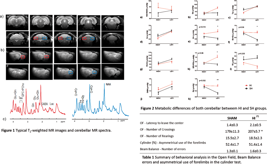
This study, to our knowledge, is the first to report extended effects of mild neonatal HI on cerebellar metabolism with extension to neuropathology of rats at their adulthood age. Taking together with behavior and motor performances, our in vivo1H-MRS findings suggest that effects of neonatal HI at PND3 on cerebellar metabolism of rats remains at adulthood. Collectively, our study provides relevant in vivo evidences that neonatal HI, one of prematurity disorders, affects brain pathology towards adulthood.
PB01-F01
NMDA attenuates the neurovascular response to hypercapnia in the neonatal cerebral cortex
1Department of Physiology, University of Szeged, Hungary
Abstract
Objectives
Spreading depolarizations (SDs) – propagating waves of almost complete neuronal depolarization associated with characteristic hemodynamic alterations-play an important role in the development of cerebrocortical lesions in traumatic brain injury and stroke. A single passing SD can abolish the vasodilatory cerebrocortical microvascular response to CO2 for hours indicating neurovascular unit dysfunction that can contribute to neuronal injury especially in metabolically already compromised cortical areas. The generation and the propagation of SDs involve glutamate release and critically depend on NMDA receptor activity, however, the selective contribution of NMDA receptor activation in altered microvascular reactivity after SD is unknown. The major objective of the present study was to investigate the effect of NMDA on hypercapnia-induced cerebrocortical microvascular responses. We utilized a neonatal piglet model, as the neonatal brain cannot yet generate SDs, so the effect of NMDA could be studied in the absence of a confounding NMDA-induced SD.
Methods
Anesthetized, mechanically ventilated newborn (<1 day old, n = 31) pigs were fitted with either closed or open cranial windows over the parietal cortex to study hemodynamic or electrophysiological responses to graded hypercapnia (5–10% CO2 ventilation) before/after topically applied NMDA (0.1–1 mM, dissolved in artificial cerebrospinal fluid; aCSF) assessed with laser-speckle imaging/contrast analysis and a 16-channel single-shank electrode, respectively.
Results
In control animals, graded hypercapnia resulted in repeatable, concentration-dependent increases in cortical blood flow (CoBF) and dilation of pial arterioles associated with changes in local field potential power in the delta and theta ranges originating in the deep cortical layers. NMDA also increased CoBF and pial arteriolar diameters that coincided with a delta-oscillation recorded at 4–600 µm cortical depth. Furthermore, after NMDA, the electophysiological response to hypercapnia was altered, whereas the hemodynamic response was virtually abolished. Pre/coapplication of MK-801 (0.1 mM, dissolved in aCSF) with NMDA prevented both the NMDA-induced increases in CoBF and also the attenuation of the microvascular reactivity to hypercapnia. In contrast, the selective neuronal nitric oxide synthase (nNOS) inhibitor (N (4S) 4 amino 5 [aminoethyl]aminopentyl N’ nitroguanidin; AAAN; 0.4 mg/kg iv) did not affect hypercapnia or NMDA-induced increases in CoBF, although it attenuated the pial arteriolar response to NMDA. However, AAAN significantly reduced the attenuation of the CoBF response to hypercapnia after NMDA.
Conclusions
NMDA receptor activation in a partially nNOS-dependent way can induce a neurovascular unit dysfunction in the newborn cerebral cortex that is similar to that observed after SD in adults. The induction of neurovascular unit dysfunction appears to be independent of the direct hemodynamic effects of NMDA-receptor activation and does not seem to require other factors associated with SD.
Support
Hungarian Brain Research Program 2.0 (2017–2.1 NKP 2017 00002), the EU-funded Hungarian grant EFOP-3.6.1-16-2016-00008 and the GINOP 2.3.2 15 2016 00034. V.K. is supported by OTKA-PD128464 from the NRDI.
PB01-F02
Neuroprotective effects of metabotropic glutamate receptor blockers on Ketamine and Dexmedetomidine induced neonatal brain injury in a Rat Model
1Tufts Medical Center and Tufts School of Medicine
Abstract
Introduction
Animal research and retrospective studies of infants and children younger than 3 years of age indicate that anesthetics and sedatives may alter synapto- and neurogenesis, and may contribute to neuronal cell death and neurological deficits including neurodevelopmental-related disorders, learning disabilities and attention deficit hyperactivity disorders (1). In neonatal rats the sudden reduced brain activity caused by anesthetic agents may lead to a maladaptive hyperexcitability and apoptosis that may be mediated by metabotropic glutamate receptor 5 (mGluR5). We hypothesized that the co-administration of a mGluR5 blocker 3-[(2-methyl-1,3-thiazol-4-yl) ethynyl]pyridine (MTEP) at the time of anesthesia will reduce or eliminate neuronal apoptosis. We focused on the striatum, which is important for brain functions ranging from motor control to cognition, including stimulus-induced learning. We present results from on-going studies aimed at characterizing the region-specific effects of MTEP on brain exposed to ketamine (Ket) and dexmedetomidine (Dex).
Methods
Following IACUC approval, we used postnatal day 7 (PND7) Sprague-Dawley neonatal rat pups in our experiments. We assessed the ability of MTEP to block the neuronal apoptosis elicited in neonatal rats by ket (20 mg/kg) and Dex (50 mcg/kg). We selected 2 different doses of MTEP (5 mg/kg (LD) and 10 mg/kg (HD) MTEP) for in vivoexperiments. Pups (n = 4/group) were intraperitoneally (IP)-injected with either saline (Veh), Ket, Ket + HD MTEP, Ket + LD MTEP, Dex, or Dex + HD MTEP. Four injections were administered at one-hour intervals, and the animals were sacrificed 3 hrs after the last injection. Immunohistochemistry was performed on brain sections using cleaved caspase-3 antibody that detects cells undergoing apoptosis. Imaging was performed using KEYENCE high-resolution digital microscope. Striatum of the second set of animals IP-injected with either saline, Ket or Ket+HD MTEP was microdissected and processed for western blot analysis.
Results
Compared to Veh (Fig 1, a) Ket increased caspase-3 expression (Fig 1, b) in the striatum. HD MTEP reduced ket-induced apoptosis of brain cells in the striatum where caspase-3 uptake was greatly down-regulated in ket+HD MTEP injected pups (Fig 1, c) compared to Ket-injected pups (Fig. 1, b). There was a similar trend in the ket+LD MTEP group. Compared to Veh, Dex also increased caspase-3 expression, and this increase was not affected by MTEP co-injection. The results of the immunohistochemical studies of Veh, Ket or Ket+HD MTEP injected animals were confirmed with western blot analysis (p < 0. 05).
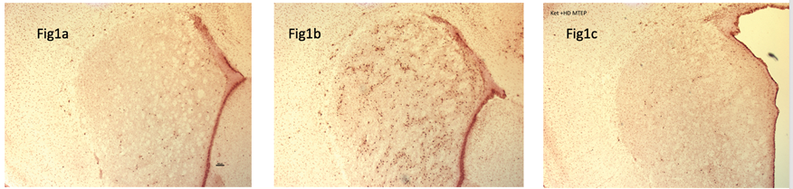
Conclusion
This finding may be a promising indication of MTEP mediated neuroprotective effects for ketamine-induced neurotoxicity in the striatum. These neuroprotective effects of MTEP for ketamine-induced brain injury were dose dependent as high dose MTEP had a significantly higher neuroprotective effect than low dose MTEP. Dex-induced neurotoxicity did not appear to be affected by MTEP in our study, suggesting a different injury mechanism for this agent. However, further studies are required to investigate the effects of MTEP on other brain regions, and to determine the clinical impact of MTEP on the sedative anesthetic effects of ketamine and neurobehavioral outcomes.
PB01-F03
Early calcium responses and AQP4 changes in reactive astrocytes after juvenile mild traumatic brain injury
1CNRS UMR 5287, University of Bordeaux, France
2Departments of Medicine and Physiology, University of California, San Francisco, CA, USA
3CNRS UMR 5297, University of Bordeaux, France
4Basic Science Department, Loma Linda University School of Medicine, CA, USA
Abstract
Objectives
Traumatic brain injury (TBI) is the first cause for emergency department visits in the pediatric population with a majority of mild TBIs. Even for mild TBI, pediatric patients suffer long-term cognitive and emotional impairments but the underlying cellular and molecular mechanisms are unknown. Astrocytes are known to respond to an injury by becoming “reactive”. We hypothesized that astrocyte dysfunction is contributing to the poor outcome at long-term, with phenotype changes and impaired calcium signaling in astrocytes. Previously, the water channel aquaporin 4 (AQP4) has been proposed to initiate calcium-signaling events in astrocytes. We hypothesized that AQP4 changes contribute to early changes in calcium signaling after mild TBI and that absence of AQP4 would restore calcium dynamics in astrocytes after injury.
Methods
We used a mild TBI model, CHILD (
Results
Using two-photon calcium imaging, functional evaluation of astrocytes was made by measurement of the calcium response to ATP in cortical astrocytes at 1 and 3 days after CHILD. Calcium responses after ATP application were delayed and longer-lasting in astrocytes after 1 day of CHILD mice compared to shams. This more sustained calcium response reflected an increased number of calcium transients compared to sham astrocytes. However, there was no change in the astrocytic calcium responses between sham and CHILD at 3 days post-injury. Despite the absence of neuronal cell death, GFAP expression and the number and length of astrocytic processes were increased in the ipsilateral cortex of CHILD animals at both time points. However, AQP4 expression exhibited biphasic changes, as observed for the changes in calcium response, showing an increase at 1 day and a decrease at 3 days after injury. We then assessed the potential role of AQP4 in calcium response by using AQP4-/- mice. Interestingly, calcium responses after ATP application were delayed in sham AQP4-/- compared to sham wild type mice, suggesting that AQP4 channels are involved in ATP-driven calcium responses. In AQP4-/- animals we did not observe any differences in the calcium responses between CHILD and sham animals (neither at 1 nor 3 days post-injury), indicating that AQP4 is necessary for the changes in calcium responses to ATP after CHILD.
Conclusions
Our results showed early major morphological and functional changes in astrocytes after mild juvenile TBI. Therefore, even mild TBI may induce astrocytopathy and present a target for treatment development.
Reference
PB01-F04
Brain structure, functional connectivity and cerebrovascular reactivity outcomes of mitochondrial treatment for developmental traumatic brain injury
1Department of Radiology and Biomedical Imaging, Yale University School of Medicine, 330 Cedar St, New Haven, CT 06520
2Department of Pharmacology, Physiology and Neuroscience, Rutgers Biomedical and Health Sciences-New Jersey Medical School, Medical Science Building, 185 South Orange Ave, Newark, NJ 07101
3Department of Biomedical Engineering, Yale University, 10 Hillhouse Ave, New Haven, CT 06520
4Department of Radiology, Rutgers Biomedical and Health Sciences -New Jersey Medical School, ADMC-5 Room 575, 30 Bergen St, Newark, NJ 07103
Abstract
Objectives
Traumatic brain injury (TBI) pathology affects multiple cellular types and hence different brain structural and neurophysiological variables. Early energy metabolic failure and mitochondrial dysfunction play a predominant role in injury outcome. But brain mitochondria are functionally diverse depending on their cellular location, cell type and brain anatomical region. Our recent MRI-based multimodal imaging has enabled the simultaneous determination of brain parenchymal microstructure, neural connectivity and cerebrovascular functional outcomes after TBI in developmental age rats. Using the developed multimodal imaging of TBI along with a mitochondria-targeted treatment, we tested the mitochondrial diversity hypothesis. Rats in the developmental age (P31) were subjected to a mild/moderate intensity TBI and treated with vehicle or Kaempferol, a mitochondrial Ca2+ uniporter channel activator and shown to improve energy metabolism, neural viability, behavior and brain responses after TBI (Murugan et al., Frontiers in Neuroscience; 2017; Chitturi et al., Neurochem Int; 2018).Kaempferol’s impact on synaptic/axonal microstructural and neurophysiological variables such as neural connectivity and cerebrovascular reactivity were determined..
Methods
Rats were subject to lateral fluid percussion injury (FPI). Adolescent imaging outcomes of developmental TBI in the rat model (2-months post-injury) were determined using functional Magnetic Resonance Imaging (fMRI) and Diffusion Tensor Imaging (DTI). Animal groups ie., TBI+vehicle (n = 5) and TBI+Kaempferol treatment (n = 8) underwent the fMRI and DTI measurements for the present study. Imaging was performed 8–10 weeks later using a Bruker 9.4T spectrometer and an ellipsoidal surface coil (5 x 3 cm). Animals were anesthetized using urethane (1.3 mg/kg body weight, intraperitoneal), and body temperature was monitored throughout the procedure and maintained at 35–37°C.
Results
fMRI based resting state functional connectivity (RSFC) – a marker of neural connectivity, was significantly increased between vehicle and Kaempferol-treated TBI animals. RSFC strength significantly improved in the cortex and hippocampus with corresponding increases in the spatial extent of the corresponding RSFC networks. However, no significant RSFC changes were observed in the thalamus. DTI measures of fractional anisotropy (FA) and mean diffusivity (MD), reporting on axonal and microstructural integrity of the brain, showed significant increases with Kaempferol treatment, with highest changes in corpus callosum, internal capsule and cingulum regions. Kaempferol treatment also improved corpus callosal FA, indicating measurable improvement in the inter-hemispheric structural connectivity. Global cerebrovascular changes assessed with C02 challenge revealed nosignificant effects with Kaempferol treatment.

Conclusion
Different multimodal imaging outcome results demonstrate the heterogeneous impact of mitochondrially targeted Kaempferol treatment across various brain compartments affecting relevant neurophysiological outcomes in vivo. The results support mitochondrial heterogeneity in the working brain and potentially help optimize mitochondrially targeted treatments.
Acknowledgements
Funding from the New Jersey Commission for Brain Injury research CBIR12PIL028 (SK), NS097750 (VS), R01 MH-067528 (FH) and P30 NS-052519 (FH).
PB01-F05
Mesenchymal storomal cell delivery through cardiopulmonary bypass for neuroprotection in a juvenile porcine model
1Children’s National Heart Institute, Children’s National Health System, Washington, DC, USA
2Center for Neuroscience Research, Children’s National Health System, Washington, DC, USA
3Department of Radiology and Nuclear Medicine, Children's National Health System Washington, DC, USA
4Departments of Pharmacology and Biochemistry and Molecular Biology, Institute for Personalized Medicine, Pennsylvania State University College of Medicine, Hershey, PA, USA
5Program for Cell Enhancement and Technologies for Immunotherapy, Division of Blood and Marrow Transplantation, Center for Cancer and Immunology Research, Children's National Health System Washington, DC, USA
6Frank Laboratory and Laboratory of Diagnostic Radiology Research, Department of Radiology and Imaging Sciences, National Institutes of Health, Bethesda, MD, USA
Abstract
Objectives
Neurodevelopmental impairment is emerging as one the most important current challenges for survivors after pediatric cardiac surgery. Cardiopulmonary bypass (CPB) can cause substantial systemic inflammation and trigger prolonged microglial activation in the brain. Mesenchymal stromal cells (MSCs) have significant immunomodulatory properties and regulate microglia activation. We hypothesize that intra-arterial MSC delivery through CPB is neuroprotective by modulating systemic and neuro-inflammatory responses.
Methods
Two-week old piglets (n = 16 total) were randomly assigned to one of 3 groups: (1) Control, (2) Deep hypothermic circulatory arrest (DHCA), (3) DHCA followed by MSC administration. In group 3, 18F-FDG or superparamagnetic iron oxide (SPIO)-labeled MSCs (10x10^6 per kg) were delivered through CPB during the rewarming period. Positron emission tomography (PET) was performed at 1 hr after MSC delivery to determine the whole body distribution of cells with 18F-FDG. Animals were sacrificed 3 hrs after CPB for the analysis with magnetic resonance imaging (MRI) and immunohistochemistry. In order to assess transcriptomic effects of MSC treatment, total RNA was extracted from cortical tissue of animals from each group and subjected to RNA sequencing, followed by differential expression analysis using the DESeq2 statistical package. Gene ontology (GO) analysis was performed using Enrichr on differentially expressed genes (DEGs) whose normalized read count displayed a rescue profile upon MSC delivery. Differential expression of key target genes was validated using qRT-PCR. Plasma cytokine/chemokine levels were determined by multiplex immunoassay. Clinically-relevant physiological biomarkers determined the effect of MSC delivery on multi-organ functions.
Results
It has been well demonstrated that intra-venous injection of MSCs resulted in high accumulation of cells primarily into lungs. In contrast our PET study showed that intra-arterial delivery through CPB uniformly distributed MSCs to all organs analyzed such as the brain, heart, and kidney except that lungs and intestine showed lower uptake. T2* weighted brain MRI showed diffuse distribution of hypointense voxels (SPIO particles) throughout the entire brain. Immunohistochemistry revealed an even distribution of SPIO-labeled MSCs within the cortex and white matter. We have previously demonstrated an increase in permeability of the blood-brain barrier after DHCA. Consistently we identified MSCs located in the extra-vascular space. Analysis of the RNA sequencing data identified 262 DEGs between the DHCA and DHCA/MSC groups. Of these, 53 upregulated genes were significantly enriched for WNT signaling in the GO analysis, indicating a potential mechanism through which MSCs mediate their neuroprotective effects. MSC delivery through CPB modulated plasma cytokine/chemokine expression following surgery. In the brain MSC treatment reduced microglia expansion/activation and inhibited caspase activation resulting from CPB. Various biomarkers after MSC delivery did not differ compared with CPB group. No evidence of either embolic events or microstrokes were observed by MRI and histology.
Conclusions
MSC delivery during CPB is highly effective and shows translational potential to minimize CPB-induced systemic inflammation and reduce microglial expansion and caspase activation in children with CHD.
PB01-F06
Cerebellar development in preterm infants at term-equivalent age: Assessment using MRI
1Neuroscience Research Institute, Gachon University, Incheon, Korea
2Division of Neonatology, School of Medicine, Ewha Womans University, Seoul, Korea
3Division of Neonatology, Gachon University, Gil Hospital, Incheon, Korea
Abstract
Introduction
The cerebellum has been suggested as a pivotal region that is linked with motor, cognitive, and behavioral impairment in preterm infants. Therefore, preterm birth often is associated with an underdeveloped cerebellum, specifically cerebellar volume reduction. The aim of our study was to investigate cerebellar development in preterm infants compared with healthy full-term infants, with adjustment for clinical risk factors that may affect cerebellar development.
Methods
Subjects: We obtained 88 infants who were obtained on 16 full-term control infant and 72 preterm infants at term-equivalent age. The mean gestational age for preterm infants and full-term infants was 28+4 weeks and 37+4 weeks, respectively. 3D T1-MPRAGE and DTI images were obtained using 3T MRI (Verio;Siemens, Germany) with a Siemens matrix coil. The 3D T1-MP RAGE imaging parameters used were as follows: TR = 1900 ms, TE = 2.97 ms, FA = 9°, pixel bandwidth = 170 Hz/pixel, total acquisition time = 3 minutes, 2 seconds, iso-voxel resolution 1.0 mm. The DTI sequence parameters were as follows: b = 0 and 700 s/mm2, number of diffusion gradient directions = 30, TR = 6600 ms, TE = 74 ms, slice thickness = 1.8 mm, FOV = 230 mm, matrix = 128 × 128, total acquisition time = 7 minutes, 36 seconds.
Data analysis: Cerebellar volume was measured by manually outlining the cerebellar tissue on each cerebellar section with the 3D Slicer, version 4.3.1 (http://www.slicer.org/). (Figure 1. A.). DTI images were processed offline with the FMRIB Software Library (FSL, Oxford, United Kingdom). To investigate the relationship between clinical variables and cerebellar volume or DTI parameters, such as FA, ADC, and axial and radial diffusivities, we selected regions of interest in the superior cerebellar peduncle (SCP, at the level of the decussation) and middle cerebellar peduncle (MCP; at the level of the pons) from the FA color map. (Figure 1. B.)
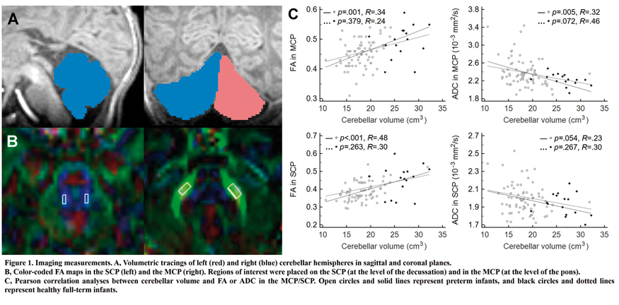
Results
Compared with full-term infants, preterm infants showed smaller cerebellar volumes and a lower FA, greater ADC, and increased radial diffusivities in the cerebellar peduncles (all P<.05) Figure 1. C presents results of the Pearson correlation analyses between cerebellar volumes and FA/ADC of the cerebellar peduncles. In preterm infants, cerebellar volume was significantly correlated with FA/ADC of the cerebellar peduncles, respectively (all P<.01), with the exception of ADC of the SCP (P = .054). No significant correlations were observed in full-term infants.
Conclusion
Preterm infants without severe brain abnormalities showed impaired cerebellar development at term-equivalent age after we controlled for PMA at the time of the scan. Cerebellar volume correlated with FA or ADC of the cerebellar peduncles in preterm infants. These findings suggest that preterm infants has potential harmful effects on cerebellar development.
PB01-G01
Hyperglycemia increases regional brain glucose consumption but not oxygen consumption or blood flow
1Dept. of Radiology, Washington University School of Medicine
2Dept. of Pediatrics, Washington University School of Medicine
3Dept. of Neurology, Washington University School of Medicine
4Dept. of Psychiatry, Washington University School of Medicine
5Dept. of Biomedical Engineering, Washington University in St. Louis
Abstract
Introduction
Studies in humans (1) and rodents (2) indicate that hyperglycemia increases brain glucose metabolism in certain brain regions. In humans this occurs predominantly in white matter. It is less clear how brain blood flow and oxygen consumption are affected. Here we measured regional brain glucose, oxygen consumption, and blood flow in humans using glucose-clamped hyperglycemia.
Methods
The plasma glucose levels of 26 healthy young adults were clamped at either euglycemia (90–100 mg/dL) or hyperglycemia (250–300 mg/dL). PET acquisition included 18FDG, H215O, O15O for the measurement of glucose metabolism, blood flow, and oxygen consumption, respectively. For relative, topographical analysis, PET images were normalized to whole-brain tracer uptake. To enable the quantification of the cerebral metabolic rate of glucose (CMRglc), arterial sampling was also collected in a subset of participants (n = 12). MRI acquisition included pseudo-continuous arterial spin labeling (ASL) for the quantification of cerebral blood flow (CBF). Bilaterally symmetric regions of interest were defined using FreeSurfer 5.3. Conditions were compared using a linear mixed model, and multiple comparisons were corrected for by controlling the false discovery rate.
Results
Hyperglycemia significantly altered the topography of brain glucose metabolism whereas the topography of oxygen consumption was unaffected, indicating a selective change in glycolysis. No changes in the topography of blood flow were observed. Quantitatively, hyperglycemia significantly increased CMRglc in several white matter regions (Figure 1A). For example, CMRglc in the deep white matter increased from 15.1 ± 1.7 to 24.8 ± 1.3 (p < 0.0001; Figure 1B). Although CMRglc did increase in the gray matter of the cerebellum and medial temporal lobe, these changes were not significant. Small, non-significant, decreases in CMRglc were found In the majority of cortical gray matter regions.
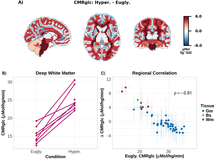
Regional changes in CMRglc were highly correlated with baseline metabolic rates, with larger changes in CMRglc occurring in regions with lower baseline CMRglc. (Figure 1C). No quantitative changes in CBF were significant.
Discussion
Despite significant hyperglycemia-induced changes in glucose metabolism, we observed no change in regional blood flow or oxygen consumption. The increase in CMRglc in the absence of an increase in oxygen consumption indicates that hyperglycemia selectively increases glycolysis, a finding consistent with earlier work in mice (2). This selective increase in glycolysis was driven by an increase in CMRglc within white matter, confirming earlier work in humans (1). The effect of hyperglycemia on white matter has so far eluded explanation and, thus, is deserving of our attention. The importance of this issue is reinforced by the observation that experimentally-induced hyperglycemia is associated with both increased glycolysis and elevated levels of amyloid-beta in mice (2). Furthermore, Type 2 diabetes mellitus, which is associated with chronic hyperglycemia, is a significant risk factor for Alzheimer’s disease.
References
PB01-G02
Acute insulin-induced hypoglycemia impairs mitochondrial respiration and vasoreactivity in brain microvasculature without altering blood-brain-barrier permeability
1Dept. of Pharmacology, Tulane University School of Medicine
2Neuroscience Program, Tulane Brain Institute, Tulane University
Abstract
Background
Microvasculature in the brain serve unique functions in providing the blood-brain barrier (BBB) and contributing to the neurovascular coupling. Furthermore, mitochondrial dysfunction plays a critical role in the cerebrovascular pathologies including diabetes and stroke. Insulin therapy for patients with diabetes carries the risk of acute hypoglycemia. Recurrent hypoglycemia increases the ischemic brain injury in subjects with diabetes receiving insulin treatment, but the underlying mechanisms were unknown.
Objectives
First, to develop a novel high-throughput assay for measuring mitochondrial respiration of brain microvessels. Second, to investigate for the first time the effect of acute hypoglycemia on mitochondrial respiratory function in the isolated brain microvasculature (ex vivo) and BBB (in vivo).
Methods
Three-months-old C57bl6 mice were divided into three groups; namely, control (euglycemia), hypoglycemia and euglycemia with hyperinsulinemia (Eug+HyperIns) (n = 6 each). Euglycemia mice were injected intraperitoneally with saline; hyperglycemia mice were injected with insulin (3U/Kg body weight) to induce and maintain 90-minutes hypoglycemic state (<70 mg/dL); and Eug+HyperIns mice were injected with insulin+20% dextrose. Brains were excised and microvessels (40µ to 100µ diameter) were isolated. Oxygen consumption rates (OCR) were measured using Seahorse XFe24 analyzer, normalized to protein, and expressed as picomoles of Oxygen consumed per µg protein. Respiratory parameters were calculated from OCRs. For two-photon imaging experiments, mice were prepared for in vivo imaging by removing a 4 mm diameter sector of bone over the somatosensory cortex and sealing it with a glass coverslip, cyanoacrylate, and dental acrylic. Mice were held hypoglycemic (using 1–2 U/kg insulin) during imaging and compared with a baseline imagining session one day before.
Results
Brain microvessels from hypoglycemia group exhibited a significant decrease in maximal respiration (by 47.3%, p = 0.01) and spare respiratory capacity (by 76%, p = 0.0004) when compared to the saline group. Similarly, microvessels from hypoglycemia group showed a tendency towards a decrease in basal respiration, protein leak, and non-mitochondrial respiration when compared with the microvessels from the control group. Western blot analysis revealed that acute hyperglycemia decreased the expression of glucose-regulated protein 75 (GRP75) and the activity of type 1 protein arginine N-methyl transferase (PRMT), two key regulators of microvascular mitochondria. Interestingly, microvessels from Eug+HyperIn mice failed to exhibit mitochondrial respiratory dysfunction and the associated molecular changes, indicating that the observed microvascular changes in response to acute hypoglycemia were induced by hypoglycemia rather than by hyperinsulinemia. Our in vivo imaging studies revealed that acute hypoglycemia induced a significant increase in cerebral microvessel diameter., which was not present in saline controls (diameter increased by 10 ± 1.7%, N = 7 mice, n = 89 microvessels, vs. 1.751 ± 1.0% N = 6 mice, n = 134 microvessels, in saline controls, p = .0007). However, BBB permeability was unaffected by hypoglycemia.
Conclusions
We conclude that acute hypoglycemia induces impaired mitochondrial respiratory dysfunction in the cerebral microvasculature of healthy mice potentially linking hypoglycemia and cerebrovascular dysfunction that may be mediated by GRP75 and PRMT. Support: National Institute of Health: National Institute of General Medical Sciences and National Institute of Neurological Disorders and Stroke (Katakam: R01NS094834); National Institute of Aging (Mostany: R01AG047296).
PB01-G03
Ketone body prevents the induction of PTZ kindling and the reduction of glucose metabolism in mouse brain
1Division of Health Sciences, Graduate School of Medicine, Osaka University, Japan
Abstract
Objectives
The ketogenic diet has been used for the treatment of intractable epilepsy. There still remain many unanswered questions regarding the antiepileptic action, a hypothesis that the changes of energy metabolism in the brain is involved in is proposed. In our previous study, aliphatic ketones, methyl ethyl ketone (MEK) and diethyl ketone (DEK), suppressed status epilepticus in lithium-pilocarpine epileptic model in rats (1) and electrical- and chemical- induced seizure in mice (2). In this study, we evaluated the effects of MEK on kindled seizure induction and the changes of cerebral blood flow and metabolism in pentylenetetrazole (PTZ)-induced kindling mice model.
Methods
Male ICR mice received daily i.p. injections of saline, MEK, or subconvulsive dose of PTZ. The behaviors were observed for 30 min after the PTZ administration. Completion of kindling is defined as the tonic-clonic seizures evoked over five consecutive days. The next day of the first tonic-clonic seizure, PTZ treated mice were divided into two groups, and saline or MEK was administered each day 15 min before PTZ injection. Two hours after the last dose of PTZ, the mice were intravenously injected with [14C]2DG or [14C]IMP for ex vivo autoradiography. All the animal experiments were approved by the Institutional Animal Care and Use Committee, Division of Health Sciences, Graduate School of Medicine, Osaka University.
Results
Compared with control group, repeated PTZ administration induced the reduction of [14C]2DG in brain, while there were no significant alterations in cerebral blood flow. The reduction of [14C]2DG uptake in PTZ treated mice occurred only in brain, and did not occur in heart and muscle. MEK pretreatment prevented the second tonic-clonic seizure and the reduction of [14C]2DG in brain by PTZ. Repeated MEK alone administration did not affect [14C]2DG in brain.
Conclusions
MEK successively prevents the development of PTZ kindling in mice. This anticonvulsant effect was accompanied by brain energy metabolism alterations and radiolabeled 2DG imaging can be useful for monitoring and assessing the therapeutic efficacy.
References
PB01-G04
Regional difference in energy metabolism in murine hippocampus
1Department of Physiology, Faculty of Welfare and Health Sciences, Oita University, Japan
2Medical Student, Oita University Faculty of Medicine, Japan
3Department of Physiology, National Defense Medical College, Japan
4JASDF Aeromedical Laboratory, Japan
5Department of Nursing, Faculty of Health Care, Kiryu University, Japan
6Department of Anesthesiology, Oita University Faculty of Medicine, Japan
7Department of Molecular Anatomy, Hamamatsu Medical School, Japan
Abstract
Objectives
Energy metabolism differs among regions in hippocampus. For example, in a murine model of kainate-induced seizure, reduction in ATP was significant in CA3 region.1 However, regional difference in correlation of hippocampal high-energy phosphates (ATP, ADP) has not been illustrated as far as the authors surveyed. The aim of the present study was to investigate regional correlation of ATP and ADP in murine hippocampus using matrix-assisted laser desorption/ionization (MALDI) imaging mass spectrometry (IMS).
Methods
An anesthetized C57BL/6NCrSlc male mouse (urethane, i.p.) was placed on the centrifuge for small animals (Tomy Seiko, Tokyo, Japan). +8Gz acceleration was applied to the mouse for 30 s to make whole brain ischemia. The brain was in-situ frozen by isopentane-propane freezing mixture (−193°C) before (sham, n = 5) and immediately after the ischemia (n = 6). Coronal sections (10 µm-thick) at the hippocampal level were made with a cryotome and thaw-mounted on indium tin oxide-coated glass slides. MALDI IMS was conducted in the negative ionization mode with 9-aminoacridine as the matrix using RapiFlex MALDI-time-of-flight mass spectrometry (Bruker Daltonics, Leipzig, Germany). Since ATP and ADP were localized in the cell layers of hippocampus,1 pixels (20 µm-pitch) in pyramidal and granule cell layers of cornu Ammonis (CA) and dentate gyrus (DG) were analyzed (40–50 pixels/region for each mouse). The energy status was evaluated by two-dimensionally plotting normalized ATP (nATP) against ADP (nADP), which gave detailed information on energy metabolism of each pixel (ATP-ADP plot); nATP=[ATP]/([ATP] + [ADP] + [AMP]), nADP = [ADP]/([ATP] + [ADP] + [AMP]). For comparison, energy charge (EC) index1,2 were also calculated; EC = ([ATP] + 1/2[ADP])/([ATP] + [ADP] + [AMP])
Results
In samples before the ischemia, ATP-ADP plot differed significantly among CA1, CA3 and DG in all mice (MANOVA, p < 0.001 for each mouse). The representative graph in a sham operated animal is shown; data points of CA3 were plotted upper left and those of DG lower right, indicating the energy status being CA3 > CA1 > DG. There were also significant differences in EC among CA1, CA3 and DG for all five mice examined (ANOVA, p < 0.001 for each mouse). EC was the highest in CA3 and the lowest in DG. After the ischemia, regional differences in ATP-ADP plot and EC were observed in all centrifuged mice (p < 0.001), although no consistent tendency was obtained regarding the order of energetic status among regions.

Conclusions
In the present study, ATP-ADP plot successfully visualized differences in correlation between ATP and ATP among hippocampal regions, which was in accordance with EC. It is of interest that regional differences were also observed immediately after the ischemia. It might suggest possible interregional differences in energetic response to ischemia-reperfusion; i.e., ATP depletion during ischemia and its recovery after reperfusion).
References
PB01-G05
Human brain gray matter energy mapcomputed on the basis of cellular staining from BigBrain
1Dept. of Biophysics, Fudan University, China
2Magnetic Resonance Research Center, Quantitative Neuroscience with Magnetic Resonance Core Center, Department of Biomedical Engineering, Department of Molecular, Cellular and Developmental Biology, Yale University, New Haven, CT 06520, USA
Abstract
Neuronal-glial cell crosstalk in cerebral cortex is vital for consciousness, which craves energy from glucose oxidation (CMRglc(ox)). Since energy availability limits coding and structural needs of cortical networks, we computed a CMRglc(ox) model of signaling and non-signaling processes where metabolic-morphometric measures from human gray matter offered validation, and PET provided 3D mapping of awake CMRglc(ox). Using a stereological cellular atlas, BigBrain, we developed BigBrainActivity_GM, a public tool referenced to Brodmann atlas, in order to explore the activity of human cytoarchitecture in 3D. Neuropil density (∼0.7 g/mL) portrayed anatomical energy demands, with remaining resources assigned to cortical firing (∼1 Hz). Energy restrictions on signaling (∼0.7CMRglc(ox)) and non-signaling (∼0.3CMRglc(ox)) demands were stable brain-wide. The model also enlightened how particular cortical regions utilize energy in supporting cognitive activity with relatively high response sparseness in cortical coding while most of others in low sparseness during resting eye-closed state. BigBrainActivity_GM with energy-constrained cortical networks enables study of neuronal-glial processes underlying human cortical function, in health and disease.
PB01-G06
Computer-aided construction of metabolic pathway models for brain energy metabolism and synaptic neurotransmission
1Weill Cornell Medicine, USA
Abstract
Objectives
To develop a computer-aided means, consistent with prior methods (Maciejewski and Rothman, 2008), to construct metabolic pathway models for brain energy metabolism and synaptic neurotransmission that are composed of individual molecular processes, strictly consistent with stoichiometric principles, and perform specific functions (e.g., packing neurotransmitter into synaptic vesicles).
Methods
A computer application automatically constructs metabolic pathway models via systematic application of stoichiometric principles given user specification of (a) relevant molecular enzymatic and transport processes, including their cellular and sub-cellular locations, and (b) key metabolic functions of the resultant models. A library of enzymatic and transport processes, consistent with public sources (e.g., BRENDA, ExPASy, Alexander et al. 2013), serves as a resource for user process and function specifications. The user defines compartments (e.g., cell types, mitochondria within cells, plasma and mitochondrial membranes, etc.), assigns enzymes and transporters to compartments, specifies the overall net reaction for the pathway (e.g., glucose oxidation), and specifies localized enzymatic and/or transport processes that must be included in the model. The application then computes the relative rate of each process within a metabolic pathway model that satisfies the user imposed compartmentalized structure and net reaction and process inclusion/exclusion constraints.
Results
Figure 1 displays details of a metabolic pathway model for glucose oxidation, GABA synthesis and GABAergic neurotransmission constructed using this computer-aided approach.

In this model, neurotransmitter GABA is released into synaptic clefts by presynaptic neurons and taken up by astrocytes. Nitrogen balance is maintained between presynaptic neurons and astrocytes by the exchange of 2 GABA molecules (via neurotransmission) for 1 glutamine molecule (via SNATs).
In astrocytes, 2 neurotransmitter GABA molecules along with 2 OG molecules enter a “GABA shunt”, i.e., a combination of GABA transaminase (ABAT) and succinic semialdehyde dehydrogenase (SSADH), to produce 2 glutamate and 2 succinate molecules. One glutamate molecule is converted to glutamine via glutamine synthetase (GS) and the other is converted to OG via glutamate dehydrogenase (GDH). One succinate molecule is transported to the presynaptic neuron (via dicarboxylate transporters) and the other is metabolized via tricarboxylic acid (TCA) cycle enzymes (supported by pyruvate derived from glycolysis) to produce 1 molecule of OG. The 2 OG produced from 1 glutamate and 1 succinate support the operation of the “GABA shunt.”
In presynaptic neurons, 1 neurotransmitter GABA molecule is produced from glutamine via phosphate activated glutaminase (PAG) and glutamic acid decarboxylase (GAD) and another is synthesized from succinate via TCA cycle enzymes (supported by pyruvate derived from glycolysis), GDH, PAG and GAD.
The net reaction for the entire metabolic pathway for GABAergic neurotransmission is simply the complete oxidative metabolism of glucose, i.e., 1 glucose + 6 O2 → 6 CO2 + 6 H2O.
Conclusions
This computer-aided approach to the construction of metabolic pathway models provides a solid foundation for developing or clarifying theories, planning experiments, and performing numerical simulations of brain energy metabolism and synaptic neurotransmission.
References
PB01-G07
Simultaneous glucoCEST and optical measurements of glucose in the brain
1Institute of Pharmacology and Toxicology, University of Zurich, Zurich, Switzerland
2Neuroscience Center Zurich, University and ETH Zurich, Zurich, Switzerland
3Department of Biochemistry and Biophysics, Perelman School of Medicine and Department of Chemistry, School of Arts and Sciences, University of Pennsylvania, Philadelphia, USA
Abstract
Introduction
Glucose is the brain's main energy source and its uptake and metabolism are sensitive biomarkers for neurodegenerative diseases, cancer and stroke. Glucose Chemical Exchange Saturation Transfer (glucoCEST) is an MRI-based approach that is used to amplify glucose detectability and it has been proposed for tumor imaging, mainly because it is radioactivity-free and there is large availability of MRI scanners [1]. Even though the main principle of glucoCEST is clear, the exact compartmental origin of the signal (i.e. vascular, extracellular, intracellular) needs further investigation in the brain [2].
3-O-methyl-D-glucose (3OMG) is used as a glucoCEST contrast agent. Furthermore, it is known to trans-accelerate the glucose transporter GLUT1 in erythrocytes. Trans-acceleration occurs when hexose molecules are presented on the trans side, which allows the transporter to return faster to the cis side with a bound substrate. In practice, the extracellular presence of 3OMG stimulates the exit of intracellular glucose leading to a decrease of the fluorescent nanosensor signal. The initial slope of the reduction due to trans-acceleration, can be used as a marker for the rate of glycolysis in astrocytes and neurons.
Objectives
The aim of this study was to assess astrocytic and neuronal glucose levels and metabolic rates upon administration of 3OMG using Single-Photon Excitation and Emission Detection (SPEED) and then correlate these measurements with glucoCEST, to determine the contribution of astrocytes and neurons to the overall brain signal.
Methods
We performed simultaneous glucoCEST and SPEED measurements of intracellular glucose concentration in the mouse neocortex upon intravenous infusion of 3OMG, insulin and glucose with galactose, successively.
For SPEED recordings, the genetically encoded Förster Resonance Energy Transfer (FRET) nanosensor FLII12Pglu600μΔ6 (FLIIP) was used [3]. Constructs coding for this sensor were injected into somatosensory cortex of adult mice using an adeno-associated viral approach and specific promotors were used for either astrocytic (short GFAP promotor) or neuronal (human Synapsin promotor) expression.
Results
We observed trans-acceleration in astrocytes (mainly expressing GLUT1) and neurons (mainly expressing GLUT3).
Insulin and glucose with galactose were used to reach the minimum and maximum of the nanosensor dynamic range, for two-point calibration. Normalization to these points revealed higher baseline FLIIP signals in astrocytes.
Conclusions
We demonstrate the feasibility of simultaneous glucose level measurements using SPEED and glucoCEST in vivo.
Fluorescence measurements during glucoCEST MRI give immediate validation of the influx of 3OMG into neurons and astrocytes, by observation of trans-acceleration. Despite lower baseline FLIIP measurements in neurons than in astrocytes, both cell types exhibited trans-acceleration.
In next steps, experiments with astrocyte-specific GLUT1 knock-out mice will be performed in order to directly investigate the role of astrocytic glucose uptake for the glucoCEST signal.
References
PB01-H01
Glucosamine a non-toxic candidate for metabolic imaging of the healthy brain
Department of brain repair and rehabilitation, Institute of Neurology, UCL
Abstract
Recently, a new MRI technique based on the detection of glucose and glucose analogues through chemical exchange has been shown to provide a read out of tissue metabolic activity, primarily in body tumors (1–3). Other molecules such as 2-DG may be better alternatives because their phosphorylated products accumulate thereby enhance the detected signal (3). However, they may be toxic at high concentration. Non-toxic alternatives, such as 3-OMG or glucosamine have also been proposed as alternative contrast agents in tumours (3). Here, glucosamine was tested as a potential candidate for metabolic imaging of the healthy brain.
A 9.4 T Agilent scanner was used to image six healthy SD rats before and after per ost (PO) administration of glucosamine. The rats were fasted overnight with water access, anesthetised with isofluorane (1–1.5%), and scanned with a 72 mm volume coil for radio frequency (RF) transmission and a two-element receive array coil.
CEST measurements were acquired using a gradient-echo sequence from a single 2 mm axial slice of the brain with a saturation train of 30 Gaussian pulses at 5.0 µT. Saturation was applied at 71 frequency offsets between −5.0 and 5.0 ppm. B0 correction used the minimum of a fitted spline. CEST images were acquired before and for 1 hour after glucosamine administration. Glucosamine enhancement plots were calculated as 1-Mz (1.0–2.5 ppm)/M_0 in three selected regions (i.e. thalamus, cortex and basal ganglia).
CEST MRI experiments were performed in 5 SD rats with a dose of 1.5 g/kg glucosamine. Another 2 rats served as controls by administrating the same amount of saline without glucosamine. Significant CEST effects were detected for more than hour following administration of glucosamine in cortex, thalamus and basal ganglia. No measureable CEST differences were obtained upon saline administration. Figure 1 the averaged signals from thalamus, cortex and basal ganglia in all rats administrated with or without glucosamine.

This study provides further evidence of the use of glucose analogues such as glucosamine to study brain metabolism. Here the signal reaches a maximum and stays around this value for up to an hour possibly indicating that the metabolic products of glucosamine are accumulated in the cells with limited phosphorylation. It is worth to point out that glucose transporters GLUT1 and GLUT2 have similar affinity for glucose however, in the case of glucosamine GLUT2 has about 20-fold higher affinity thus making this agent more efficient to be used clinically (3). Future studies aim to use this agent clinically for assessing both healthy brain metabolism and metabolic disturbances such as brain tumours.
References
PB01-H02
Neuronal energy metabolism in the mouse brain measured by1H-[13C] MRS at 14.1T under [3-13C]-Lactate infusion: a feasibility study
1Laboratory of Functional and Metabolic Imaging, Ecole Polytechnique Fédérale de Lausanne (EPFL), Lausanne, Switzerland
2Center for Biomedical Imaging (CIBM), Ecole Polytechnique Fédérale de Lausanne (EPFL), Lausanne, Switzerland
3Faculty of Medicine, University of Geneva, Geneva, Switzerland
Abstract
Objectives
The aim of this study was to demonstrate the feasibility of in vivo indirect1H-[13C]-MRS to characterize13C turnover curves of cerebral lactate C3, alanine C3, glutamate and glutamine carbon positions and other labeled amino acids in the mouse brain at 14.1T following brain uptake and metabolism of infused [3-13C]-lactate (Lac) and subsequently determine brain metabolic rates.
Methods
All adult C57/BL6 (4 males and 2 females, 25.5 ± 2.2 g) were studied according to the local animal experimental guidelines and approved by the federal authorities.
MRS experiments were performed on a 14.1T magnet with a 26 cm horizontal bore (Varian/Magnex) using a home-built1H surface coil in quadrature (two 9 mm inner diameter loops) combined with a linear13C coil (9 mm diameter) as transceiver. In order to increase13C measurement sensitivity, a localized proton-detecting carbon-editing sequence, i.e. BISEP-SPECIAL (1 and references therein) was used.
[3-13C]lactate injection bolus dosage was calculated to reach 10% fraction enrichment (FE) based on the brain lactate level measured from the baseline acquisition without13C-editing. A step-wise exponential decaying bolus was given in 15 minutes with five consecutive rate steps of 3 minutes and thereafter followed by a continuous administration of a [3-13C]-lactate solution (10% w/v 99% isotopic enrichment) mixed with 20% w/v unlabeled glucose and pH adjusted to 7.
A one compartment model of brain energy metabolism was adapted from a previous study (1), as shown in Figure1B.

Results and Discussion
The quality of the obtained spectra (Figure 1A) enabled the visualization of cerebral LacC3 immediately after starting [3-13C]-lactate administration. Subsequently, alanine (Ala) C3 and glutamate (Glu) C4 appeared. Moreover, the turnover of Glu C4 and glutamine (Gln) C4, Glu+Gln C3 (GlxC3) and Glu+Gln C2 (GlxC2) could be resolved with a time resolution of 11 min, during ∼120 minutes of lactate infusion (Figure 1C). Using the measured LacC3 FE as input function, the one-compartment model was fitted to the measured GluC4, GlnC4 and GlxC3 turnover curves resulting in VTCA = 0.59 ± 0.07µmol/g/min, Vx = 1.97 ± 0.98µmol/g/min, VNT = 0.23 ± 0.10µmol/g/min and dilN = 0.35 ± 0.01.
Conclusions
We conclude that despite the low isotopic enrichment resulting from LacC3 uptake and metabolism in the mouse brain, localized in vivo1H-[13C]-MRS at 14.1T enabled a reliable measurement of the dynamic13C labeling of amino acids and the determination of downstream brain metabolic rates consistent with previous glucose infusion studies (1). This opens the way to specific mice studies of blood lactate contribution as a fuel to brain energy metabolism.
Acknowledgements
The work was supported by the Center of Biomedical Imaging (CIBM) of the École Polytechnique Fédérale de Lausanne (EPFL), the Université de Lausanne (UNIL), the Université de Genève (UNIGE), the Hôpitaux Universitaires de Genève (HUG) and the Centre Hospitalier Universitaire Vaudois (CHUV), the Leenaards and Jeantet Foundations.
Reference
PB01-H03
Nutritional ketosis increases NAD+/NADH ratio in healthy human brain: an in vivo study by31P-MRS
1Center for Biomedical Imaging, Ecole Polytechnique Fédérale de Lausanne, Lausanne, Switzerland
2Clinical Development Unit, Nestlé Research Center, Lausanne, Switzerland
3Nestlé Institute of Health Sciences SA, Lausanne, Switzerland
4Nestlé Health Science, Epalinges, Switzerland
Abstract
Objectives
Ketone is an alternative brain energy source. Interventions using ketones or their precursors have shown therapeutic potential in several neurometabolic disorders1, demonstrating the possible value of ketones as an alternative source of brain energy. Remarkably, under healthy homeostatic conditions, an increase in brain ketones proportionally decreases brain glucose utilization2. It is not clear if a shift in energy substrate from glucose to ketones has any further benefit or metabolic consequence in healthy human brain beyond providing energy. In fact, energy production through ketones metabolism toward acetyl-CoA generation is distinct from using glucose and requires a lower utilization of the oxidized form of nicotinamide adenine dinucleotide (NAD+). Therefore, one can speculate that an increase in ketolysis may spare NAD+ and lead to a greater brain NAD+/NADH ratio, a potential mechanism that is becoming recognized3,4.
Therefore, to test if ketones can increase the brain NAD+/NADH ratio in human or affect other energy metabolic pathways, we conducted a 31P-MRS study at 7T.
Methods
25 healthy individuals (26.6 ± 6.0 years) provided written informed consent and consumed 250 mL Peptamen® (Nestlé Health Science SA), a liquid nutrition product containing 10 g of medium chain triglycerides (MCT), which are efficient ketone precursors.31P MRS was conducted before and after the uptake of the product.
MR experiments were performed on a 7T/68 cm MR scanner (Siemens Medical Solutions, Erlangen, Germany) with a 1H-31P surface coil for the occipital lobe.
Four31P acquisitions were performed: 1) a pulse acquire sequence without the saturation transfer; 2) saturation pulse applied at γ-ATP(−2.5 ppm) with a saturation time of 8.25 ms (stead-states measurement) 3) saturation pulses applied at 12.2 ppm (control measurement for Pi) 4) saturation pulses applied at 2.5 ppm (control measurement for PCr). Acquisition 1 is used to measure all 31P resonance signals including NADH, NAD+. Acquisition 2–4 are used to measure forward rate constants of creatine kinase and ATP synthase. All spectra were analyzed by LCModel.
Results
After the intake of Peptamen®, the main observation was a significant change in NAD metabolites levels: NAD+ was increased by 3.4% while the level of NADH was reduced by 13% resulting in a 18% increase of the redox ratio NAD+/NADH (p = 0.01; Figure 1a). To further demonstrate that the variations in NAD levels were independent of the fitting model, a non-flat difference spectrum was shown in Figure 1b, supporting an elevated signal for NAD+ and a diminished signal for NADH. No change could be detected in PCr, Pi, their ratios or metabolic fluxes for ATP production.
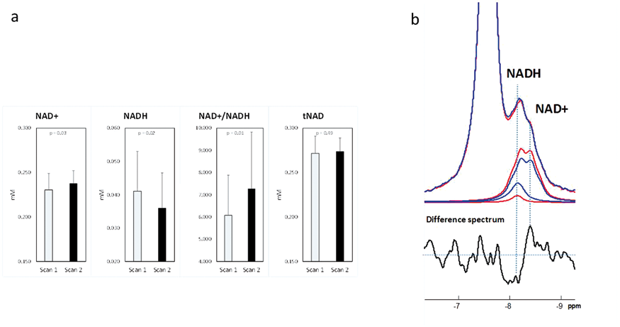
Conclusion
We conclude that a nutritional ketone intervention, beyond providing an alternative source of energy to the brain, offers the potential to boost the NAD+/NADH redox state and may provide additional benefits to the brain such as protection from oxidative stress and inflammation.
References
PB01-H04
Mitochondrial dysfunction contributed to cerebral metabolic abnormalities detected by Proton MR Spectroscopy in patients with mitochondrial encephalomyopathy subtypes
1Department of Pathology,Nanjing Drum Tower Hospital, China
2Department of Neurology, Nanjing Drum Tower Hospital, China
Abstract
Aims
Growing evidence indicates that cerebral metabolic features, evaluated by proton magnetic resonance spectroscopy (1H-MRS), are sensitive to early mitochondrion dysfunction associated with mitochondrial encephalomyopathy (ME). Although1H-MRS metabolite ratios of lactate (lac)/Cr, N-acetyl aspartate (NAA)/creatine (Cr), total choline (tCho)/Cr and myoinositol (mI)/Cr that are measured in the infarct-like lesions may reveal evidence of metabolite changes in ME, the pathological basis of the1H-MRS in indicating the disease progression of ME subtypes is unknown.
Methods
In the present study, cerebral metabolic features of pathologically diagnosed ME cases (19 Mitochondrial encephalomyopathy, lactic acidosis, and stroke-like episodes (MELAS) and 9 chronic progressive external ophthalmoplegia (CPEO) and 23 healthy controls were investigated using1H-MRS. Receiver operating characteristics (ROC) analysis was used to evaluate the diagnostic power of the cerebral metabolites. Histochemical evaluation was carried out on muscle tissues derived from biopsy, in order to assess the abnormal mitochondrial proliferation. The association between cerebral metabolic and mitochondrial cytopathy was examined by correlation analysis.
Results
Patients with MELAS or CPEO exhibited a significantly higher Lac/Cr ratio and a lower NAA/Cr ratio compared with controls. The ROC curve and/or the ratio of Lac/Cr indicated prominent discrimination between MELAS or CPEO and healthy control subjects, whereas the NAA/Cr ratio may present diagnostic power in the distinction of MELAS from CPEO. Lower NAA/Cr ratio was associated with higher Lac/Cr in MELAS, but not in CPEO. Furthermore, higher ragged red fibers (RFFs) percentages were associated with elevated Lac/Cr and reduced NAA/Cr ratios, particularly in MELAS. This association was not noted in the case of mI/Cr ratio.
Conclusions
Mitochondrion cytopathy (lactic acidosis and RRFs on muscle biopsy) was associated with neuronal viability but not glial proliferation, notably in MELAS. Mitochondrial neuronopathy and neuronal vulnerability appears to be important causes in the pathogenesis of MELAS, particularly of the signature stroke-like episodes.
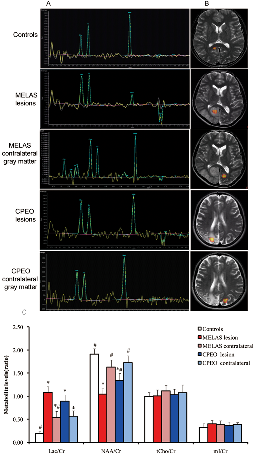
Keywords
cerebral metabolic abnormalities, mitochondrial cytopathy, 1H-MRS, MELAS, CPEO
PB01-H05
Assessing cerebral metabolism and physiology in post-stroke mice by dynamic oxygen-17 MRI
1Department of Biomedical Engineering, Case Western Reserve University, United States
2Department of Radiology, Case Western Reserve University, United States
3Department of Medicine, Case Western Reserve University, United States
4The Harrington Heart & Vascular Institute, University Hospitals Case Medical Center, United States
5Department of Neurological Surgery, Case Western Reserve University, United States
6Department of Neurology, Case Western Reserve University, United States
7Department of Physiology and Biophysics, Case Western Reserve University, United States
Abstract
Objectives
Metabolic evaluation of post-stroke brain can more accurately delineate the salvageable penumbra due to the direct association between tissue metabolism and viability. Further, blood-brain-barrier (BBB) disruption has been associated with increased risk of hemorrhagic transformation. Hence, the assessments of metabolic and BBB integrity can provide a comprehensive evaluation of the risks and benefits for thrombolytic therapy. Here, we aimed to evaluate the efficacy of using17O-MRI derived cerebral metabolic rate of oxygen (CMRO2) for identification of viable tissue in post-stroke mouse brain, and the potential of using17O-labeled water as a tracer for simultaneous assessment of cerebral perfusion and BBB integrity.
Methods
C57BL/6 mice (3–6 months old) underwent 60-min of middle cerebral artery occlusion (MCAO). MRI studies were performed on a 9.4T scanner at 2, 24, and 72 hours after MCAO. T2-, diffusion-, and perfusion-weighted images were acquired. A subgroup of mice (N = 4) underwent17O-water injection studies, while the remaining mice (N = 4) underwent17O-gas inhalation studies. For the injection study, a bolus of 100 µL17O-water (20% enrichment) was injected via tail vein with continuous acquisition of dynamic17O-data before, during, and 20 min after the injection.17O-water uptake and washout rate were quantified by fitting17O-signal with an exponential model. For the inhalation study, a gas mixture of17O2/N2 (30%:70%) was delivered through a nose cone for 2 to 3 min, and metabolic generation of17O-water was tracked by dynamic17O-data acquisition before, during, and after the inhalation. CMRO2 was calculated using the method proposed by Zhang et al1.17O data acquisition used a fast imaging method described previously2.
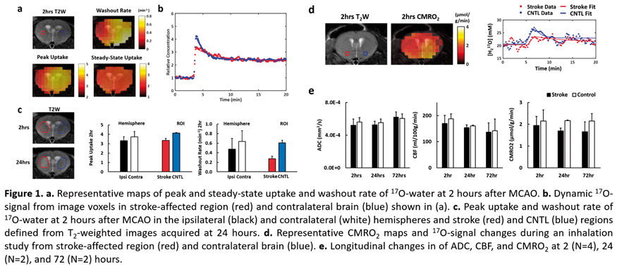
Results
T2-weighted images showed no obvious differences between the two hemispheres at 2 hours but hyper-intensity in the stroke-affected region at 24 hours. However, the ipsilateral hemisphere showed reduced peak17O-water uptake comparing to the contralateral hemisphere (3.32 ± 0.44 vs 3.73 ± 0.58), and a slower washout rate (0.47 ± 0.23 vs 0.63 ± 0.20 min−1) at 2 hours. Further, the washout rate in the infarct core, regions with hyper-intensity in T2-weighted images acquired at 24 hours, was only 57% of the entire ipsilateral hemisphere.
In the inhalation group, 2 mice showed ∼20% reduction of CMRO2 in the ipsilateral hemisphere at 2 hours after MCAO and died within 24 hours. The remaining 2 mice showed similar CMRO2 between two hemispheres at the acute stage and survived the subsequent scans. However, CMRO2 in stroke-affected region decreased to 78% of the contralateral hemisphere at 24 hours, suggesting infarct expansion. ADC in stroke-affected region decreased initially by 8% but was normalized at 72 hours. CBF in the stroke-affected region decreased by 10% at the acute stage, and was partially restored to 95% at 24 and 72 hours.
Conclusions
Our results suggest that significantly reduced CMRO2 at acute stage is associated with higher mortality rate. Reduction of17O-water uptake and washout in the stroke-affected region could be associated with reduced CBF or the development of cerebral edema. This study demonstrates the potential of17O-MRI to provide comprehensive assessment of cerebral physiology and metabolism in post-stroke brain.
References
PB01-H06
Metabolic alterations in the hippocampus, cortex and hypothalamus of mice exposed to long-term high-fat diet
1Department of Experimental Medical Science, Faculty of Medicine, Lund University, Sweden
2Wallenberg Centre for Molecular Medicine, Lund University, Sweden
3Laboratory for Functional and Metabolic Imaging, École Polytechnique Fédérale de Lausanne, Switzerland
4Instituto de Investigaciones Biomédicas “Alberto Sols” CSIC-UAM, Spain
Abstract
Objectives
Metabolic syndrome and diabetes impact brain function and metabolism. While it is well established that rodents exposed to diets rich in saturated fat develop brain dysfunction, contrasting results abound in the literature, likely as result of exposure to different high-fat diet (HFD) compositions and for varied periods of time. In the present study, we tested the hypothesis that the severity of the metabolic syndrome is related to the degree of metabolic alterations, as well as brain function deficits. For that, mice were fed diets containing different amounts of fat during 6 months, which lead to different degrees of glucose intolerance and insulin resistance (Soares et al., 2018).
Methods
We investigated alterations of spatial memory by measuring Y-maze spontaneous alternation, metabolic profiles of the hippocampus, cortex and hypothalamus by1H magnetic resonance spectroscopy (MRS) at 14.1 T, and levels of proteins specific to synaptic and glial compartments in mice fed diets with 10, 45 or 60% of fat during 6 months. Significant changes were considered for P < 0.05 in ANOVA and LSD post-tests.
Results
Increasing the dietary amount of fat from 10% to 45% or 60% resulted in obesity accompanied by increased leptin, fasting blood glucose and insulin, and reduced glucose tolerance. In comparison to controls (10%-fat), only mice fed the 60%-fat diet showed increased fed glycaemia, as well as plasma corticosterone that has a major impact on brain function. HFD-induced metabolic profile modifications measured by1H MRS were observed across the three brain areas in mice exposed to 60%- but not 45%-fat diet. Namely, there was a significant HFD-induced increase in concentrations of glutamine, N-acetylaspartate, N-acetylaspartylglutamate, glutathione, taurine, choline-containing compounds and glucose in the hippocampus; increase in concentrations of N-acetylaspartylglutamate, phosphorylethanolamine and glucose in the cortex; and increase in concentrations of myo-inositol, choline-containing compounds, phosphorylethanolamine, and lactate in the hypothalamus. Interestingly, the three brain regions showed a HFD-induced decrease of phosphocreatine-to-creatine ratio, suggesting energy metabolism deficits. Compared to controls, both HFD groups displayed impaired hippocampal-dependent memory, and reduced levels of synaptic proteins in the hippocampus. Immunoreactivity against GFAP and/or Iba-1 in the hypothalamus was higher in mice exposed to HFD than controls, suggesting occurrence of astrogliosis and neuroinflamation.
Conclusions
A 60%- but not 45%-fat diet altered metabolic profiles in the hippocampus, cortex and hypothalamus, although both HFD levels induced memory impairments and synaptic changes in the hippocampus, and gliosis in the hypothalamus. Thus, we conclude that there are different mechanisms underlying HFD-induced metabolic and synaptic changes.
Reference
PB01-H07
Metabolic profile alterations in the hippocampus of mice under chronic high-fat and high-sucrose feeding
1Department of Experimental Medical Science, Faculty of Medicine, Lund University, Lund, Sweden
2Wallenberg Centre for Molecular Medicine, Lund University, Lund, Sweden
Abstract
Objectives
Type 2 Diabetes (T2D) is associated to increased risk of dementia. However, the mechanisms implicated in T2D-induced brain dysfunction are not fully understood. T2D and obesity promote a low-grade chronic inflammation which leads to impaired neuronal insulin signaling, synapse deterioration and memory loss (de Felice & Ferreira, 2014). We aimed at identifying early T2D-induced metabolic profile alterations in the hippocampus, which might be related to memory dysfunction.
Methods
We investigated metabolic profiles in the hippocampus of male and female C57BI6/J mice exposed to a High Fat- High Sucrose diet (HFHSD; 60% lard-based fat, 20% sucrose) by means of1H magnetic resonance spectroscopy (MRS) at 9.4 T. MRS scans and glucose tolerance tests were performed before (baseline; 10-weeks old) and after 1, 2, 4, 8, 16 weeks of HFHSD exposure in a longitudinal way. Memory performance was measured at the end of the study through object relocation and novel object recognition tasks with one hour of interval between training and testing trials.
Results
Overweight, increased blood glucose, glucose intolerance and insulin resistance characteristic of T2D were all patent after 4 weeks of HFHSD feeding, when compared to age-matched controls (fed a 10%-fat diet without added sucrose). HFHSD-exposed mice showed poorer memory performance in both behavior tests, relative to controls. The hippocampal metabolic profile was transiently modified HFHSD-fed mice, when compared to controls: reduced concentration of glutamine was observed at 2 (P < 0,001) and 4 (P < 0,0001) weeks of HFHSD; reduced concentration of ascorbate was observed a 4 weeks of HFHSD (P < 0.01). These metabolic changes were not observed at 8, 16 or 24 weeks of HFHSD.
Conclusions
HFHSD feeding modified levels of glutamine and ascorbate in the hippocampus, suggesting alterations of the glutamate-glutamine cycle and glutamatergic neurotransmission, and of redox homeostasis (Girault et al., 2019). These alterations were transient, suggesting that the metabolism in the hippocampus is adaptable to T2D.
Reference
PB01-J01
Metabolic basis of deactivation and activation in default mode and sensory networks
1Yale University, USA
2University of Copenhagen, Denmark
3University of Louvain, Belgium
4Université de Montréal, Canada
Abstract
Objectives
In task-based functional MRI (fMRI) studies, a task-positive region is where the BOLD signal is greater during experimental versus rest blocks, and a task-negative region is where the BOLD signal is smaller during experimental versus rest blocks. The task-negative region is also referred to as the default mode network (DMN). Functional MRS (fMRS) using short echo-time(TE) non-edited 1H-MRS protocols, has reproducibly detected lactate increase during sensory-induced activations (1-3) and evoked modulations of the BOLD signal (4-6). Here we investigated the metabolic basis of functional paradigms in non-DMN and DMN areas using concurrent 1H-MRS acquisitions of J-edited lactate and diffusion-weighted water (i.e., BOLD signal) at 4 T.
Methods
Twenty healthy volunteers (10 male, age 29 ± 2) participated in 2 fMRI and 2 fMRS runs spanning over 3 days. We used fMRI at 3 T to localize the visual cortex (VC, non-DMN area) and the posterior cingulate cortex (PCC, DMN area), using an activation (flashing checkerboard, 8 Hz, visual angle 15 × 18°) and deactivation (auditory emotion perception) paradigm, respectively. Functional runs consisted of three 2.7 min VC (or PCC) stimulation blocks interleaved with three 2.7 min fixation (or rest) blocks. For PCC deactivation, participants listened to audio-emotion portrayals (7) with eyes closed, and were asked to press the response button each time a specific emotion, contempt, disgust or surprise, was identified. A single fMRS voxel was placed around the functionally localized area in VC and PCC. The fMRI and fMRS experiments were performed on a 3 T Siemens Prisma scanner and 4 T Bruker spectrometer, respectively. For fMRI, we used T2*-weighted EPI (648 scans, TR = 1.5 s, TE = 30 ms, 2 × 2 × 2 mm voxels). For fMRS, we used J-difference editing (180 paired spectra, TR = 2.7 s, TE = 144 ms, VC volume = 16.7 ± 1.7 cm3, PCC volume = 25.4 ± 1.4 cm3). Each pair of the water-suppressed J-edited spectra was followed by the diffusion-weighted STEAM (8) water spectra acquisition (TE = 20 ms, b = 1400 s/mm2) with 200 ms delay. J-edited spectra were preprocessed and corrected for BOLD linewidth narrowing. Lactate levels were estimated using LCM and normalized to the corresponding NAA levels. To estimate the T2*, we applied water peak linewidth linear approximation using logarithm of the water FID (4-6).
Results
Preliminary analyses revealed diffusion-weighted water (i.e., BOLD signal) increase in VC during visual stimulation as compared to fixation (Fig. 1; t = 2.7, p < .05, ΔBOLD = 0.8 ± 0.5%), which was associated with increased lactate levels (t = 3.2, p < .05, Δlactate = 7.2 ± 2.4%). In PCC, we found BOLD and lactate decreases during audio-emotion task as compared to rest (Fig. 1; BOLD: t = 2.1, p < 0.05, ΔBOLD = −0.5 ± 0.2%; lactate: t = 1.9, p < 0.05, Δlactate = −3.4 ± 2.0%). The ΔBOLD is somewhat smaller due to the partial volume effect and diffusion weighting applied to suppress the signal from large vessels (9,10).

Conclusions
In VC (non-DMN area, activation), we detected increase of BOLD signal and lactate level during task. In PCC (DMN area, deactivation), we identified decrease of BOLD and lactate during task. These results suggest similar degree of task-dependent aerobic glycolysis in both DMN and non-DMN areas.
PB01-J02
Dynamic brain functional connectivity change analysis based on random matrix theory
1Duksung Women's University
Abstract
Functional magnetic resonance imaging (fMRI) data sets are large and characterized by complex dependence structures driven by highly sophisticated neurophysiology and aspects of the experimental designs. fMRI studies often include multiple sessions, and temporal dependencies may persist between the corresponding estimates of mean neural activity. Further, spatial correlations between brain activity measurements in different locations are often present in fMRI data. We develop a method that leverages the special structure of our covariance model, enabling relatively fast and efficient change-point estimation. We propose a statistical approach for characterizing patterns of change in the temporal evolution of FC measures. First, for each subject and each of the 91 pairs of brain regions, we quantify the evolution of FC using a time varying cumulative covariance matrix. We apply the Tracy-Widom transformation (Tracy and Widom 1996) for the largest eigenvalue of the covariance matrix ratio up to time t, and that after time t, for each time-point t. For resting state fMRI data, the covariance function is probably one of the most important quantities of interest. Change-point analysis based on the maximum eigenvalue and canonical correlation approach are useful tools in situations where high-dimensional data are collected. We further examine dynamic FC properties by estimating change points and performing group comparisons. Using our proposed method, we conduct simulation study and analyze fMRI data from a study of epilepsy patients.
PB01-J03
Resting state fMRI in stroke patients with individual ROI segmentation
1Research institute for brain and blood vessels Akita
Abstract
Introduction
Resting state fMRI (rsfMRI) is now widely performed for the clinical application[1]. Although it is suitable for group analysis, the signal difference is too small to diagnose individual patient. Diagnosis capability may be reduced by structural normalization. Individual segmentation for region of interest (ROI) might be helpful to improve the diagnosis capability. In this study, we have examined the capability of individual ROI for stroke patients in acute and subacute phase.
Materials & Methods
Six patients with cerebral infarction in the left penetrating branch area were included. MRI was examined in acute (within two weeks after stroke onset) and subacute phase (one month after onset) with a 3 T MRI. Five minutes of rsfMRI was performed by GE-EPI sequence (TR/TE = 3000/30 ms, resolution 3'3 mm2). Functional connectivity (ROI to ROI) was evaluated by Conn v18.a software with MATLAB[2]. Seed ROI was selected at the postcentral gyrus in right healthy hemisphere. For individual ROI analysis, segmentation was processed by the Freesurfer v6.0 using anatomical T1-weighted image acquired by 3D MPRAGE sequence[3]. ROI was selected using Destrieux atlas in Freesurfer (aparc.a2009 s). For normalized brain analysis, ROI selected by FSL Harvard-Oxford Atlas maximum likelihood cortical atlas in Conn (atlas) was also used. Subject motion correction, slice timing correction, coregistraion, normalization and smoothing (8'8 mm) were calculated by Conn.
Results
Seed ROI of Freesurfer atlas is not well segmented on normalized brain, whereas individual segmentation is well. At the acute phase, twenty-five of significant connectivity (p-FDR < 0.05) ROI were detected in individual ROI analysis, whereas 15 of Freesurfer atlas and 9 of Conn atlas in normalized brain analysis. Connectivity difference between acute and subacute phase was well shown in individual ROI than normalized brain analysis.
Discussion
More detected area and higher T value was shown in individual than normalized brain analysis using ROI in Freesurfer. It should be due to the precise segmentation in each patient. Misregistration of the tissue was reduced in individual ROI analysis. In normalized brain analysis, ROI should be covered such misregistration. Therefore, bigger ROI such as Conn atlas is required. For the precise analysis, small ROI and individual segmentation should be useful.
References
PB01-J04
Resting state and task BOLD fMRI signals for patients with AVM
1CAI, Dept. of Radiology, WVU, USA
2Surgery, WVU, USA
Abstract
Objectives
We hypnotized mechanisms of BOLD for task and resting state are different and investigated the BOLD signals of brain motor functions and the corresponding functional connectivity (fc) for AVM patients by finger tapping fMRI and resting state fMRI (rsfMRI) methods.
Methods
17 patients with AVMs in left brain hemispheres and 10 healthy subjects were included in this study. rsfMRI and the finger tapping fMRI were performed by using a 3 T scanner with a 12-channel head matrix coil and a multi slice 2D EPI. Group independent component (IC) analyses on the data of rsfMRI and the task fMRI were performed by using FSL [1] and AFNI [2], and the component (i.e., the network) related to sensory motor network (SMN) from rsfMRI data or the component associated to primary motor cortices (PMC) from the fMRI data were found from the ICs, then the functional and connectivity maps were exported to AFNI to get the signals (activation volumes). The ratios of the signals for the left (i.e., AVM) sides over the right sides were calculated.
Results
Both the motor function and corresponding fc of the health group displayed a bilateral symmetry (Fig 1 the top row) and the corresponding L/R side ratios as expected as ones were listed in the top section of Table 1. The functional volumes for the AVM group however demonstrated a dominated pattern (Fig. 1 the bottom row) in the lesion side (i.e., the left) but an almost bilateral symmetry for the corresponding fc. The ratios of the AVM group were also listed in the bottom section of the table.
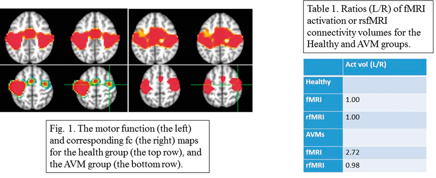
Conclusions
BOLD fMRI signal is dominated by the increase of cerebral blood flow (CBF) during performing a task [3], but the presence of an AVM results in an abnormal CBF pattern. The effects of AVM on the BOLD fMRI signal and fc maybe linked with the influences of the “steal effect” of the CBF from AVM. We found BOLD activation volume of motor function in the non-AVM hemisphere was significant smaller than the one in the lesion hemisphere, which suggests the AVMs only increasing the BOLD fMRI signal by increasing more change of CBF in the AVM side for the finger task. This enhancement may link to either functional reorganization or CBF redistribution.
References
PB01-J05
The effects of masking therapy in tinnitus patients are characterized by longitudinally altered local activity and functional connectivity
1Beijing Friendship Hospital, Capital Medical University
Abstract
Introduction
Masking therapy serves as an effective way to induce relief of adverse tinnitus sensations. However, the neural mechanism of effective masking therapy in tinnitus remains incompletely understood.
Methods
In this study, 30 tinnitus patients completed resting-state functional magnetic resonance imaging scans at baseline and after 12 weeks of masking therapy intervention. Thirty age- and gender-matched healthy controls were also longitudinally scanned twice over a 12-week interval. Data were analyzed by ALFF (amplitude of low-frequency fluctuation) and seed-based functional connectivity (FC) to measure significantly altered local brain spontaneous activity and its connections.
Results
Interaction effect between the two groups and two scans in local neural activity assessed by ALFF values were observed in the left parahippocampal gyrus and right superior temporal gyrus (STG), the key region of the primary auditory cortex). Importantly, local functional activity in the left parahippocampal gyrus were higher than normal controls at baseline, but significantly reduced and approached normal levels after treatment. Conversely, activity in the right STG significantly increased and extended beyond a relatively normal range after masking intervention. These changes were found to be positively correlated with tinnitus relief. FC between the left parahippocampal gyrus (seed) and the left right anterior cingulate cortex and left middle cingulate cortex were observed higher in tinnitus patients after treatment.
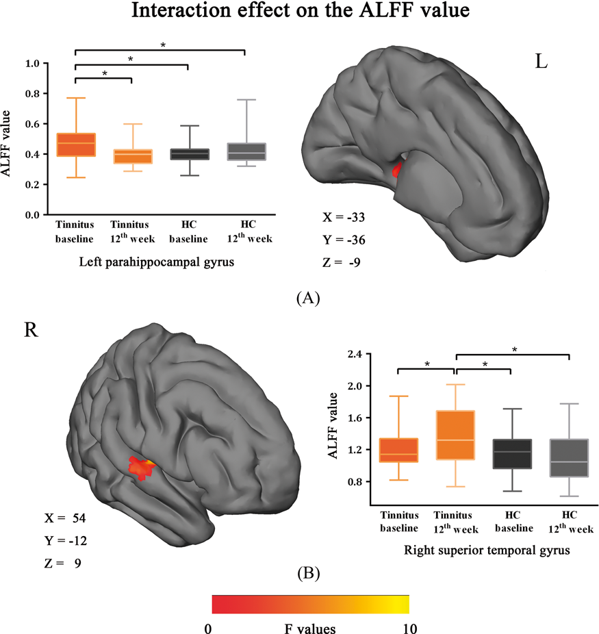
Conclusion
We prove our hypothesis that masking therapy can selectively modulate neural activity associated with the limbic system and auditory cortex. Such an understanding would be crucial to identifying biomarkers for monitoring the treatment efficacy of tinnitus masking therapy.
Keywords
Tinnitus, Masking therapy, fMRI, ALFF, Functional connectivity, Neural biomarker
PB01-J06
Baseline functional connectivity features of the neural network nodes can predict improvement after masking therapy in tinnitus patients
1Dept. of Radiology, Beijing Friendship Hospital, Capital Medical University, Beijing, China
Abstract
Introduction
Previous neuroimaging studies have reported neural activity alterations after treatment of tinnitus. This is the first study to analyze the predictive value of the baseline functional connectivity features of the neural network nodes for the outcome prediction of masking therapy.
Methods
The resting-state functional magnetic resonance imaging (fMRI) data of 27 untreated tinnitus patients and 27 matched healthy controls were analyzed in this study. We calculated the graph-theoretical metric degree centrality (DC) within a standardized brain atlas to characterize the functional connectivity of the neural network nodes. Changes in the Tinnitus Handicap Inventory (THI) score were used to evaluate treatment outcome after a 12-week masking therapy.
Results
The DC value of ten brain nodes in tinnitus patients were significant increased at baseline. Five nodes, including right insula, inferior parietal lobule (IPL), bilateral thalamus and left middle temporal gyrus, exhibited significant correlations of their DC at baseline with treatment-induced THI changes in tinnitus patients. Receiver operating characteristic curve analyses revealed well performance of the five brain regions in classifying better effect of therapy. Area under the curve (AUC) of the right IPL and thalamus reached the highest value (AUC = 0.818), with identical sensitivity and specificity of 80.0% and 76.5%. Moreover, the right thalamus was identified as the optimal regressor as determined by stepwise regression.

Conclusion
Our study further supported the involvement of fronto-parietal-cingulate network in mediating tinnitus, and provided DC value of the right thalamus at baseline as an object neuroimaging-based indicator for predicting better efficacy of masking therapy.
Keywords
Tinnitus, Masking therapy, fMRI, Functional connectivity, Degree centrality, Neural biomarker, Graph-theory
PB01-J07
Measurements of CMRO2 and its relationship with CBF in hypoxia with an extended calibrated-BOLD method
1Center for MRI Research, Peking University, Beijing, China
2Peking-Tsinghua Center for Life Sciences, Academy for Advanced Interdisciplinary Studies, Peking University, Beijing, China
3Beijing City Key Lab for Medical Physics and Engineering, Institute of Heavy Ion Physics, School of Physics, Peking University, Beijing, China
Abstract
Objectives
Cerebral blood flow (CBF) and cerebral metabolic rate of oxygen (CMRO2) are physiological parameters that not only reflect health and disease of the brain but also jointly contribute to the blood-oxygen-level-dependent (BOLD) signals vastly applied to evaluate brain functions. Nevertheless, there remains unsolved issues concerning the CBF-CMRO2 relationship in a working brain at various oxygen states. In particular, CMRO2 responses to functional tasks in hypoxia are less studied due to the lack of a reliable dynamic CMRO2 quantification method. This study extended the calibrated-BOLD model to incorporate dynamic CMRO2 quantification in hypoxia. The extended model was applied to investigate the effects of mild hypoxia on CBF and CMRO2 responses to graded visual stimuli.
Methods
The extended calibrated-BOLD model
The original calibrated-BOLD model suggested by Davis et al.1 assumes that arterial blood is fully oxygenated, and the BOLD signal comes from changes in blood volume and the concentration of deoxyhemoglobin of venous blood. A scaling factor, M is used to calibrate the change of the BOLD signal against its baseline state. The M factor can be obtained via gas-related physiological challenges. In hypoxia, however, not only that the deoxyhemoglobin from both arterial and venous blood affect the BOLD signal, but also that the hypoxic M factor cannot be easily obtained through physiological calibrations due to disturbed flow and metabolism states. Thus, the original calibrated-BOLD method is not valid for hypoxia.
This extended calibrated-BOLD model calculates an “effective” concentration of deoxyhemoglobin which properly weights deoxyhemoglobin from both arterial and venous blood (i.e. [dHb] = 0.3[dHb]a + 0.7[dHb]v) and scales the hypoxic M factor in relation to the normoxic M factor according to equation 4 in Lin et al.2
The experiment
Fifty subjects participated in normoxia (21%O2 + 79%N2) and hypoxia (12%O2 + 88%N2) visual tasks and a CO2 challenge (5%CO2 + 21% O2 + 74%N2). The visual task was consisted of three 60 s sessions alternating between resting and checkerboard stimulation flickering at 1, 4 or 8 Hz.
A pulse sequence which simultaneously acquires CBV, CBF and BOLD-weighted signals was implemented on a 3.0 T MRI system. A single slice passing through the visual cortex was imaged. Acquisition parameters, procedures for preprocessing and quantification of visual task-induced δCMRO2in normoxia were adopted from Lin et al.2 δCMRO2 in hypoxia was estimated with the extended calibrated-BOLD method described above. The flow-metabolism coupling ratio, n = δCBF/δCMRO2, was calculated.
Results
The average δCBV, δCBF, δBOLD, δCMRO2 and n, evoked by the visual tasks in normoxia and mild hypoxia were shown in Figure 1. Mild hypoxia, compared with normoxia, caused significant alterations in both the amplitude and the trend of the CMRO2 responses but had no impact on the CBF responses to the same tasks. Specifically, smaller task-induced increases of CMRO2 were observed in mild hypoxia compared with normoxia.
Conclusions
In this study, an extended calibrated-BOLD model was developed to incorporate dynamic CMRO2 quantification in hypoxia. Effects of hypoxia to the CBF-CMRO2 relationship during graded visual stimulation was studied. Our observations suggested an altered flow-metabolism relationship in hypoxia compared to normoxia.

PB01-J08
Relating lateralized mq-BOLD-based relative oxygen extraction fraction to areas of elevated DSC-based oxygen extraction capacity in asymptomatic unilateral carotid artery stenosis
1Department of Neuroradiology, Technical University of Munich, Munich, Germany
2MRRC, Yale University, New Haven, CT, United States
3Clinic for Radiology, Technical University of Munich, Munich, Germany
4Institute of Clinical Medicine, Aarhus University, Aarhus, Denmark
5Philips Healthcare, Hamburg, Germany
6Clinic for Neurology, Technical University of Munich, Munich, Germany
Abstract
Objectives
Adequate oxygen supply is fundamental to meet cerebral metabolic demand. Oxygenation information can be assessed voxel-wisely by different MRI-based techniques. Multiparametric quantitative BOLD (mq-BOLD) is based on R2’ and estimates relative oxygen extraction fraction (rOEF),1 while parametric modeling of dynamic susceptibility contrast (DSC) data yields oxygen extraction capacity (OEC, proposed as maximum possible oxygen extraction OEFmax).2 We have recently reported ipsilaterally elevated OEC in unilateral internal carotid artery stenosis (ICAS) patients, whereas rOEF lateralization was not found, both in accordance with recent studies.3-5 The sensitivity of OEC to hemodynamic impairments seems to be higher, but so far the relation between these two approaches is unclear. We have therefore compared rOEF and OEC in patients with unilateral ICAS and healthy controls (HC).
Methods
We performed MRI in 59 participants (29 asymptomatic, unilateral ICAS-patients, age = 70.1 ± 4.8 y and 30 age-matched HC, age = 70.3 ± 7.3 y) on a Philips 3 T Ingenia. Maps of mq-BOLD-based rOEF were obtained by quantitative R2, R2* and DSC-based relative cerebral blood volume (rCBV) mapping1, whereas OEC was derived by parametric modeling of DSC data2 (Fig.1A,C). To investigate the correspondence of rOEF and OEC, individual masks of increased-OEC in grey matter (GM) were defined for each participant and each hemisphere using the first (OECQ1), second (OECQ2 = median) and third (OECQ3) quartile of OEC as thresholds, respectively (Fig.1B). Lateralization of rOEF between left and right hemispheres (LH/RH) within each OEC-based VOI was determined for each participant using the lateralization index (LI):
LIrOEF[%] = 100% × |[mean(rOEFRH)-mean(rOEFLH)]/[mean(rOEFRH) + mean(rOEFLH)]|.
rOEF lateralization between hemispheres was compared between ICAS-patients vs. HC within all different masks of increased-OEC.
Results
Individual patients’ rOEF and OEC maps show some similarities (Fig.1A,C). On group level of all ICAS-patients, rOEF lateralization between hemispheres is weak in whole GM and within areas of moderately increased OEC (OECQ1), and does not differ significantly from HC (two-sample t-test, whole GM: p = 0.30 and OECQ1: p = 0.15). However, in areas of OEC elevations (OECQ2, OECQ3), significant rOEF lateralization (OECQ2: p < 0.05 and OECQ3: p < 0.01) was found in unilateral ICAS compared with HC (Fig.1D). Thus, stronger rOEF lateralization was found in areas of increased OEC.

Discussion
In accordance with previous reports,4,5 lateralization of rOEF between hemispheres in whole GM was weak for ICAS-patients and similar to HC. However, investigation of rOEF within areas of elevated OEC (OECQ2,OECQ3) revealed significantly increased rOEF lateralization in ICAS (Fig.1D). rOEF lateralization is most pronounced in the area of highest OEC elevations (OECQ3) (Fig.1D). Thus, both oxygenation related parameters show similarities to some extent. However, differences between rOEF and OEC are striking and point to possible contributions of oxygen diffusivity.6
Conclusion
We successfully analyzed oxygenation impairments in unilateral ICAS and did not find general lateralization of rOEF in accordance with current literature. Application of individual masks of increased-OEC in GM unveiled significantly increased rOEF lateralization in unilateral ICAS, demonstrating sensitivity of mq-BOLD to oxygenation impairments in ICAS-patients.
References
PB01-J09
Recovery of cerebrovascular reactivity after treatment of asymptomatic carotid artery stenosis is assessable by non-invasive breath-hold fMRI within global watershed areas
1Department of Neuroradiology, Technical University of Munich, Munich, Germany
2MRRC, Yale University, New Haven, CT, United States
3Clinic for Radiology, Technical University of Munich, Munich, Germany
4Philips Healthcare, Hamburg, Germany
5Institute of Clinical Medicine, Aarhus University, Aarhus, Denmark
6Clinic for Neurology, Technical University of Munich, Munich, Germany
7Institute of Radiopharmaceutical Cancer Research, Helmholtz-Zentrum Dresden-Rossendorf, Dresden, Germany
Abstract
Objectives
Accounting for approximately 10% of all strokes,1 severe internal carotid-artery stenosis (ICAS) is a major public health issue. The average 2-year mortality after the invasive treatment is very high with 32%,2 which creates the need for non-invasive methods to support treatment decisions and evaluate treatment efficacy.3,4 A highly promising biomarker of vascular health is cerebrovascular reactivity (CVR),3,4 however, commonly employed methods are either invasive acetazolamide injections or complicated gas applications.3-8 We therefore used an easily-applicable breath-hold fMRI (BH-fMRI) scheme for CVR measurements. To maximize sensitivity and ensure specificity, we evaluated CVR within global watershed areas (gWSAs) in ICAS-patients before and after treatment and in healthy controls (HC).9
Methods
Thirty-three participants (16 asymptomatic, unilateral ICAS-patients, age = 71.4 ± 5.8 y and 17 HC, age = 70.8 ± 5.3 y) underwent MRI on a 3 T Philips Ingenia with written informed consent. All participants were scanned twice, patients before and at least three months after treatment (by stenting or endarterectomy), HC with a similar follow-up delay. The BOLD-based BH-fMRI scheme comprised five breath-holds of 15 s, each. CVR-maps were calculated by data-driven analysis10 (Fig.1B,C). Artefact-affected CVR-maps were excluded based on visual ratings (CP,SK,JG). To investigate the role of chronic vasolidation,5 dynamic susceptibility contrast (DSC) MRI was additionally acquired in both scans to calculate relative cerebral blood volume (rCBV) maps11. Lateralization between hemispheres was calculated in MNI-space by mean parameter-values within GM of gWSAs for each participant (Fig.1A). ICAS-patients were evaluated within hemispheres ipsilateral and contralateral to the stenosis.
Results
Exemplary data of an ICAS-patient shows impaired CVR before treatment, which improves after treatment (see arrows in Fig.1B,C). On group level, CVR is significantly decreased in the ipsilateral hemisphere before treatment (Fig.1D, p = 0.0038). After treatment, CVR lateralization was significantly reduced (p = 0.0495) towards more symmetrical values between hemispheres (p = 0.25). Similarly, rCBV was ipsilaterally increased in ICAS before treatment and more symmetrical after treatment (data not shown). HC data was symmetrical between hemispheres at all scans (Fig.1E, p > 0.60).
Discussion
As hypothesized, BH-fMRI based evaluation of CVR lateralization within gWSAs was sensitive to subtle impairments in asymptomatic ICAS without compromising its specificity, as affirmed by symmetrical HC results (Fig.1E). Decreased CVR along with increased rCBV before treatment is associated with chronic vasodilation.5 Consistent with current literature, CVR recovery was detected after ICAS-treatment,4-8 demonstrating improved haemodynamic status. Compared to more accurate CVR-measurements with CO2 application and end-tidal gas analysis,3,12 breath-holds remain a viable alternative being much more tolerable and easily applicable at low costs within clinically feasible scan times.
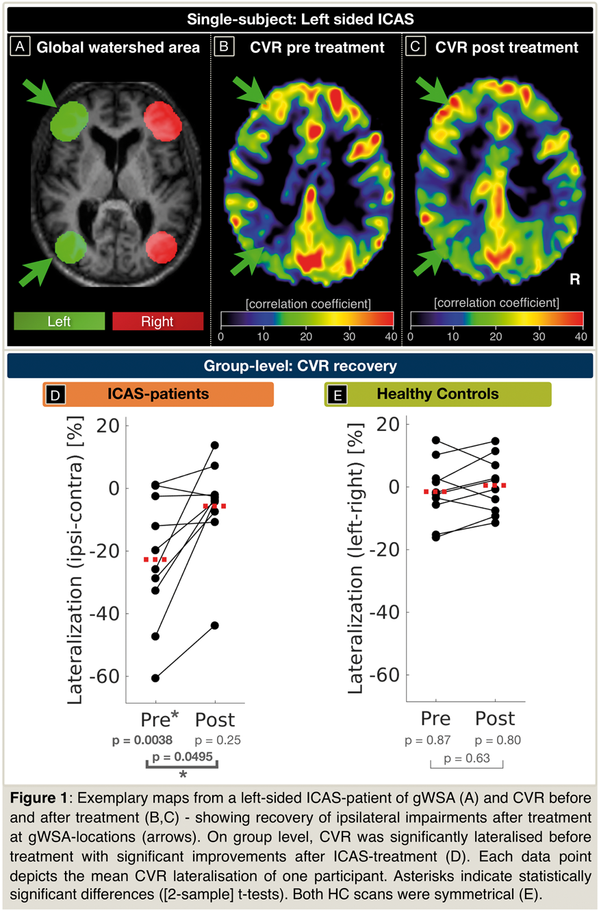
Conclusion
We successfully analyzed haemodynamic impairments by non-invasive BH-fMRI in ICAS-patients. Evaluation within gWSAs was sensitive to even subtle CVR changes. In accordance with current literature, our results showed lateralization of CVR before ICAS-treatment and its recovery after treatment – pointing to a potential application of BH-fMRI based CVR-imaging.
References
PB01-J10
Relationship between breath-hold reactivity dynamics and the amplitude of resting-state fluctuations in bold fMRI
1Institute for Systems and Robotics and Department of Bioengineering, Instituto Superior Tecnico, Universidade de Lisboa, Lisbon, Portugal
2Neurology Department, Hospital Egas Moniz, Centro Hospitalar de Lisboa Ocidental; CEDOC – Nova Medical School, New University of Lisbon, Lisbon, Portugal
3Imaging Department, Hospital da Luz, Lisbon, Portugal
Abstract
Objectives
BOLD-fMRI is often used to probe cerebrovascular health, either by stimulating the vasculature to measure cerebrovascular reactivity (CVR), for example using a breath-hold (BH) task, or by assessing the spontaneous signal fluctuations in resting-state (rs-fMRI). Several studies have found a relationship between the CVR amplitude and that of rs-fMRI fluctuations1–3. However, none of these studies explicitly assessed the temporal dynamics of CVR. Here, we aim to investigate the relationship between the temporal dynamics of CVR measured using BH-fMRI with the amplitude of BOLD fluctuations measured using rs-fMRI.
Methods
29 older adults were studied on a 3 T Siemens system (50.8 ± 7.8 years old), including 17 patients with small vessel disease. Structural images included T1-weigthed MPRAGE (1 mm isotropic) and BOLD-fMRI data were acquired using a GE-EPI sequence (TR/TE = 2500/30 ms, 40 slices, 132 volumes, 3.5 × 3.5 × 3.0 mm3) during a BH task (BH-fMRI, 3 cycles of 15 s BH after inspiration, alternated with normal breathing following inspiration/expiration auditory cues) and also during resting-state (rs-fMRI, ∼6.5 min). All data were analyzed using FSL, ANTs and R software. BOLD fMRI data were corrected for EPI distortions using a fieldmap. For BH-fMRI, after motion-correction and high-pass filtering, general linear model (GLM) analysis was performed including as regressors of interest a sine and a cosine at the task frequency and corresponding 1st harmonics. After model fitting and statistical inference, voxelwise maps were derived for the CVR amplitude and time-to-peak (TTP)4. For rs-fMRI, after motion-correction, regression of motion parameters and a second order polynomial for removal of low-frequency drifts was performed. The fractional amplitude of low frequency fluctuations (fALFF) was then computed in specific frequency bands, yielding: fALFF (0.01–0.10 Hz), fALFF1 (0.010–0.023 Hz), fALFF2 (0.023–0.073 Hz) and fALFF3 (0.073–0.20 Hz). All metrics were averaged across gray matter (GM) and white matter (WM). Pairwise Pearson correlation analysis was subsequently performed between CVR – TTP and fALFF metrics (fALFF, fALFF1, fALFF2 and fALFF3), averaged across GM and WM in each participant.
Results
Figure 1 displays the Pearson correlation between CVR-TTP and all fALFF metrics. Statistically significant correlations were found for TTP and fALFF and fALFF1 metrics (p < 0.05) in both GM and WM, and for TTP and fALFF3 in GM (r values ranged from 0.38 to 0.53). Similar relationships were found both in healthy participants and patients.
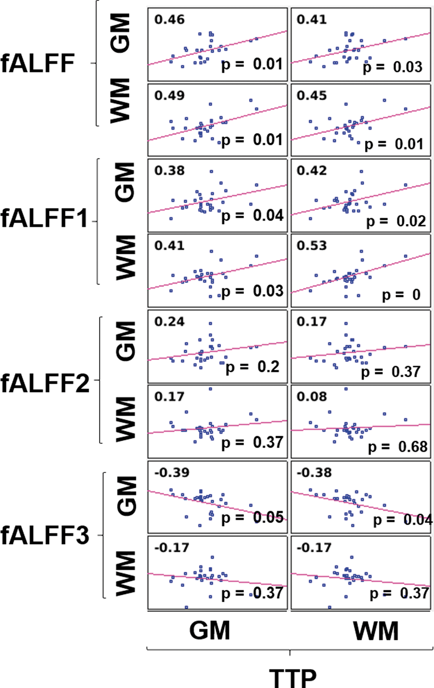
Conclusions
We found that the latency of BOLD responses to a BH task was significantly associated with the fractional amplitude of resting-state BOLD fluctuations, particularly within the lower frequency band (0.01–0.10 Hz). The temporal aspect of BH-CVR is often overlooked and unexplored, and further work is necessary to understand the physiological meaning of these relationships. Nevertheless, our work highlights the role of CVR temporal dynamics in the understanding of BH-fMRI studies and their relation with rs-fMRI measurements.
Acknowledgements
This work was funded by FCT grants PD/BD/135114/2017, PTDC/BBB-IMG/2137/2012, and UID/EEA/50009/2013.
References
PB01-J11
Activation of mirror neuron system during gait observation in sub-acute stroke patients and healthy persons
1Department of Physical Therapy, Faculty of Health and Welfare, Tokushima Bunri University, Japan
2Department of Neurosurgery Institute of Biomedical Sciences, Tokushima University Graduate School, Japan
3Department of Radiology Institute of Biomedical Sciences, Tokushima University Graduate School, Japan
4Tokushima University Hospital, Japan
Abstract
Objectives
The observation of walking improves gait ability in chronic stroke survivors1). It has also been suggested that activation of the mirror neuron system contributes to this effect2). However, activation of the mirror neuron system during gait observation treatment in sub-acute stroke patients and healthy persons has not been assessed to date. The objective of this study was to clarify the activation of mirror neuron system during gait observation in sub-acute stroke patients and healthy persons.
Methods
In this study, we sequentially enrolled five sub-acute stroke patients who had undergone gait training and nine healthy persons. We used fMRI to detect neuronal activation during gait observation.
Results
During the observation period in the stroke group, neural activity in the left inferior parietal lobule, right and left inferior frontal gyrus, right postcentral, right paracentral lobule, right and left precuneus, left inferior temporal lobule, left middle occipital gyrus and left lingual gyrus was significantly higher than during the rest period. In the healthy group, neural activity in the left inferior parietal lobule, left inferior frontal gyrus, left middle frontal gyrus, left superior temporal lobule, right and left middle temporal gyrus, right superior parietal lobule, right angular gyrus and left precentral gyrus was significantly higher than during the rest period.

Conclusions
The results indicate that the mirror neuron system was activated during gait observation in sub-acute stroke patients who had undergone gait training and also in healthy persons. Our findings suggest that gait observation treatment may provide a promising therapeutic strategy in sub-acute stroke patients who have experienced gait training.
References
PB01-J12
Dependence of slice selection on cerebral blood flow signals during visual stimulation: a study using simultaneous ASL and bold measurement
1National Institute of Radiological Sciences, QST
2Research Center for Child Mental Development, Chiba University
3Siemens Healthcare K.K.
Abstract
Objectives
The blood-oxygenation-level-dependent (BOLD) effect on the signal measured with MRI has been widely used to map patterns of neural activation during activation experiments or resting state. Recently, a new sequence has enabled simultaneous acquisition of the arterial spin labelling (ASL) and BOLD signals by reducing the interference of both signals within one repetition time, and an evaluation of relationship between the cerebral blood flow (CBF) obtained by the ASL and BOLD may provide new additional information such as vascular dysfunction and Alzheimer-related brain changes. However, in the measurement of ASL, delay of the labeled water arrival often affects the reliability of CBF estimates, and this effect may change depending on a slice orientation. In this study, we investigated the effect of slice orientation on ASL signal changes due to a visual stimulation using images acquired by the simultaneous ASL-BOLD measurements.
Methods
ASL and BOLD signals were acquired simultaneously for 7 min using the prototype sequence of Dual-spiral Q2TIPS (WIP, Siemens) (TR = 2000 ms, TE = 5, 30 ms, TI1 = 700 ms, TI2 = 1500 ms) at the transaxial slice and 45° oblique (almost parallel to calcarine sulcus) slice orientations on eight healthy volunteers performing a visual stimulation task with a checkerboard. A time series of CBF-related value (FLOW) and BOLD signal were calculated from the tagging and control signals obtained by the first and second echo of each time point, respectively. Correlation maps of the FLOW and BOLD images were generated by calculating a cross-correlation coefficient between the stimulation paradigm and each time series of FLOW or BOLD signal in each voxel. In these correlation maps, voxels having high correlation coefficient (r > 0.5) were detected as high-correlated area, and distribution of FLOW high-correlated area for each slice orientation was evaluated by the ratio of number of the high-correlated voxels between the FLOW and BOLD (F/B area ratio). The FLOW change during the activation was also compared by calculating the average of FLOW change from the baseline for 60 voxels with highest correlation coefficient.
Results
In the oblique slice orientation, high-correlated area was observed in the visual cortex of the occipital lobe both in the BOLD and FLOW images. However, in the transaxial slice orientation, high-correlated area in the FLOW images was smaller than that in the BOLD images, showing a discrepancy in correlation maps between the BOLD and FLOW images. The F/B area ratio of the oblique slice orientation was 34 ± 14%, and much larger compared with that of the transaxial slice orientation (7.8 ± 10%). There was no significant difference in the FLOW signal amplitude during the activation between both slice orientations.
Conclusions
Although the FLOW signal amplitude during the activation showed no differences, higher correlation with the activation were seen by the oblique slice orientation than that by the transaxial slice orientation. The results suggest that scans at the oblique slice orientation may be useful to obtain changes in the ASL signals more reliably.
PB01-K01
A Frequency-domain machine learning (FML) method for dual-calibrated MRI estimation of oxygen extraction fraction (OEF) and cerebral metabolic rate of oxygen consumption (CMRO2)
1CUBRIC Cardiff University Brain Research Imaging Centre, School of Psychology, Cardiff, UK
Abstract
Objectives
Non-invasive mapping of the cerebral metabolic rate of oxygen consumption (CMRO2) with dual-calibrated MRI 1 has great potential in the study of the brain in health and in disease. However, this method suffers instability in OEF0 estimates. Here we present a machine learning approach that reduces the variance and bias in OEF estimates. The proposed method takes advantage of the inherent input noise immunity of artificial neural networks (ANN) 2, and the feature learning ability of frequency-domain deep learning 3.
Methods
Simulated data were used to train a cascade of two ANNs (2 hidden-layers with 50 nodes each and relu activation function) to estimate resting CBF and CMRO2, from which OEF was calculated. MRI data were simulated using standard physiological models 2 and constrained to be physiologically plausible. The input feature vectors for the ANNs were constructed from the first 15 points of the FFT of the BOLD and ASL timeseries data (magnitude and phase), and end-tidal respiratory data. Additional data was simulated to compare the performance of the FML method with previously published regularised non-linear least squares (R-NLS) methods 4. Each method was also applied to in-vivo data acquired in healthy volunteers (n = 30, 15 male, mean age 32.41 ± 6.13 years), dual-calibrated MRI data acquisition as previously published 4.
Results
The simulated data predicts a significant improvement, compared to R-NLS methods, in OEF RMSE and proportional bias (slope of the regression of estimated OEF against true OEF). Using an ensemble of (40) networks further reduces the predicted OEF RMSE with only a modest increase in the bias, see figure 1.

Data fitting was substantially faster using the FML method, 15–30 seconds per dataset versus 17–18 minutes for R-NLS. Regression of estimated OEF values against a published empirical model of OEF variation 5 (80.5–1.75 × [Hb] – 0.317 × CBF) produced R2 values of 0.40 and 0.29 (p < 0.01) for the FML and R-NLS methods respectively, suggesting an improvement in sensitivity to physiological variation for the proposed method. The inter-subject COV for OEF, CBF and CMRO2 estimates were 8.7%, 15.1%, and 10.2% for FML and 13.8%, 15.1%, and 12.6% for the R-NLS method. The reduced COV for OEF estimates observed with the FML method is consistent with lower COV reported with PET measurements (7.3 ± 1.2 %)6.
Discussion
The proposed method of estimating OEF demonstrates a significant improvement in computational speed, predicted RMSE, and proportional bias compared to existing analysis methods. In-vivo results suggest good sensitivity to physiological variation and reduced inter-subject variation. FML appears to be a rapid and robust method that should facilitate online analysis of CMRO2 data, an important step in transitioning dual-calibrated fMRI from a research method to a clinical tool.
References
PB01-K02
A pilot study to characterize the sensitivity of MRI measures of cerebral blood flow and brain tissue oxygenation to a hyperventilation challenge
1Dept. of Neuroscience, Brighton and Sussex Medical School, University of Sussex, UK
2Clinical Imaging Sciences Centre, Brighton and Sussex Medical School, Brighton, University of Sussex, UK
3School of Life Sciences, University of Nottingham, Nottingham, UK
4Trinity Biomedical Science Institute, Trinity College Dublin, Dublin, Ireland
Abstract
Introduction
Hyperventilation physiologically induces a fall in Cerebral Blood Flow (CBF) and a compensatory rise in Oxygen Extraction to maintain adequate supply of oxygen to the brain tissue. Brain oxygenation extraction is proportional to venous deoxyhaemoglobin concentration and hence can be estimated with MRI by exploiting the sensitivity of the reversible transverse relaxation rate R2’ (R2’ = R2*-R2) to venous deoxyhaemoglobin concentration1. A novel MRI protocol (FLAIR-GESEPI-ASE) based on an asymmetric spin echo (ASE) sequence with CSF suppression and prospective correction for the effect of macroscopic magnetic field gradients, has been recently developed to allow estimation of R2’ with very short acquisition times and without respiratory gas challenges calibration2. R2’ is assumed proportional to both oxygen extraction fraction (OEF) and deoxygenated blood volume (DBV), and the OEF component can be separated from DBV through modelling of the signal2. In order to better understand the sensitivity of the sequence to changes in OEF, we assessed the effects of an hyperventilation challenge on R2’ and OEF parameters, whilst in parallel we also measured its effects on Cerebral Blood Flow using pseudo Continuous Arterial Spin Labelling (pCASL).
Methods
Six healthy male participants (median age [range] = 23.5 [21-32] years) received an MRI scan at 3 T. The participants performed intensive hyperventilation by increasing their ventilatory rate and depth aiming for ETCO2 of less than 20 mmHg assessed via capnograph during scans. The MR protocol acquired at rest (baseline) and repeated during the hyperventilation challenge, lasted 8 minutes and included a high-resolution T1-weighted volume (MPRAGE), followed by FLAIR-GESEPI-ASE (positioned to include the central part of the brain), and pCASL scans. ASE data were processed as previously described2. Mean R2’, OEF and DBV were extracted for each regions of interests: frontal, parietal, occipital, temporal lobes, after conservative grey-matter masking (threshold = 0.8). ASL image acquisition and processing was done according to the consensus paper of the ASL community3.
Results
The observed changes in respiratory parameters after hyperventilation relative to baseline, were (median [range]): Respiratory Rate, 81% increase [37-188]; ETCO2, 54% decrease [42-61]. ETCO2 after hyperventilation was reduced to 16.1 (13.1–18.2) mmHG.
In all regions, hyperventilation induced a decrease in CBF (Median [range] across regions = −34 % [-37.9:-30.6 %]; Wilcoxon signed rank test, p < 0.001) and an increase in R2’ (median [range] across regions = 20.5% [8.7–26.34]; Wilcoxon signed rank test, p < 0.05). There was a trend-level increase in OEF in the temporal lobe (median 7.6%; Wilcoxon signed rank test, p = 0.075) but not in the other regions.
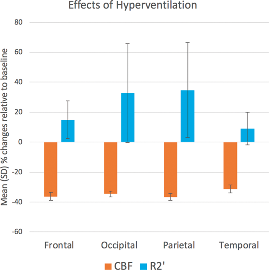
Conclusions
We observed a reduction in CBF paralleled by an increase in measures of oxygen extraction during intensive hyperventilation, in line with our predictions. Our data showed that R2’ was most sensitive to the changes in oxygenation induced by hyperventilation, particularly in the occipital and parietal regions. Future work towards optimisation of the modelling could contribute to further clarify the exact contribution of OEF to the R2’ signal.
References
PB01-K03
Does methylene blue affect brain perfusion and oxygenation in the healthy human brain? a multi-modal MRI study
1Dept. of Neuroscience, Brighton and Sussex Medical School, University of Sussex, UK
2Dept.of Neuroimaging, IoPPN, King's College London, UK
Abstract
Objectives
Methylene Blue (MB) is a compound with complex pharmacological properties, which include pleoiotropic and hormetic effects. MB easily crosses the blood brain barrier and due to its in-vitro antioxidant and metabolic enhancing effects on mitochondrial complexes1, it is regarded as a putative neuroprotective agent. The effects of MB on haemodynamics and metabolism have not been directly studied in vivo in the human brain. Here we aim to evaluate the effects of two doses of MB on cerebral blood flow (CBF) and oxygen extraction. For this purpose, we respectively employed 3D pseudo-continuous Arterial Spin Labelling (3D-pCASL) and an Oxygen Extraction Fraction (OEF) mapping technique based on Asymmetric Spin Echo (ASE)2. This method exploits the sensitivity of the reversible transverse relaxation rate R2’ to venous deoxyhaemoglobin concentration; and allows the estimation of oxygen extraction fraction through modelling of the voxel-wise R2’ values.
Methods
Eight healthy female subjects [age: 27.75 (22-46) years] were randomised to three sessions a week apart, each consisting of a 30 minute MRI scan (3Tesla GE MR750), following a slow intravenous administration of either 50 mL 5% glucose solution, (low MB dose, 0.5 mg/kg) or 50 mL 5% glucose solution (high MB dose, 1.0 mg/kg). Whole-brain CBF maps were generated with a 6 min 3D-pCASL sequence, with four control-label pairs and a proton density image; following recently published recommendations3. ASE data were acquired with TR/TE = 3100/74 ms, 10 repetitions, t = 4 ms, 16 echo-shifts. Only the first 8 shifts were considered optimal for the R2’ and OEF estimation. Field maps were also acquired to correct for macroscopic magnetic field inhomogeneities. Voxel-wise R2’ and OEF values were then estimated according to Blockley et al.2,4 Mean global CBF, R2’, and OEF values in the grey matter were extracted with FSL and compared across conditions using non-parametric tests (Wilcoxon signed ranks test).
Results
We found significant reductions in the global CBF after both low (Wilcoxon Signed Rank, p = 0.012) and high MB doses (Wilcoxon Signed Rank, p = 0.05) compared to placebo. No differences in CBF were found when comparing the two doses of MB, however the most widespread CBF reduction was observed after the low MB dose. We observed a dose-dependent reduction of global OEF, but not of R2’, after MB (High dose MB < Low dose MB < placebo; Wilcoxon Signed Rank, p = 0.05).
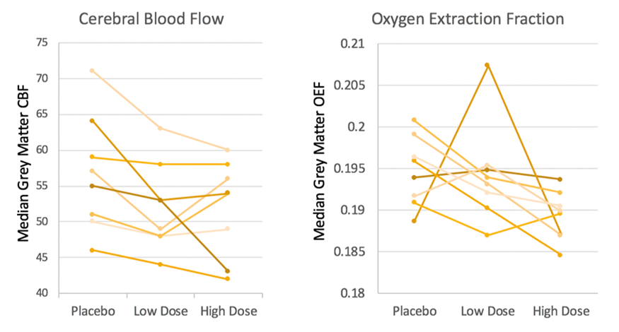
Conclusions
Our results show evidence for MB dependent reductions in CBF. This was paralleled by a dose-dependent reduction in OEF, as estimated through modelling of R2’. The latter finding must be considered cautiously as the ASE approach is a rapidly evolving methodology. If confirmed, these findings in healthy human subjects would indicate that MB overall reduces brain oxygen consumption which would be in contrast to previously reported observations in rodents5.
References
PB01-K04
Arterial spin labeling MR imaging at short post-labeling delay reflects CBF/CBV verified by 15O-PET in cerebrovascular steno-occlusive disease
1Dept. of Neurosurgery, Yamagata University of Faculty of Medicine, Japan
Abstract
Although positron emission tomography (PET) and single-photon emission computed tomography (SPECT) remain the gold standard modalities for evaluating the cerebral circulation, we are currently exploring the utility of arterial spin labeling (ASL) magnetic resonance imaging, which enables the noninvasive and convenient measurement of regional cerebral perfusion. ASL imaging is strongly influenced by the arterial transit time (ATT), especially under conditions of cerebral ischemia. We must therefore consider the post-labeling delay (PLD). ASL images with a short PLD carry a risk of overestimating the cerebral blood flow (CBF) reduction in cerebrovascular steno-occlusive disease, as previously reported. We must therefore also clarify whether or not ASL images at a short PLD have utility. In this study, we performed ASL imaging with multiple PLDs and compared the findings to oxygen-15 (15O)-PET in order to clarify the role of short-PLD ASL in cerebrovascular steno-occlusive disease. Fifty-three patients (29 males and 24 females; mean age [range] 50.3 ± 23.6 [7-83] years) with cerebrovascular steno-occlusive disease were included in this study, which was conducted from April 2013 to March 2017. Twenty-six patients had moyamoya disease, 25 had cervical internal carotid artery (ICA) stenosis/occlusion, and 2 had intracranial ICA/middle cerebral artery (MCA) stenosis. To compare the ASL findings with each parameter of PET, the right-to-left (R/L) ratio, defined as the right MCA value/left MCA value, was calculated. We demonstrated a more significant correlation between ASL images at a short PLD and cerebral blood flow/cerebral blood volume of 15O-PET, which may indicate cerebral perfusion pressure (r = 0.79, p < 0.001). A receiver operating characteristic curve analysis indicated that ASL images at PLD 1.0 and 1.5 s were more accurate than at PLD 2.0–3.0 s for the detection of a ≥ 10% change in the PET CBF. In contrast, the regression coefficients of ASL images at shorter PLDs were higher than at longer PLDs (PLD 1.0 s: 2.86, PLD 2.0 s: 1.25). ASL images at shorter PLDs may be useful at least as a screening modality to detect changes in the cerebral circulation in cerebrovascular steno-occlusive disease; indeed, the degree of change in the cerebral circulation may be more substantial at a shorter PLD than at a longer PLD. Although ASL might have utility for screening cerebral circulation, similar to SPECT or PET, we must evaluate ASL images at multiple PLDs while considering the ATT of each case at present.
PB01-K05
Quantitative evaluation of cerebral blood flow by enhanced arterial spin labeling (eASL) technique in patients with steno-occlusive disease
1Division of Neurosurgery, Department of Sensory and Locomotor Medicine, Faculty of Medical Sciences, University of Fukui, Fukui, Japan
2Department of Radiology, Faculty of Medical Sciences, University of Fukui, Fukui, Japan
3Biomedical Imaging Research Center, University of Fukui, Fukui, Japan
Abstract
In recent years, it is a prosperous development of the pulsed Continuous Arterial Spin Labeling (pCASL) technique using a 3Tesra-Magnetic Resonance Imaging (3 T-MRI) for cerebral circulation evaluation. The pCASL can be repeatedly performed in a short period of time, as it is noninvasive and does not employ ionizing radiation. On the other hand, because there are individual differences in the collateral circulation path, one of the problems of pCASL is that the reliability of quantitative evaluation of cerebral blood flow (CBF) is low. Various improvements have been made to the algorithm of pCASL in order to solve this problem. Our institution also reported that pCASL with multiple post label delay (PLD) acquisitions increased the reliability of quantitative evaluation of CBF. The aim of this study is to investigate the accuracy of this new method we named enhanced ASL (eASL) for quantitative evaluation of CBF in comparison with 15O-positoron emission tomography (PET).
Methods
Twelve patients with unilateral stenoocclusive lesions in the major cerebral arteries (10 men; age 71 ± 2 years) who were admitted to our hospital between April 2013 to March 2016 were included in this prospective study. All patients underwent eASL and Single-photon emission computed tomography (SPECT) to evaluate preoperative cerebral hemodynamic state quantitatively. Among all patients, the patients who had severe cerebral hemodynamic impairment also underwent 15O-PET studies to evaluate the other parameters such as oxygen extraction fraction (OEF), cerebral metabolic rate of oxygen (CMRO2), and cerebral blood volume (CBV).
Results
Although eASL overestimated regional CBF values, there was significant correlation (r = 0.60, y = 0.9 × -1.8) between the quantitative CBF values obtained using eASL and 15O-PET.
Conclusion
Although eASL tends to be slightly overestimated, it seems to be a useful method.
PB01-K06
Arterial spin-labeling magnetic resonance imagings in stroke patients with atherosclerotic middle cerebral artery disease
1Dept. of Neurology, JCHO Osaka Hospital, Japan
Abstract
Objectives
Arterial spin-labeling (ASL) perfusion MRI with image acquisition at two post-labeling delay (PLD) times could detect cerebral perfusion deficit and collateral flow1, non-invasively, and previous reports including us have shown that hemodynamic impairment was closely related with the development of internal watershed (WS) infarction in patients with internal carotid artery disease1,2. In the present study, we investigated stroke patterns and cerebral hemodynamic states in symptomatic patients with unilateral middle cerebral artery (MCA) disease.
Methods
Eleven patients (7 male: aged 40–85 y.o.), who were diagnosed as non cardio-embolic multiple cerebral infarctions in the unilateral hemisphere with occlusion or severe stenosis ( > 70%) at M1 portion of the ipsilateral MCA on MRA imaging, underwent ASL perfusion MRI with two PLD times at 1525 (early) and 2525 (late) ms1,2, and classified into three groups2; I) normal perfusion group (n = 2): normal perfusion in both early and late images, II) compensated perfusion group (n = 7): compromised perfusion in the early image that is alleviated in the late one, III) poor perfusion group (n = 0): compromised perfusion both in the early and late images, as described previously2. From the lesion patterns on diffusion-weighted MRI, each stroke episode was subdivided into the pial artery infarcts (PI), perforating artery infarcts (PAI) including lenticulo-striate artery (LSA), or internal WS infarctions in the centrum semiovale (CSO) or corona radiata (CR) areas, according to the previous reports3-5.
Results
In two patients with normal perfusion on both images (group I), one patients developed multiple PIs in the MCA territory, suggestive of artery-to-artery embolism, and another patient developed PAI (LSA) infarction. In 9 patients with impaired perfusion in the early, but not in the late image (group II), one patient developed lone PI, and seven patients (78%) developed internal WS infarctions in the CR area. with concomitant PI in one and PAI in two patients. No patient was classified as group III. We could not detect internal WS in the CSO in this study.
Conclusions
The present study might support the current notion that hemodynamic alterations were closely related with the internal WS infarctions in the corona radiata area, where is supposed to the borderzone of the perfusion territories between deep and superficial perforators of the MCA5, in patients with an M1 occlusion or severe stenosis.
References
PB01-K07
Reliability and reproducibility of pseudo-continuous arterial spin labeling (pCASL) derived measurements of cerebral blood flow: a replication study
1School of Biomedical Engineering and Imaging Sciences, King's College London, London SE1 7EH, UK
2Univ Lyon, CarMeN Laboratory, INSERM, INRA, INSA Lyon, Université Claude Bernard Lyon 1, Lyon, France
3Siemens Healthcare SAS, Saint-Denis, France
4King's College London & Guy's and St Thomas' PET Centre, St Thomas' Hospital, London SE1 7EH, UK
5Department of Clinical Neurophysiology and Epilepsy, Guy's and St Thomas' NHS Foundation Trust, London, UK
6Department of Biomedical Engineering, Centre for Medical Engineering, School of Biomedical Engineering & Imaging Sciences, King’s College London, King’s Health Partners, St Thomas’ Hospital, London, SE1 7EH, UK
Abstract
Objectives
Arterial spin-labeling (ASL) is a noninvasive MR imaging technique, used to measure cerebral blood flow (CBF) without the injection of contrast agents. Though several studies on reproducibility of ASL exist, local replication of any specific acquisition protocol is essential prior to clinical implementation. Many current ASL protocols using 3D readouts employ a highly segmented acquisition that has the disadvantages of greater motion sensitive and reduced temporal resolution. Here we report our investigation into the reproducibility of pCASL-derived cerebral blood flow (CBF) on a Siemens PET-MR scanner with a 3D sequence using a reduced 2-shot segmentation, providing a fast 18 sec temporal resolution that is better suited to studies involving breathing challenges.
Material and methods
Thirteen subjects (median ± interquartile range age 31 ± 16.5, three males) were scanned in three separate sessions within four hours on a 3 T Siemens PET-MR system. A 3D T1-weighted MPRAGE (1 mm3 voxels) was acquired during first session and used for co-registration and region-of-interest (ROI) definition. Grey matter and white matter were segmented using FSL-FAST (https://fsl.fmrib.ox.ac.uk/fsl/fslwiki/); 95 ROIs were obtained from structural data using MAPER (Heckemann, 2010).
For perfusion imaging, pCASL was used (Wu, 2007): 2-shot, 4 × 4 × 5.5 mm3 resolution, TR = 4.5 s, TE 17.32 ms, 1.8 s labeling, 2.1 s post-labeling delay; 1 M0 image and 25, 22 and 12 tag/control pairs acquired in scanning sessions 1, 2 and 3 respectively. Distortion correction was performed with FSL-topup.
Perfusion data was processed and quantified using FSL’s Oxford ASL v3.9.13 (Chappell, 2009), co-registered to the first session perfusion images and then to the T1-weighted MPRAGE space.
Average CBF measurements were calculated for each anatomical ROI, whole grey and whole white matter. Reproducibility and reliability were evaluated with intraclass correlation coefficient (ICC), within-subject coefficient of variation (wsCV), and Bland–Altman plots.
Results
Figures A-C show Bland–Altman plots of the grey matter CBF measurements from all sessions. A negligible bias was found between the three sessions WsCVs and ICCs are listed in Fig.D. PCASL had excellent reliability with ICCs higher than 0.85 and very good reproducibility with wsCVs lower than 10% in whole grey and white matter and representative deep grey matter ROIs.
Discussion
This study shows the implemented 2-shot pCASL sequence provides CBF measurements with excellent reliability and very good reproducibility with three tests within three hours, similar to previous pCASL test-retest studies (Chen et al., 2011). Regional heterogeneity was observed with better reliability in basal ganglia compared to in thalamus. Further investigations will investigate heterogeneity of bolus arrival time across brain regions and its influence on the reproducibility of regional CBF measurements (Wu, 2014).

References
PB01-K08
Hyperventilation-induced reduction of cerebral blood flow measured with pseudo-continuous arterial spin labeling (pCASL)
1School of Biomedical Engineering and Imaging Sciences, King's College London, London SE1 7EH, UK
2Univ Lyon, CarMeN Laboratory, INSERM, INRA, INSA Lyon, Université Claude Bernard Lyon 1, Lyon, France
3Siemens Healthcare SAS, Saint-Denis, France
4King's College London & Guy's and St Thomas' PET Centre, St Thomas' Hospital, London SE1 7EH, UK
5Department of Clinical Neurophysiology and Epilepsy, Guy's and St Thomas' NHS Foundation Trust, London, UK
6Department of Biomedical Engineering, Centre for Medical Engineering, School of Biomedical Engineering & Imaging Sciences, King’s College London, King’s Health Partners, St Thomas’ Hospital, London, SE1 7EH, UK
Abstract
Objectives
The cerebral vasculature adapts cerebral perfusion to variations in ventilatory volume and composition of inspired gases. Modulation of respiration induces a vasomotor response resulting in quantitative changes in cerebral blood flow (CBF). Hyperventilation induces hypocapnia by increasing elimination of carbon dioxide, leading to a cerebral vasoconstriction, with invasive measurements reporting a ∼30% decrease in CBF (Kety and Schmidt, 1946). Here we quantify the effect of hyperventilation on CBF using pseudo-continuous arterial spin labeling (pCASL).
Methods
Eleven participants (median ± interquartile range age 31 ± 16.5, three males) were studied on a 3 T Siemens Healthcare PET-MR system. 3D T1-weighted MPRAGE (1 mm3 voxels) was used for co-registration and region-of-interest (ROI) identification using FSL-FAST (https://fsl.fmrib.ox.ac.uk/fsl/fslwiki/).
Normal breathing and 3-min hyperventilation (HV) intervals were interleaved. HV consisted of paced breathing at a rate of 30-breaths/minute, as per standard clinical electroencephalography (EEG) practice, with continuous auditory cue (i.e. “in, out, in, out…”).
Asl
was acquired using pCASL (Wu, 2007): 2-shots, 4 × 4 × 5.5 mm3 resolution, TR = 4.5 s, TE 17.32 ms, 1.8 s labeling, 2.1 s post-labeling delay; 1 M0 image and 12, 10, and 7, 18 tag-control pairs for preHV (4 min), HV, rest (2 min) and post-HV (6 min) intervals respectively. Data was distortion corrected using FSL-topup.
Perfusion quantification used FSL Oxford-ASL v3.9.13 (Chappell, 2009), co-registered to the structural space. GM CBF measurements were extracted and averaged across each interval (rest or HV). Paired t-test were used to assess significance of inter-interval CBF differences.
Results
Good compliance with the task was observed, with marked EEG slowing. Hyperventilation significantly lowered CBF measurements with a median decrease of 42% in GM (IQR = 16%) after HV1. After 2 min of rest, CBF did not return to its basal value and was 19% lower than baseline. The following sessions of HV induced a similar decrease compared to the basal CBF value (i.e. median ± IQR; 45 ± 10% for HV2; 40 ± 12% for HV3). A long-term effect of hyperventilation is observed on the rest sessions, with a 28% and 30% decrease in CBF in rest2 and rest3 relative to baseline, respectively. Eight minutes after the last HV phase, CBF was still 11% lower than its basal value.
Discussion
We confirm in this study the detection of hyperventilation-induced decrease of absolute CBF with pCASL MRI (Tancredi, 2013), noting marked between-subject variability. Decreases persist for several minutes after the cessation of overbreathing. Our hyperventilation – pCASL protocol with simultaneous PET-MR holds promise as a method to study the neurochemistry of seizures (Bartenstein, 1993).
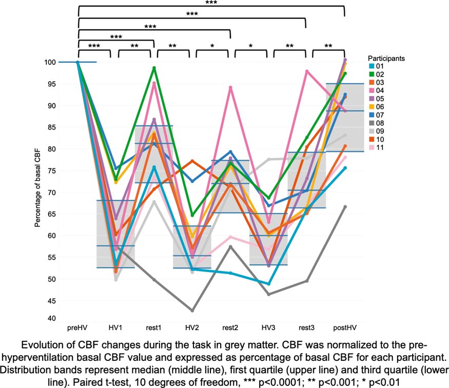
References
PB01-K09
Changes in cerebral blood flow during pregnancy detected by arterial spin labeling MRI in healthy subjects and in the patients with moyamoya disease
1Department of Neurosurgery, Tokyo Medical and Dental University
2Department of Perinatal and Women’s Medicine, Tokyo Medical and Dental University
Abstract
Purpose
The physiology of cerebral hemodynamics change during pregnancy is still unclear. The objective of this study is to evaluate the maternal cerebral blood flow (CBF) change measured by arterial spin labeling (ASL) MRI in healthy subjects and in the patients with moyamoya disease (MMD). The safety of applying high magnetic field MRI to the pregnant woman is also confirmed.
Subjects and methods
This study was approved by the ethical committee of the institute. MRI studies were performed using a 3.0 Tesla MR scanner. Three dimensional pulsed continuous ASL was performed to the healthy non-pregnant women (Group A, n = 10), healthy pregnant women (Group B, n = 10), and pregnant women with MMD (n = 16). All MMD patients also had ASL-MRI more than 6 months before or after the pregnant period. Regional and global CBF (ml/100 g/min) were compared by 3D-SSP based ROI analysis.
Results
There was no adverse event observed during scan and during pregnant course after scan. Also, no fetal abnormality was observed. In healthy groups, global and cortical CBF (ml/100 g/min) showed no significant change during pregnant period. The global CBF of group A and B were 58.4 ± 10.9 and 62.1 ± 9.7, respectively (p = 0.45). Contrarily, the global CBF significantly dropped during pregnancy in the MMD patients (non-pregnant v.s. pregnant: 53.8 ± 9.8 v.s. 45.2 ± 6.6, p = 0.002). Among the MMD patients, 7 had multiple scans in different trimester of pregnancy and showed lower CBF in the later trimester (2nd trimester v.s. 3rd: 54.3 ± 4.8 v.s. 44.0 ± 6.7).
Conclusions
ASL-MRI could perform safely and non-invasively to the pregnant women. This is the first report to evaluate both global and regional CBF of the pregnant the result indicated that cerebral hemodynamic may change differently between healthy women and the patients with MMD.
PB01-K10
T2-weighted imaging may be indicative of drug distribution in glioblastoma patients
1Precision Neurotherapeutics Innovation Program, Department of Neurological Surgery, Mayo Clinic, Phoenix, AZ, USA
2Brain Barriers Research Center, Department of Pharmaceutics, University of Minnesota, Minneapolis, MN, USA
3Department of Neurological Surgery, Mayo Clinic, Rochester, MN, USA
4Department of Radiation Oncology, Mayo Clinic, Rochester, MN, USA
Abstract
Objectives
Dogma in clinical neuro-oncology holds that gadolinium (Gd) contrast on T1-weighted magnetic resonance imaging (MRI) in tumor regions confirms that the blood-brain barrier (BBB) is locally compromised, and thus drug is being distributed within these tumor regions[1-2]. However, the poor response to targeted therapies delivered intravenously highlights the importance of the local microenvironmental heterogeneity and diffuse disease beyond enhancing regions[1-2]. This study investigated whether non-invasively acquired imaging features can provide an informative snapshot of the tumor microenvironment allowing for an accurate prediction of drug distribution.
Methods
Eight patients with presurgical radiographic evidence suggesting a brain tumor were included in this Phase 0 trial. Malignancies present in the recruited cohort included 7 gliomas (1 Grade I, 3 Grade III, and 3 Grade IV) and 1 metastatic adenocarcinoma. Presurgery T1-weighted, T1-weighted Gd contrast enhanced (T1Gd), T2-weighted Gd contrast enhanced (T2Gd), and T2-weighted Fluid Attenuated Inversion Recovery (T2FLAIR) MRIs were acquired. All images underwent bias correction using the N4 algorithm[3], standardization of intensities, and registration. Prior to incision, patients received doses of cefazolin, an antibiotic, and levetiracetam, an anti-seizure drug. These two drugs were chosen for their current clinical use and differing BBB penetrances. Cefazolin has only a 6% penetrance, while levetiracetam has 80% penetrance[4-5]. After both drugs were administered, multiple blood samples were acquired at least 20 minutes apart to measure plasma drug concentration. Multiple image-localized biopsies were collected from each patient using a surgical navigation system for a total of 27 biopsies across all patients. Biopsies and plasma samples were analyzed for drug concentration using liquid chromatography mass spectrometry (LCMS). Biopsy drug levels are reported as Brain-Plasma Ratio (BPR), the ratio of biopsy concentration relative to plasma concentration. Mean image intensity was extracted from an 8 × 8 mm window surrounding the biopsy location on each image. Regression analysis was performed to determine which combination of image types were linearly predictive of BPR for both drugs. Image-localized biopsies were categorized according to their T1Gd and T2Gd/T2FLAIR appearance: non-enhancing (NE), T2Gd/T2FLAIR hyperintense but non-enhancing on T1Gd (F + NE), and contrast enhancing (CE). BPR and image intensities were then analyzed according to the radiographic signature associated with each biopsy location.
Results
Regression analysis revealed that T2Gd intensity was linearly predictive of cefazolin BPR and FLAIR intensity was linearly predictive of levetiracetam BPR (p = 0.009 and 0.041, respectively). Grouping samples according the the radiographic appearance revealed patterns that were similar between BPR and image intensities. Levetiracetam BPR had a similar pattern of values to that of FLAIR intensities and cefazolin BPR had a similar pattern to T2, further supporting the regression analysis results.
Conclusions
Local concentrations of drug may be related to T2-weighted signals (T2Gd and T2FLAIR) rather than the Gd distribution on T1Gd images. Future work includes evaluating advanced quantitative imaging including perfusion and diffusion-weighted imaging.
References
PB01-K11
The feasibility of deep learning to predict final stroke lesion using baseline diffusion-weighted imaging only in non-recanalized acute ischemic stroke patients
1Radiology department, Stanford University, USA
2Neurology department, Stanford University,USA
3Electrical engineering department, Stanford University, USA
Abstract
Objective
We aim to investigate if baseline diffusion-weighted imaging (DWI) can predict final stroke lesion in acute ischemic stroke patients.
Methods
Acute ischemic stroke patients were reviewed and selected from Imaging Collaterals in Acute Stroke (iCAS) study and from DEFUSE2 study. Patients who underwent baseline MRI including perfusion-weighted imaging, diffusion-weighted imaging, angiography; and follow up imaging with angiography 24 hours after arrival and T2-FLAIR 3–5 days after stroke onset; and who had a TICI score ≤2 a or no arterial recanalization within 24 hours were included. The ground truth was defined as the final stroke lesions on follow-up T2-FLAIR, which were manually delineated by readers blinded to clinical information. A 2.5 dimension U-net model was trained using baseline DWI b1000 and apparent diffusion coefficient (ADC) as input. The model was trained based on a mixed loss function of cross-entropy and dice score. Five-fold cross-validation was performed to evaluate the predictions. The optimal threshold for the U-net model was determined by the largest Youden’s Index of the receiver operating curve (ROC), and then dice score, sensitivity, specificity, positive predictive value, and volume difference were calculated based on the optimal threshold. The model prediction was also compared with clinically used threshold of Tmax > 6 s plus ADC < 620 using paired sample Wilcoxon test.
Results
In total, 65 patients were eligible for inclusion. (32 males, age 65 ± 16, onset-to-imaging time 5.6 (IQR 4.5–7.5) hours, median NIHSS 14 [IQR 11–19], 3-month mRS 4 [IQR 2–4], final stroke lesion volume 107 (IQR 57–228) ml). The U-net model had an area under curve of 0.851 vs AUC of Tmax 0.8. Compared with Tmax and ADC, the U-net model had a dice score of 0.48 [IQR 0.27–0.65] vs 0.42 [IQR 0.23–0.57] (p = 0.025), sensitivity of 0.74 [IQR 0.46–0.92] vs 0.76 [IQR 0.65–0.89] (p = 0.18), specificity of 0.90 [0.84–0.96] vs 0.83 [IQR 0.76–0.90] (p < 0.001), positive predictive value of 0.43 [IQR 0.18–0.63] vs 0.30 [IQR 0.14–0.53] (p = 0.002), volume difference of 55 [IQR -6-128] ml vs 124 [IQR 55–208] ml (p < 0.001) and absolute volume difference of 88 [IQR 37–156] ml vs 141 [IQR 83–264] ml (p < 0.001). The metrics of the U-net model were calculated based on the optimal threshold of the ROC curve.
Conclusion
The U-net model with DWI and ADC only as input may help estimate final stroke lesion without major reperfusion, which may be useful when perfusion scans are not available. Further validation and improvements are required.
PB01-K12
Evaluation of tissue reversibility by relative apparent diffusion coefficient value in acute ischemic stroke patients treated with successful thrombectomy
1Dept. of Strokology, Stroke Center, St. Marianna University Toyoko Hospital, Kawasaki, Japan
Abstract
Background
By recent advance of endovascular thrombectomy, we have often experienced acute ischemic patients who have diffusion weighted imaging (DWI) reversal lesions after earlier successful recanalization. We retrospectively investigated the relationship between apparent diffusion coefficient (ADC) thresholds of tissue infarction and time from onset to recanalization in acute ischemic stroke patients.
Methods
We assessed 24 patients who have occlusion of internal carotid artery (n = 11) and the main trunk of middle cerebral artery (n = 13) and obtained recanalization of TICI2 b (n = 12) and TICI3 (n = 12) by thrombectomy and performed MRI before and after treatment. Relative ADC (rADC) value were calculated for initial DWI lesions and around hypoperfused regions. We evaluated rADC values in infarcted and non-infarcted area and analyzed the relationship between rADC thresholds of tissue infarction and time.
Results
The mean time from onset to recanalization was 209 minutes and mean initial NIHSS was 15.4. The mean rADC value was 0.633 in infarcted lesions and 0.905 in non-infarcted area (p < 0.001). The thresholds for rADC value for infarction by the area under the curve derived from receiver operating characteristic curve analysis were 0.769 in the area which recanalized under 180 minutes form the onset, 0.792 in that from 180 to 240 minutes, and 0.798 in that over 240 minutes.
Conclusion
The estimatio n of rADC value may be useful in predicting the likelihood of DWI lesion reversal. Marked decreasing of rADC value which is under thresholds of infarction indicated irreversible damage of ischemic tissue regardless of early successful recanalization.
PB01-L01
The effects of acute dopamine depletion on restingstate functional connectivity and striatal glutamate in healthy humans
1Department of Psychiatry, University of Toronto Withdrawn
PB01-L02
Radiation-induced and non-radiation-induced carotid atherosclerosis: computer-aided sonographic evaluation for plaque characterization and stroke risk assessment
1Department of Health Technology and Informatics, The Hong Kong Polytechnic University, Hong Kong
2Department of Clinical Oncology, The University of Hong Kong, Hong Kong
Abstract
Objectives
Cancer patients with previous neck radiotherapy and patients with cardiovascular risk factors (CVRF) such as diabetes and hypertension prone to have carotid atherosclerosis that leads to development of carotid plaques. The vulnerability and composition of carotid plaque are highly associated with the risk of stroke. However, the mechanisms of radiation-induced and non-radiation-induced carotid atherosclerosis are different. Ultrasound is a common imaging method to evaluate carotid atherosclerosis. This study aimed to use a computer-aided assessment method to analyze carotid ultrasound images and characterize the carotid plaques in post-radiotherapy patients and patients with CVRF.
Methods
We recruited 107 nasopharyngeal carcinoma (NPC) patients with previous neck radiotherapy (RT) (68 males, 39 females, mean age = 58.4 years old) and 110 patients with at least one CVRF (44 males, 66 females, mean age = 60.7 years old). Each patient had a carotid ultrasound examination to identify any carotid plaque. When a carotid plaque was identified, ultrasound image of the carotid plaque was acquired for image analysis. Ultrasound images of identified carotid plaques were analyzed with the Image Pro Plus 6.0 software. The brightness level of the ultrasound images was normalized before image analysis. The plaque component was categorized into five types based on the grey scale ranges: blood = 0-9; lipid = 10–31; muscle = 32-74; fibrous tissue = 75-111; calcification = 112-255 (1,2). The plaque components were color-coded and the area in percentage of each component within the plaque was calculated.
Results
Carotid plaques were found in 86 post-RT NPC patients (80.4%) and 42 patients with CVRF (38.2%). There were 247 carotid plaques identified in the 86 post-RT NPC patients with an average of 2.9 plaques in each patient, whereas 59 carotid plaques were identified in the 42 CVRF patients with an average of 1.4 plaques in each patient. The differences in the incidence of carotid plaque and the average number of plaque in patients were statistically significant between the two patient groups (p < 0.05). Carotid plaques in CVRF patients tended to have more lipid than those in post-RT NPC patients (p < 0.05). However, carotid plaques in post-RT NPC patients had significantly more calcification than those in CVRF patients (p < 0.05) (Figure 1).

Conclusions
Radiation-induced carotid atherosclerosis had a higher incidence of plaque formation than non-radiation-induced carotid atherosclerosis. However, the radiation-induced carotid plaques had more calcification and tended to be more stable, whereas non-radiation-induced carotid plaques had more lipid and was more vulnerable. Thus, patients with CVRF tended to have higher risk of stroke than post-RT NPC patients.
References
PB01-L03
Ultrasonographic micro-calcification indicate vulnerability of carotid atherosclerosis plaque
1Department of Vascular Ultrasonography, Xuanwu Hospital, Capital Medical University, Beijing 100053, China
Abstract
Objective
Intraplaque hemorrhage (IPH) and ulceration with cap rupture are considered as the main characteristics of vulnerable plaque. Whereas, the impact of calcification to the plaque vulnerability is still controversial based on previous studies. The aim of this study was to investigate the association of calcium configurations and IPH/ulceration via ultrasound.
Methods
There were two hundred and sixteen consecutive patients performed with carotid endarterectomy (CEA) recruited in this study. All patients accepted prior ultrasonography (USG) exam and then confirmed with computed tomography angiography (CTA) or digital subtraction angiography (DSA). Ultrasonographic characteristics of plaque were recorded including plaque cap morphology, echogenicity and calcifications which categorized as superficial, deep, micro and mixed types. All plaque samples were collected for histology after CEA.
Results
Patients enrolled were aged from 38 to 82 with vascular risk factors. There were 63 plaques had IPH (29.2%) and 96 had ulceration (44.4%). No differences of stenosis degree, superficial or deep calcification were found between IPH or ulceration groups. However, the incidence of hypoechoic (P < 0.01), presence of calcification (P < 0.01) and micro-calcification (P < 0.01) were higher in lesions with IPH, while the cap-rupture (P < 0.01) and micro-calcification (P < 0.01) were also higher in lesions with ulceration (

Conclusions
The plaque calcium was not a protective factor for plaque vulnerability. The micro-calcification might be a marker of vulnerable plaque both in IPH and ulceration.
References
PB01-L04
The correlation of carotid web and ischemic stroke evaluated by ultrasonography
1Dept. of vascular ultrasonography, Xuanwu Hospital, Capital Medical University, Beijing, China
2Dept. of radiology, Xuanwu Hospital, Capital Medical University, Beijing, China
Abstract
Objective
Carotid web is an intimal variant of fibromuscular dysplasia and is related to recurrent ischemic stroke. Computed tomography (CTA) has been accepted as a superiority for diagnosing the carotid web, whereas ultrasound recognition was underestimated due to the lack of the knowledge of its imaging characteristics. The aim of this study was to investigate the ultrasonographic features of carotid web and analyze the correlation of carotid web and ischemic stroke (IS).
Methods
A total of 24 patients with final diagnose of carotid web were retrospectively enrolled from January 2018 to November 2018. All patients were performed with ultrasonography (USG) first, then confirmed with CTA. According to the presence of infarction in the involved territory, they were divided into IS and no IS (NIS) groups. The acute angle between web and carotid wall was measured on picture archiving and communication systems (PACS) afterwards. The ultrasonographic characteristics of carotid web were compared between two groups.
Results
There were 13 patients diagnosed with carotid web by initial USG examination (54.2%), but 11 patients (45.8%) misdiagnosed as ulcer-plaque (9 patients) or dissection (2 patients) prior to final diagnosis. Therefore, a total of 24 patients were enrolled (10 in IS group, 14 in NIS group). In ultrasound scanning, the web was shown as a diaphragm-like tissue with hyperechoic, and a vortex was usually found in the acute angle under color mode. Although there were no differences of the web length (P = 0.76), thickness (P = 0.53) and carotid stenosis degrees (P = 0.34) between two groups, the presence of thrombus (80.0% vs 35.7%, P = 0.047) and the rate of acute angle ≤ 70° (90% vs 57.1%, P = 0.033) were higher in IS group (

Conclusion
Since ultrasound imaging is the first line in clinic, here we provide valuable information of ultrasonographic features for carotid web. By differentiating from ulcer plaque or dissection, carotid web was illustrated as a diaphragm-like tissue with high echo. The acute angle ≤ 70° and web thrombus were correlated with ischemic stroke.
PB01-L05
Lipid core plaque evaluation using NIRS-IVUS predicts thromboembolic complications in patients undergoing carotid artery stenting
1Department of Neurosurgery, Nara Medical University, Japan
Abstract
Introduction/Purpose
Carotid artery stenting (CAS) is a less invasive alternative to carotid endarterectomy, but it is essential to prevent thromboembolic complications. Intravascular near-infrared spectroscopy (NIRS) is a novel imaging modality that can detect a lipid core plaque (LCP) within an atherosclerotic plaque. Recently, IVUS and NIRS were combined in a single catheter so that the 2 modalities could be acquired simultaneously. In the present study, we have evaluated whether LCP evaluation using NIRS-IVUS predicts thromboembolic complications rather than preoperative magnetic resonance (MR) plaque imaging in patients undergoing carotid artery stenting (CAS).
Materials and Methods
Fifty-seven patients (48 males; mean age 74 years old) who undergoing CAS were enrolled. NIRS-LCP (lipid core burden index; LCBI, maximum LCBI; maxLCBI, maxLCBI at MLA) and MR plaque vulnerability (plaque SIR at MLA, plaque SIR at the area, TOF high intensity) were compared with clinical results.
Results
In DWI positive group, LCBI, maxLCBI, and maxLCBI at MLA were significantly higher than that in DWI negative group (p = 0.001, p = 0.001,p = 0.001 respectively). There wassignificant correlation between LCBI value and plaque SIR. In contrast, maxLCBI value was significantly decreased after stent placement and post-balloon dilatation (p < 0.001).
Conclusion
NIRS-IVUS can accurate LCP evaluation and can detect CAS procedural high risk plaque. NIRS-IVUS is a useful tool for preventing thromboembolic complication in patients undergoing CAS.
PB01-L06
Hyperventilation test with indocyanine green kinetics under near-infrared spectroscopy predicts cerebral hyperperfusion and indicate an efficacy of staged strategy of carotid artery stenting
1Department of Neurosurgery, Nara Medical University, Japan
Abstract
Objective
Cerebral hyperperfusion syndrome (CHS) is a serious complication following carotid artery stenting (CAS), but definitive early prediction of CHS has not been established. Staged CAS strategy can avoid hyperperfusion phenomenon (CHP) have been reported. Here, we evaluated whether indocyanine green kinetics and nearinfrared spectroscopy (ICG-NIRS) with hyperventilation (HV) and the breath-holding (BH) test can predict CHP after CAS and can prove the efficacy of staged CAS strategy.
Methods
The blood flow index (BFI) ratio during HV and BH was monitored using ICG-NIRS in 161 patients scheduled to undergo CAS. HPS high risk patients, whose asymmetry index (AI) was no more than 0.8 with cerebrovascular reactivity (CVR) of less than 10% on preoperative single-photon emission computed tomography were included (n = 23). Preoperative CVR and the postoperative AI were also assessed with SPECT and before and after percutaneous angioplasty (PTA) / CAS and the correlation with the BFI HV/rest ratio, BFI BH/rest ratio was evaluated between direct CAS group (n = 16) and staged CAS group (n = 7). Staged CAS were performed 3 weeks after PTA.
Results
In direct CAS group, eight cases (50%) showed CHP, and two (13%) showed CHS after CAS, however, no one showed CHP/CHS in staged CAS group (p = 0.007). A BFI HV/rest ratio and a BFI BH/rest ratio were significant differences between the groups as well as postoperative AI.
Conclusions
HV and BH test under ICG-NIRS is a useful tool for detection of hyperperfusion phenomenon and can indicate the effect of staged CAS strategy in patients who underwent CAS.
PB01-L07
Angiographic circulation time and cerebral blood flow during balloon test occlusion of the internal carotid artery
1Department of Neuroendovascular Therapy, Kohnan Hospital, Japan
2Department of Neurosurgery, Kohnan Hospital, Japan
3Department of Neurosurgery, Tohoku University Graduate School of Medicine, Sendai, Japan
4Department of Neurosurgery, Akita University Graduate School of Medicine, Akita, Japan
Abstract
Angiography-based balloon test occlusion (BTO) has been empirically used to predict tolerance to permanent carotid artery occlusion. We tested the hypothesis that the laterality between the hemispheric circulation time (HCT) of contrast medium at the cerebral angiography would reflect bilateral asymmetry in the cerebral blood flow (CBF) during the BTO.
Thirty-one consecutive patients who underwent the BTO of the internal carotid artery were analyzed. The HCT was defined as the interval of time-to-peak in time-density curve between the middle cerebral artery and the cortical veins. Difference of the HCT between the occluded and non-occluded side was calculated at the carotid or dominant vertebral angiograms obtained during the BTO. We estimated the correlation between the difference of the HCT and the bilateral asymmetry in the CBF, which quantitatively determined with single-photon emission computed tomography.
The HCT was 5.3 ± 1.5 seconds and the regional CBF was 41.3 ± 11.3 ml/100 g/min in the occluded side, whereas 3.6 ± 0.9 seconds and 48.4 ± 14.9 ml/100 g/min in the non-occluded side, respectively. The difference of the HCT was strongly correlated with the asymmetry ratio of the CBF (r2 = 0.89, P < 0.0001).
Measurement of the cerebral circulation time based on angiography can provide valuable information of cerebral hemodynamics.
PB01-L08
2D X-ray angiography perfusion and transcranial cerebral oxygen saturation monitoring during carotid artery stenting for the detection of post-stenting hyperperfusion
1Dept. of Neurosurgery, Tokyo Dental College Ichikawa General Hospital, Japan
Abstract
Objectives
The purpose of this study was to evaluate the efficacy of 2D X-ray angiography perfusion software (2D-Perfusion) and transcranial regional cerebral oxygen saturation (rSO2) monitoring via near-infrared spectroscopy (INVOS) to detect hyperperfusion after carotid artery stenting (CAS).
Methods
This study included four patients who underwent CAS for severe carotid artery stenosis. 2D-perfusin studies were performed before and after CAS, and a time-density curve was constructed for each hemisphere. The mean transit time (MTT) and time to peak (TTP) were obtained1). Differences in these parameters between affected and unaffected hemispheres were compared between before and after CAS. During CAS procedure, rSO2 monitoring was simultaneously performed2).
Results
A 86 years old male patient underwent CAS for progressing stroke due to severe carotid artery stenosis. Before CAS, both of MTT and TTP of affected hemisphere were prolonged, but after CAS, both parameters of not only affected but also unaffected side were shortening. Corresponding to these changes, rSO2 of both sides were increased, which supposed to be the possibility of hyperperfusion. The patient was under deep sedation for a few days to prevent the devastating complication. After that, the patient recovered without any neurological deficits. Other three patients underwent CAS in the chronic phase, and only affected side of parameters were improved after CAS in all cases, and no significant increase of rSO2 were observed in these cases. After procedures, all patients were uneventful.
Conclusion
2D-perfusion study and rSO2 monitoring during CAS were convenient and useful for the detection of hemodynamic changes of hemispheres.
Reference
PB01-L09
Cerebral hemodynamic disturbance in dural arteriovenous fistula with retrograde leptomeningeal venous drainage: a prospective study using 123I-iodoamphetamine single photon emission computed tomography
1Department of Neurosurgery, University of Yamanashi, Yamanashi, Japan
Abstract
Object
The severity of cerebral hemodynamic disturbance caused by retrograde leptomeningeal venous drainage (RLVD) of a dural arteriovenous fistula (dAVF) is related to neurological morbidity and unfavorable outcome. However, the cerebral hemodynamics of this disorder have not been elucidated well. The aim of this study was to assess the relationship between the cerebral venous congestive encephalopathy represented as a high-intensity area (HIA) on T2-weighted MR images and the cerebral hemodynamics examined by 123I-iodoamphetamine (IMP) single photon emission computed tomography (SPECT), as well as the predictive value of 123I-IMP SPECT for the development and reversibility of venous congestion encephalopathy.
Methods
Based on the pre- and posttreatment T2 HIAs associated with venous congestion encephalopathy, patients were divided into 3 groups: a normal group, an edema group, and an infarction group. The regional cerebral blood flow (rCBF) at the region with RLVD was analyzed by 123I-IMP SPECT, and the results were compared among the groups.
Results
There were 11, 6, and 3 patients in the normal, edema, and infarction groups, respectively. No patients in the normal group showed any symptoms related to venous congestion. In contrast, all patients in the edema and infarction groups developed neurological symptoms. The rCBF in the edema group was significantly lower than that in the normal group, and significantly higher than that in the infarction group. The cerebral vascular reactivity (CVR) of the infarction group was significantly lower than that of the normal and edema groups. After treatment, the neurological signs disappeared in the edema group, but only partial improvement was seen in the infarction group. The rCBF also significantly increased in the normal and edema groups, but not in the infarction group.
Conclusions
Quantitative rCBF measurement is useful for evaluating hemodynamic disturbance in dAVF with RLVD. The reduction of rCBF was strongly correlated with the severity of venous congestive encephalopathy, and loss of CVR is a reliable indicator of irreversible venous infarction caused by RLVD.
PB01-L10
Exploring the effect on cerebral blood flow after acupuncture in elderly normal subjects using 99 mTc-ECD imaging
1Department of Nuclear Medicine, Tri-Service General Hospital, National Defense Medical Center, Taipei, Taiwan
2Bai-Han Chinese Medicine Clinic, Taipei, Taiwan
3Yusheng Chinese Medicine Clinic, Taipei, Taiwan
Abstract
Objectives
In the past few decades, there has been an increasing demand of traditional Chinese medicine. Based on general conception, traditional Chinese treatment is more focused on patients' holistic physical condition, which gradually works up to getting the whole body in balance. Acupuncture is widely applied in the combined treatment of cerebrovascular accident clinically, but the effect of acupuncture on cerebral blood flow is not clear. The aim of this study was to explore the effect on cerebral blood flow after acupuncture in elderly normal subjects using 99mTc-ECD imaging.
Methods
This protocol was approved by the Institutional Review Board of Tri-Service General Hospital, National Defense Medical Center, Taipei, Taiwan (approval number: 1–107-05-161). We choose Shousanli (LI 10) for acupuncture point in this study, which located at the radial side of the forearm and 2 inches below the transverse cubital crease. A total of 10 elderly normal subjects (age: 55–72, 58 ± 11) received 99mTc-ECD imaging two times. The baseline 99mTc-ECD imaging started 30–80 minutes after 20 mCi 99mTc-ECD injection. After four weeks, the second 99mTc-ECD imaging was performed after receiving left Shousanli (LI 10) acupuncture for 20 minutes, the 99mTc-ECD imaging was also performed 30–80 minutes after 20 mCi 99mTc-ECD injection. All images were spatially normalized into Montreal Neurological Institute (MNI) template, and 15 VOIs in the AAL template were applied for uptake ratio (global mean as reference). The difference uptake ratio (DUR) between baseline (URb) and after acupuncture (URa) were compared. [DUR% = URa – URb / URb *100]
Results
The results showed increase cerebral blood flow in the hippocampus (7.37), putamen (7.27), amygdala (6.17), olfactory (6.09), caudate (5.78), thalamus (5.05), insula (4.06), cingulum (3.53), precuneus (2.27), cerebellum (2.19) and temporal (0.11). Decreased cerebral blood flow was also noted in the occipital (−3.05), parietal (−2.44), superior frontal (−2.23) and middle frontal (−1.89).
Conclusion
Preliminary results show that cerebral blood flow can be stimulated by administering acupuncture on Shousanli (LI 10), which functional neuroimaging may provide scientific evidence for the therapeutic potential of acupuncture.
PB01-L11
Management of intraoperative technological advances [intraoperative MRI, neuronavigation system using PET, and 5-aminolevulinic acid (5-ALA)–induced fluorescence image-guided surgery] for glioblastoma
1Department of Neurological Surgery, Kagawa University Faculty of Medicine, Japan
Abstract
Objective
Glioblastoma (GBM) is the most aggressive neoplasm among the gliomas and is characterized by histopathologic intratumoral heterogeneity, with respect to tumor morphology. The maximum resection of GBM is the standard therapy and is expected to improve prognosis. Image-guided surgery using a neuronavigation system is the standard technique for glioma surgeries. Because of the brain shift occurring during surgery, neuronavigation systems are limited by the proper presence and metabolism of GBM cells. Therefore, intraoperative technologies, such as 5-ALA fluorescence and intraoperative MRI (IoMRI), are employed. Radiotracers are used during positron emission tomography (PET) for metabolic and molecular imaging and assist the evaluation of glioma metabolism. We compared the effectiveness of these intraoperative technologies.
Methods
Between January 2016 and December 2018, 45 patients with gliomas underwent IoMRI. After excluding patients who underwent biopsy only and those who could not undergo multiple PET studies (MET, FLT, and FMISO), 15 patients were selected for 5-ALA fluorescence-guided resection of GBM. Each patient received 5-ALA approximately 3 h before surgery, and a modified neurosurgical microscope was used for intraoperative visualization of 5-ALA-induced fluorescence. We graded fluorescence level as strong, vague, or none. Following tumor resection, we identified the fluorescence level and evaluated the residual volume of gadolinium-enhanced T1WI (T1-Gd) on IoMRI and at each PET study. After calculating the extent of resection (EOR) for T1-Gd on a MET PET study, we selected an end of radiation (EOR) of 96%. Subsequently, we compared the residual volume on T1-Gd for IoMRI and each PET study, between EOR ≥ 96% and EOR < 96%.
Results
We detected strong 5-ALA fluorescence during induction and before tumor resection in all 15 (100%) patients with a newly-diagnosed and histopathologically-confirmed GBM. Following tumor resection, we noted an EOR ≥ 96% for T1-Gd in 8 cases (vague, 2; none, 6) and an EOR < 96% for T1-Gd in 7 cases (vague, 4; none, 3). The compared median residual volume (mL) with no fluorescence between EOR ≥ 96% and EOR < 96% for T1-Gd were T1-Gd (0.08, 0.19), MET (0.79, 0.2), FLT (0.5, 0.17), and FMISO (0.29, 0.16). For MET, 7 and 8 cases had an EOR ≥ 96% (vague, 2; none, 5) and EOR < 96% (vague, 4; none, 4), respectively. The compared median residual volume (mL) with no fluorescence between EOR ≥ 96% and EOR < 96% for MET were T1-Gd (0.03, 0.21), MET (0.13, 1.17), FLT (0.11, 0.73), and FMISO (0.02, 0.51).
Conclusions
It is challenging to fuse IoMRI images with multiple preoperative PET images during navigation. Moreover, GBM cells are difficult to distinguish in cases without 5-ALA fluorescence. For cases without 5-ALA fluorescence, the residual volume of T1-Gd, MET, FLT, and FMISO focusing on the MET was lesser than T1-Gd. Extracting the accumulation of MET facilitated the maximum resection of GBM.
PB01-T01
Bidirectional control of neurovascular coupling by pyramidal cells
1UPMC Univ Paris 06, INSERM, CNRS, Neuroscience Paris Seine-Institut de Biologie Paris Seine (NPS-IBPS), Sorbonne Université, Paris
Abstract
Tight coupling between neural activity and cerebral blood perfusion is essential for brain function and integrity. Neuronal activity induces a neurovascular response consisting of a local and transient increase in blood perfusion surrounded by a delayed hypoperfusion underpinned by vasoconstrictions. These vasoconstrictions might be essential for an optimal spatiotemporal match between energy demand and energy supply and are notably impaired in Alzheimer's disease. Yet, despite this physiopathological importance, the cellular and molecular mechanisms governing vasoconstrictions remain largely unexplored.
Objective
In the cerebral cortex, pyramidal neurons constitutively expressing the rate limiting cyclooxygenase-2 (COX-2) can induce an increase in blood perfusion through the release of prostaglandin E21. Nonetheless, prostaglandin E2 is also capable to induce vasoconstrictions2. This suggests that pyramidal neurons could also induce a hypoperfusion. Thus, we aimed at determining whether pyramidal neurons can induce both vasodilations and vasoconstrictions.
Methods
Pyramidal neurons were specifically activated in cortical brain slices of 3-week-old Emx1-Cre::Ai32 mice by optogenetic photostimulations. The triggered vascular responses were visualized by infrared videomicroscopy and analyzed using pharmacological tools. Pyramidal neurons were also analyzed by single-cell RT-PCR after patch-clamp3 to determine their molecular equipment in vasoactive messengers.
Results
We show that depending on the frequency of photostimulation, pyramidal neurons can induce either vasodilations or vasoconstrictions. At low frequency, they induce vasodilations whereas at a high frequency they triggers vasoconstrictions. We previously showed the role of prostaglandin E2 in the vasodilatations induced by pyramidal cells. We now disclose that COX-2 derived prostaglandins produced by these neurons are also responsible for the neurogenic vasoconstrictions observed at high frequency. The molecular characterization by single-cell RT-PCR of these neurons reveals that pyramidal cells are equipped for the synthesis of both vasodilatory and vasoconstrictive prostaglandins.
Conclusion
These results indicate that pyramidal neurons can both initiate and terminate the neurovascular response. It remains to determine how the bidirectional control of this neurovascular response evolves with age and in Alzheimer's disease.
References
PB01-T02
Coupling of neuronal activity to blood flow in dendrodendritic synapses is mediated by postsynaptic gabaergic cells in olfactory bulb
1Dept. of Radiology, University of Pittsburgh, USA
2Dept. of Bioengineering, University of Pittsburgh, USA
3Center for Neuroscience Imaging Research, Institute for Basic Science, Suwon, Korea
4Depar. of Biomedical Engineering, Sungkyunkwan University, Suwon, Korea
Abstract
Objectives
Recent optogenetic studies demonstrate that direct photo-stimulation of channelrhodopsin (ChR2) expressing GABAergic cells evokes blood flow responses (1–3). However, the mechanism of evoked blood flow in these ChR2-expressing inhibitory mouse models may be different from synaptically activated GABAergic cells since ChR2 is permeable to Na+, K+ and Ca2+. In the present study we, therefore, examined whether synaptically activated GABAergic cells can evoke blood flow responses.
Methods
Isoflurane-anesthetized (∼1.2%) Sprague-Dawley rats were used. In the olfactory bulb, GABAergic cells can be exclusively and synaptically activated via reciprocal dendrodendritic synapses in the external plexiform layer (EPL). These synapses are formed between the lateral dendrites of excitatory output mitral cells (MCs) and dendrites of inhibitory granule cells (GCs). Excitation of MCs releases glutamate from their lateral dendrites, which excites postsynaptic GCs to drive GABA release back onto the lateral dendrites; thus, MCs activity is reciprocally inhibited. A stimulation electrode was implanted in a bundle of axons from MCs (lateral olfactory tract, LOT) to antidromically excite MCs and trains of rectangular pulses (100 or 200 us, −200 uA, 40 Hz for 64 s every 5 min) were delivered to the electrode for activating the dendrodendritic synapses in EPL. Blood volume and blood flow responses were determined by a contrast agent-based functional magnetic resonance imaging (fMRI) and laser-Doppler flowmetry (LDF), respectively. Neuronal activity was also recorded during LDF measurement. LOT-stimulation evoked hemodynamic responses were compared before and after application of drugs on the bulb surface.
Results
As already reported (4), maximal increase in blood volume was evoked in EPL during LOT stimulation. This fMRI response was significantly suppressed when NMDA or AMPA glutamate receptor antagonists APV (25 mM) or NBQX (5 mM), respectively, were topically applied. Consistent results were obtained for neuronal activity and blood flow responses evoked by LOT stimulation. We then examined whether astrocyte activity driven by glutamate uptake was responsible for the evoked hemodynamic responses, as reported in the glomerular layer of the bulb (5). The topical application of astrocyte-specific glutamate transporter inhibitor DHK (1 mM) did not suppress the evoked neuronal activity and blood flow responses in EPL, but rather enhanced them. Additionally, we examined whether nitric oxide, the predominant vascular mediator in the cerebellum (6) and cortical GABAergic cells (3), initiated hemodynamic response. A nitric oxide synthase inhibitor L-NNA (1 mM) did not change the evoked neuronal activity or blood flow responses in EPL.
Conclusions
Activity of presynaptic excitatory cells and astrocyte activity driven by glutamate uptake are not enough for the LOT-stimulation evoked hemodynamic responses in EPL; but excitation of postsynaptic GABAergic cells increases blood flow and volume.
Supports
NIH grants (NS094404 to AV, EB018903 and EB003324 to MF) and a grant from the Institute for Basic Science (IBS-R15-D1 to SGK).
References
PB01-T03
Linear and non-linear temporal features of vascular responses evoked by photo-stimulation in excitatory and inhibitory optogenetic mouse models
1Dept. of Radiology, University of Pittsburgh
2Dept. of Bioengineering, University of Pittsburgh
3Dept. of Neurobiology, University of Pittsburgh
Abstract
Introduction
Recent in vivo studies using optogenetic strategies have revealed that inhibitory neurons can have a profound impact on local blood flow regulation. While it is known that sustained activity from excitatory neurons produce sustained vascular responses, the temporal contribution and features of vascular responses evoked by inhibitory neuron activity are less clear. The goal of this work is to examine the temporal properties of vascular responses evoked by optogenetic stimulation of excitatory and inhibitory neurons with various pulse durations and frequencies. This analysis will help understand the relationship between neuro-vascular coupling mechanisms engaged by these different neuronal populations.
Methods
Transgenic mice expressing Channelrhodopsin-2 (ChR2) in either excitatory neurons (Thy1-ChR2-YFP, n = 4) or inhibitory neurons (VGAT-ChR2-YFP, n = 4) were used for experimentation. Optogenetic stimuli (473 nm and 1 mW for 1–10 sec at 5 Hz, or 1sec at 5–40 Hz) were delivered by optic fiber to whisker somatosensory cortex. Measurements of changes in CBF and CBV were acquired by laser Doppler flowmetry (LDF) and optical imaging of intrinsic signal (OIS; 570 nm), respectively, under awake or light-ketamine anesthesia (30 mg/kg) conditions. Somatosensory stimulation (whisker air puffs) was used as a reference.
Results and Discussion
Under ketamine anesthesia, optogenetic stimulation of Thy1 mice (excitatory) at 5Hz and various pulse durations (2,10 or 30 ms) or frequencies produced responses with similar temporal characteristics that peaked 4–5 seconds after onset (Figure 1A). Similar optogenetic stimulation in VGAT mice (inhibitory) produced slightly slower responses that peaked 5–7 seconds after onset (Figure 1B). Under awake conditions, optogenetic stimulation of Thy1 mice produced similar temporal responses but with higher amplitude (Figure 1C), while optogenetic stimulation of VGAT mice produced responses that peaked faster and showed post-stimulation undershoot (Figure 1D). Further, increasing the stimulus duration did not show sustained vascular responses in VGAT mice, especially under awake conditions (Figure 2). Preliminary linearity analyses show that the vascular responses from Thy1 mice (excitatory) behave in roughly linear fashion, while the responses from VGAT mice (inhibitory) do not, especially under awake conditions. These results reinforce the notion that excitatory and inhibitory neurons engage different neuro-vascular mechanisms with different temporal properties. We are currently performing these experiments under blockade of glutamatergic and gaba receptors (NBQX, AP5, BMI) to observe responses mostly from the optogenetic expressing neuronal populations and minimize their impact/contribution from the connected network.
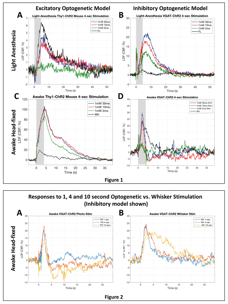
PB01-T04
Vasodilatory response to optogenetic activation of neurons or astrocytes in the anesthetized mouse cortex
1Department of Neurology, Keio University School of Medicine, Tokyo, Japan
2Tomita Hospital, Aichi, Japan
3Graduate School of Informatics and Engineering, University of Electro-Communications, Tokyo, Japan
4Department of Neuropsychiatry, Keio University School of Medicine, Tokyo, Japan
5Division of Interdisciplinary Medical Science, Tohoku University Graduate School of Medicine, Miyagi, Japan
6Department of Functional Brain Imaging Research, National Institute of Radiological Sciences, Chiba, Japan
Abstract
Objectives
Cerebral blood flow (CBF) is controlled by cell-to-cell cooperation of neurons and astrocytes. We previously showed optogenetic stimulation to neurons or astrocytes both similarly lead to focal and propagated increases in local CBF. Here, we aimed to determine the signaling mechanisms involved in the CBF responses to optogenetic stimulation.
Methods
Transgenic mice expressing a light-gated cation channel, channelrhodopsine-2 (ChR2), into cortical neurons (N = 43) or astrocytes (N = 34) were used for the experiments. A portion of the animal’s skull (3 mm in diameter) was removed under anesthesia, and sulforhodamine 101 was intraperitoneally injected to fluorescently label blood plasma. Photo-stimulation to a focal region of the cortex was evoked with a Xenon lamp equipped in the microscope, and diameter responses of the cortical vessel were sequentially captured with two-photon microscopy in vivo.
Results
Photo-stimulation to the ChR2-expressing neurons evoked significant dilation of the penetrating arteries nearby the stimulated cortex, whereas the ChR2-astrocytic stimulation provoked significant vasodilation of the both pial and penetrating arteries. These observation indicate that cortical vasodilation evoked by the astrocytic stimulation involves localized and conducted mechanisms, whereas the vasodilatory responses to the neurons were driven by localized mechanisms in the parenchyma.
Conclusions
Cell-type specific mechanisms of vasodilation participate in the optogenetically-evoked CBF responses.
PB01-T05
The differential contribution of vascular gap junctions to pial and penetrating artery dilation induced by forepaw electrical stimulation in isoflurane-anesthetized rats
1Department of Autonomic Neuroscience, Tokyo Metropolitan Institute of Gerontology
2Graduate School of Informatics and Engineering, The University of Electro-Communications
3Brain Science Inspired Life Support Research Center, The University of Electro-Communications
Abstract
Objectives
Somatosensory stimulation has been demonstrated to induce an increase in cerebral blood flow (CBF) in the representative regions of the somatosensory cortex. An increase in CBF is accompanied by the parenchymal and pial artery dilation. Somatosensory stimulation-induced arterial dilation is believed to be propagated through the vascular endothelium from the cerebral parenchyma to the cortical surface1. Furthermore, it has been suggested that vascular gap junctions transmit vasodilation signals in vitro; however, their roles in vascular regulation in vivo remain unclear. The present study aimed to investigate whether vascular gap junctions contribute to the regulation of the pial and penetrating arteries during somatosensory stimulation-induced neuronal activity.
Methods
Male Wistar rats (age 7–10 weeks) were artificially ventilated under isoflurane anesthesia (1.5%–1.7%). For somatosensory stimulation, the left forepaw was electrically stimulated (1.5 mA, 0.5 ms, and 10 Hz, for 5 s). Rhodamine-labeled Ficoll dissolved in saline was intravenously administered, to fluorescently label the cerebral vasculature. Imaging of the artery in the forelimb area of the right somatosensory cortex was performed via a cranial window using two-photon microscopy, and the diameter response to the stimulation was measured. In separate experiments, CBF was measured using a laser speckle flowmeter, and the somatosensory-evoked potentials were recorded from the right somatosensory cortex. To block vascular gap junctions, 100-mg/kg carbenoxolone (CBX) was intravenously administered.
Results
The forepaw electrical stimulation induced an increase in the diameter of the pial artery by 7.0% and of the penetrating arteries by 5.0% of the pre-stimulus diameter, without changing the systemic arterial blood pressure. The stimulation increased regional CBF by 24.1% of the pre-stimulus levels, and induced somatosensory-evoked potentials. Following CBX administration, pial artery dilation during forepaw stimulation was reduced to 3.2%. In contrast, CBX showed no effect on the extent of penetrating artery dilation or of CBF increase. Further, CBX demonstrated no influence on the resting level of the blood pressure or the magnitude of somatosensory-evoked potentials, suggesting that CBX does not affect the parenchyma functions but possibly had an effect on the propagation of cerebral vascular responses.
Conclusions
These results suggest that vascular gap junctions contribute to pial artery dilation but not to penetrating artery dilation during somatosensory stimulation-induced neuronal activity. The differential contributions of gap junctions to the pial and penetrating arteries may be explained by differences in their major regulatory mechanisms of the cerebrovascular tones2. Gap junctions in the endothelium might contribute to propagation of the vasodilatory signals. Further studies are, however, warranted to establish the cell types involved and their respective roles in cerebrovascular regulation.
References
PB01-T06
Stimulation-induced vasodilation and vasoconstriction depend on conducted vascular responses in cerebral capillaries
1Department of Neuroscience, Faculty of Health and Medical Sciences, University of Copenhagen, Denmark
Abstract
Functional neuroimaging, such as fMRI, is based on coupling neuronal activity and accompanying changes in cerebral blood flow (CBF) and metabolism. However, the relationship between CBF and events at the level of the penetrating arterioles and capillaries is not well established. Recent findings suggest an active role of capillaries in CBF control, and pericytes on capillaries may be major regulators of CBF and initiators of functional imaging signals. Here, using fast volume-scanning two-photon microscopy of brains in living mice, we demonstrate that both whisker pad stimulation and local ejection of gliotransmitter ATP evoke conducted vasodilation followed by vasoconstriction, starting mostly at the first- or second-order capillaries. These conducted vascular responses propagate in a slow speed of 3–20 µm/s. Vasodilation and vasoconstriction by both stimulation methods initiate at similar location in capillaries. Our data support an active role of pericytes in cerebrovascular control, and indicate that purinergic signaling involves in slow conducted vascular responses in capillaries. Thus, conducted vascular responses in capillaries may be a previously unidentified modulator of cerebrovascular function and functional neuroimaging signals.
PB01-T07
Hippocampal neurovascular coupling is reduced compared to visual cortex
1University of Sussex
Abstract
Objectives
Neurovascular coupling has been predominantly studied in primary sensory cortices, where neuronal activation leads to increased cerebral blood flow and a large influx of oxygenated blood. However, neural activity may be less well-coupled to increased blood flow in other brain areas. For example, local field potentials are highly correlated with fMRI/BOLD signals in sensory cortex, but not in the hippocampus (Ekstrom, 2010). This suggests that neurovascular coupling may differ between sensory cortex and hippocampus, which we investigated in this study.
Methods
We used 2-photon imaging to record fluctuations in neuronal activity and microvascular diameter in primary visual cortex (V1) and the hippocampus of awake, behaving mice, as well as combined laser doppler flowmetry and haemoglobin spectroscopy to record baseline and stimulus-induced alterations in macroscopic haemodynamics.
Results
Compared to V1, the hippocampus had lower resting blood flow and blood oxygen saturation, despite similar rates of oxygen consumption. Furthermore, individual blood vessels dilated significantly less frequently and to a smaller extent in the hippocampus compared to V1, despite equivalent-sized calcium responses. Finally, increases in regional oxygen consumption led to smaller macroscopic increases in blood flow in the hippocampus compared to V1.
Conclusions
These data suggest that not only is the hippocampus under-supplied with oxygen compared to primary sensory cortex at rest, but it is also less able to increase local blood flow in response to increased neuronal activity. These deficits in neurovascular coupling could contribute to the vulnerability of the hippocampus to hypoxia by reducing the ability of hippocampal neurons to match energy supply with demand.
Reference
PB01-U01
Two-photon and functional ultrasound transfer functions: from neuronal calcium to local capillary and regional vascular responses
1INSERM-U1128
2Penn State University
3Université Paris Descartes-UMR 8145
Abstract
Objectives
Blood-flow based imaging techniques indirectly report neuronal activity, involving a series of cellular interactions named neurovascular coupling1 (NVC). Despite the recent advances of these mesoscopic imaging techniques2–5, the quantitative relationship between neuronal activation and vascular responses collected at the microscopic or mesoscopic scale remains elusive.
Methods
We recently developed a single chronic mouse preparation and a sedation protocol suitable for multi-modal imaging, two-photon Laser Scanning Microscopy (TPLSM) and function ultrasound imaging (fUS). We used natural sensory stimulations (short odour inhalations) and imaged neuronal and vascular responses in the olfactory bulb (OB). Neuronal calcium responses were recorded in the most sensitive glomerulus to ethyl tiglate. Vascular responses were measured either locally with TPLSM or in the entire OB slice comprising the responding glomerulus with fUS. Transfer Functions (TFs) have been computed on the collected database.
Results
TFs from neuronal to capillary vascular responses proved to be surprisingly robust with respect to different stimulation parameters, such as the odor duration (from a single sniff to 5 sec), concentration or the odorant molecule. These “microscopic” TFs were also reliable across time and across different mice. Coalignment of our fUS and TPLSM apparatus enabled us to center a specific fUS voxel onto the responsive glomerulus imaged with TPLSM. fUS acquisitions allowed to calculate the mesoscopic TF between glomerular calcium responses and single voxel responses, providing the first “hemodynamic response function” (HRF) for fUS data. This HRF remained robust from single sniff to 5 second inhalations. It could also be used to link local calcium responses to global fUS responses from the entire OB slice comprising the responsive glomerulus.
Conclusions
The microscopic TF between calcium and capillary responses, in our experimental conditions, was much faster than previous found HRFs based on BOLD and CBV fMRI in humans and rodents6–8. This finding was confirmed with the single voxel or the global fUS mesoscopic HRF. To conclude, the fUS HRFs may provide a new tool to compare NVC in control and pathological animal models.
References
PB01-U02
The cortical vasculature of mouse affects the pattern of hemodynamic responses under dual sensory stimulus
1Center for Neuroscience Imaging Research (CNIR), Institute for Basic Science (IBS), Suwon, 16419, Rep. of Korea
2Dept. of Biomedical Engineering, Sungkyunkwan University, Suwon, 16419, Rep. of Korea
3Biomedical Institute for Convergence at SKKU (BICS), Sungkyunkwan University, Suwon, 16419, Rep. of Korea
4Dept. of Biological Science, Sungkyunkwan University, Suwon, 16419, Rep. of Korea
5Samsung Advanced Institute of Health Science and Technology (SAIHST), Sungkyunkwan University, Suwon, 16419, Rep. of Korea
Abstract
Objectives
Barrel and visual cortex of mouse brain is known to be supplied by two different arteries, i.e., middle cerebral artery (MCA) and posterior cerebral artery (PCA)1. Studies before observed changes of hemodynamic response in cortices supplied by MCA2,3. In this study, we quantified the amount of ‘artery-sharing’ of two different cortices and observed the differential hemodynamic responses under single and dual sensory-stimulus. The goal of this study is to investigate whether the vasculature of two cortices is responsible for the pattern of hemodynamic responses upon each stimulus.
Methods
Thinned-skull surgery was done to mice, and after 1w recovery we measured HbT by using intrinsic optical signal (IOS) imaging system. Both barrel and visual cortex were stimulated by giving either or both whole-whisker and visual stimulation (with LED flickering) and imaged simultaneously under ketamine/xylazine anesthesia. To compare the difference of hemodynamic response between single and dual stimuli without adaptation factor, stimulus was given in random order. Afterwards, we measured the distance between the focus pixel upon stimulation and branch pixels on arteries, then quantified the participation of different arteries under single and dual stimulation condition.
Results
Under single stimuli, each cortex did not show any significant correlation with specific artery. However, when two stimuli were given concomitantly, both barrel and visual cortex showed dependence to MCA but not to PCA. We could infer that both barrel and visual cortices became ‘MCA-dependent’ when dual stimuli were given, and the amount of ‘artery-sharing’ is responsible for the degree of hemodynamic responses within each animal.
Conclusions
Unique cortical vasculature of individual mouse is responsible for the pattern of hemodynamic response under dual stimulation, unlike when single stimulus is given. Thereby ‘artery-sharing’ need to be considered when quantifying the cerebral blood flow and metabolism under dual sensory stimulus.
References
PB01-U03
An oligarchy of no-producing neurons controls basal and evoked blood flow in the cortex
1Dept. of Neural Engineering and Neurosurgery, The Pennsylvania State University
2Depts. of Engineering Science and Mechanics, Neurosurgery, and Biomedical Engineering, The Pennsylvania State University
Abstract
Objectives
Changes in cortical neural activity are coupled to changes in local arterial diameter and blood flow. However, the neuronal types and signaling mechanisms that control basal and evoked changes in arterial diameter are not well understood. Elucidating which neurons can exert control over arterial diameters in the awake brain is important for interpreting the hemodynamic signals measured with fMRI1, as well as for understanding the declines in basal cerebral blood flow that precede dementia2.
Methods
We used local pharmacological infusions3 and cell-type specific chemogenetics (DREADDS) to manipulate neural activity in the somatosensory cortex of awake mice through a chronic, thinned skull window. Using two-photon microscopy4, we longitudinally assayed the impact of these manipulations in multiple vessels. We quantified basal arterial diameter and responses evoked to voluntary locomotion5, which drive large increases in neural activity in the somatosensory cortex.
Results
Bi-directional manipulations of all neural activity in increases or decreases in basal arterial diameter. Surprisingly, while chemogenetic manipulation of pyramidal neurons drive large changes in neural activity (>50% changes in the gamma band of the LFP), these large changes had little effect on basal or evoked arterial diameter. In contrast, manipulation of the activity of nNOS-positive neurons or local pharmacological manipulation of NO signaling had minimal impact on overall neural activity, but had large effects on the arterial diameter. Finally, we manipulated the activity of a subset of nNOS-positive interneurons that express the neurokinin-1 receptor6 with infusions of substance P or an antagonist. These infusions resulted in little change in neural activity, but substantial changes in basal arterial diameter, suggesting that this small group of neurons exercises considerable control over the cerebral vasculature.
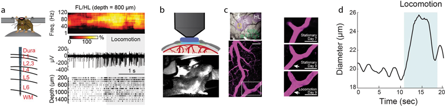
Conclusions
Hemodynamic signals have traditionally been interpreted as reporters of bulk neural activity, with increases in neural activity thought to be directly correlated with increases in vessel dilation and blood flow. Our data establishes a different model, where basal and evoked blood flow are controlled by a sparse subset of neurons (an ‘oligarchy’ independent of the population activity).
References
PB01-U04
Deciphering the role of neurons and vessels in neurovascular coupling in cerebrovascular disease through aging
1Laboratory of Cerebrovascular Research, Montreal Neurological Institute, McGill University, QC, Montreal, Canada
2Ecole Polytechnique Montréal, Department of Electrical Engineering, Montréal, QC, Canada
Abstract
Background
Neurovascular coupling (NVC) refers to the tight relationship between local neuronal activity and changes in glucose and oxygen supply from cerebral blood flow (CBF). NVC forms the basis of functional brain imaging techniques, such as fMRI. fMRI is increasingly used as a diagnostic tool in brain diseases, including in Alzheimer’s disease (AD) and other forms of dementia associated with cerebrovascular alterations. However, the challenge in discriminating the respective contribution of neuronal dysfunction from comorbid cerebrovascular disease in NVC responses in AD has recently been recognized.
Hypothesis and Objectives
As NVC requires an intact neurovascular unit, we hypothesize that a compromised brain circulation may potentially lead to misinterpretation of fMRI data when used as a neural proxy. Hence, using a transgenic mouse model of cerebrovascular alterations reminiscent to those found in AD, we assessed the reliability of NVC in detecting changes in neuronal activity by concurrently measuring sensory-evoked neuronal and hemodynamic responses.
Methods
Whisker-evoked changes in neuronal activity (local field potentials), cerebral blood flow (CBF, laser-speckle contrast imaging and laser Doppler flowmetry) and cerebral blood volume (CBV, optical imaging of intrinsic signals) were measured at 3, 6 and 9 months of age in the somatosensory cortex of transgenic mice overexpressing transforming growth factor-b1 (TGF mice) and wild-type (WT) controls instrumented with a chronic cranial window and an intracortical microelectrode. Spatial and executive memories were measured in the Morris water maze and novel object recognition tests, respectively.
Results
At three months of age, TGF mice displayed impaired hemodynamic responses to whisker stimulation compared to WT mice. Particularly, the evoked responses in oxy- and deoxyhemoglobin were reduced and delayed in TGF mice, together with decreased CBF responses. These differences were exacerbated with aging. In contrast, whisker-evoked neuronal responses were similar in TGF and WT mice, at all ages. Through aging, TGF mice did not develop cognitive deficits.
Conclusions
Cerebrovascular alterations without neuronal dysfunction resulted in age-dependent alterations in NVC responses in TGF mice which progressed through aging, pointing to a lack of correlation between hemodynamic and neuronal signals. The results underscore that caution is warranted in the use and interpretation of clinical brain imaging data in patients with cerebrovascular diseases.
Acknowledgements
Supported by a CIHR grant (MOP-142417, EH), and an FRQS fellowship (MB).
PB01-U05
Noradrenergic deficit affects whisker-evoked neurovascular coupling responses
1Laboratory of cerebrovascular research, Montreal Neurological Institute, McGill University, Qc, Canada
2Ecole Polytechnique Montreal, Department of Electrical Engineering, Montreal, Qc, Canada
Abstract
Background
Neurovascular coupling (NVC) defines the coupling between neuronal activity and the resulting hemodynamic response, it is spatially circumscribed to the activated brain region. Activation of noradrenaline (NA)-containing neurons, originating from the locus coeruleus, is correlated with sleep/wake patterns and promotes arousal. Accordingly, NA released in the cortex is a potent modulator of local cortical activity. NA input to the cortex has been also shown to modulate cortical cerebral blood flow (CBF) (1), and NA deficits are observed in several pathological conditions, including Alzheimer’s disease.
Hypothesis and Objectives
In the cortex, little is known about the effects of varying levels of neuronal modulators, such as NA, on the hemodynamic response to whisker stimulation. Through volume transmission, NA release in the cortex can affect neuronal network activity and the resulting vascular response. Accordingly, loss of NA input to the cerebral cortex could impact sensory-evoked NVC.
Methods
Neurovascular coupling responses to whisker deflection (piezo actuator, 4Hz, 5 s) were imaged using optical imaging of intrinsic signals (oxy-, deoxy- and total hemoglobin changes) and cerebral blood flow (CBF) using laser speckle contrast imaging in wild-type mice, through a cranial window over the somatosensory cortex. NA denervation was induced by injection of a specific neurotoxin, DSP-4. Concomitant neuronal activity (local field potentials) was recorded by a microelectrode placed in the somatosensory cortex.
Results
NA denervation, following DSP-4 injection, resulted in a significantly increased CBF response to whisker stimulation, as compared to the baseline responses (peak response: 8.2 ± 0.5% versus 13.4 ± 1%, p < 0.01, n = 7) in the core of the responsive area of the barrel cortex. This increase was also found in the surrounding region (peak response: 4.6 ± 0.5% versus 8.7 ± 1%, p < 0.05, n = 7), suggesting a larger extent of the response. While more subtle, similar effects of NA deficits were evidenced on oxy- and deoxy-hemoglobin levels.
Conclusions
Our results confirm the key role of NA pathway in the NVC response to sensory stimulation (2), and further highlight the powerful effect of NA denervation on the resulting hemodynamic responses. Further analysis of the electrophysiological recordings will allow deciphering the vascular versus neuronal components of the NA modulation of the NVC responses.
Acknowledgements
Supported by a CIHR grant (MOP-142417, EH).
References
PB01-U07
In vivo veritas: importance of in situ readouts of neurovasccular dysfunction
1Sunnybrook Research Institute
2Yale School of Medicine
Abstract
Objectives
Understanding of the progression of neurovascular dysfunction following brain injury is fundamental for the development of effective therapies. Several approaches have been applied in recent work to monitor the in vivo functioning of neurons, glia and vasculature in the chronic stage following insult and to measure the efficacy of therapeutic interventions. In this work, we focus on contrasting the sensitivity of morphological vs. functional readouts of of neurovascular damage in three different preclinical models of brain injury.
Methods
We examine the sensitivity of morphological (i.e. immunohistopathology) vs. functional (Local Field Potentials (LFP) and pseudo Continuous Arterial Spin Labelling (pCASL) MRI) assays of neurovascular damage and/or recovery in rodent models of stroke and traumatic brain injury TBI).
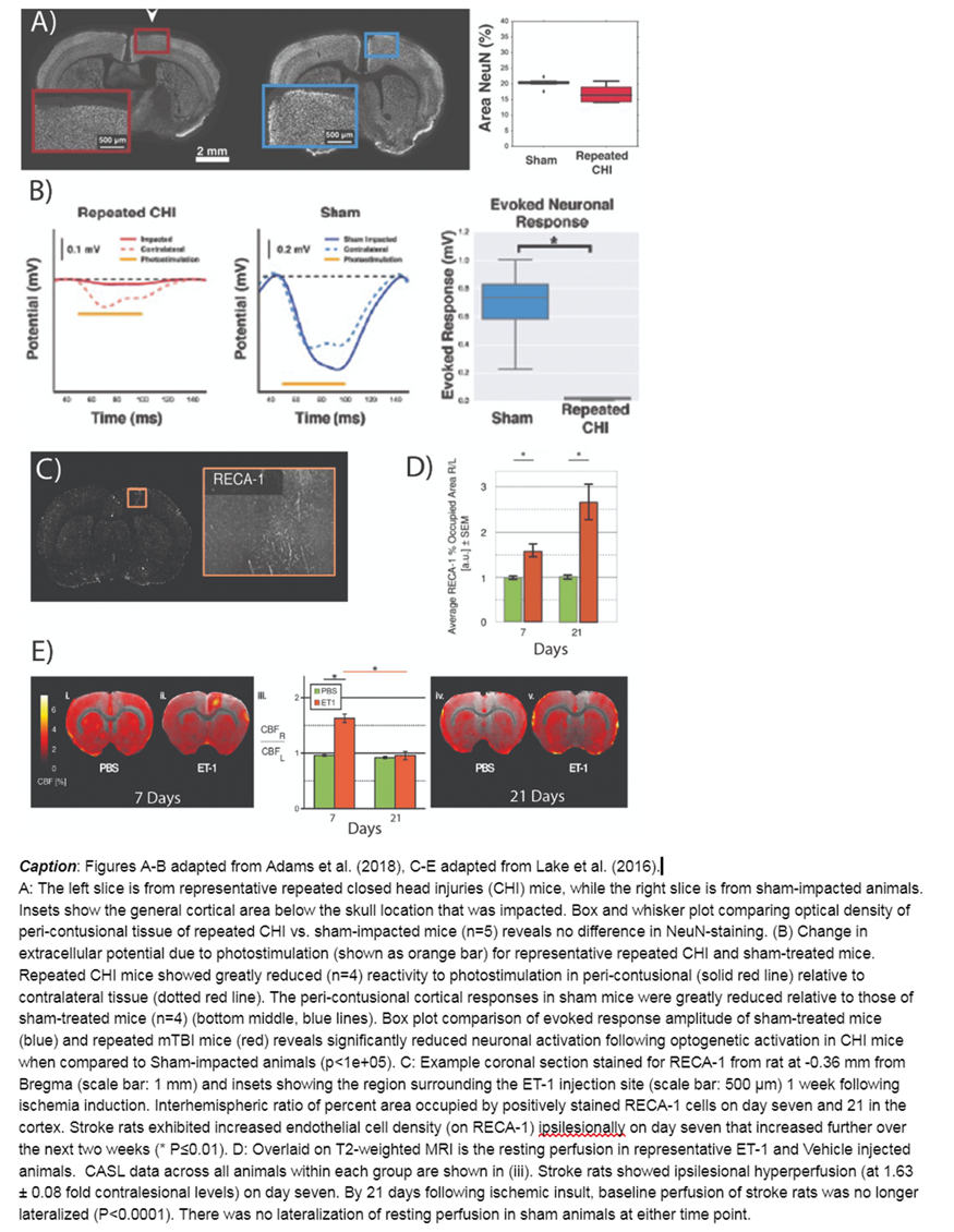
Results
In Adams et al.(1), two weeks following repeated TBI, LFP recordings performed in vivo from the peri-contusional cortex detected a significant decrease in neuronal excitability. In contrast, pathological analysis on the same tissue (MAP2, Neurofilament, NeuN and TUNEL staining) did not reveal any changes in dendritic or axonal density, or neuronal survival. In Lake et al.(2), we assessed neuronal and vascular function in vivo as well as morphology ex vivo, one and three weeks following focal cortical ischemia by cortical injection of endothelin-1. Histopathological analysis revealed comparable perilesional neuronal damage (NeuN), astrogliosis (GFAP) and vascular remodelling (RECA-1) at both time points, indicating no recovery. Contrarily, electrophysiological analysis and pCASL-MRI revealed neuronal hypoexcitability and hypoperfusion at one week post-ischemia, followed by neuronal hyperexcitability and partial recovery of somatosensory responses associated with normoperfusion three weeks after stroke. Hayward et al.(3) observed that in the thalamus, following mild ischemia induced by transient carotid artery occlusion, CBF drops sub-acutely (2 to 4 days) bilaterally then recovers contralesionally but not ipsilesionally 90 days thereafter. In contrast, endothelial marker RECA-1 revealed prominent ipsilesional increase in endothelial cell density and vascular branching at 90 days post stroke.
Conclusions
The spatio-temporal mismatch between morphological and functional signals suggests that the commonly used histopathological markers of neuronal and vascular state are challenged to detect the changes in neuronal activity and cerebrovascular perfusion in the acute and chronic stage following cerebrovascular damage. These observations stress the importance of functional readouts when investigating neurovascular coupling in pathological conditions.
References
PB01-U08
Increased susceptibility of ischemic penumbra to optogenetic functional activation-induced peri-infarct spreading depolarizations
1Departments of Radiology, Massachusetts General Hospital, Harvard Medical School, USA
2Department of Neurosurgery, Yamaguchi University, Japan
3Departments of Neurology, Massachusetts General Hospital, Harvard Medical School, USA
4Martinos Center for Biomedical Imaging, Massachusetts General Hospital, Charlestown, MA, USA
Abstract
Objectives
Peri-infarct depolarizations (PIDs) are believed to contribute to injury progression and worsen the outcome. We have previously shown that functional activation of peri-infarct cortex by somatosensory stimulation triggered PIDs when the activated cortex was within a critical range of ischemia. Optogenetic stimulation is an alternative to directly activate neurons, bypassing the subcortical sensory relays and eliminating potential confounders. We employed minimally invasive optogenetic cortical activation through intact skull in transgenic mice expressing channelrhodopsin-2 in neurons (Thy1-ChR2-YFP) and examined the threshold to induce PIDs by optogenetic functional activation.
Methods
We occluded the middle cerebral artery distally using a microvascular clip (dMCAO) in Thy1-ChR2-YFP mice (age ∼10 weeks, male, n = 10). Optogenetic stimuli (470 nm) of core, penumbra and mild ischemic cortex were delivered in random order at 1 to 10 mW, ramped up every 30 seconds, using a frequency of 8 Hz and pulse duration of 6 ms. PIDs were detected by optical intrinsic signal (OIS) and laser speckle imaging. We also examined the effect of optogenetic stimuli in sham-operated mice (n = 5, Thy1-ChR2-YFP no ischemia) and in wild-type (C57BL/6, n = 3).
Results
Optogenetic functional activation of penumbra triggered a PID at a much higher rate than activation of contralateral homotopic cortex (64% vs. 8%, p = 0.004), and at much lower stimulus intensities (6.5 vs. >10 mW; p = 0.004). The PID rate was also increased in the core compared with contralateral homotopic cortex (33% vs. 0%, p = 0.042) with lower intensity thresholds (8.7 vs. >10 mW; p = 0.042). In contrast, ipsilateral non-ischemic cortex had reduced susceptibility to functional activation-induced PIDs (0% vs. 46%, p = 0.015), and elevated threshold compared with contralateral homotopic cortex (>10 vs. 7.7 mW; p = 0.015). Spatial analysis of CBF showed that functional activation-induced PIDs were triggered from penumbra when residual CBF within the stimulated cortex was approximately 31%. The magnitude of evoked potentials did not explain the differences between ischemic and non-ischemic hemispheres. Interestingly, sham-operated mice (i.e. non-ischemic) also developed spreading depolarizations in response to optogenetic functional activation mimicking physiological firing rates.
Conclusions
Our data show that functional activation of ischemic penumbra triggers PIDs at a much higher rate and lower threshold than homotopic non-ischemic cortex. Moreover, functional activation is capable of triggering SDs even in non-ischemic brain tissue. These data have relevance for the origins of migraine aura.
PB01-U09
Neurovascular sequelae of repeated mild traumatic brain injury in an optogenetic mouse model
1Sunnybrook Research Institute
2Mouse Imaging Centre, Toronto Centre for Phenogenomics, Hospital for Sick Children
Abstract
Objectives
Up to 90% of traumatic brain injury cases are mild TBI (mTBI)1: while these injuries produce no contrast on structural neuroimaging and elicit only transient behavioural changes, they elevate the risk of latter cognitive decline, especially with repeated exposure to mTBI2. Notwithstanding, there is a paucity of data on the neurovascular functioning following repeated mTBI. This work aims to address this gap through in vivo neuronal and vascular recordings of optogenetic activation in mice exposed to repeated closed head injury (CHI) and administered delayed, low-dose of L-655,708, an inverse agonist of a5 subunit-containing GABA-A receptor3.
Methods
Mice expressing channelrhodopsin-2 (ChR2) in pyramidal neurons (007612, Jackson Labs) were given three impacts at +1.5 mm ML, +0.5 mm AP (1.5 mm tip diameter, 2 m/s impactor speed, 1 mm depth, 0.2 s dwell time) via a cortical impactor (Leica Impact One stereotaxic actuator) with a three-day inter-impact interval. Three days post third impact, a subset of mice were subcutaneously implanted with an osmotic pump (Alzet model 1002) containing either L-655,708 (200µg/kg/day) or vehicle (50% saline, 50% DMSO). Two weeks post-third impact, a 2 mm diameter cranial window was implanted over the injury for two-photon fluorescence microscopy (2PFM) via an Olympus FVMPE-RS microscope and local field potential (LFP) recordings via HEKA EPC-10 (Figure 1A-B). Under alpha-chloralose anesthesia, red blood cell (RBC) speed of cortical penetrating vessels (10–25 per subject) labeled with 70 kDA Texas Red dye was recorded via 800–900Hz linescans at 900 nm during periods of raster-scanned 458-nm photostimulation centered over the vessel (2 s duration, 45 s inter-stimulus interval, 0.6 mW/mm2, 120µm diameter, 500 ms repetition time, 0.5µm pixel spacing), repeated 3x/vessel (Figure 1A-C). RBC speed was estimated from linescans using LSPIV4 and response to photostimulation ascertained using pre- and post-stimulus variance testing and z-scoring. Responses were defined as post-stimulus onset periods exhibiting both p≤0.05 and z>2 for at least 5 s.
Results
Robust local field potentials were recorded in both Sham and TBI mice during photostimulation. (3.5±0.6 mV, Figure 1B). In contrast, the incidence of responses in the RBC speed was significantly lower in CHI vs. sham mice (Figure 1D, p = 0.01). Furthermore, preliminary results indicate that this attenuation in cerebrovascular reactivity of the peri-contusional cortex may be recovered through chronic, delayed low dose GABA-A receptor inverse agonism via L-655,708 administration (Figure 1D, p = 0.2).
Conclusions
Two weeks post final impact, repeated CHI resulted in decreased reactivity of individual cortical penetrating vessels of the peri-contusional cortex to optogenetic activation despite robust accompanying local field potentials. This cortical vascular reactivity compromise was partially rescued by treatment of L-655,708. Further work will focus on optimization of this treatment paradigm to maximize both behavioural and neurovascular recovery post repeated mild TBI.

References
PB01-V01
Time-evolution biomarkers of wake-up stroke
1Health Research Institute of Santiago de Compostela (IDIS)
Abstract
Objectives
Wake-up stroke, defined as the situation where patients go to sleep normal and awaken with stroke symptoms, represents roughly 15–30% of strokes, and present a management dilemma for acute stroke providers. The concept of wake-up stroke has been developed in relation to the limitations of treatment with the recombinant tissue plasminogen activator (rt-PA) and has conditioned the potentiation of neuroimaging techniques. The most clinical aspects and those that can help to know the mechanisms that condition it and to prevent its appearance, or its consequences, are more unknown [1,2]. In the present work, we analysed the clinical variables and outcomes of awake and wake-up stroke patients in order to study possible risk factors on nocturnal vascular events.
Methods
We conducted a retrospective study enrolling ischemic stroke (IS) and non-traumatic intracerebral hemorrhage (ICH) patients admitted to the Stroke Unit in the last 10-years. Baseline demographic variables, clinical characteristics and molecular markers by 3-month neurologic outcome in IS and ICH. The main endpoint was poor functional outcome at 3-months (mRS≥3). To estimate the odds ratios of each independent variable in the patient’s worsening in the first 3-months, we used logistic regression model
Results
We included 5284 patients; 4251 IS (90.3% awake, 9.7% wake-up) and 954 ICH (95.4% awake, 4.6% wake-up). Wake-up IS patients showed a greater severity at admission, but they evolved similarly to awake IS. Wake-up IS were independently associated with markers of inflammation [higher axillary temperature (OR-2.15, CI-95% 1.00–1.56), fibrinogen (OR-0.99; CI-95% 0.99–1.00), C-reactive protein (OR-1.14; CI-95% 1.01–1.17)], and plasma concentrations of interleukin-6 (OR-1.04; CI-95% 1.00–1.23). However, these associations were dependent on low levels of vitamin-D (Figure 1). An ICH amyloid hemorrhage showed 6-times more risk during sleep.

Conclusion
Low plasma levels of vitamin D (≤9 ng/mL) multiply by 30 the risk of wake-up IS. We did not detect any biomarker associated with the risk of nocturnal ICH onset.
References
PB01-V02
MicroRNA profiles in peripheral neutrophils of acute ischemic stroke patients and their predicted gene targets
1Institute of Cerebrovascular Disease Research and Department of Neurology, Xuanwu Hospital of Capital Medical University, China
Abstract
Aim
Inflammatory response including neutrophils activation underwent phenotypic and functional changes following ischemic stroke (IS). Moreover, several microRNAs (miRNAs) have been reported to be differentially expressed in immune cells of acute IS patients and predicted to involve in pathways including immune activation. In this study we sought to determine the miRNAs that are differently expressed in circulating neutrophils of acute IS patients and explore potential mechanisms underlying phenotypic transformation of neutrophils following acute IS.
Methods
In this study, we screened differentially expressed miRNAs in circulating neutrophils of acute IS patients and healthy controls using miRNA microarray analysis, and validated their expressions by quantitative reverse-transcriptase polymerase chain reaction assays (qRT-PCR). mRNAs of N1/N2 marker CD16/CD206 in circulating neutrophils were quantified using qRT-PCR and their association with candidate miRNA was analyzed. Furthermore, we performed gene ontology (GO) and Kyoto encyclopedia of genes and genomes (KEGG) pathway analyses to explore genes and pathways predicted to be regulated by the candidate miRNAs. mRNAs of specific genes targeted by candidate miRNAs were confirmed by qRT-PCR and their correlations with performed as well.
Results
Notably, our results disclosed that 3 miRNAs, i.e. miR-30b, miR-193a and miR-7 were differentially downregulated in circulating neutrophils of acute IS patients. In addition, N1marker CD16 mRNA was positively associated with miR-193a and miR-7 levels while N2 marker CD206 was inversely correlated with miR-30b. Further GO and KEGG pathway analyses showed that genes predicted to be regulated by differentially expressed miRNAs were significantly enriched in ubiquitin-like protein conjugating enzyme, ubiquitin conjugating enzyme activity and ubiquitin mediated proteolysis pathways. qRT-PCR validated the upregulation of UBE2D2 and UBE2V2 expressions following acute IS. Moreover, mRNA expression of both UBE2D2 and UBE2V2 were inversely associated with miR-193a levels. Whether miR-193a involved in neutrophils phenotypic transformation following acute IS via regulating UBE2D2 and UBE2V2 expression deserved further in vivo and in vitro investigation.
Conclusion
In conclusion, we identified differentially expressed miRNAs, i.e. miR-30b, miR-193a and miR-7 in peripheral neutrophils of acute IS. Further bioinformatics and correlation analysis supported the hypothesis that miR-193a may involve in neutrophils phenotypic transformation via regulating UBE2D2 and UBE2V2 expression following acute IS.
PB01-V03
Genetic variant RNF213 c.14576G>A in intracranial atherosclerosis of the anterior and posterior circulation
1Department of Neurosurgery, Faculty of Medicine, The University of Tokyo, Tokyo, Japan
2Department of Neurosurgery, Teraoka Memorial Hospital, Fukuyama, Hiroshima, Japan
Abstract
Objectives
Intracranial atherosclerosis of the anterior circulation (anterior ICAS) and posterior circulation (posterior ICAS) are thought to involve different pathogeneses and risk factors.1,2 Recently, we identified a genetic variant which has a significant association with ICAS.3,4 The variant was ring finger protein 213 (RNF213) c.14576G>A (rs112735431) which was originally identified as a susceptibility genetic variant for moyamoya disease (MMD).5,6 The present study investigated the association of RNF213 c.14576G>A with anterior and posterior ICAS.
Methods
A total of 221 study participants (43 with anterior ICAS, 61 with posterior ICAS, 12 with extracranial atherosclerosis (ECAS), 5 with MMD, and 100 control subjects) were recruited from April 2015 to October 2015. Genetic analysis of RNF213 c.14576G>A and association study with these cerebrovascular diseases were performed.
Results
RNF213 c.14576G>A was present in 10/43 patients in the anterior ICAS group and 4/5 in the MMD group, but in no patient in the posterior ICAS and ECAS groups. c.14576G>A was found in 2/100 patients in the control group. RNF213 c.14576G>A showed significant association with anterior ICAS (allele count, P = 3.9×10⁻⁵, odds ratio = 13.0, 95% confidence interval = 2.8–60.8; prevalence of carriers of c.14576G>A, P = 2.6×10⁻⁵, odds ratio = 14.8, 95% confidence interval = 3.1–71.3). However, RNF213 c.14576G>A showed no association with posterior ICAS. RNF213 c.14576G>A also had a significant association with MMD and no association with ECAS.
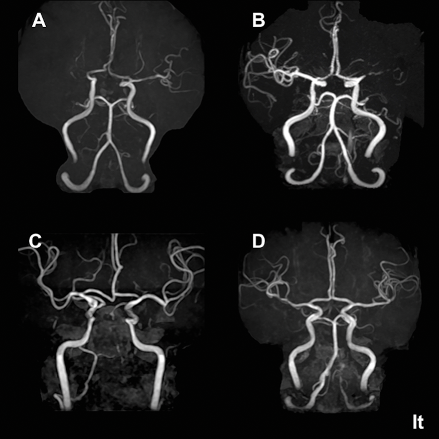
Conclusions
RNF213 c.14576G>A is a specific genetic risk factor for anterior ICAS but not for posterior ICAS. Evaluation of RNF213 c.14576G>A may lead to more accurate prediction and prevention of anterior ICAS and stroke.
References
PB01-V04
Accurate etiology diagnosis in patients with stroke and atrial fibrillation: a role for brain natriuretic peptide
1Dept. of Neurology, Nippon Medical School
Abstract
Background
Atrial fibrillation (AF) is the leading cause of cardioembolic stroke (CES), and patients with stroke and AF are frequently assumed to have CES. However, stroke patients often have accompanying risk factors other than AF, and strokes presumably due to atherosclerotic pathophysiologies in large or small vessels can also occur in patients with AF. The aims of the present study were to clarify the prevalence of and factors related to a non-cardioembolic features on imaging in acute stroke patients with AF.
Methods
From March 2011 through May 2017, consecutive acute ischemic stroke patients with AF were retrospectively recruited from the prospective registry. The concomitant presence of non-cardioembolic features on admission was defined as: 1) having a sole ischemic lesion in perforating artery territory (basal ganglia, corona radiata, thalamus, and brainstem) and maximum infarct diameter <15 mm (small vessel occlusion [SVO] feature) and/or 2) the ischemic lesion was restricted to a sole arterial territory with >50% stenosis of the responsible artery (large artery atherosclerosis [LAA] feature). The frequency of and factors associated with co-existing SVO/LAA features were assessed.
Results
A total of 560 consecutive patients with AF and acute stroke (237 women; median age 78 [interquartile range 71–85] years; National Institutes of Health stroke scale [NIHSS] score 9 [3-20]) were enrolled. Of these, 42 (7.5%, 95% confidence interval 5.3–9.7%) had co-existing SVO/LAA features. The CHA2DS2-VASc score (5 [2-6] vs. 4 [2-5], p = 0.024) was higher, and the NIHSS score (4 [2-13] vs. 10 [3-20], p = 0.005) and levels of D-dimer (1.0 [0.6–1.7] µg/ml vs. 1.4 [0.9–3.2] µg/ml, p = 0.001) and brain natriuretic peptide (BNP, 124 [58-211] pg/ml vs. 202 [109-376] pg/ml, p = 0.001) were lower in patients with co-existing SVO/LAA features than those without. Paroxysmal AF was more common in patients with co-existing SVO/LAA features than those without (p = 0.007). Multivariable logistic regression analysis showed that the BNP level (OR 0.80, p = 0.029 per 100 pg/ml increase) was independently and negatively associated with co-existing SVO/LAA features and receiver operating characteristic curve analysis revealed the practical cut-off BNP value was 130 pg/ml (sensitivity 54% and specificity 68%).
Conclusion
SVO/LAA features were found in 7.5% of acute stroke patients with AF. A relatively low BNP level on admission was independently associated with co-existing SVO/LAA features. Thorough examination for a more appropriate etiology may be particularly necessary in acute stroke patients with AF and a relatively low BNP level.
PB01-V05
Biomarker for diagnosis of intracrebral hemorrhage in the early stage
1Dept. of Neurosurgery, Sendai Medical Center
2Dept. of Neurosurgical Engineering and Translational Neuroscience, Tohoku University Graduate School of Biomedical Engineering
3Dept. of Neurosurgery, Tohoku University Graduate School of Medicine
4Dept. of Neurosurgery, Kohnan Hospital
Abstract
Background
Correct diagnosis of cerebral stroke type, hemorrhagic or ischemic, is essential in the early stage to establish the optimum treatment. The diagnosis is mainly determined based on imaging studies. Other more available diagnostic methods are desirable, such as blood sample examination. Lectin-like oxidized low density lipoprotein receptor-1 (LOX-1) is very important in vascular dysfunction induced by oxidized low-density lipoprotein (LDL), including cell apoptosis. The present study evaluated LOX-1 as a biomarker for cerebral stroke.
Subjects and Methods
Patients with newly diagnosed stroke were prospectively enrolled between February and July 2014. LOX-1 serum level was measured twice, within 24 hours and 2 months after the onset.
Results
A total of 16 patients were enrolled; 7 patients with intracerebral hemorrhage (ICH) and 9 patients with cerebral infarction. Median LOX-1 values of patients with ICH and infarction in the acute phase were 1825.8 and 593.9 pg/mL, respectively, significantly higher in patients with ICH than in patients with infarction (P < 0.0001).
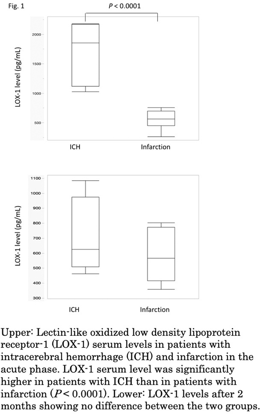
Conclusion
LOX-1 serum level has potential as a biomarker of ICH.
PB01-V06
Distinctive transcriptomic profiles for ischemic stroke etiologies and intracerebral hemorrhage in whole blood and leucocyte subtypes
1Department of Neurology, School of Medicine, University of California at Davis, Sacramento, CA, USA
2Department of Medicine, University of Alberta, Edmonton, Canada
Abstract
Objectives
Differentiating intracerebral hemorrhage (ICH) from ischemic stroke (IS) can be challenging when imaging is not readily available. Ruling out ICH is essential for early thrombolytic or endovascular IS therapy to be initiated. Understanding transcriptome differences between ICH and IS, and between IS etiologies, can lead to a better knowledge of the molecular and cellular pathways involved in the response to the acute brain injury caused by ICH and IS. Therefore, in this study we sought to characterize the transcriptomic profiles in blood from cases with ICH and different IS etiologies to identify acute molecular changes in isolated neutrophils and monocytes and in whole blood.
Methods
Peripheral blood was drawn from cases with diagnosed ICH (6) and IS (33) (cardioembolic, large vessel disease and lacunar) in the first 30 ± 20 hours after the onset of symptoms. We performed whole-genome RNA sequencing of whole blood (WB), and of isolated neutrophils and monocytes. Control cases (10) with vascular risk factors (diabetes and/or hypertension and/or hypercholesterolemia) were included (VRFC). A linear regression model including diagnosis and sample subtype with p < 0.05 and |fold-change|>1.2 was used for identifying differentially expressed (DE) genes. Gene ontology and pathway enrichment were performed for investigating their biological context (Benjamini-Hochberg p < 0.05).
Results
339, 371, 386 genes were DE between cardioembolic IS, large vessel IS and lacunar IS compared to VRFC in monocytes; 1188, 1035, 1276 in neutrophils, and 1464, 2405 and 748 in WB respectively. In ICH vs VRFC 1408 were DE in neutrophils, 500 in monocytes, and 3124 in WB. Most of the changes are cell-type specific and involve several cell-specific pathways, along with different signal transduction and immune response pathways per leucocyte type. For example, in ICH compared to VRFC, about half of the over-represented pathways were unique to either monocytes or neutrophils. 65% of the pathways over-represented in WB were not over-represented in monocytes or neutrophils.
Additional analyses will also allow us to demonstrate alternative splicing differences between cell subtypes to further refine the molecular signatures of both diseases.
Conclusions
Gene expression changes in neutrophils and monocytes after ICH and IS subtypes. The unique molecular signatures in these blood cell types underscore their involvement in IS and ICH pathophysiology. The large number of unique pathways in whole blood not detected in monocytes or neutrophils signifies the contribution of other cell types to the ICH and IS responses.
References
PB01-V07
Hemodynamic or thromboembolic stroke – what have we learned from cardiac surgery?
1Neurobiology Research Unit, Rigshospitalet, Copenhagen University Hospital
2Department of Cardiothoracic Anaesthesiology, Rigshospitalet, Copenhagen University Hospital
3Department of Anesthesia, Center of Head and Orthopedics, Rigshospitalet, Copenhagen University Hospital
4Department of Radiology, Rigshospitalet, Copenhagen University Hospital
5Department of Biostatistics, Rigshospitalet, Copenhagen University Hospital
6Department of Cardiothoracic Surgery, Rigshospitalet, Copenhagen University Hospital
Abstract
Introduction
Stroke is a common disease caused by some degree of focal cerebral ischemia. The thromboembolic occlusion of cerebral vessels is a well known cause of stroke. Here the occlusion of the cerebral vessel may arise from an embolus from the heart or from effects of a localised endothelial lesion in the cerebral or extracranial arteries. The hemodynamic stroke caused by stenosis of a cerebral or extracranial vessels and episodic blood pressure drop, has generally been considered less frequent (1, 2). But, fluctuations in blood pressure with episodic drop may lead to manifest neurological deficits, transitory or permanent.
Objectives
To use results obtained from our recent study on cerebral lesions following cardiopulmonary bypass surgery to discuss the general likelihood of hemodynamic vs. thromboembolic stroke
Methods
In a patient- and assessor-blinded randomized trial, patients were allocated to a higher (70–80 mm Hg) or lower (40–50 mm Hg) target for mean arterial pressure by the titration of norepinephrine during cardiopulmonary bypass. Pump flow was fixed at 2.4 L·min−1·m−2. The primary outcome was the total volume of new ischemic cerebral lesions (summed in millimetres cubed), expressed as the difference between diffusion-weighted imaging (DWI) conducted preoperatively and again postoperatively between days 3 and 6 (3). In a secondary analysis we compared cerebral metabolism using magnetic resonance spectroscopy (4). Further secondary analyses investigated postoperative cognitive dysfunction (POCD) and its association with DWI detected brain lesions (5).
Results
Among the 197 enrolled patients, mean (SD) age was 65.0 (10.7) years in the low-target group (n = 99) and 69.4 (8.9) years in the high-target group (n = 98). Overall, DWI revealed new cerebral lesions in 52.8% of patients in the low-target group versus 55.7% in the high-target group (P = 0.76). The primary outcome of volume of new cerebral lesions was comparable between groups, 25 mm3 in the low-target group versus 29 mm3 in the high-target group (P = 0.99). MRS revealed a decrease in GM-NAA/Cr in the high-target group, (P = 0.014). In a subset of the patients (89 in the low-target and 80 in the high-target group) no statistically significant between-group differences were found when comparing the overall rate of POCD or the proportion of patients with a domain-specific deterioration over the pre-defined critical level in 7 individual test variables at discharge.
Conclusions
Overall, we found no major neuroradiological or cognitive outcome differences when targeting higher vs lower blood pressure during full-flow cardiopulmonary bypass. Still, a tendency was observed towards a better outcome in the low-target group both for primary and secondary outcomes. These insights obtained from cardiac surgery research indicate that stroke caused by a hemodynamic mechanism in the elderly population seems to be uncommon in contrast to the much more frequent thromboembolic stroke.
References
PB01-V08
Significance of microembolic signals in the acute period for stroke subtypes
1Dept. of Neurology, Tokyo Women's Medical University,Japan
Abstract
Purpose
Microembolic signals (MES) on Transcranial Doppler (TCD) ultrasonography are often detected in acute ischemic stroke patient. However, significance of MES for stroke subtypes including embolic stroke of undetermined source (ESUS) remains unclear. The aim of this study is to clarify prevalence and prognostic value of MES in each stroke subtype.
Methods
Between April 2017 and July 2018, 164 patients with acute ischemic stroke who admitted within a week from onset were enrolled in this study. We monitored the flow signal of the ipsilateral middle cerebral artery (MCA) for 60 minutes. After excluding 105 patients (transient ischemic attack (TIA), n = 40; posterior circulation stroke, n = 20, unsuccessful TCD monitoring, n- = 45), 59 patients with acute ischemic lesions in the anterior circulation were included for further analysis. They were classified into the stroke subtypes according to TOAST classification, large-artery atherosclerosis (LAA, n = 13), small-vessel disease (SVD, n = 15), cardioembolism (CE, n = 11), and embolic stroke of undetermined source (ESUS, n = 20). The incidence of MES in each stroke subtype on admission and ischemic stroke recurrence at 3-month were evaluated.
Results
The detection rate of MES was 53.8% in LAA, 45.5% in CE, 26.7% in SVD, and 50% in ESUS. Although MES was often observed in LAA and ESUS, no significant difference was seen between stroke subtypes (p = 0.450). The recurrence of ischemic stroke in three months is higher in patients with MES (17.4%, 3/23) than those without (0%, 0/29) (p = 0.045).
Conclusion
MES was detected in all type of ischemic stroke, and majority of LAA and ESUS showed MES in the acute stage. The presence of MES could be a predictor for recurrent stroke.
PB01-V09
Inflammatory biomarkers at the infarct during large vessel occlusion in human stroke patients
1Dept. of Neurology, University of Kentucky, USA
2Dept. of Neuroscience, University of Kentucky, USA
3Dept. of Neurosurgery, University of Kentucky, USA
4Dept. of Radiology, University of Kentucky, USA
5College of Nursing, University of Kentucky, USA
Abstract
Objective
In the setting of mechanical thrombectomy for emergent large vessel occlusion (ELVO), we have for the first time collected and evaluated blood immediately distal and proximal from the removed intracranial thrombus. These samples provide a unique resource in evaluating acute gene expression changes at the time of ischemic stroke. The purpose of this study was to evaluate gene expression changes occurring within the thrombus and across the occlusion in the intravascular space in acute ischemic stroke patients.
Methods
We developed the Blood and Clot Thrombectomy Registry and Collaboration (BACTRAC) protocol: an IRB-approved tissue banking strategy for ELVO (clinicaltrials.gov NCT03153683). We evaluated relative concentrations of gene expression in 84 inflammatory molecules in thrombi removed from adults who received thrombectomy for ischemic stroke, in static blood distal to thrombus, and in peripheral circulation.
Results
We analyzed the first 39 subjects (age = 67 ± 13.2, 16 males) in the BACTRAC registry. Results from microarray analyses demonstrate that 21 genes (CCL1, CCL11, CCL13, CCL17, CCL26, CCL8, CSF3, CX3CL1, CXCL1, CXCL9, IFNA2, IL13, IL17C, IL17F, IL1A, IL27, IL3, IL33, LTA, TNFSF11, and TNFSF13) had at least 25 mean fold change in the distal blood compared to the peripheral blood. Fourteen genes (AIMP1, CCL11, CCL13, CCL15, CCL16, CCL23, CCR2, CCR4, CCR8, CD40LG, CXCL10, IL10RA, IL15, and TNFSF13B) had at least 10 mean fold change in the thrombus compared to the peripheral blood. Overall, these genes are associated with chemo-attraction and cell proliferation of monocytes, neutrophils, and T cells.
Conclusion
These findings provide a novel insight into the initial pathology of large vessel stroke in humans, particularly in regard to identifying acute gene expression changes that occur during stroke.
PB01-V10
Pro-BNP: a simple and useful predictor of stroke risk after transient ischemic attack
1Hospital Clínico Universitario de Santiago, Stroke Unit, Neurology Department, Santiago de Compostela, Spain
2Health Research Institute of Santiago de Compostela (IDIS), Hospital Clínico Universitario, Clinical Neurosciences Research Laboratory, Santiago de Compostela, Spain
Abstract
Background
Transient ischemic attacks (TIAs) are predictors of cerebral infarction which can be prevented if the appropriate therapeutic measures are taken. Elevated levels of B-type natriuretic peptide (BNP) and its prohormone (pro-BNP) are associated with an increased risk of ischemic stroke. The limited reliability to predict the risk of stroke after a TIA justifies the objective of our study to determine the role of pro-BNP in patients with TIAs.
Methods
From our prospective stroke registry, we performed a retrospective study in all patients with the diagnosis of TIA admitted to the Stroke Unit of our Hospital between January 2008 and March 2018. Pro-BNP was determined in the first hours after TIA. The endpoint was the development of stroke during the follow-up.
Results
A total of 381 patients (227 men and 154 women) were included. Mean time of follow-up was 36.8 ± 16.4 months. 224 patients were hospitalized due to a stroke during the follow-up, and 157 were not. After the TIA, the stroke was hemorrhagic in 9.8% of patients and ischemic in 90.2%, with 25.4% atherothrombotic ischemic strokes, 29.6% of cardioembolic strokes, 4.9% lacunar strokes, and 30.3% strokes of undetermined origin. The time from the TIA to the stroke was less than 24 hours in 159 patients (71.0%), between day 2 and 7 in 38 patients (17.0%), and greater than 7 days in 27 patients (12.0%). Pro-BNP serum levels were higher in patients who suffered a stroke compared to those who did not (p < 0.001). We also found greater levels of this marker the earlier the stroke happened (p = 0.024). A cut-off point of 800 pg/mL of pro-BNP predicted a stroke with a sensitivity of 64% and a specificity of 79% (p < 0.001), and was independently associated with higher risk of stroke after a TIA (OR: 6.65, p<0.001). This association persisted for different etiopathogenic TIA groups (cardioembolic: OR 26.12, p<0.001; undetermined: OR 4.87, p = 0.006; atherothrombotic: OR 1.67, p = 0.044).
Conclusions
The early determination of pro-BNP is a simple and very useful alternative to predict the prognosis after TIA regardless of the etiopathogenesis of the TIA.
PB01-V11
Cortical venous reddening is a predictor for remote cerebral infarction after STA-MCA bypass surgery in atherosclerotic occlusive cerebrovascular disease patients
1Dept. of Neurosurgery, Kumamoto University, Japan
2Dept. of Neurosurgery, Saiseikai Kumamoto Hospital, Japan
Abstract
Objective
The superficial temporal artery (STA)–middle cerebral artery (MCA) anastomosis (STA-MCA bypass) was currently performed to prevent of atherosclerotic occlusive cerebrovascular disease. However, the benefit of the bypass surgery is still controversial. To ensure consistent surgical benefits, understanding the mechanisms of perioperative cerebral infarction (CI) is required. In addition, appropriate patient selection procedures should be determined to decrease the rate of perioperative stroke including CI. We retrospectively investigated patients who underwent bypass surgery at our hospital and determined that the patients who presented with cortical venous reddening after STA-MCA bypass during the surgery developed perioperative CI.
Methods
A total of 45 consecutive patients who underwent STA-MCA bypass surgery were retrospectively investigated. Twenty-five of the 45 patients underwent STA-MCA bypass for atherosclerotic occlusion or stenosis of the internal carotid artery or MCA. Preoperative iodine-123-N-isopropyl-iodoamphetamine-single photon emission computed tomography (123I-IMP-SPECT) was performed with and without acetazolamide administration. Change in color of the cortical veins was observed on recorded surgical videos, and its correlation with perioperative stroke including CI was investigated.
Results
We experienced two cases of perioperative extensive CI at a region remote from the site of anastomosis of STA-MCA bypass. In both cases, retrospective investigation of surgical videos demonstrated reddening of cortical veins soon after the anastomosis procedure. Of all 45 patients, postoperative CI and venous reddening were observed in only these two cases.
Conclusion
We determined that the patients presenting with cortical venous reddening after STA-MCA bypass eventually developed perioperative CI. Cortical venous reddening may be an important predictor for the occurrence of CI after STA-MCA bypass surgery for patients with atherosclerotic occlusive cerebrovascular disease.
PB01-V12
Brain volume as a determinant of functional outcome after acute ischemic stroke
1Harvard Medical School, USA
2Massachusetts Institute of Technology, USA
3German Centre for Neurodegenerative Diseases (DZNE), Germany
Abstract
Objectives
Stroke is one of the leading causes of disability in adults [1]. However, given the heterogeneity of the observed phenotypes, factors contributing to stroke outcome are still poorly understood. Brain volume has been associated with overall brain health, life-time exposures to vascular risk factors, and the brain’s resilience to function loss due to acute injury [2-4]. However, it has not been assessed if brain volume is associated with functional outcome after acute ischemic stroke (AIS).
Methods
We conducted a cross-sectional analysis of the MRI-GENetics Interface Exploration (MRI-GENIE) study [5], a multi-site, international hospital-based study of 884 AIS patients (mean age 64.8 ± 14.4 years; 59% male) with clinical brain MRI obtained on admission for index stroke. Functional outcome assessment was determined using the modified Rankin Scale (mRS) score (0–6; 0: asymptomatic; 6 death) recorded between 60–190 days after stroke [6]. Demographics and other clinical variables including acute stroke severity (determined using the National Institutes of Health Stroke Scale (NIHSS) score), vascular risk factors, and etiologic stroke subtypes based on the Causative Classification of Stroke (CCS) were recorded during admission. We assessed brain volume in this large, multi-site cross-sectional cohort using a fully-automated deep-learning volumetric analysis of clinical magnetic resonance imaging (MRI) scans.
Results
In a generalized linear model, brain volume (per 154.1cc) was significantly (P<0.05) associated with age (beta −0.212 (per 14.4 years)), male sex (beta 1.167) and history of smoking (beta −0.124). Utilizing these results in a multivariable outcome model, brain volume was an independent predictor of mRS (beta −0.429), with reduced odds of worse long-term functional outcomes (OR: 0.79, 95% CI 0.70–0.90) in those with larger brain volumes on the MRI scans obtained at the time of stroke (see Figure).
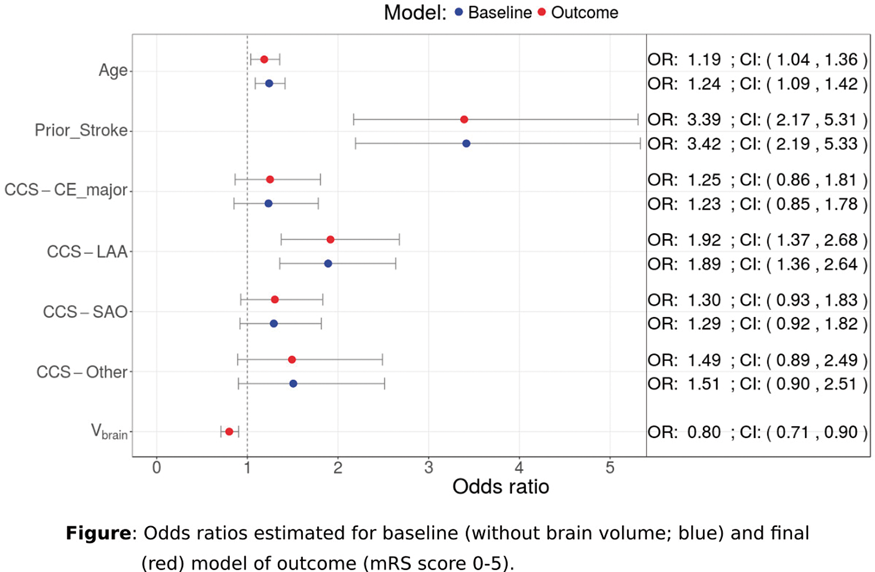
Conclusion
Here, we showed that brain volume serves as independent determinant of functional post-stroke outcome which can be assessed in the acute stage, and where having a larger brain volume at the time of stroke purports a protective mechanism. This relationship between brain volume and functional post-stroke outcomes points at the role of effective brain reserve and/or overall brain health in facilitating post-stroke recovery.
References
PB01-W01
Cerebral blood flow during balloon occlusion test of the internal carotid artery
1Dept. of Neurosurgery, Nagoya city university, Japan
Abstract
Background
Balloon occlusion test (BOT) of the internal carotid artery (ICA) is crucial before sacrificing it and various criteria for sufficient collateral flow have been proposed. This study was conducted to show the reliability of fully automated quantification of regional cerebral blood flow (rCBF) in BOT of the ICA. We also show the usefulness of ratio of rCBF during BOT to rCBF at rest (BOT/rest ratio = rCBF during BOT/rCBF at rest) rather than asymmetry index (AI) during BOT (AI = occluded-side rCBF/contralateral rCBF).
Methods
In the last 2 years, we performed the BOT on 10 consecutive patients (4 with intracranial aneurysms and 6 with head and neck tumors). During the BOT, mean stump pressure (MSTP) of the ICA was monitored. We measured cerebral blood flow (CBF) with technetium-99m hexamethylpropylene amine oxime single-photon emission computed tomography at rest and during BOT. rCBF was determined using 3-dimensional stereotaxic region of interest template (3DSRT) which automatically divided CBF into 12 segments. We defined hypoperfusion segment as BOT/rest ratio <0.9 or AI <0.9.
Results
When the BOT/rest ratio was used as a hypoperfusion parameter, the number of hypoperfusion segments was significantly greater in patients with an MSTP ≤50 mm Hg than in patients with an MSTP >50 mm Hg. However, only AI during BOT did not reflect MSTP significantly.
Conclusions
The evaluation of CBF changes in BOT using 3DSRT and the BOT/rest ratio were useful because of objective comparison.
PB01-W02
Monitoring cerebral blood flow and critical closing pressure in stroke patients
1Optics at Athinoula A. Martinos Center for Biomedical Imaging, Department of Radiology, Massachusetts General Hospital and Harvard Medical School, USA
2Biomedical Engineering Department, Boston University, USA
3Department of Neurology, Brigham and Women's Hospital, USA
4Department of Neurosurgery, Brigham and Women's Hospital, USA
5Instituto Nacional de Astrofísica, Óptica y Electrónica (INAOE), Mexico
Abstract
Objectives
Diffuse correlation spectroscopy (DCS) is a relatively new near-infrared spectroscopy (NIRS) method which allows for measurement of an index of cerebral blood flow (CBFi) continuously and non-invasively in humans[1,2]. From the measure of pulsatile blood flow (pCBFi), we have recently proposed to use DCS to quantify critical closing pressure (CrCP). CrCP is the arterial blood pressure (ABP) at which blood flow ceases. It is needed to assess cerebral perfusion pressure and is related to intracranial pressure (ICP). These are important biomarkers which, if continuously monitored may guide blood pressure targets and help improve stroke patient’s outcomes. Alternative methods to measure CBF and CrCp are either invasive or, like transcranial Doppler ultrasound (TCD), not optimal for continuous monitoring. DCS offers the advantages of being portable, easy to apply, suitable for continuous bedside monitoring, and does not interfere with conventional angiography. In this work, for further validation, we applied DCS in stroke patients, while simultaneously acquiring TCD.
Methods
We recruited 15 acute ischemic stroke patients at Brigham and Women’s Hospital. In each patient, we performed up to three 30 min. simultaneous DCS and TCD sessions during the first few days post stroke. The study protocol was reviewed and approved by the local IRB.
Pulsatile ABP was measured non-invasively with a Finapres (Finapres Medical Systems, Netherlands). TCD pulsatile blood flow (pBFV) was measured through bilateral temporal bone windows using two ultrasound transducers. The DCS optical sensor was applied on the forehead on the stroke side. Our custom DCS system includes a long coherence laser at 785 nm, four single-photon counting detectors, and a custom FPGA-based correlator which allows us to acquire the arrival time of each photon and recover the autocorrelation function at 100 Hz. Since the SNR at such low integration time was too low, we implemented an algorithm that averages the optical intensity autocorrelation curves at the same instant in the cardiac cycle across 50 heartbeats, using ABP as the reference. The averaged autocorrelation curve was then fitted to the solution of the correlation diffuse equation to derive pCBFi with sufficient SNR and a high number of points along a cardiac cycle.
Results
Arterial pulsation measured with the three modalities, ABP, TCD and DCS, showed remarkably similar shape. Absolute CBFi significantly correlated with TCD blood flow velocity. CrCP derived from DCS showed a strong correlation with the CrCp derived from TCD. The correlation was higher in the non-stroke side, with R2 = 0.69 and slope = 0.89, than in the stroke side, with R2 = 0.52 and slope = 0.91. Figure 1 shows the CBFi and CrCP scatter plots across all subjects and all measurement sessions.
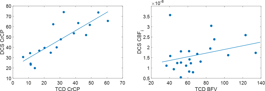
Conclusions
These results give us confidence that DCS could be a viable alternative to TCD to monitor CBF and CrCP in neuro-ICU patients. Our next step is to correlate these data with clinical outcomes. Our future work is to refine the CrCP algorithm and to further validate the CrCP on patients with invasive ICP monitoring.
References
PB01-W03
High glucose and low ASPECTS are associated with intracranial hemorrhage after endovascular reperfusion therapy for acute ischemic stroke in the stent retriever era
1Doctoral program in Clinical Sciences, Graduate School of Comprehensive Human Sciences, University of Tsukuba, Japan
2Department of Neurosurgery, Faculty of Medicine, University of Tsukuba, Japan
3Division for Stroke Prevention and Treatment, Department of Neurosurgery, Faculty of Medicine, University of Tsukuba, Japan
Abstract
Objectives
Intracranial hemorrhage (ICH) is the most feared complication of endovascular reperfusion therapy (EVT). In the era of tissue plasminogen activator, some risk factors such as hypertension, high serum glucose level were known. The purpose of this study is to investigate the predictors of ICH after EVT in the modern stent retriever era by using a multi-center retrospective observational registry.
Methods
A total of 343 consecutive patients who underwent EVT with a diagnosis of anterior circulation cerebral infarction between January 2015 and March 2018 were enrolled from 5 neurovascular centers in Japan. ICH was defined as any type of intracranial hemorrhage evaluated on follow-up computed tomography or magnetic resonance imaging at 24 hours after EVT. According to the Heidelberg bleeding classification, we also investigated the parenchymal hematoma type2 (PH2), which indicates the hematoma occupying 30% or more of the infarcted tissue with obvious mass effect.
Results
Among 343 patients (median age, 75 years; 126 women [36.7%]), 284 patients (85.0%) underwent EVT with the stent retriever. ICH was observed in 105 patients (30.6%), and PH2 in 10 patients (2.9%).
Concerning
ICH, body weight (55.1 ± 13.2 kg vs 58.6 ± 11.9 kg, p = 0.02) and DWI-ASPECTS (median 6 vs 7, p = 0.046) were significantly lower in patients with ICH than in those without. Serum glucose level on admission (median 134 mg/dl vs 124 mg/dl, p = 0.001), rate of systemic heparinization during EVT (66.7% vs 53.5%, p = 0.03), and door-to-reperfusion time (median 145 minutes vs 125 minutes, p = 0.01) were significantly higher in patients with ICH than in those without. The rate of functional independence (modified Rankin scale score, 0 to 2) at discharge was significantly lower in patients with ICH than in those without (25.7% vs 39.9%, p = 0.01). The rate of stent-retriever use was not statistically different in patients with and without ICH (88.5% vs 83.5%, p = 0.3). In binomial logistic regression analysis, glucose level (OR: 1.010, [95%CI: 1.002–1.018], p = 0.02), and DWI-ASPECTS (OR: 0.784, [95%CI: 0.646–0.952], p = 0.01) were independent predictors of ICH.
Concerning PH2, serum glucose level (median 164 mg/dl vs 127 mg/dl, p = 0.007) and door-to-reperfusion time (median 174 minutes vs 128 minutes, p = 0.02) were significantly higher in patients with PH2 than in those without. The rate of functional independence at discharge was significantly lower in patients with PH2 than in those without (0% vs 36.6%, p = 0.02). The rate of stent-retriever use was not statistically different in patients with and without PH2 (80.0% vs 85.2%, p = 0.6).
Conclusions
This study suggested that low body weight, low ASPECTS, high serum glucose level on admission, heparinization during EVT, and long door-to-reperfusion time were associated with ICH after EVT for anterior circulation cerebral infarction in the stent retriever era. High serum glucose level, and low DWI-ASPECTS were independent predictors of ICH according to binomial logistic regression analysis. High serum glucose level and long door-to-reperfusion time were also associated with PH2.
PB01-W04
Recanalization of the aneurysm originating from the posterior communicating artery after coil embolization: Incidence and Risk Factors
1Dept. of Neurosurgery, Korea University, South Korea
Abstract
Purpose
Recanalization is a major problem in the embolization of cerebral aneurysms. We aimed to analyze the recanalization of the aneurysm originating from the posterior communicating artery and its risk factors after coil embolization.
Materials and Methods
This retrospective study included 37 aneurysms originating from the posterior communicating artery treated with coil embolization. Cumulative medical records and radiological data were evaluated. Recanalizaiton was defined as flow filling at the neck of aneurysms or aneurysms sac and coil compaction on MRA or angiography. To calculate the packing density, volume of aneurysms, and volume of coils, we used an on-line system available at www.angiocalc.com. The recanalization rate and risk factor of recanalization was analyzed.
Results
Recanalization developed in 13 (35.1%) out of 37 patients. Multiple logistic regression analysis indicated that recanalization of coiled aneurysm was associated with fetal origin posterior communicating artery presentation (P = 0.045) and packing density ≥ 18% (P = 0.037).
Conclusion
Our data suggest that presenting with fetal origin posterior communicating artery and low packing density (<18%) are predisposed recanalization of aneurysms originating from the posterior communicating artery after coil embolization.
PB01-W06
Evaluation of cognitive function using neural network analysis before and after revascularization surgery for internal carotid artery stenosis
1Dept. of Neurosurgery, Kobe University Graduate School of Medicine, Japan
2Dept. of Neurosurgery, Kobe City Nishi-Kobe Medical Center, Japan
Abstract
Objectives
Internal carotid artery stenosis (ICS) can lead to cognitive impairment as well as ischemic stroke. Although carotid revascularization surgery, such as carotid endarterectomy (CEA) and carotid artery stenting (CAS), can prevent future strokes, the effect of revascularization on cognitive function is controversial. In recent years, the analysis of functional connectivity (FC) in resting-state functional MRI (rs-fMRI) has been used to investigate the effects of cognitive interventions. In this study, cognitive function is evaluated in ICS patients undergoing revascularization surgery with rs-fMRI.
Methods
We proespectively enrolled 17 ICS patients, who were expecting the intervention of CEA or CAS. Cognitive assessment, including the
Results
After revascularization surgery, significant improvement in the score of MMSE (28.1 vs 29.1, P = 0.01) and MoCA-J (24.0 vs 26.7, P = 0.001) was found. No significant difference was found in the score of the FAB (16.2 vs 16.8, P = 0.09) between before and after surgery. As for the subscore of MoCA-J, Serial Subtractions (2.41 vs 2.88, P = 0.016), Verbal Fluency (0.47 vs 0.76, P = 0.02) and Memory (2.24 vs 3.53, P = 0.0012) improved significantly after surgery. According to the analysis of FC, ICS patients showed increase of connectivity between PCC and posterior cingulate gyrus, and between PCC and precuneus postoperatively at 3 months.
Conclusion
Revascularization surgery for ICS improves cognitive function. Increase of connectivity between PCC and posterior cingulate gyrus/precuneus may contribute to the cognitive improvement.
PB01-W07
Etiology of cortical microinfarcts can be determined by diagnostic scores either for cerebral amyloid angiopathy or microembolism using 3T MRI
1Department of Neurology, Mie University Graduate School of Medicine, Mie, Japan
2Department of Radiology, Mie University Graduate School of Medicine, Mie, Japan
3Department of Advanced Diagnostic Imaging, Mie University Graduate School of Medicine, Mie, Japan
Abstract
Background
Cortical microinfarcts (CMIs) are recently detectable on high-resolution magnetic resonance imaging (MRI). The major causes of CMIs are cerebral amyloid angiopathy (CAA) and microembolism. Cerebral amyloid angiopathy is associated with dementia, and microembolism may be a risk of large ischemic stroke. The distinction between CAA and microembolism as a cause of CMI is important for their prevention and treatment.
Purpose
To differentiate between CMIs due to CAA and those due to microembolism on 3 tesla MRI.
Methods
We retrospectively screened 655 patients who consulted the Department of Neurology and Memory Clinic of our hospital. They underwent Mie University protocol for detection of CMIs on 3T MRI which includes 3-dimensional double inversion recovery (DIR), 3-dimensional fluid attenuated inversion recovery (FLAIR) and susceptibility-weighted imaging (SWI). Among them, 119 patients had at least one small cortical infarct less than 10 mm in diameter. These patients were classified into the embolic stroke group (Emboli-G) and the CAA group (CAA-G) which fulfilled the modified Boston criteria for possible or probable CAA. The small cortical infarcts were classified into three lesion types: Type A was a lesion less than 5 mm within the cortical gray matter; Type B was a lesion less than 5 mm involving both cortical and subcortical area; Type C was a lesion larger than 5 mm. We compared lesion type and distribution of cortical infarcts between the groups. Then, we contrived a radiological diagnostic score based on the obtained data.
Result
We enrolled 43 patients with Emboli-G and 27 patients with CAA-G. Patients with CAA-G tended to have only type A lesions (P<0.01), and patients with Emboli-G tended to have at least one type B (P<0.01) or type C lesions (P<0.01). Mean counts of cortical lesions in the Emboli-G was significantly higher than those of the CAA-G (5.5 vs 3.1; P<0.01). Mean diameter of cortical lesions in Emboli-G was significantly larger than those of CAA-G (4.0 mm vs 3.3 mm; P<0.01). Regarding the distribution of cortical lesions, the CAA-G showed occipital lobe predominance (P<0.01) and Emboli-G showed parietal lobe predominance (P = 0.04). We devised a radiological diagnostic score to distinguish whether it is CMI due to microembolism or CAA. The score comprises 4 variables including location (with or without a Type B lesion), size (with or without a Type C lesion), distribution (frontal or parietal lobe dominance in CMI score for predicting microembolism, occipital lobe dominance in CMI score for predicting CAA) and counts (with or without multiple lesions; ≥ 3). The score 3 or 4 strongly suggest microembolism or CAA in each score (Specificity were 92% and 91% respectively). The area under the curve (AUC) is 0.85(95% CI: 0.76–0.94) in score for microembolism, whereas AUC is 0.87(95%CI: 0.78–0.95) in score for CAA.
Conclusion
Our radiological diagnostic score may be useful to differentiate CMIs due to CAA from those caused by microembolism on 3T MRI.
PB01-W08
Foreign bodies are present in thrombi mechanically extracted from patients suffering acute ischemic stroke and who underwent endovascular treatment
1Department of Experimental Cardiology, Erasmus MC Rotterdam, The Netherlands
2Department of Radiology, Haaglanden Medisch Centrum, The Netherlands
3Department of Radiology, Erasmus MC Rotterdam, The Netherlands
4Department of Neurology, Erasmus MC Rotterdam, The Netherlands
Abstract
Background
During endovascular treatment procedures for acute ischemic stroke (AIS), stent-retrievers may be re-introduced as multiple attempts are often needed to achieve complete reperfusion. In this paper we describe the occurrence of foreign bodies in retrieved thrombi.
Methods
Thrombi (n = 281) were collected directly after thrombectomy from MR CLEAN registry hospitals in the Netherlands. Of these, 16 thrombi were still entrapped within in the stent-retriever. Regular thrombi were sectioned and prepared for cross-sectional light microscopy (LM). Thrombi still entrapped in their stent-retriever were prepared as a whole for scanning electron microscopy (SEM). All thrombi were systematically inspected for the presence of foreign body’s with either LM or SEM. For LM, a thrombus was considered positive if the foreign body was found within the thrombus. For SEM, a thrombus was considered positive if the foreign body was entwined within the thrombus, not merely adherent to the surface.
Results
Foreign bodies, some consisting of thread- (Fig 1AB) or sheet-like structures (Fig 1C) were found in 19 of 281 thrombi 7%). Of these, 4 were found in the 16 SEM specimens (25%).
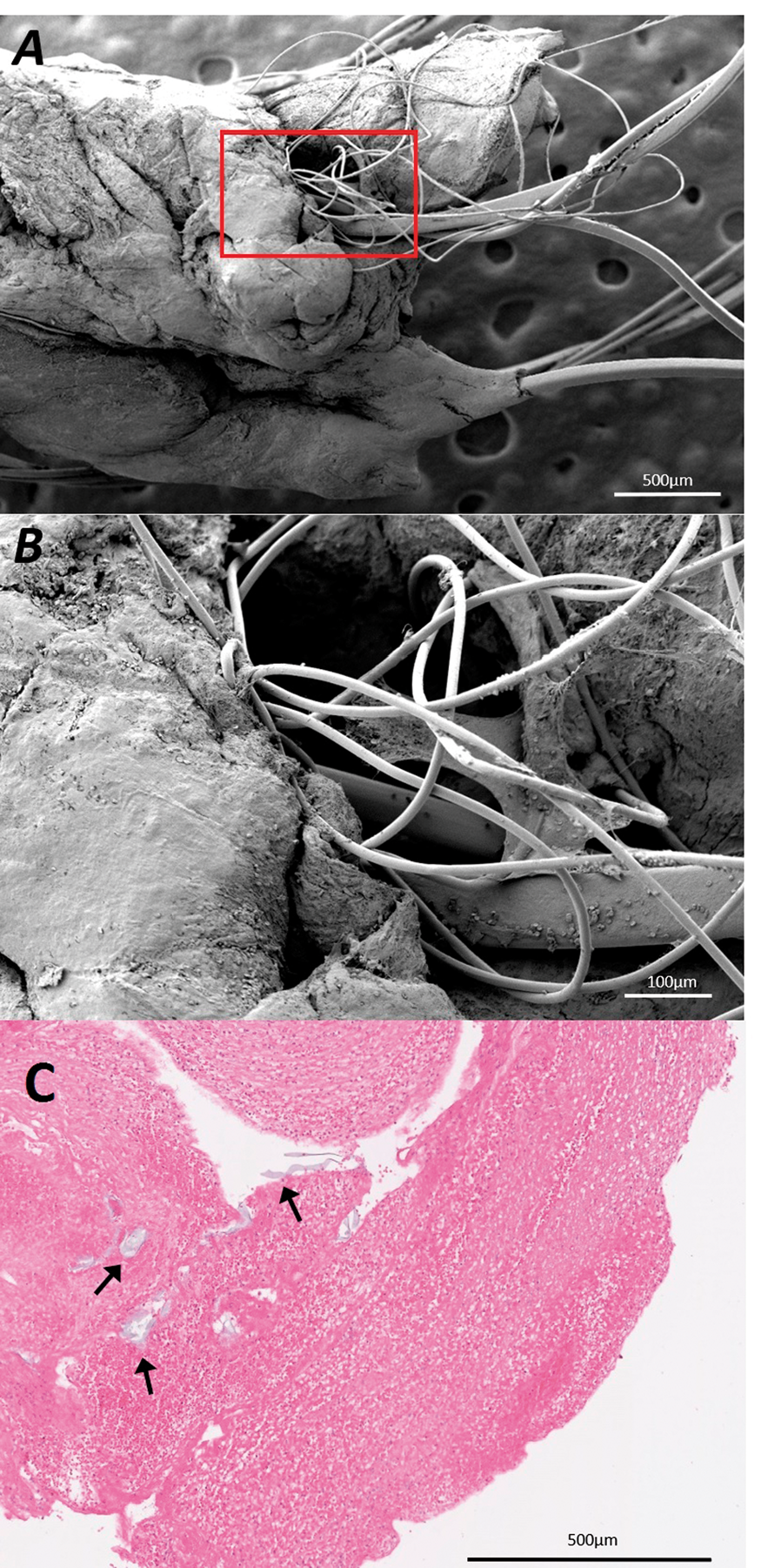
Conclusion
Foreign bodies are found in a substantial proportion of thrombi retrieved by mechanical thrombectomy for AIS. Neuro-interventionalists should be aware of the possibility of inadvertently introducing foreign bodies during thrombectomy procedures.
PB01-X01
Comprehensive analysis of RNF213 variants in patients with moyamoya disease (MMD) and intracranial artery stenosis (ICS) by target sequencing
1Department of Neurosurgery, Faculty of Medicine, The University of Tokyo, Tokyo, Japan
2Department of Neurosurgery, Japan Community Healthcare Organization Tokyo Shinjuku Medical Center, Tokyo, Japan
3Kanto Neurosurgical Hospital, Kumagaya, Saitama, Japan
4Department of Neurosurgery, Fuji Brain Institute and Hospital, Fujnomiya, Shizuoka, Japan
Abstract
Objectives
Moyamoya disease (MMD) is an idiopathic cerebral vasculopathy characterized by stenosis and occlusion at the terminal portion of internal carotid arteries in the circle of Willis accompanied by the development of collateral vessel network. RNF213 p.R4859K (rs112735431) was identified as a founder variant for MMD in East Asian population. In addition, we recently reported that RNF213 p.R4859K is also strongly associated with Intracranial artery stenosis (ICS) which is diagnosed mainly as atherosclerosis but not MMD. However, the disease susceptibility of the other RNF213 variants have not been fully clarified. In the present study, we comprehensively investigated MMD- and ICS- associated RNF213 variants other than p.R4859K.
Methods
Ninety-five patients with MMD, 168 patients of ICS and 141 controls (total 404 subjects) were enrolled in this study. Genomic DNA was extracted from the peripheral blood leukocytes of enrolled subjects. All coding exons of RNF213 were sequenced by next generation sequencing. Minor allele frequencies (MAF) of all variants detected were acquired by comparison against ExAC and 1000 genomes databases. Variants with MAF ≥0.01 and <0.01 were defined as common variants and rare variants respectively. Association studies were performed between patients and controls for nonsynonymous variants detected in the subjects. Chi-squared test was used for the studies, and p < 0.05 was considered significant. For investigation of association between rare variants in our subjects and diseases, we reviewed RNF213 rare variants previously reported in MMD patients and compared with rare variants in our subjects.
Results
Ninety-four missense variants and 58 silent variants (total 152 variants) were detected in our all subjects. The association studies revealed p.R4859K was most significantly associated with both MMD and ICS (p = 7.47×10⁻³¹ and 1.37×10⁻⁸ respectively). Two and 15 missense rare variants were identified in MMD and ICS patients without p.R4859K respectively. Among them, one rare variant (p.T3365I) in our MMD patients and 2 rare variants (p.C167R and p.L2405F) in our ICS patients were consistent with previously reported rare variants in MMD patients. On the whole, variants in MMD patients were clustered in the C-terminal region of RNF213 gene, and variants in ICS patients were in contrast distributed closer to the N-terminal.
Conclusions
Association studies showed the importance of RNF213 p.R4859K in intracranial major artery stenosis. We identified 3 candidate rare variants associated with MMD and ICS. The location of variants in RNF213 might be associated with the phenotype of the intracranial major artery stenosis. Validation study in a larger cohort and molecular biological functional analysis are necessary in future.
References
PB01-X02
Impaired endothelial progenitor cell differentiation based on IL-10 secretory insufficiency from peripheral blood cells in Moyamoya disease
1Department of Neurology, Tokai University, Japan
2Department of Physiology, Tokai University School of Medicine
3Department of Regenerative Medicine Science, Tokai University School of Medicine
4Department of Neurosugery, Tokai University, Japan
Abstract
Recently, it has been reported that a polymorphism of RNF213 gene or the numerical reduction of circulating endothelial progenitor cells (EPCs) might induce its abnormal vascular phenotype as the pathogenesis of MMD. However, the etiology and pathogenesis of Moyamoya disease (MMD) are still unknown.
In this study, we investigated the biological interacts of EPCs contributing to vascular formation in MMD patients using the newly developed vasculogenic culture of peripheral blood mononuclear cells (PBMNCs). In EPC colony forming assay, the cultured PBMNCs exhibited the reduction of definitive EPC colony forming potential in MMD patients, although the increase in healthy controls. In secretory cytokines of the cultured PBMNCs measured by
PB01-X03
Incidence, clinical presentation and risk factor of watershed shift phenomenon after superficial temporal artery-middle cerebral artery anastomosis for adult moyamoya disease
1Department of Neurosurgery, Kohnan Hospital, Sendai, Miyagi, Japan
2Department of Neurosurgery, Tohoku University Graduate School of Medicine, Sendai, Miyagi, Japan
3Department of Diagnostic Radiology, Tohoku University Graduate School of Medicine, Sendai, Miyagi, Japan
4Division of Epidemiology, Department of Health Informatics and Public Health, Tohoku University School of Public Health, Graduate School of Medicine, Sendai, Miyagi, Japan
Abstract
Objectives
Superficial temporal artery-middle cerebral artery (STA-MCA) anastomosis is a standard surgical management for adult moyamoya disease (MMD) patients, but focal cerebral hyperperfusion (CHP) and cerebral ischemia are potential complications of this procedure. Recent hemodynamic analysis on the acute stage after revascularization surgery for MMD revealed more complex and unique pathophysiological condition so called ‘watershed shift (WS) phenomenon’, which is defined as a paradoxical decrease of cerebral blood flow (CBF) at adjacent cortex near the site of local CHP. The objective of this study was to clarify the exact incidence, clinical presentation, and the outcome of WS phenomenon after STA-MCA anastomosis for adult MMD.
Methods
Among 74 patients with MMD undergoing STA-MCA anastomosis with indirect pial synangiosis for 78 affected hemispheres, 60 adult patients operated on 64 hemispheres completed the serial quantitative CBF analysis by N-isopropyl-p-[123I] iodoamphetamine single-photon emission computed tomography(123I-IMP-SPECT) in the acute stage after revascularization surgery. The local CBF was quantitatively measured at the site of anastomosis and the adjacent cortex by auto-radiographic method before surgery, as well as one and seven days after surgery. Then we investigated the incidence, clinical presentation, and risk factors of WS phenomenon.
Results
WS phenomenon was evident in 7 patients (7/64 hemispheres; 10.9%) after STA-MCA anastomosis for adult MMD. None of the patients manifested neurological deterioration due to WS phenomenon, but one patient developed reversible ischemic change by diffusion-weighted magnetic resonance imaging at the site of WS phenomenon, indicating incomplete infarction. Multivariate analysis revealed lower preoperative CBF value was significantly associated with occurrence of WS phenomenon (20.3 ± 7.70 ml/100 g/min in WS-positive group vs 31.7 ± 8.81 ml/100 g/min in WS-negative group, p = 2.3 × 10⁻³).
Conclusions
The incidence of WS phenomenon was as high as 10.9% after STA-MCA anastomosis for adult MMD. Clinical outcome of WS phenomenon was generally favorable, but WS phenomenon could have a potential risk for peri-operative cerebral infarction. Thus we recommend routine CBF measurement in the acute stage after revascularization surgery for adult MMD to avoid peri-operative complications such as local CHP and cerebral ischemia caused by WS phenomenon. Concomitant detection of WS phenomenon with local CHP is critical because standard blood pressure lowering to counteract with local CHP should be contraindicated in the presence of WS phenomenon.
References
PB01-X04
Bilateral hypoperfusion of the cerebellum following pontine infarction
1Dept. of Radiology, Akita University Graduate School of Medicine, Japan
2Dept. of Radiology and Nuclear Medicine, Research Institute for Brain & Blood Vessels-Akita, Japan
Abstract
Objectives
The aim of this study was to evaluate whether perfusion of bilateral cerebellum can be reduced in patients with pontine infarction and which side of them will decrease more severely.
Methods
Between April 2013 and March 2018, patients with pontine infarction who underwent single photon emission computed tomography (SPECT) scanning using 123I-N-isopropyl-p-iodoamphetamine (123I-IMP) at our institution were included. A patient with midline pontine infarction was excluded. 24 patients were included in the final analysis. Three-dimensional steteotaxic region of interest (ROI) template (3D-SRT)1) was used for ROI analysis and the following values were caluculated; total count value of right side of cerebellum (CRt cerebellum), total count value of left side of cerebellum (CLt cerebellum), total count value of right side of occipital lobe (CRt occipital) and total count value of left side of occipital lobe (CLt occipital). Perfusion of a cerebellum was evaluated using two of following methods. First, we obtained side-to-side perfusion differences by an asymmetry index (AI). AI = (CRt cerebellum – CLt cerebellum)/ {(CRt cerebellum + CLt cerebellum)/2}. Second, to assess hypoperfusion of each side of the cerebellums, normalized cerebellar blood flow (NCBF) was calculated standardized using the both side of occipital lobe. NCBF_Rt = CRt cerebellum/ {(CRt occipital + CLt occipital)/2} or NCBF_Lt = CLt cerebellum/ {(CRt occipital + CLt occipital)/2}. These data were analyzed using either the Welch's t test, the Student's t test or the Fisher's exact test as appropriate. For each of the cases, the AI and the NCBF were considered significant when 2 standard deviation above the mean of control group.
Results
The study population consisted of 24 patients (2 women, 22 men). The mean patient age was 71 years (range, 56–83 years). 11 patients had right infarction and 13 patients had left infarction. The mean AI value was 0.009 ± 0.025 (p < 0.01) in patients of right pontine infarction and −0.011 ± 0.034 (p = 0.51) in patients of left pontine infarction. Contralateral hypoperfusion of the cerebellum was in 9 patients (37%) and ipsilateral hypoperfusion of the cerebellum was in 2 patients (8%) with pontine infarction. AI showed contralateral perfusion of the cerebellum was decreased stronger than ipsilateral perfusion of the cerebellum. NCBF_Rt was 1.033 ± 0.067 (p = 0.11) and NCBF_Lt was 1.025 ± 0.076 (p < 0.05) in patients with the right pontine infarction. NCBF_Rt was 1.018 ± 0.087 (p = 0.06) and NCBF_Lt was 1.031 ± 0.098 (p = 0.06) in patients with the left pontine infarction. Bilateral perfusion of the cerebellum were decreased significantly in 11 (46%) patients. Results revealed that bilateral perfusion of the cerebellum were decreased significantly in patients with pontine infarction.
Conclusions
Bilateral perfusion of the cerebellum were decreased stronger in the patients with pontine infarction than in the control. Contralateral hypoperfusion was usually stronger than ipsilateral hypoperfusion but ipsilateral hypoperfusion was rarely stronger than contralateral hypoperfusion. It is necessary to take the pontine infarction into account as the cause of contralateral hypoperfusion of the cerebellum.
Reference
PB01-X05
Ischemic stroke due to varicella zoster virus vasculopathy: clinical, laboratory, and imaging features
1Dept. of Neurology, Tokyo Women's Medical University
2Dept. of Neurology, Tokyo Women's Medical University Yachiyo Medical Center
Abstract
Background
The varicella zoster virus (VZV) is a highly neurotropic virus that, after the primary infection, remains latent in ganglionic neurons and can reactivate years later, resulting in various neurologic complications such as Guillain-Barré syndrome, aseptic meningitis, myelitis, and cranial nerve palsies (e.g., Ramsay-Hunt syndrome, optic neuritis). Less commonly, ischemic stroke can occur secondary to VZV vasculopathy produced by direct VZV infiltration of cranial arteries. Here, we report a single-center experience of four patients who had ischemic stroke due to virologically-verified VZV vasculopathy.
Methods
We retrospectively reviewed our single-center registry of consecutive patients with acute ischemic stroke who had been hospitalized between April 2009 and January 2018, and identified 4 patients (2 men and 2 women; age range, 38–63 years) with stroke due to VZV vasculopathy. We described clinical features, laboratory and radiological examination findings, treatment, and functional outcomes. VZV infection was verified through detecting VZV DNA by polymerase chain reaction (PCR) and/or the anti-VZV IgG antibody by means of an enzyme immune assay (EIA) in the cerebrospinal fluid (CSF)
Results
Three patients were taking immunosuppressive agents, including prednisolone and/or methotrexate, at baseline. Each patient had a characteristic skin rash prior to stroke, with the interval from rash to stroke onset ranging from 13 to 122 days. Two patients experienced antecedent cranial nerve palsies; one had the IIIrd, VIIth, IXth, and Xth nerve palsies and the other had the IVth nerve palsy before stroke. Cerebral infarctions were located in the anterior circulation lesion (n = 1), in the posterior circulation lesion (n = 2), and in both lesions (n = 1). Intracranial arterial stenosis was only identified in one patient on magnetic resonance angiography. A high plasma d-dimer level was detected in one patient, whereas high β-thromboglobulin and platelet factor 4 levels were detected in two patients. As a result of combined therapies with acyclovir, steroid, and antithrombotic agents, neurological symptoms markedly improved in three patients, whereas one patient was left with moderate hemiplegia.
Conclusions
Cranial nerve palsies may be prodromal symptoms of VZV-associated stroke. Increased levels of thrombotic markers may support the use of antithrombotic agents, although the benefit of combined treatment should be determined through larger studies.
PB01-X06
Concurrent medullary infarction with ruptured vertebral artery
1Dept. of Neurosurgery, Nara Medical University, Japan
Abstract
Background
Medullary infarction (MI) occasionally occurs after intervention for ruptured vertebral artery dissection (VAD); however, it is difficult to determine whether MI is a treatment-related complication or is due to dissection.
Objective
The aim of this study was to measure the prevalence of concurrent MI and perforating artery involvement with ruptured VAD.
Methods
Thirty-four patients underwent direct surgery or endovascular intervention for ruptured VAD. To survey the concurrent MI with ruptured VAD, we analyzed 21 patients who underwent MRIs, excluding those with posttrapping due to the inability to identify the cause of MI. Furthermore, to survey the perforating artery involvement with ruptured VAD, we analyzed 9 patients who underwent MRIs after trapping and 19 who underwent digital subtraction angiography (DSA) procedures after proximal clipping.
Results
Among the 21 patients, including 4 who underwent MRIs before intervention and 17 who underwent MRIs after proximal clipping as the surrogate marker, there was no MI observed. The perforating arteries were estimated to be involved in 18 of 28 patients with the VAD according to MRIs and DSAs after intervention (64.3%).
Conclusion
No patient was identified as having concurrent MI with ruptured VAD. Considering the rarity of concurrent MI and the frequency of perforating artery involvement, we might have to choose treatment options such as flow diverters stents for preserving perforating arteries to achieve better outcomes when treating ruptured VAD.
PB01-X07
In-stent restenosis after vertebral artery ostium stenting: A five-year follow-up cohort study
1Department of Vascular Ultrasonography, Xuanwu Hospital, Capital Medical University, Beijing 100053, China
Abstract
Background
Vertebral artery ostium stenosis (VAOS) is contributed to majority of posterior circulation ischemia based on the results of previous literatures. Stenting has become an alternative treatment method for VAOS in recent decades. However, in-stent restenosis (ISR) remains as an unsolved issue after the procedure. This cohort study was designed to investigate the risk factors related to ISR in VAOS patients.
Methods
Patients with VAOS stenting from January 2013 to December 2014 were recruited in this retrospective study. All stenting procedures were conducted by experienced interventional neurosurgeons (performed at least 30 cases annually). Balloon-expanding stent was implanted to alleviate target artery stenosis. All patients underwent scheduled clinical and imaging follow-up. Vascular ultrasound was utilized as the primary follow-up imaging modality, while computed tomography angiography or digital subtraction angiography was performed when necessary. Univariant analysis was applicated to identify the possible risk factors including clinical characteristics, concomitant artery lesions and stent parameters. Then the Cox regression analysis was used to calculate the hazard ratio (HR) of the possible risk factors. ISR was defined as the in-stent stenosis >50%.
Results
A total of 400 consecutive patients (from a population of 8145 patients with VAOS) were finally recruited in this study including 216 with drug-eluting stents and 184 with bare metal stents. There were 325 (81.3%) patients underwent a mean 14 months (range 3–66) follow up and 116 (35.7%) patients diagnosed with ISR. Symptomatic ISR occurred in 8 patients with the complain of dizziness but free of stroke. Univariant analysis showed stent type, contralateral vertebral artery occlusion and target vessel tortuosity were the risk factors related to ISR (P<0.05) (
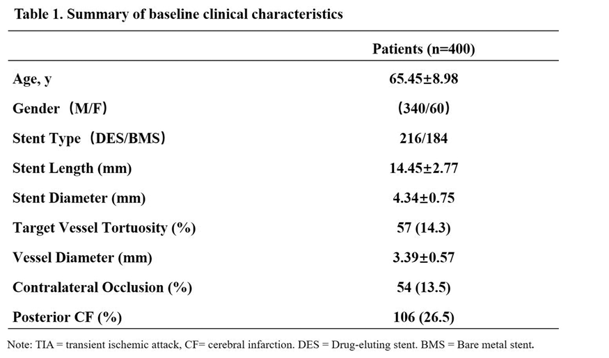
Conclusion
This study demonstrated that bare metal stent had more ISR than drug-eluting stent in VAOS. Contralateral vertebral occlusion and tortuose target vertebral could increase the risk of ISR. Implanting stents in patients under these circumstances should be carefully considered. A more aggressive medicine protocol during the follow-up may be required due to the high-risk of ISR.
References
PB01-X08
Importance of mucin tumor markers and successful treatment with DOACs in cancer-assiciated thrombosis patients with stroke
1Department of Neurology, Tokai University Hachioji Hospital, Japan
Abstract
Background
Trousseau’s syndrome defined as the cancer-associated thrombosis (CAT) due to hypercoagulability may not cause only venous thromboembolism (VTE), but also arterial thromboembolism including stroke. The cancer-associated stroke is sometimes hardly diagnosed and classified as one category of the embolic stroke of undetermined source (ESUS). Although resent clinical trials of several direct oral anticoagulants (DOACs) failed to show their efficacy for prevention of ESUS, DOACs have been demonstrated to be comparable with low-molecular heparin in prevention of VTE associated with cancer. Various types of tumor markers as well as blood coagulation markers may provide clues to the correct diagnosis of malignancy. Because most cases with CAT are associated with adenocarcinoma, we thought that mucin molecules released into the blood may play an important role not only as diagnostic markers, but also as causal factors in CAT.
Purpose
The purposes of the present study were two-folds; First, we tried to elucidate roles of mucin tumor markers in the hypercoagulable state and stroke. Second, we compared effectiveness of DOACs and other antithrombotic drugs in treatment for CAT.
Subjects and methods
We enrolled 30 sequential patients with cancer-associated ischemic stroke (77.5 ± 7.6 year old, 16 males and 14 females) prospectively. Most of their pathological diagnosis were adenocarcinoma; 8 gastric cancer (ca.), 7 colorectal ca., 3 lung adenocarcinoma, 3 gynecological ca., 3 prostate ca., 3 pancreatic ca., 2 cholangiocarcinoma, 2 hepatocellular ca., and others. Then, we reviewed their clinical features; 1) brain MRI findings, 2) incidence of VTE and nonbacterial thromboendocarditis (NBTE) detected with the echography, 3) correlation between coagulation markers (FDP, D-dimer, TAT and PIC) and tumor markers (CEA, CA19-9, and CA125), 4) efficacy and safety of antithrombotic drugs.
Result
Their brain MR images showed 2 patterns of infarcts; large territorial infarcts with distinct borders, suggesting cardiogenic embolization with massive thrombi, and scattered small infarcts which might be caused by arterial fragmented thrombi. VTE was observed in more than 50% patients, while NBTE was detected only in one patient by the transthoracic echocardiography. 2) There was a significant correlation between CEA and D-dimer/PIC (Pearson’s coefficient: 0.7173/0.7061), CA19-9 and D-dimer (0.40649), CA125 and D-dimer/PIC (0.5017/0.4330). Regarding antithrombotic treatment, direct oral factor Xa inhibitors were prescribed in 7 patients with DVT. Some of them who underwent resection of tumors had a long survival. Warfarin, heparin (i.v. or s.c), antiplatelets, and recombinant thrombomodulin were used in 6, 4, 2, and one patient, respectively. However, no treatment was applied in 10 patients, because hemorrhage could not be controlled in most of these patients.
Discussion
Serum levels of the mucin molecules were correlated with hypercoagulability in the CAT patients with stroke. These data suggested that the circulating mucin macromolecules per se may be involved in thrombogenesis, presumably by activating prothrombin directly or through p-selectin mediated interactions with platelets and endothelial cells. The treatment with the factor Xa inhibitors could be a promising strategy for CAT patients with VTE and stroke, further multi-center prospective studies are warranted.
PB01-X09
Pre-stroke modified Rankin Scale is useful in patient selection for mechanical thrombectomy regardless of age
1Department of Stroke Medicine, Kawasaki Medical School, Okayama, Japan
2Department of Neurology, Osaka University Graduate School of Medicine, Osaka, Japan
3Department of Neurosurgery, Kawasaki Medical School, Okayama, Japan
Abstract
Background
The usefulness of mechanical thrombectomy (MT) for acute ischemic stroke has recently been shown. Some patients with large artery occlusion have poor pre-stroke conditions, such as older age and poor activity of daily living in the real world, but they were excluded in recent randomized controlled trials. We aimed to clarify whether patients with poor pre-stroke conditions can benefit from MT.
Methods
This is a single-center, retrospective study of 108 patients with acute ischemic stroke who underwent MT between April 2015 and July 2018. Good clinical outcome was defined as modified Rankin Scale (mRS) 0–2 at 90 days, and poor clinical outcome was defined as mRS 3–6 at 90 days. We evaluated prognostic impact of patient’s background factors.
Results
Thirty-six patients (33.3%) had good clinical outcome and 72 patients (66.7%) had poor clinical outcome. Pre-stroke mRS was independently associated with good clinical outcome (odds ratio 0.27, 95% confidence interval 0.12–0.60, P = 0.001). In patients with pre-stroke mRS 0–1, 50.7% had good clinical outcome, and there was no significant difference in prognosis between patients aged <80 years and those aged ≧80 years (P = 0.60). A total of 56.4% of patients with pre-stroke mRS ≧2 was graded as mRS 5–6 at 90 days.
Conclusion
Patients with pre-stroke mRS 0–1 are expected to have a good prognosis after MT regardless of their age. Patients with pre-stroke mRS ≧2 might have an extremely poor prognosis, and we should be more careful in patient selection for MT.
PB01-X10
Brain tissue pulsations in acute haemorrhagic and ischaemic stroke: case studies
1University of Leicester, UK
2University Hospitals of Leicester NHS Trust, UK
3Nihon Kohden, Japan
4Broadview Labs, USA
5University of Washington, USA
Abstract
Objectives
Healthy brain tissue pulsates with the cardiac cycle [1], but whether brain tissue pulsations (BTPs) are affected in acute stroke has yet to be investigated. This series of case studies provides preliminarily data exploring whether Transcranial Tissue Doppler (TCTD) estimates of BTPs differ between affected and unaffected hemispheres in acute ischaemic and haemorrhagic stroke patients. The aim of this study was to compare pulsation patterns observed in acute stroke with those of similarly aged healthy subjects. This study is the first to explore tissue pulsatility in acute stroke patients to investigate the potential of TCTD as a diagnostic tool for stroke assessment.
Methods
TCTD measurements of brain tissue motion were obtained from a small number of acute ischaemic and haemorrhagic stroke patients for comparison with equivalent measurements obtained from healthy volunteers. Measurements were acquired using a Spencer Technologies transcranial Doppler ultrasound system, equipped with a 2 MHz ultrasound probe. For each participant, eight seconds of ultrasound were recorded and analysed using MATLAB. This analysis was used to display BTPs from 30 depths ranging from 2–8 cm within the brain. The collected BTP waveform shapes, timing, and displacements were then qualitatively reviewed by three independent observers to compare BTP waveforms between healthy and stroke subjects, affected and unaffected hemispheres, and ischaemic and haemorrhagic stroke.
Results
Here we present a qualitative comparison of brain tissue pulsations from three female subjects; a 58 year old healthy participant, a patient with a right-side acute ischaemic stroke (aged 69 years), and a patient with a right-side acute haemorrhagic stroke (aged 63 years). Both patients were monitored between 18 and 24 hours after stroke symptom onset. Overall, stroke patients displayed differing brain tissue pulsation patterns between their affected and unaffected sides. Pulsation patterns in the healthy subject were smooth and symmetric between the left and right sides. It was also noted that tissue pulsations in the healthy subject were highly correlated with depth and had consistent time-to-peak values (the time taken for the peak displacement to be reached in each cardiac cycle). In our stroke patients, unaffected hemispheres tended to generate similar pulsation patterns as healthy subjects with consistent time-to-peak values in at least the first 15 gates, whereas affected hemispheres were characterised by fluctuating or divergent time-to-peak values. In the stroke subjects we studied, median displacements were lower than seen in our healthy comparator.
Conclusion
This case report is the first to investigate BTPs in acute stroke. We identified a number of abnormal waveform features, which may merit further investigation. A larger cohort of patients will be required to systematically identify changes in brain tissue motion during ischaemic and haemorrhagic stroke, and to confirm whether differences between healthy and stroke subjects, or comparison of asymmetry between hemispheres, provides sufficiently high sensitivity and specificity for diagnostic use.
Reference
PB01-X11
Clinical evaluation of intravenous rt-PA therapy for acute ischemic stroke in patients beyond the age of 90
1Dept. of Neurology, Okayama Medical Center, Japan
Abstract
Objectives
This study aimed to evaluate the prognostic value after intravenous recombinant tissue plasminogen activator (rt-PA) therapy for acute ischemic stroke patients beyond the age of 90.
Methods
We studied 163 consecutive patients admitted from October 2005 to June 2018 in our hospital. We compared among patients aged >90 and 3 younger age groups (20-64, 65–74, 75–89). We examined the gender, risk factors, NIHSS on admission and after 7 days, the percentage of effect after 7 days (remarkable: NIHSS 0–2 or >10 improve, effective: NIHSS 3–5 or >5 improve, unaltered: −4 < NIHSS < 4, worse: NIHSS > −5, mortality), the good prognosis (mRS under 2 after 3 months) and symptomatic intracerebral hemorrhage.
Results
The following results were obtained: (20-64, 65–74, 75–89, >90) respectively, number (35, 39, 77, 11), men (%) (82.9, 64.1, 54.5, 45.5), smoking (%) (42.9, 25.6, 9.1, 9.1), hypertension (%) (37.1, 69.2, 72.7, 90.9), NIHSS on admission (median) (7, 10, 9, 13), NIHSS after 7 days (median) (2, 4, 6, 9), remarkable effect after 7 days (%)(85.7, 66.6, 54.6, 54.6), good prognosis after 3 months (%)(72.3, 64.1, 46.8, 18.2), symptomatic intracerebral hemorrhage (%) (0, 7.7, 7.8, 0).
Conclusions
The risk of symptomatic hemorrhage is not higher than in younger patients. However, functional outcomes are lower.
PB01-X12
Early seizure and clinical outcomes after acute ischemic stroke: the Fukuoka Stroke Registry
1Department of Cerebrovascular Medicine, St. Mary's Hospital, Japan
2Department of Medicine and Clinical Science, Graduate School of Medical Sciences, Kyushu University, Japan
3Department of Health Care Administration and Management, Graduate School of Medical Sciences, Kyushu University, Japan
4Center for Cohort Study, Graduate School of Medical Sciences, Kyushu University, Japan
Abstract
Objectives
The impact of early seizure after acute ischemic stroke on post-stroke clinical outcomes is still controversial. We aimed to elucidate the association between early seizure and clinical outcomes,including functional outcome, stroke recurrence, and all-cause mortality, after acute ischemic stroke.
Methods
From the Fukuoka Stroke Registry, a multicenter hospital-based stroke registry in Fukuoka, Japan, we included a total of 7,072 patients with acute ischemic stroke who had had neither previous stroke nor epilepsy and were hospitalized within 24 h of onset in 7 stroke centersfrom June 2007 to May 2018. Patients were divided into seizure group and non-seizure group, according to the incidence of seizure within the first 7 days after stroke onset. Study outcomes were unfavorable outcomes, including poor functional outcome(modified Rankin Scale score ≥3 at 3 months)and stroke recurrence, and all-cause mortality during follow-up period (3.6 ± 2.8 years). We evaluated the associations between early seizure and poor functional outcomeby logistic regression analysis and between early seizure and stroke recurrence or all-cause mortalityby Cox proportional hazards model. Covariates were potential confounding factors, such as age, sex, conventional risk factors, stroke severity, stroke subtypes, reperfusion therapy, pre-stroke independency, and cortical symptomson admission. To control confounding variables, we further performed propensity score-matched study in 116 pairs of patients with and without early seizure on the basis of 1:1 matching.
Results
The mean age was 72.5 years, and 41.9% were women. Of all patients, early seizure occurred in 117 patients (1.7%). Cardioembolic stroke was more prevalent (46% vs 26%, p < 0.001) and NIH stroke scale on admission was higher (median 13 vs. 3, p < 0.001) in patients with early seizure than those without. Frequency of poor functional outcome was more frequent (65% vs. 34%, p < 0.001) in patients with early seizure. However, after adjusting for confounding factors, odds ratio [95% confidence interval] of poor functional outcomedid not significantly increase in patients with early seizure(0.98 [0.57–1.70]). During the follow-up period, 1,190 patients recurred stroke (90 /100 person year) and 1,811 patients died (226/100 person year). After adjusting for confounding factors, the hazard ratio of all-cause mortality significantly increased (1.41 [1.05–1.88]), while the ratio of stroke recurrence did not (0.76 [0.39–1.46]), in patients with early seizure. These trends were also maintained in propensity score-matched case control study.
Conclusions
Early seizure after acute ischemic stroke was associated with all-cause mortality, but not with poor functional outcome at 3 months and stroke recurrence.
PB02-B01
Injury-induced multipotent stem cells: basic characteristics and future perspective
1Department of Neurosurgery, Hyogo College of Medicine, Japan
2Institute for Advanced Medical Sciences, Hyogo College of Medicine, Japan
Abstract
Objectives
Using a mouse model of cerebral infarction, we previously demonstrated that injury/ischemia-induced stem cells developed in the post-stroke area. Because the stem cells had the same characteristics as neural progenitors, we initially named them injury/ischemia-induced neural stem/progenitor cells. However, subsequent studies showed that these stem cells could differentiate into various cells, including neural lineage cells. Thus, we presently call them injury-induced multipotent stem cells (iSCs). Recently, we found that brain pericytes could revert to iSCs in the human brain following cerebral infarction. This study aimed to elucidate the basic characteristics of human iSCs and discuss their therapeutic perspectives on stroke treatment.
Methods
To examine the traits of human iSCs, we obtained brain samples from post-stroke areas in patients requiring both decompressive craniectomy and partial lobectomy for diffuse cerebral infarction. The Ethics Committee of our college reviewed and approved this study. Post-stroke human brain tissue sections were mechanically dissociated, and the resulting cell suspensions were incubated under adherent culture conditions. Polymerase chain reaction (PCR), immunohistochemistry, and microarray were performed to evaluate basic characteristics of iSCs.
Results
The PCR analysis revealed that putative iSCs did not express astrocytic and endothelial lineage markers. However, they expressed pericytic and neural crest lineage markers, suggesting that human iSCs likely originate from brain pericytes, as do mouse iSCs. The immunohistochemistry study found that iSCs could differentiate into neural and mesenchymal lineage cells. The results indicate that iSCs are cell populations sharing the characteristics of neural and mesenchymal stem cells. Recent analyses also suggest that iSCs have the potential to differentiate into electrophysiologically functional neurons.
Conclusions
iSCs share characteristics of neural and mesenchymal stem cells. Because human iSCs can differentiate into various cells, including neurons, iSCs have the potential to repair the damaged central nervous system following ischemic stroke.
PB02-B02
Neurogenesis of muscle-derived stem cells to neural-like cells by retinoic acid
1Department of Molecular Science Technology
Abstract
Objectives
Several stem cells in the human body are to maintain and repair organs or tissues. Skeletal muscles derived muscle-derived stem cells (MDSCs) are widely and easily available and possess the plasticity to differentiate into neurons and glial cells such as oligodendrocytes and astrocytes in response to stimulation with growth factors and other signalling molecules. Retinoic acid (RA) has been shown to have important regulatory functions during embryonic development. Thus our aim was to identify RA that might induce neuronal differentiation of MDSCs, with the ultimate goal of treating neurodegenerative diseases.
Methods
In this work, RA were treated in MDSCs to assess the applicability of RA for a neurogenic differentiation. Then, lineage-specific differentiation was monitored using the neuronal markers neuron-specific enolase (NSE) and neuron-specific βIII tubulin (Tuj1), the oligodendrocyte marker, 2’,3’-cyclic nucleotide 3’-phosphodiesterase (CNPase) and the astrocytic marker glial fibrillary acidic protein (GFAP).
Results
These neural marker proteins were not detectable in MDSCs cultured in the absence of RA. However, MDSC treated with RA showed abundant positive cells against the NSE and Tuj1. Additionally, CNPase-positive cells were observed, indicating oligodendrocyte lineage cells. Meanwhile, GFAP positive cells were not detected. The mRNA profile of MDSC treated with RA included higher expression of NSE compared with those of non-treated cells in real-time PCR.
Conclusions
In conclusion, MDSCs differentiated principally into neural-like cells in the presence of RA. These results support the idea that MDSCs are a potentially rich source of precursors that can be induced to differentiate into neural-like cells using RA.
PB02-B03
Intracerebral ventricularly (ICV) administration of human neural stem cells ameliorate brain injury after cardiac arrest
1Department of Neurosurgery, University of Maryland School of Medicine
2Department of Anatomy Neurobiology, University of Maryland School of Medicine
3Department of Orthopaedics, University of Maryland School of Medicine
4Department of Biomedical Engineering, Johns Hopkins University School of Medicine
5Department of Anesthesiology & Critical Care Medicine, Johns Hopkins University School of Medicine
Abstract
Cardiac arrest (CA) has an incidence of 359,800 annually. Among survivors of CA, brain injury is the biggest impediment to functional recovery. Currently, neither pharmacological intervention nor therapeutic hypothermia can reverse the neural injury caused by CA. Stem cell therapy holds significant promise in the neuronal repair after brain injury. Neural stem cells (NSCs) are capable of differentiating into neurons, astrocytes, and oligodendrocytes, and has been reported to significantly improve the functional outcome in stroke, hypoxic-ischemic encephalopathy, and traumatic brain injury. We aim to evaluate the neuroprotective effects of human NSCs (hNSCs) on the acute stage of brain injury after cardiac arrest (CA) in a rodent model of CA after the return of spontaneous circulation (ROSC). 12 Long–Evans rats (4 Male and 8 female, average 376 g) subjected to 8-min asphyxia-CA were randomly assigned to receive hNSCs or control group. hNSCs were slowly infused into the left lateral ventricular (P, 1.2 mm, L 1.5 mm, D 4 mm) at 3 hours post-ROSC with 20 uL of cell suspension (containing 2X10⁵ PKH26 pre-labeled hNSCs). Neurological deficit score (NDS) was used to evaluate the neurological outcome. 72-hour after ROSC was set as the primary endpoint. Additional two rats subjected to 8-min asphyxia-CA were euthanized at 4 weeks after ROSC to confirm the survival and function of transplanted pre-labeled hNSCs by brain slides and whole cell patch clamp. The survival rate in the NSC group (71.4%) were higher than the control group (60.0%). The neurological functional recovery in the hNSCs group (median NDS was 42, 53, 56, and 63 at 6 h, 24 h, 48 h, and 72 h after ROSC) was also improved compared with the control group (median NDS was 36, 48, 48, and 48 at 6 h, 24 h, 48 h, and 72 h after ROSC) (Table 1). Immunofluorescent co-staining of PKH26 and NeuN verified the neuronal differentiation from translated PKH26 –positive hNSCs in the hippocampus CA1 and cortex 4 weeks after CA via ICV administration. The whole cell patch clamp technique confirmed the spontaneous firing activity was recorded in cell-attached mode from the functional mature neurons derived from transplanted cells at 4 weeks after ROSC. Spontaneous excitatory synaptic current represented that the cell receives synaptic inputs from other neurons. Transplanted hNSCs therapy markedly improved survival and neurologic outcomes after CA. The neuroprotective effects may be related with the cell replacement and modulation of growth factors.
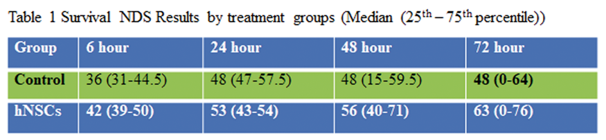
The work was supported in part by R01HL118084 and R01NS110387 from United States National Institutes of Health (NIH) (both to XJ).
References
PB02-B04
In vivo analysis of mRNA-ITGA4-engineered mesenchymal stem cell docking to inflamed endothelium after their intra-arterial transplantation in animals with focal brain injury
1NeuroRepair Department, Mossakowski Medical Research Centre, PAS, Warsaw, Poland
2Russel H. Morgan Department of Radiology and Radiological Sciences, Division of MR Research, The Johns Hopkins University School of Medicine, Baltimore, USA
Abstract
Objectives
: Cell therapy with the use of mesenchymal stem cells (MSCs) is currently one of the most dynamically developing branches of regenerative medicine. Due to anti-apoptotic properties, high paracrine activity and multidirectional differentiation capability MSCs are currently evaluated in experimental and clinical trials of treatment of the most common central nervous system (CNS) diseases where there is no effective alternative therapy available [1]. However, the homing of systemically administered MSCs to lesioned tissue is not sufficient which may be related to the low expression of relevant receptors and adhesion proteins. Thus we decided to induce the expression of VLA-4 protein, which is responsible for adhesion and diapedesis of leukocytes, on the surface of human MSCs and assumed that it may have a positive effect on homing and transmigration of genetically engineered cells into desired area after their transplantation [2].
Methods
: Overexpression of α4 subunit of VLA4 integrin in human bone marrow mesenchymal stem cells (hBM-MSCs) was obtained by mRNA-ITGA4 based transfection. For in vivo assessment hBM-MSCs labelled with ION (Molday, BioPAL) were transplanted into the right internal carotid artery of rats subjected to ouabain-induced brain damage. Biodistribution of engrafted cells in the brain was monitored by MRI and immunohistochemically.
Results
: Flow cytometry analysis confirmed the presence of ITGA4 protein in 73% of hBM-MSCs 24 hours after mRNA transfection, in comparison to 4% in naive counterparts. After intra-arterial administration both ITGA4 overexpressing hBM-MSCs and naive cells were routed immediately into the right hemisphere of the brain. The homing of ITGA4 expressing cells was more effective than control ones, as evidenced by higher intensity of MRI signal at the completion of injection. Immunohistochemical analysis of rat brain revealed that modified and naive hBM-MSCs remained inside the cerebral blood vessels 48 hours after transplantation. Three days after infusion hBM-MSCs showed the signs of diapedesis, with about 50% of transplanted cells present in perivascular space.
Conclusions
: The transfection with mRNA is an effective way to induce expression of ITGA4 in hBM-MSCs, which translates to their increased adhesion activity and more effective accumulation of transplanted cells in the cerebral blood vessels within the area of brain injury. Transplanted hBM-MSCs are capable to exit the lumen of the blood vessels and they mostly localize in the perivascular space, which is a central location for neurovascular unit. It might be a natural niche for MSCs, which has some relations to pericytes, and they may exert their therapeutic effect through changing properties of perivascular space and by this way to improve function of neurovascular unit.
References
PB02-B05
Regenerative potential of canine glial restricted progenitors transplanted in a mouse model of demyelinating diseases
1NeuroRepair Department, Mossakowski Medical Research Centre, Polish Academy of Sciences, Warsaw, Poland
2Department of Experimental Pharmacology and Small Animal Magnetic Resonance Imaging Laboratory, Mossakowski Medical Research Centre, Polish Academy of Sciences, Warsaw, Poland
3Vetregen Laboratory and Stem Cell Bank for Animals, Warsaw, Poland
4Institute for Cell Engineering, Division of MR Research, The Russell H. Morgan Department of Radiology and Radiological Science, Johns Hopkins University School of Medicine, Baltimore, MA, USA
Abstract
Objectives
Amyotrophic lateral sclerosis (ALS) is a progressive neurodegenerative disease with no effective cure. Disease is characterized by a progressive loss of upper and lower motor neurons [1]. In the last years there is a growing interest in stem cell transplantation as a therapeutic strategy for ALS. Recent reports also emphasize the role of glia in ALS pathology. Therefore transplanting functional glia from healthy donors could restore myelin sheath in recipient’s nervous system. Dogs with canine degenerative myelopathy (DM) and myelin basic protein (MBP)-lacking mutant mice (shiverer) demonstrate ALS-like phenotype. To overcome immunorejection caused by xenotransplantation, shiverer mice are routinely crossed with rag2 immunodeficient mice, thereby generating a convenient model for evaluating myelinating potential of newly introduced, healthy canine glial restricted progenitors (cGRPs) [2].
Methods
Transplantation experiments were performed using cGRPs extracted from brains and spinal cords of dog foetuses. Cells were grown in vitro in a standardized media (DMEM/F12 + B27 + N2 + heparin + bFGF) until sufficient expansion and transplanted intraventricularly into shiverer/rag2-/- mice pups (P2) – the immunocompetent model of congenital demyelination. The following stereotaxic coordinates were used: AP:0.6, ML:1.0/-1.0, DV:0.8 from bregma; AP:0.8, ML:1.0/-1.0, DV:0.8 from lambda. 200 000 cells suspended in 2 µl of PBS were injected into each hemisphere of P2 shiverer/rag2-/- mice. Prospective myelination was analysed using MRI, electron microscopy and confocal microscopy following immunohistochemical staining. T2 weighted images of shiverer/rag2-/- mice brains were taken 18, 31 and 40 weeks after transplantation. Lifespan of each recipient mouse was monitored throughout the study.
Results
Electron microscopy analysis and immunohistochemical study revealed myelination in corpus callosum and spinal cord of shiverer/rag2-/- mice after cGRP transplantation. The migration pattern of infused cells varied depending on the injection site (near bregma or close to lambda) however, the myelination rate was comparable in both cases yet it differed between animals. Myelin was visible in MRI and under electron and confocal microscopy after staining as early as 18-weeks post transplantation and remained on a constant level by the end of the experiment. Among the graft recipients about 50% of mice demonstrated lifespan longer than 200 days – a mean lifespan of non-transplanted, control shiverer/rag2-/- mice.
Conclusions
Canine-derived glia restricted progenitors are able to produce myelin following transplantation into the mice model of demyelinating diseases. It seems that GRP transplantation could be an experimental approach for treatment of demyelinating diseases.
Acknowledgements
This work was supported by NCR&D Grants: STRATEGMED I No. 1/233209/12/NCBR/2015 “GRP&ALS” and POWER Och!Dok program.
References
PB02-B06
Application of human glial restricted progenitors for treatment of amyotrophic lateral sclerosis in SOD1/RAG2 mice – limitations and perpectives
1NeuroRepair Department, Mossakowski Medical Research Centre PAS
2Department of Experimental Pharmacology and Small Animal Magnetic Resonance Imaging Laboratory, Mossakowski Medical Research Centre PAS
3Division of MR Research, Institute for Cell Engineering, The Russell H. Morgan Department of Radiology and Radiological Science, Johns Hopkins University School of Medicine
Abstract
Objectives
Amyotrophic Lateral Sclerosis (ALS) is a neurodegenerative disease with no effective cure. Presently, research aims at different approaches of stem cell therapy, however it must be stressed that transplantation of motor-neurons (MNs) seems to be hard if not impossible due to the length of their axons. Thus, interesting concept aims at glia, that in healthy organism are responsible for maintaining homeostasis and supporting of neurons. It is believed that glia failure may lead to degenerative changes in ALS1–3. Therefore, replacement of malfunctioning glia with healthy population of cells appears adequate therapeutic approach. It was previously described that GRPs transplanted intraventricularly into mouse model of demyelination (shiverer mice) revealed myelinating and therapeutic potential4. In our project we aimed to evaluate the efficacy of human glial restricted progenitor (GRPs) transplantation in immunocompetent model of ALS (SOD1/rag2 mice) with high or low copies of SOD1 transgene.
Methods
400 000 of hGRPs (Q-cells, QTherapeuthics) were transplanted intra-ventricularly into both hemispheres of 2-days old SOD1/rag2 mice pups. Efficacy of GRPs treatment was evaluated with histological (Nissl staining) analysis of motor-neuron number of in spinal cord of transplanted mice, immunohistochemical and Western Blot analysis of misfolded SOD1 (msSOD1) protein (that is accumulated in brain and spinal cord of mice with SOD1 mutation). Neurodegenerative changes in brain motor-nuclei were estimated in MRI.
Results
The number of MNs in transplanted animals was reduced comparing to rag2 mice but remained at similar level in comparison to non-transplanted SOD1/rag2 mice. MsSOD1 protein was visible both in transplanted and non-transplanted animals. MRI analysis revealed the presence of neurodegenerative changes in motor related nuclei in brains of transplanted high copy number mice. Human GRPs transplantation did not rescue the ALS phenotype of SOD1/rag2 mice. Transplantation of GRPs did not prolong the mice lifespan when compared to non-transplanted controls.
Conclusions
The failure of GRP transplantation to prolong animal lifespan and to improve motor-neuron condition in SOD1/rag2 mice might be caused by inappropriate distribution of infused cells. The hostile environment of recipient mice and spreading of pathological misfolded SOD1 protein to the transplanted glial progenitors may be another cause of disappointing therapeutic effect of GRPs transplantation. To achieve the positive effect of GRP transplantation on the course of the disease in SOD1/rag2 mice the multiple sites of cell injection and the higher number of cells is propounded.
Supported by a NCR&D grant for STRATEGMED project: “GRP&ALS"
References
PB02-B07
MRI-guided intra-arterial and intrathecal delivery for global targeting of glial progenitors in canine ALS-like degenerative myelopathy
1School of Medicine, Collegium Medicum, University of Warmia and Mazury, Department of Neurosurgery, Olsztyn, Poland
2Russell H. Morgan Dept. of Radiology and Radiological Sciences, Johns Hopkins University, Baltimore MD, United States
3Cellular Imaging Section and Vascular Biology Program, Institute for Cell Engineering, Johns Hopkins University, Baltimore MD, United States
4Mossakowski Medical Research Center, Polish Academy of Sciences, NeuroRepair Department – Warsaw, Poland
5Central Clinical Hospital of Ministry of the Interior and Administration in Warsaw, Poland
6Faculty of Veterinary Medicine, University of Warmia and Mazury, Department of Surgery and Roentgenology with the Clinic – Olsztyn, Poland
7Faculty of Veterinary Medicine, University of Life Sciences – SGGW, Department of Morphological Sciences – Warsaw, Poland
Abstract
Objectives
Stem cell transplantation has been shown efficacious in animal models of neurological diseases; however, in case of diseases with disseminated pathology such as ALS, delivery of cells and their broad distribution remains to be a challenge. To address this problem, we exploited two most promising delivery routes: intrathecal, into cerebrospinal fluid-filled (CSF) spaces and intraarterial into cerebral vasculature. The goal of this study was to investigate feasibility and safety of both routes for MRI-guided transplantation of glial restricted precursors (GRPs) in dogs suffering from naturally occurring ALS-like disease degenerative myelopathy (DM).
Methods
Dogs from various breeds, with confirmed DM were enrolled for transplantation study. Canine GRPs were labeled with superparamagnetic iron oxide nanoparticles (SPIO;Biopal) and suspended in PBS for intraarterial transplantation or in hyaluronic acid-based hydrogel (HyStem;EsiBio) for intrathecal injection. For
Results
Intraarterial: Dynamic MR imaging during intraarterial injection of GRPs revealed hypointense signal in the ipsilateral hemisphere (
Intrathecal
Dynamic MRI imaging during intrathecal infusion of hydrogel-embedded GRPs revealed hypointense signal that gradually extended during hydrogel injection. Hypointensities were expanding rostrally and caudally from the catheter tip (

Conclusion
Both intraarterial and intrathecal routes of cell delivery proved to be safe. Real-time MRI-guided, cell transplantation allowed immediate visualization and feedback for precise delivery of cells. Intraarterial injection while safe, resulted in rather poor retention of cells in the brain indicating the need for cell engineering to enhance endothelial capture. Intrathecal injection was more effective and embedding the cells in the hydrogel was essential to keep the cells at the injection site and prevent sedimentation or outflow with circulating CSF. These two minimally invasive procedures may be used as a separate therapeutic strategies or simultaneously for global stem cells delivery in ALS and other neurological disorders.
Authors declare no conflict of interests.
Funding
STRATEGMED1/233209/12/NCBR/2015, 12/EuroNanoMed/2016.
PB02-B08
Viability and migration ability in vitro of human polymorphonuclear leukocytes loaded with synthetic microcapsules
1University of Pennsylvania, Philadelphia, PA, USA
2RASA Center in Tomsk, Tomsk Polytechnic University, Tomsk, Russian Federation
3RASA Center, Kazan Federal University, Kazan, Russian Federation
4Saratov State University, Saratov, Russian Federation
5Massachusetts General Hospital, Harvard Medical School, Charlestown, MA, USA
6Rutgers University, Piscataway, NJ, USA
7Queen Mary University of London, London, United Kingdom
Abstract
Objectives
Targeted drug delivery to brain is a chief medicinal objective, because most potent therapeutics are unable to cross the blood-brain barrier (BBB) following systemic administration. Polymorphonuclear leukocytes (neutrophils) have been investigated as potential carriers for drug delivery as they are naturally recruited across the BBB during neuro-inflammatory conditions such as stroke or Alzheimer diseases.
Methods
Microcapsules (2 microns) were fabricated using alternating adsorption layers of polyelectrolytes onto calcium carbonate micro-particles used as a sacrificial template. Human neutrophils were isolated from the blood of healthy donors and then incubated with the FITC-labeled microcapsules at various cell/capsule ratios for 20 minutes at 37°C. Immediately after incubation, the cells were washed and further cultured for 1 to 4 hours. Human neutrophils were analyzed for viability using flow cytometry with a ΔΨm-sensitive MitoTracker dye, intracellular incorporation of microcapsules was confirmed by transmission electron microscopy. The migration ability of the cells through a 3 -μm-pore polycarbonate membrane was assessed using a HTS Transwell plate with IL-8 as a chemoattractant. Cells were allowed to migrate for 1 hour at 37°C and the fraction of migrated cells to the bottom chamber were quantified by flow cytometry.
Results
Electron microscopy of neutrophils demonstrated that 1-5 capsules per cell were internalized and appeared as variable oval and more complex-shaped structures 0.5–2.5 µm in size with an electron-dense contour. Flow cytometry showed that ∼95% of neutrophils remained viable after 4 hours of incubation in the absence or presence of capsules. The average fractions of neutrophils loaded with microcapsules were 52%, 47%, and 48% cells at the cell/capsule ratios of 1:0.5, 1:1, and 1:1.5, respectively. The average number of viable neutrophils bearing the capsules did not change significantly during the 4-hour incubation and varied from 37% to 45% irrespective of the cell/capsule ratios studied. Flow cytometry data demonstrated that on average 10% of the neutrophils loaded with microcapsules maintained their ability to migrate through a 3-μm-pore towards chemoattractant, which is similar to unloaded neutrophils.
Conclusions
Human neutrophils can be loaded with synthetic microcapsules and maintain their viability for up to 4 hours in vitro. The internalized microcapsules remain non-degraded intracellularly, which is important for the possible use of these capsules as drug cargos. Neutrophils loaded with microcapsules also demonstrated their ability to migrate through a 3-μm-pore. Our findings support the potential use of live neutrophils as natural transporters of encapsulated bioactive compounds, such as nucleoside-modified mRNA, for targeted delivery in neuro-inflammatory diseases, as stroke or Alzheimer diseases. These studies are ongoing.
Funding Sources
This work was supported by the Program for Competitive Growth at Kazan Federal University; Tomsk Polytechnic University Competitiveness Enhancement Program grant (VIU-SEC B.P. Veinberg-210/2018); Russian Science Foundation grant No. 17-15-01111 (DNA), NIH R01 1R01NS096237 (DNA.); NIH RO1 HL086621 (AJG), NIH R01-AI124429 (DW).
PB02-B09
Neuroprotective effects of human dental pulp stem cells after acute cerebral ischemia
1Department of Neurological Science, Graduate School of Medicine, Nippon Medical School
2Department of Biochemistry and Molecular Biology, Nippon Medical School
Abstract
Objective
To prevent brain injury after ischemia, numerous experimental studies have focused on cellular therapies by using stem cells. Human dental pulp stem cell (DPSC) is an attractive cell source because it can be easily obtained as medical waste without ethical and logistical problems. In this study, we investigated the effects of intravenous DPSC administration after transient focal ischemia in rats.
Methods
Male Sprague-Dawley rats underwent transient 90 min middle cerebral artery occlusion (MCAO)(n = 8, each). DPSCs (1 × 10⁶) or vehicle were administered via the femoral vein at 0 h or 3 h after ischemia-reperfusion. Infarct volume, neurological deficits, and immunological analyses were performed at 24 h and 72 h after reperfusion.
Results
DPSCs transplanted at 0 h after reperfusion significantly reduced infarct volume and reversed motor deficits at 24 h and 72 h recovery (p < 0.01, p < 0.01). DPSCs transplanted at 3 h after reperfusion also significantly reduced infarct volume and improved motor function compared with vehicle groups at 24 h and 72 h recovery (p < 0.01, p < 0.05). Further, DPSC transplantation significantly inhibited microglial activation and pro-inflammatory cytokine expression compared with controls at 72 h after reperfusion (p < 0.01, p < 0.01). Moreover, DPSCs attenuated neuronal degeneration in the cortical ischemic boundary area (p < 0.01).
Conclusions
The present study shows that systemic delivery of human DPSCs after reperfusion reduced ischemic damage and improved functional recovery in a rodent model, with a clinically relevant therapeutic window. The neuroprotective action of DPSCs may relate to the modulation of neuroinflammation during the acute phase of stroke.
PB02-B10
Human bone marrow mesenchymal stem cells or extracellular vesicles transplanted intra-arterially modulate immune response evoked by focal brain injury in rat recipients
1NeuroRepair Department, Mossakowski Medical Research Centre, PAS, Warsaw, Poland
2Department of Experimental Pharmacology, Mossakowski Medical Research Centre, PAS, Warsaw, Poland
3Department of Women's and Children's Health, University of Padua, Padua, Italy
4Russel H. Morgan Department of Radiology and Radiological Science, Division of MR Research, The Johns Hopkins University School of Medicine, Baltimore, USA
Abstract
Objectives
Ischemic stroke is the third cause of death and main cause of disability in people after 65 years old in the developed countries [1]. Cell death in the injured region of the brain activates the production of DAMPs (damage-associated molecular patterns) and activates immune reactions which lead to further progression of damaged tissues [2]. To search for the potential immunomodulatory therapy, mesenchymal stem cells (MSCs) could be potentially used in ischemic stroke disorders [3]. Recent studies demonstrated that therapeutic effect of MSCs is mainly associated with their paracrine activity and at least partially related to the release of extracellular vesicles (EVs). The aim of the study was to compare the immunomodulatory properties of human bone marrow mesenchymal stem cells (hBM-MSCs) and extracellular vesicles derived from these cells in a model of rat focal brain injury.
Methods
The in vitro studies were performed on hBM-MSCs (Lonza) and EVs isolated from the culture media of these cells. hBM-MSCs were stained with iron nanoparticles conjugated with rhodamine B (Molday ION, BioPAL) and EVs were labelled with lipophilic dye PKH26 (Sigma). The in vivo studies were performed on adult male Wistar rats with focal brain insult evoked by injection of 1μl/50 nmol ouabain into the right hemisphere. Two days after ischemic injury 5x10⁵ hBM-MSCs labelled with Molday ION or 1.3x10⁹ EVs stained with PKH26 were transplanted into right internal carotid artery of Wistar rats. The bio-distribution of injected hBM-MSCs was analyzed using MRI and the presence of hBM-MSCs or EVs in rat brain was explored by confocal microscopy. The cellular immune response in the right hemisphere of experimental animals was evaluated by immunohistochemical stainings. Moreover, the production of cytokines and chemokines in the rat brain was detected using Bio-Plex Pro™ Cytokine, Chemokine and Growth Factor Assay (BioRad).
Results
hBM-MSCs or EVs derived from them transplanted intra-arteriary into focal injured rat brain were visible the injured hemisphere near the ischemic lesion. Immunohistochemical analysis of different cell subsets observed in the rat brain revealed that injection of hBM-MSCs or EVs reduced the number of activated astrocytes (GFAP+), microglia (ED1+) and leukocytes (CD45RA+), including CD8+ T cells, evoked after brain damage. Additionally, transplantation of hBM-MSCs or EVs leaded to the decrease of cytokines: IL-1α, IL-1β, IL-4, IL-6, IFN-γ, TGF-β2 and chemokines: CXCL1, MIP-1α, MIP-3α, MCP-1 in comparison to untreated rats with focal brain injury.
Conclusions
Our studies revealed that hBM-MSCs or EVs transplanted intra-arterially modulate immune response in rat brain caused by focal brain injury. It seems that extracellular vesicles have similar immunomodulatory properties as their cells of origin.
Supported by the Polish NCR&D grant STRATEGMED1/235773/19/ NCBR/2016 ‘‘EXPLORE ME’’
References
PB02-B11
Long-term survival and differentiation of human neural stem cells in the ischemic stroke brain of rhesus monkey without immunosuppression
1National Primate Research Center, Korea Research Institute of Bioscience and Biotechnology, Ochang, Korea
2Medical Research Institute, Chung-Ang University College of Medicine, Seoul, Korea
3Department of Radiology, Chungbuk National University College of Medicine, Cheongju, Korea
Abstract
Stem cell transplantation therapy may have great potential to therapeutic strategy in ischemic stroke. In the present study, we induced ischemic stroke in rhesus monkeys by using endovascular middle cerebral artery occlusion (MCAO) technique and inoculated human neural stem cells (hNSCs) without immunosuppression in middle cerebral artery or femoral vein at 8 weeks after induction of ischemic stroke. During 24 months after hNSCs xenograft, the survival, differentiation and potential tumorigenesis of hNSCs were evaluated with magnetic resonance imaging (MRI), histopathological and immunohistochemical analyses. hNSCs colonized successfully and survived into the ischemic brain for 24 months in the absence of immunosuppression. Most hNSCs were located in and around stroke lesion and differentiated into neurons. There was no significant difference in the number of hNSCs that placed in the brain between the two delivery methods. In addition, none of the inoculated hNSCs were bromodeoxyuridine positive in the brain, indicating no proliferation and tumor formation. This study serves as a proof of principle and provides evidence that inoculated hNSCs could migrate into ischemic stroke lesion of non-human primate brain, and survive and differentiate into neurons in the absence of immunosuppression.
PB02-B12
Oligodendrocyte precursor cell transplantation promotes oligodendrogenesis and synaptogensis via CXCL12/CXCR4 and netrin-1/DCC axises in mice after cerebral ischemia
1School of Biomedical Engineering, Shanghai Jiao Tong University
2Shanghai Ruijin Hospital, School of Medicine, Shanghai Jiao Tong University
Abstract
Objectives
Ischemic stroke induces injury to the white matter. Insufficient recruitment of oligodendrocyte precursor cells (OPCs) to the demyelinated area and the lack of differentiated oligodendrocytes are associated with worsened outcomes of stroke.1 OPCs transplantation showed elicit extensive and re-myelination in congenitally dysmyelinated brain.2 However, whether and how OPCs transplantation affect ischemic stroke is unclear. Here, we investigated the effect of OPC transplantation at the sub-acute phase after ischemic stroke using a mouse model of transient middle cerebral artery occlusion (MCAO).
Methods
Adult male C57BL/6 mice (n = 52) were subjected to 60-min MCAO. OPCs were isolated from the cerebral cortex of newborn rats, and stereotactically injected into perifocal region of mice at 1 week after MCAO. Atrophy volume and neurobehavioral outcomes were determined at 5 weeks after MCAO. Oligodendrogenesis and synaptogenesis were examined by immunostaining at 5 weeks after MCAO. Mechanistic study was further conducted by inhibition of CXCL12-CXCR4 and netrin-1/DCC signaling.
Results
OPC transplantation reduced brain atrophy at 5 weeks after tMCAO. Mice received OPC transplantation performed better in rotarod test, passive avoidance and T-maze test at 5 weeks after ischemic stroke, as well as up-regulated the expression of CXCL12 compared to the control (p < 0.05). Administration of CXCR4 antagonist AMD3100 eliminated the beneficial effect of OPCs. Immunostaining results showed that OPC treatment improved synaptogenesis in vivo and in vitro. The mechanistic study further demonstrated that siRNA knockdown of DCC, a receptor of the axon-guiding protein Netrin-1, abolished OPC-induced neurite growth and synaptogenesis of neurons in the co-culture system.
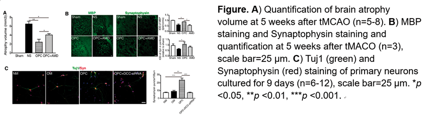
Conclusion
We showed that OPC transplantation reduced brain atrophy and promoted oligodendrogenesis and synaptogenesis in the sub-acute phase after ischemic injury via CXCL12/CXCR4 and Netrin-1/DCC axis, suggesting a new avenue for the treatment of ischemic stroke.
References
PB02-B13
Oligodendrocyte precursor cell transplantation protects blood-brain barrier integrity via Wnt/beta-catenin signaling after cerebral ischemia in mice
1Department of Neurology, Ruijin Hospital, School of Medicine, Shanghai Jiao Tong University, China
2Med-X Research Institute and School of Biomedical Engineering, Shanghai Jiao Tong University, China
Abstract
Objective:
Ischemic stroke increases blood-brain barrier permeability.1 Oligodendrocyte precursor cells can maintain the integrity of blood-brain barrier during the development.2 However, the function of oligodendrocyte precursor cell transplantation in blood-brain barrier permeability after ischemic stroke is unknown. Here, we investigate whether oligodendrocyte precursor cell transplantation protects blood-brain barrier integrity and promotes post-ischemic stroke recovery
Methods
Adult male ICR mice (n = 68) underwent 90 min transient middle cerebral artery occlusion. Ischemic mice received stereotactic injection of oligodendrocyte precursor cells (6 × 10⁵). Brain infarct volume, neurobehavioral outcomes, blood-brain barrier permeability, and β-catenin level were evaluated. β-catenin inhibitor and Wnt7a protein were used to explore the underlying mechanism.
Results:
Oligodendrocyte precursor cell transplantation alleviated infarct and edema, and promoted neurological recovery after focal ischemia (p < 0.05). Oligodendrocyte precursor cell transplantation reduced blood-brain barrier leakage via increasing claudin-5, occludin, Wnt7a and β-catenin expression compared to the control (p < 0.05). Administration of β-catenin inhibitor eliminated the beneficial effects of oligodendrocyte precursor cell transplantation. Wnt7a protein treatment increased β-catenin and claudin-5 expression in endothelial cells after oxygen glucose deprivation (p < 0.05), which was similar to the effect of the conditioned medium treatment of oligodendrocyte precursor cells on endothelial cells.
Conclusion:
Our results demonstrated that oligodendrocyte precursor cell transplantation protected blood-brain barrier in the acute phase of stroke via activating Wnt/β-catenin pathway of endothelial cells. Oligodendrocyte precursor cell transplantation promoted functional recovery in ischemic mice and was a novel therapy for ischemic stroke.
References
PB02-B14
Endothelial progenitor cell transplantation alleviated ischemic brain injury via reducing astrocyte-derived C3
1Department of Neurology, Zhongshan Hospital, Fudan University, Shanghai 200032, China
2Med-X Research Institute and School of Biomedical Engineering, Shanghai Jiao Tong University, Shanghai 200030, China
Abstract
Background
Complement activation is involved in the pathogenesis of ischemic stroke. Attenuating complement activation was crucial for the reduction of ischemic brain injury1. Studies showed that endothelial progenitor cell
Methods
We established transient middle cerebral artery occlusion model in adult male CD-1 mice (n = 111) and transplanted EPCs into the peri-infarct region immediately after middle cerebral artery occlusion. Modified neurological severity scores, brain infarct volume and atrophy volume were examined after brain ischemia. The expression of C3, C3aR and pro-inflammatory factors TNF-a, IL-1b, IL-6 were further examined to explore the role of EPCs on the induction of neuroprotection after focal ischemia.
Results
EPC transplantation reduced the modified neurological severity scores and brain infarct and atrophy volume in ischemic mice compared to the control (p < 0.05). C3 and C3aR expression in the ipsilateral hemisphere of the brain were up-regulated from day 1 up to days 14 (p < 0.05). EPC transplantation reduced astrocyte-derived C3 expression from day 3 to day 14 (p < 0.05). It was noted that C3aR expression at the day 14 was reduced in EPC group after ischemic brain injury (p < 0.05). EPC transplantation also reduced inflammatory cytokines TNF-a, IL-1b, IL-6 expression at 3 and 14 days after cerebral ischemia (p < 0.05).
Conclusions
EPC transplantation reduced astrocyte derived C3 expression in the brain after ischemic stroke, together with decreased C3aR and inflammatory response contributing to neurological function recovery. Our results suggest that modulating complement C3/C3aR pathway is a novel therapeutic target for the ischemic stroke.
References
PB02-B15
Regeneration-associated cell transplantation leads to tissue recovery in acute ischemic stroke mice
1Department of Neurology, Tokai University, Japan
2Department of Physiology, Tokai University School of Medicine
3Department of Regenerative Medicine Science, Tokai University School of Medicine
Abstract
Many cell-based therapeutic strategies have been investigated for vascular and tissue regeneration after ischemic stroke. We have developed a novel cell population, called regeneration-associated cells (RACs), by quality- and quantity-controlled culture of unfractionated mononuclear cells. The trans-arterially injections of RACs were performed into 10-week-old syngeneic male mice at 1, 3, 5 or 7 days after permanent middle cerebral artery occlusion (MCAO) due to find the optimal timing for administration in terms of outcome at day 21. Next, we tested the effects of RACs injection at day 1 after MCAO on neurological deficits, infarct volume, and mediators of vascular regeneration and anti-inflammation at days 7 and 21. Infarct volume at day 21 was significantly decreased by transplantation of RACs at day 1 or 3. RACs injected at day 1 decreased the infarct volume at day 7 and 21. Angiogenesis and anti-inflammatory mediators, VEGF and IL-10, were increased at day 7, and VEGF was still maintained at day 21. We also examined significantly enhanced ink perfusion in vivo, tube formation in vitro, and definitive endothelial progenitor cell colonies in colony assay. These results pointed out that RAC transplantation in MCAO models promoted significant recovery of neural tissues through intensified anti-inflammatory and angiogenic effects.
PB02-B16
Clot derived contaminants in transplanted cells enhance inflammation at peri-stroke area after bone marrow mononuclear cell transplantation in murine stroke model
1Dept. of Regenerative Medicine Research, Institute of Biomedical Research and Innovation, Hyogo, Japan
2Fraunhofer Research Institution for Marine Biotechnology and Cell technology(EMB), Luebeck, Germany
3Fraunhofer Institute for Molecular Biology and Applied Ecology IME – ScreeningPort, Hamburg, Germany
Abstract
Objectives
Hematopoietic stem cell transplantation is known to induce angiogenesis at ischemic tissue followed by functional recovery1. However, the potential of stem cell transplantation is not always fully exploited and significant numbers of clinical trial, especially transplantation of cells isolated by automatic cell separator that has no function to remove clot derived cells, failed to show its therapeutic effect. The purpose of this study is to investigate the factors that inhibit the effect of hematopoietic stem cell transplantation on tissue repair.
Methods
The results of previous clinical trials were re-examined in order to identify the underlying cause of the poor outcomes of autologous bone marrow mononuclear cell transplantation. To investigate the difference in the effect on stroke outcome between cell preparation procedure, bone marrow mononuclear cells were isolated manually or using automatic cell separator and injected intravenously into mice at day 2 after induction of stroke. Clot derived cells were also injected intravenously into post-stroke mice and investigated its effects on activation of inflammatory responses.
Results
Patients who received bone marrow cells contaminated by a significant number of platelets and red blood cells, mainly originated from clots formed at the buffy coat, showed little functional recovery. Bone marrow cells separated by an automatic cell isolator enhanced inflammatory response at peri-stroke area and did not attenuate brain atrophy after stroke, though manually isolated cells did not enhance inflammation and showed its therapeutic effects. Injection of clot derived cells also enhanced inflammation at peri-stroke area.
Conclusions
Enhanced inflammatory response after stroke is known to be one of the negative factors on stroke outcome. Bone marrow aspiration fluid contains various cells/substances, including connective tissue, bone fragment and lipids, and these substances trigger clot formation. Our findings indicated that clot-derived contaminants in transplanted cells is one of the factors that impairs the effect of the cell therapy and can explain the series of negative results of clinical trials, especially those that make use of an automated stem cell separator which lacks the ability to remove clot-derived contaminants.
Reference
PB02-B17
A novel regenerative associate cells transplantation may be applied in patients with acute ischemic stroke
1Dept. of Neurology, Tokai University, Japan
2Dept. of physiology, Tokai University, Japan
3Dept. of regenerative medicine science, Tokai University, Japan
Abstract
Purpose
Treatment of acute ischemic stroke (IS) has largely been limited to strategies to restore blood flow. Pharmacological recanalization and mechanical embolectomy have been the mainstay for acute treatment. However, the short time frame for safe intervention is limited. Hence, it needs to be established a novel therapy targeted on neuroprotection and angiogenesis. We are developing autotransplantation of regenerative associate cells (RAC) for acute ischemic stroke patients. RAC are mononucleosis group and they include endothelial progenitor cells (EPC). We cultivated these cells by Quality and quantity (QQ) culture, which we uniquely developed. QQ culture brings up EPC with high capacity of angiogenesis, and provides extremely optimal environment for angiogenesis by proliferation of anti-inflammatory macrophages and regulatory T cells. Here, we investigated availability of QQ culture at IS patients.
Subjects and Methods
Twenty-five acute IS patients who admitted our hospital were recruited in the study. Peripheral blood sampling were collected at less than three days after IS (group day3) and approximately ten days after IS (group day10). Blood were cultivated in QQ culture, and RAC were analyzed by the capacity of colony formation and flow cytometry. CD206, CD34, CD133 were used for cell surface marker to assess differentiation function and kinetics of RAC. 24 healthy people underwent the same analysis.
Results
CD206 positive cells decreased in group day3, but we could get high quality RAC as healthy people by QQ culture. Contrary to it, CD133 positive cells decreased after culture in group day3.
Conclusion
We could get high quality RAC by QQ culture in day3 and day10 after IS onset as well as healthy people. We believe that the study can develop to the realization of RAC autotransplantation as a clinical trial.
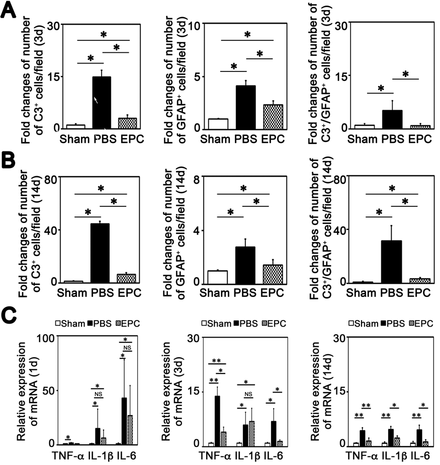
PB02-B18
Preliminary data of phase I clinical trial of intracerebral transplantation using bone marrow stromal cell (BMSC) against acute ischemic stroke (RAINBOW study)
1Dept. Neurosurgery, Hokkaido University, Japan
2Dept. Neurosurgery, Toyama University, Japan
Abstract
Background
Recent breakthrough in cell therapy is expected to reverse the neurological sequelae of stroke. Prior studies have demonstrated that bone marrow stromal cells (BMSCs) have therapeutic potential against stroke, however, there are several problems remain unsolved. In this study, we investigated the use of autologous BMSC transplantation for acute ischemic stroke with several new aspects as a next-generation cell therapy for treating stroke. This study is called the Research on Advanced Intervention using Novel Bone marrOW stem cell (RAINBOW, UNIN ID: UMIN000026130).
Methods/Design
RAINBOW is a phase 1, open-label, uncontrolled, dose-response study, with the primary aim to determine the safety of the autologous BMSC administered to the patients with acute ischemic stroke. Estimated enrollment is 6–10 patients suffering from moderate to severe neurological deficits. Approximately 50 mL of the bone marrow is extracted from the iliac bone of each patient 15 days or later from the onset, and BMSCs are cultured with allogeneic human platelet lysate (PL) as a substitute for fetal calf serum and are labeled with superparamagnetic iron oxide for cell tracking using magnetic resonance imaging (MRI). BMSCs are stereotactically administered around the area of infarction in the subacute phase. Each patient will be administered a dose of 20 or 50 million cells. Neurological scoring, MRI for cell tracking, 18F-fuorodeoxyglucose positron emission tomography, and
Discussion
This is a first-in-human trial to use labelled BMSC to the patients with acute ischemic stroke. We expect that intraparenchymal injection can be a more favorable method for cell delivery to the lesion and improvement of the motor function. Moreover, it is expected that the bio-imaging techniques can clarify the therapeutic mechanisms.
PB02-B19
The international cooperative Phase II trial of modified stem cells (SB623) transplantation therapy for traumatic brain injury -Participation in a double-blind, controlled study (NCT02416492)-
1Department of Neurosurgery, JCHO Tokyo Shinjuku Medical Center, Tokyo, Japan
2Department of Neurosurgery, The University of Tokyo Hospital, Tokyo, Japan
Abstract
Background
This trial was conducted for patients with traumatic brain injury (TBI) to evaluate the safety and efficacy of stereotaxic intracranial administration of allogenic modified bone marrow-derived mesenchymal stem cells (SB623) developed by “ SanBio Co., Ltd “. In Japan, five institutions including the University of Tokyo Hospital participated in the trial within a total of 27 facilities including overseas.
Clinical design
The subjects were TBI patients who had more than 1 year of persisting motor deficits. The damaged brain area causing dysfunction was confirmed by MRI or CT and their Glasgow outcome score ranged 3-6 points (i.e. moderate or severe disability), having Motricity Index 10-81 (UE Scale) and/or 10-78 (LE Scale). A total of 61 subjects were enrolled from Japan, the US, and Ukraine. Subjects underwent stratified randomization using baseline GOS-E score to assign in a 1:1:1:1 ratio to either 2.5x10⁶, 5.0x10⁶, 10x106 SB623 cells or surgical sham procedure. Safety was also assessed in all subjects. Efficacy analyses were conducted on the modified intent-to-treat population of randomized subjects completing surgery (N = 61). Primary efficacy outcome was mean change from baseline in the Fugl-Meyer Motor Scale score (FMMS) of SB623-treated subjects compared to controls at 24 weeks.
At the University of Tokyo Hospital we managed a total of 4 male cases (from22 to 36 years old). Surgical treatment is performed by a neurosurgeon in accordance with the case assignment. SB623 were transplanted into the brain around the injured area using stereotaxic surgery for patient allocated to SB623 group. Other medical staff, as well as patients, were blinded to the allocation results. The motor function evaluation was done by a rehabilitation specialist.
Results
Since the observation period is under way for 1 year, the results are still veiled. At 24 weeks the study met its primary endpoint showing a statistically significant improvement: SB623 patients achieving an average 8.7 point improvement from baseline in the FMMS, versus 2.4 in the control group. The safety data showed that SB623 was well tolerated and no new safety warnings were detected.
Discussion
Participating in a global clinical trial, a total of 4 cases with TBI including the first case in Japan was treated in our hospital. Indeed the result is promising. However, regarding the mechanisms of improvement of motor function, we do not got any evidence except for some speculations. It is critical to analyze the data in the broadest aspect and consider all possible functional interactions in the central nervous system. Based on this experience, we would like to discuss the current status and some specific issues of regenerative medicine.
Reference
PB02-C01
The influence of acute and chronic dietary sodium on cerebrovascular reactivity
1Kinesiology & Applied Physiology, University of Delaware, Newark, DE, USA
2Department of Pharmacology, Physiology & Neuroscience, Rutgers University, Newark, NJ, USA
Abstract
Acute (3) and chronic (4) high dietary sodium (Na+) transiently reduces peripheral blood vessel function in humans, but it is unknown whether dietary Na+ affects cerebral blood vessel function. Rodent models demonstrate that high dietary Na+ intake reduces middle cerebral artery (MCA) function (2). A cerebrovascular reactivity test (CVRT) to increased carbon dioxide (CO2; hypercapnia) is a validated method for assessing the cerebrovasculature in humans (1). However, the effects of acute and chronic high dietary Na+ on CVRT in humans is unknown.
Objective
We tested the hypothesis that acute and chronic high dietary Na+ impairs CVRT in young adults.
Methods
For the acute study, 20 healthy adults (11F/9M, age: 25 ± 4 yrs; BMI: 24 ± 1 kg/m2; BP: 105 ± 2/58 ± 2 mmHg, mean ± SD) were provided either a low- (LSM: 138 mg Na+) or high- (HSM: 1,495 mg Na+) Na+ meal in two randomized visits separated by one week for males. Females were tested during the early follicular phase of their menstrual cycle. For the chronic feeding study, three healthy adults (3 M, age 23 ± 1 yrs; BMI: 24 ± 2 kg/m2; BP: 113 ± 6/58 ± 7 mmHg) underwent ten days of low- (LSD:1 g Na+ /day), medium- (MSD: 2.3 g Na+ /day) or high- (HSD: 7 g Na+ /day) Na+ diets, in a randomized order. The diets were separated by a month. Urinary Na+ excretion was measured to ensure compliance. For both studies, participants laid supine while transcranial Doppler was used to assess right MCA velocity. Serum Na+ and CVRT (%MCA velocity/ΔCO2) to hypercapnia was measured. For the acute study, we assessed serum Na+ and CVRT at baseline, 30 minutes post- and 60 minutes post-meal. Data were analyzed using two-way repeated measures ANOVA (meal x time). For the chronic feeding study, we assessed serum Na+ and CVRT on the tenth day of each diet. Data were analyzed using one-way repeated measures ANOVA. Tukey post hoc comparisons were used when appropriate.
Results
In the acute study, serum Na+ was elevated at 30- (LSM:140.0 ± 0.5 vs HSM:141.2 ± 0.3 mmol/L, p < 0.01) and 60-minutes post meal (LSM:140.2 ± 0.6 vs HSM:141.7 ± 0.8 mmol/L, p < 0.001) following the HSM. Despite elevated serum Na+, the HSM did not alter CVRT to hypercapnia (30 min- LSM: 2.7 ± 0.9 vs HSM:2.5 ± 1.0%/mmHg; 60 min- LSM:2.6 ± 0.7 vs HSM:2.8 ± 0.8 %/mmHg, p > 0.05). In the chronic study, although not statistically significant, 24-hour urinary Na+ excretion increased in stepwise fashion from the low, to the medium, to the high Na+ diet (LSD: 45 ± 23, MSD: 138 ± 44, HSD:350 ± 220 mmol, p = 0.18). Serum Na+ was not different amongst the three diets (LS:138 ± 1.0 vs MS:139 ± 0.3 vs HS:140.0 ± 1.8 mmol/L, p = 0.60). There was a significant effect of the diet (p = 0.04) on CVRT to hypercapnia (LSD:2.0 ± 0.1 vs MSD:1.7 ± 0.1 vs HSD:2.8 ± 0.4 %/mmHg).
Conclusion
These data suggest that a high sodium meal does not acutely influence cerebrovascular reactivity and the preliminary chronic data projects that chronic high sodium consumption may alter cerebrovascular reactivity to hypercapnia in young adults.
References
PB02-C02
Blood pressure profiles obscure pulsatility and resistance indices when detecting cerebrovascular regulation differences between brain stem and cortex circulations during sit to stand
1Dept. of Pharmacology, Physiology & Neuroscience, New Jersey Medical School, Rutgers University, Newark, NJ, USA
2War Related Illness and Injury Study Center, Dept. of Veterans Affairs, East Orange, NJ, USA
3National University of Ireland Galway, Ireland
Abstract
Differences in the regulation of blood flow in the various vascular beds within the human brain is an important area that may provide relevant clinical information for assessing cerebrovascular disease risk. The pulsatility and resistance indices (PI and RI) are used clinically in many vascular beds (e.g. umbilical cord, fetal brain, salivary glands, etc.) to assess physiological state; however, these measures are significantly impacted by blood pressure (BP) profiles. Thus, in order to study the relationship between PI/RI and cerebral flow in various vascular beds we compared responses during an induced BP drop during repeated sit to stand maneuvers. We recorded the cerebral blood flow velocity (CBFV) of anterior, middle, and vertebral arteries (ACA, MCA, and VA) simultaneously using transcranial Doppler. The BP, heart rate, and end-tidal CO2 (ETCO2) were also continuously monitored in 27 young healthy subjects (age 30 ± 11 yrs; 14 females). All subjects repeated the sit to stand maneuver (3-min sitting followed by 1-min standing) three times. We calculated cerebral PI = (Systolic CBFV − Diastolic CBFV)/Mean CBFV, and RI = (Systolic CBFV − Diastolic CBFV)/ Systolic CBFV; as well as the BP PI profile (BPPI) = (Systolic BP − Diastolic BP)/ Mean BP, and BP RI profile (BPRI) = (Systolic BP − Diastolic BP)/ Systolic BP. The second-by-second responses to standing are presented in Figure 1 (Mean ± SEM), with initiation of stand at the 60th second. The Tiecks dynamic autoregulatory index (ARI), static CBFV and cerebral vascular resistance (CVR) responses (Δ%nCBFV and Δ%nCVR) were also calculated (Note: nCBFV = normalized CBFV = the percentage ratio of the mean CBFV to the baseline time-averaged value, nCVR = normalized CVR = Mean BP/nCBFV); and the static values were calculated from the 30th-55th (seated) and 90th-105th (standing) second time-windows (Figure 1). The following values of results are presented in Mean ± SD. The ARI was greater in the VA (4.1 ± 1.6) than the ACA and MCA (3.0 ± 1.7 and 2.9 ± 2.1; P < 0.01), suggesting improved autoregulation. Similarly, the CBFV was better maintained when standing in the VA (Δ%nCBFV, −0.2 ± 6.5%) than in ACA and MCA (−2.8 ± 6.3% and −2.9 ± 6.5%, P < 0.05), without sex difference. Similarly, the CVR increase was lower in the VA (Δ%nCVR, 1.2 ± 5.7) than in ACA and MCA (3.9 ± 5.6% and 4.1 ± 6.8%, P < 0.05) (Figure 1 A-B). The standing also produced a significant decrease in ETCO2 (−1.1 ± 1.2 mmHg). These data suggest that VA CBFV was better maintained when standing, suggesting that cerebral autoregulation may be better in the brain stem circulation than in the cortex circulation. However, we did not see any differences in PI and RI responses across the three vessels, suggesting they are primarily driven by their BP profiles, as the shapes of their responsive waveforms are very similar (Figure 1 D-F). These results collectively suggest the interpretation of PI and RI results may not be sensitive enough to pick up regional differences in cerebral blood flow regulation characteristics, whether examining dynamic or static response.

PB02-C03
Characterization of effects of head of bed position changes on the pulsatility of blood flow measured by fast diffuse correlation spectroscopy
1HemoPhotonics S.L., Castelldefels (Barcelona), Spain
2ICFO-Institut de Ciències Fotòniques, The Barcelona Institute of Science and Technology, Barcelona, Spain
3San Raffaele Scientific Institute, Neurology Department, Milan, Italy
4Children's Hospital of Philadelphia, Division of Neurology, Philadelphia, USA
5Institució Catalana de Reserca i Estudis Avançats (ICREA), Barcelona, Spain
Abstract
Objectives
Various conditions such as acute ischemic stroke, chronic obstructive sleep apnea and traumatic brain injury can lead to alterations in the cerebral vasoreactivity. The microvascular cerebral blood flow (CBF) response to head of bed (HoB) position changes measured with slow devices (≤1 Hz) has been shown to be a biomarker related to these alterations [1-3]. Recent developments allow for fast diffuse correlation spectroscopy (DCS) measurements (∼30-100 Hz) to resolve pulsatile CBF [4]. In this work, we present the effect of HoB position changes on the pulsatility of the fast blood flow signal and estimations of the critical closing pressure (CrCP) in healthy volunteers. Ultimately, this may allow for the introduction of new biomarkers to evaluate cerebral health, for prognostics and therapy planning.
Methods
The pulsatile blood flow was measured on the frontal lobes of twenty-one healthy volunteers during a HoB challenge while metronome guided controlled breathing using fast DCS. Various physiological parameters such as blood pressure, heart rate and respiratory parameters were monitored continuously alongside the fast DCS measurement. Based on the beat-to-beat signal of CBF and blood pressure, the CrCP was calculated [5].
Results
The changes in the CBF as well as the additional physiological changes during the HoB challenge are in accordance with the literature. The changes of CrCP are shown in Figure 1 demonstrating a statistically significant drop in CrCP in the 30° position compared to the baseline in supine position. As expected, it has returned back to the baseline values in healthy subjects.
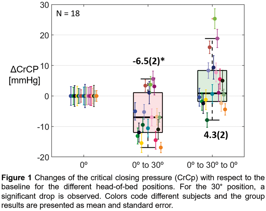
Conclusions
Pulsatile cerebral blood flow can be measured by fast diffuse correlation spectroscopy during HoB position changes and be analyzed at heart rate and respiratory rate. Furthermore, the fast signal allows to derive an estimate of CrCP. This has implications in understanding the physiological basis of the CBF response to HoB alterations and, when combined with traditional slower measures, may turn out to be a more precise biomarker of cerebral well-being.
Acknowledgements & disclosures
European Union’s Horizon 2020 project “BitMap: Brain injury and trauma monitoring using advanced photonics” (No. 675332), Fundació CELLEX Barcelona and the Obra social “laCaixa” Foundation (LlumMedBcn). UW is the CEO, has equity ownership in HemoPhotonics S.L. and UW, JF are employees. Their role has been defined by the BitMap project and was reviewed by the European Commission.
References
PB02-C05
The gender difference of cortical oxygenation in prefrontal cortex and motor-related area during cardiopulmonary exercise test
1Graduate School of Health and Welfare, Niigata University of Health and Welfare
2Institute for Human Movement and Medical Science, Niigata University of Health and Welfare
Abstract
Objectives
Previous studies using near-infrared spectroscopy (NIRS) during incremental load exercise have shown significant increases in oxygenated hemoglobin (O2Hb) in the prefrontal cortex (PFC) and in deoxygenated hemoglobin (HHb) in the motor cortex for males (Jung et al., 2015). Previous studies on gender differences in cortical oxygenation during incremental load exercise showed significantly higher O2Hb and HHb in males than in females (Auger et al., 2016). However, gender differences in cortical oxygenation were not investigated in multiple cortical areas. This study investigated gender difference in cortical oxygenation changes in the PFC and motor-related areas during cardiopulmonary exercise test (CPX).
Methods
Twenty-four male (age, 20.7 ± 0.4; weight, 64.7 ± 2.0; height, 172.2 ± 5.9) and 27 female (age, 20.3 ± 0.6; weight, 51.2 ± 5.2; height, 159.0 ± 5.2) healthy young adults participated in this study. CPX was performed using a cycle ergometer, involving incremental loading, corresponding to 20 watts/min, after 4-min of rest and warming-up (W-up) each. Cortical O2Hb and HHb were measured using NIRS (LABNIRS, Shimadzu) during CPX. The regions of interest were the right (R-PFC) and left PFC (L-PFC), supplementary motor area (SMA), and primary motor cortex (M1). We subsequently determined anaerobic threshold (AT), respiratory compensation (RC), and peak oxygen uptake (Peak) points using previously reported methods (Wasserman et al. 1964) using an exhaled gas analyzer (AE-310, Minato Medical Science). O2Hb and HHb differences from average of rest were calculated using average values over 4-min of rest and W-up, and those over 5 seconds at the AT, RC, and Peak points. Average O2Hb and HHb values at rest, W-up, AT, RC, and Peak points were compared between males and females using analysis of variance for split-plot factorial design of time factor (rest/W-up/AT/RC/Peak) and gender (male/female). When a significant main effect was identified, a post hoc analysis was conducted using Student's t-test or Mann-Whitney's U test.
Results
O2Hb in the L-PFC was significantly higher (p < 0.05) at W-up, AT, RC, and Peak points in males than in females. O2Hb in the R-PFC, SMA, and M1 was significant higher (p < 0.05) at the AT, RC, and Peak points in males than in females. HHb in the L-PFC and R-PFC was significant higher (p < 0.05) at the Peak points in males than in females. HHb in the SMA and M1 was significant higher (p < 0.05) at the AT, RC, and Peak points in males than in females.
Conclusions
O2Hb and HHb in the L-PFC, R-PFC, SMA, and M1 showed gender differences. HHb gender differences in motor-related areas from the AT to Peak points were different from those in the L-PFC and R-PFC.
References
PB02-C06
Sympathoexcitation during the cold pressor test does not result in cerebral vasoconstriction in young, healthy individuals
1Department of Pharmacology, Physiology and Neuroscience, Rutgers, The State University of New Jersey, Newark, NJ, USA
2War Related Illness and Injury Study Center, New Jersey Veterans Affairs Healthcare System, East Orange, NJ, USA
Abstract
Objectives
Sympathetic neural control of the cerebral vasculature in humans is still highly debated1. We investigated whether the sympathoexcitatory cold pressor test elicited vascular constriction in the internal carotid artery (ICA), middle cerebral artery (MCA), and anterior cerebral artery (ACA) of 19 young, healthy participants (ICA and MCA: n = 12 male; 7 female; ACA: n = 7 male; 5 female).
Methods
Participants were instructed to place their hand in ice water (∼4°C) for up to 3 minutes during continuous internal carotid duplex ultrasonography, transcranial Doppler ultrasonography of the MCA and ACA, blood pressure (Finapres), ECG, and end-tidal CO2. Participants also underwent a cerebrovascular reactivity test where they breathed a gas mixture containing 5% CO2, 21% O2, and balanced nitrogen for 2 minutes followed by 2 minutes of mild hyperventilation. The slope between end-tidal CO2 and MCA and ACA velocities were used as an index of cerebrovascular reactivity. To account for end-tidal CO2 changes during the cold pressor test, individual cerebrovascular reactivity indices for the MCA and ACA were used to correct cerebral flow velocity. Since we did not obtain cerebrovascular reactivity of the ICA, the average MCA and ACA reactivity indices were used to correct ICA blood flow. The percent change in ICA flow, MCA and ACA velocities between baseline and the cold pressor were used to assess the effects of sympathoexcitation on cerebral blood flow.
Results
Preliminary results show a significant increase in mean arterial pressure during the cold pressor test (men = +16.8 ± 4.2 mmHg; women = +14.6 ± 2.2 mmHg; p < 0.001). There were non-significant increases in MCA velocity (men = +5.1 ± 13.6% vs women = +5.5 ± 13.3%; p = 0.119), ACA velocity (men = +0.3 ± 24.0% vs women = +4.1 ± 11.9%; p = 0.718), and ICA blood flow (men = +1.7 ± 15.5% vs women = +0.8 ± 14.0%; p = 0.728) when participants placed their hand in ice water. There was a trend for decreased end-tidal CO2 during the cold pressor (men = -2.2 ± 0.9 mmHg; women = -0.4 ± 0.6 mmHg; p = 0.067). When correcting for end tidal CO2 changes, there was a significant increase in MCAv (men = +8.2 ± 10.0%; women = +7.2 ± 13.0%; p = 0.01), a non-significant increase in ACAv (men = +6.9 ± 20.4%; women= +7.2 ± 12.9%; p = 0.205), and a non-significant increase in ICA blood flow (men = +6.2 ± 12.9%; women = +4.3 ± 15.7%; p = 0.133). Increases were not significantly different between the ICA and MCA or ACA (MCA: p = 0.460; ACA: p = 0.526). There was no significant difference in ICA diameter between baseline and the cold pressor test (baseline: men = 5.2 ± 0.5 mm; women = 4.6 ± 0.5 mm; cold pressor: men = 5.3 ± 0.6 mm; women = 4.6 ± 0.5 mm; p = 0.686).
Conclusions
Based on these findings it appears that sympathoexcitation results in increased blood flow in the ICA, MCA and ACA possibly attributed to increased cortical activity such as sensory and pain stimuli associated with the hand submerged in ice water. Since ICA flow increased and the diameter remained unchanged, the increased MCA and ACA velocities cannot be explained by cerebral vasoconstriction.
Reference
PB02-C07
Veterans with gulf war illness and small fiber neuropathy demonstrate worse cerebral blood flow regulation
1Dept. of Pharmacology, Physiology & Neuroscience, Rutgers New Jersey Medical School, Newark, NJ, USA
2War Related Illness and Injury Study Center, Dept. of Veterans Affairs, East Orange, NJ, USA
3National University of Ireland Galway, Ireland
4Dept of Neurology, Massachusetts General Hospital, Harvard Medical School, Boston, MA, USA
Abstract
Introduction
Gulf War Illness (GWI) affects ∼20% of veterans who returned from the 1990-91 conflict with symptoms including fatigue, pain and cognitive difficulties. Recent evidence suggests impairment of autonomic funciton1 and cerebral autoregulation.2 There is conflicting evidence on levels of peripheral neuropathy in these veterans. The goal of this work is to examine if veterans with GWI with co-morbid small fiber peripheral neuropathy (SFPN) demonstrate impairments in cerebral blood flow regulation.
Methods
28 Veterans (4 females; Age 55 ± 7 yrs) with GWI as determined by Kansas questionnaire3 participated. To determine if veterans had SFPN, a skin biopsy was performed on the distal leg, 10 cm above the lateral malleolus using local anesthesia to determine epidermal nerve fiber density. Densities <5th centile of predicted laboratory norms were categorized as SFPN. Seven veterans were classified as SFPN based on significantly reduced nerve fiber density (0.3 ± 0.8%, P < 0.001) compared to 17 classified as normal (64.5 ± 20.4%). Four were borderline and excluded from this analysis. We recorded cerebral blood flow velocity of the middle cerebral artery (MCA) using transcranial Doppler, as well as blood pressure (finometer), heart rate, and end-tidal CO2. To examine cerebral blood flow regulation, veterans performed three sit to stand maneuvers (3-min sitting followed by 1-min standing). To assess cerebrovascular response to changes in end-tidal CO2, veterans breathed room air, followed by 8% CO2, 21% O2, balance nitrogen and then hyperventilated to reduce end tidal CO2 ∼10 mmHg.
Results
Veterans with GWI + SFPN demonstrated reduced autoregulation index (3.3 ± 1.6) compared to GWI (5.9 ± 1.7). Upon standing, the GWI + SFPN and GWI groups demonstrated similar decreases in blood pressure (−21 ± 6 vs −18 ± 6 mmHg), however MCA flow velocity was significantly lower in the GWI + SFPN group (87.3 ± 2.2% of sitting baseline) compared to the GWI group (93.4 ± 6.6%, P = 0.05). Steady state values calculated as the average 30-55 sec after standing were not different between groups. Cerebrovascular reactivity to CO2 was also not significantly different between groups (GWI + SFPN: 1.9 ± 0.5 %/mmHg vs GWI: 1.5 ± 0.4 %/mmHg). A linear regression between fiber density and autoregulatory index demonstrated a significant positive correlation (R = 0.498, P = 0.029).
Discussion
These preliminary results suggest that veterans with GWI and SFPN appear to have worse cerebral autoregulation but maintain intact cerebrovascular reactivity to CO2. The correlation between nerve fiber density and autoregulatory index suggests that progressive nerve loss is associated with increasing cerebral blood flow regulation dysfunction. However these data must be interpreted with caution due to the small number of veterans with SFPN participating in the study. This work was funded by the CDMRP GWI Research Program.
References
PB02-D01
Vascular reactivity modulation of isolated rat carotid arteries by novel c-Jun N-terminal kinase inhibitor IQ-1S
1Siberian State Medical University
2Kizhner Research Center, Tomsk Polytechnic University, Tomsk, Russia
3Cardiology Research Institute, Tomsk NRMC, Tomsk, Russia
4Research Institute of Biological Medicine, Altai State University, Barnaul, Russia
5Department of Microbiology and Immunology, Montana State University, Bozeman, USA
6Cardiovascular Research Center, Cardiology Division, Massachusetts General Hospital, Harvard Medical School, Charlestown, Massachusetts, USA
Abstract
Objectives
A novel dual NO-donating oxime and c-Jun N-terminal kinase (JNK) inhibitor IQ-1S was shown to protect against stroke injury in mice [1]. Objective of the study was to assess IQ-1S effects on the vascular reactivity of isolated rat carotid arteries.
Methods
Common carotid arteries were isolated from 11–13-week-old Wistar rats and endothelium was denuded. Modulating effects of IQ-1S (100 μM) on isometric force of the vascular rings were studied in conditions of normoxia, hypoxia, and reoxygenation by organ bath myography with Myobath II and LAB-TRAX-4/16 (Germany). All chemicals were obtained from Sigma Aldrich except IQ-1 S, which was synthesized, as described [2]. Reference constrictions taken for 100% were triggered by high-K+ (30 mM) depolarization (high-K+) or phenylephrine (1 μM). Impact of the following ion channel blockers on IQ-1 S effects were studied: L-type calcium channel blocker nifedipine (5 and 50 nM for phenylephrine- and high-K+-induced constrictions, respectively), non-selective blocker of potassium channel tetraethylammonium (TEA, 5 mM), and selective blocker of voltage-dependent potassium channels 4-aminopyridine (4-AP, 1 mM). All results are expressed as mean ± SD. Statistical analysis was performed with Mann-Whitney U test and Wilcoxon matched pairs test. Differences with p < 0.05 were considered significant.
Results
IQ-1S decreased amplitude of constrictions induced by high-K+ (normoxia: 66.3 ± 6.8%; hypoxia: 68.3 ± 4.0%; reoxygenation: 13.5 ± 4.5) and phenylephrine (normoxia: 45.5 ± 9.4%; hypoxia: 15.3 ± 6.3%; reoxygenation: 4.2 ± 1.6%, n = 6, p < 0.05 for differences between oxygenation conditions). Nifedipine enhanced IQ-1S-mediated relaxation at high-K+ (normoxia: 43.3 ± 7.9%; hypoxia: 30.3 ± 7.5%; reoxygenation: 2.01 ± 1.1%) and phenylephrine (normoxia: 22.9 ± 5.1%; hypoxia: 9.0 ± 7.3%; reoxygenation: 2.5 ± 2.3%, n = 6, p < 0.05 for differences between oxygenation conditions). TEA attenuated IQ-1S-mediated relaxation in the presence of high-K + (normoxia: 75.6 ± 6.1%; hypoxia: 87.4 ± 8.3%; reoxygenation: 69.5 ± 7.5%) and phenylephrine (normoxia: 52.8 ± 5.7%; hypoxia: 65.7 ± 9.4%; reoxygenation: 40.8 ± 10.1%, n = 6, p < 0.05 as compared with the corresponding values without TEA). In normoxia, 4-AP attenuated IQ-1S-mediated relaxation of carotid arteries contracted by high-K+ (75.2 ± 1.5%, n = 6, p < 0.05 as compared with the corresponding values without 4-AP). 4-AP decreased IQ-1S-mediated relaxation (83.6 ± 10.7% and 56.7 ± 8.3%) in hypoxia and reoxygenation (93.4 ± 10.3% and 48.11 ± 8.1%) compared to high-K+- and phenylephrine-induced contractions, respectively (p < 0.05).
Acknowledgements
The study was supported by the Ministry of Education and Science of the Russian Federation (No. 4.8192.2017/8.9); the study of hypoxia/reoxygenation responses was supported by the Russian Science Foundation (No. 17–15-01111).
References
PB02-D02
Inhibition of retinal and cerebral microvascular glycogen utilization promotes capillary constrictions in mice
1Institute of Neurological Sciences and Psychiatry, Hacettepe University, Turkey
2Department of Neurology, Faculty of Medicine, Hacettepe University, Turkey
3Department of Psychiatry, Faculty of Medicine, Hacettepe University, Turkey
Abstract
Objectives
Glycogen stored in perivascular astrocyte processes may be the principal endogenous source of energy to prevent microvascular impairment under energy-deprived conditions. In retinal and cerebral ischemia, energy substrates alternative to decreased blood glucose may diminish the ischemia-induced capillary constrictions. To test this hypothesis, we studied the effects of preventing glycogen degradation by a glycogen phosphorylase inhibitor
Methods
Swiss albino mice (30-35 g) received DAB intracerebroventricularly (0.75 µL, 0.25 M, n = 3 for each group). After 0.5, 1, 3, 6, 9, and 24 hours animals were sacrificed. Another group was subjected to proximal Middle Cerebral Artery Occlusion (MCAo) by intraluminal filament model one hour after DAB injection (n = 3). To study retinas, DAB (3 μl, 2.5 M) was intravitreally injected. Two hours after DAB delivery, mice were either sacrificed (n = 3) or subjected to retinal ischemia (n = 3) for 30 min and then sacrificed. Coronal brain sections (50 µm-thick) and whole mount retinas were labeled with Lycopersicon esculentum Lectin (LEL) for visualizing vessels, and constrictions were quantified by a semi-stereological method (total online images of 10 3D-disectors (240 × 160 × 40µm) for each hemisphere/brain sections, spanning MCA territory and whole retinas.
Results
DAB injection caused significantly higher mean number of microvascular constrictions in 0.5, 1, 3, 9, 24 hours in ipsilateral (n/mm2±SEM; 327.3 ± 27, 283.7 ± 17, 312.1 ± 15, 225.0 ± 5, 195.2 ± 9) and contralateral (210.3 ± 43, 236.2 ± 4, 258.4 ± 14, 144.9 ± 29.0, 166.8 ± 3) hemispheres, when compared to intracerebroventricular vehicle injections (91.2 ± 7). One hour after DAB injection, 90 minutes of MCAo and 3 hours of recanalisation were performed, which significantly increased the number of constrictions (653.1 ± 48) compared to the non-ischemic hemisphere (467.4 ± 52); and corresponding 6th hour of DAB treatment (295.4 ± 52). In whole-mount retinas, DAB treatment significantly increased the number of constrictions 30 min after ischemia compared to the constrictions at 30 minutes in untreated ischemic retinas (571 ± 29 vs. 245 ± 20 constrictions, p < 0.001, ANOVA Tukey’s test).
Conclusions
These data suggest that inhibiting glycogen utilization by DAB induces capillary contractions and significantly aggravates the ischemia-induced microvascular constrictions.
Keywords
Cerebrovascular diseases, stroke, microvascular constrictions, glycogen,
Acknowledgement
Financially supported by Hacettepe University Scientific Research Projects Coordination Unit, Project Number: THD-2017-14025 and TDK-2019-17652.
References
PB02-D03
Pericytes maintain basal capillary tone and blood flow resistance in the brain
1Center for Developmental Biology and Regenerative Medicine, Seattle Children's Research Institute
2Department of Pediatrics, University of Washington
3Department of Neuroscience, Medical University of South Carolina
Abstract
More than 90% of the brain's vasculature is comprised of capillaries, optimized to allocate blood to all brain cells. Yet, the regulation of blood flow through complex capillary networks remains poorly understood. We used single-cell optical manipulations in vivo to examine the role of alpha-smooth muscle actin-negative pericytes, the predominant mural cell type covering brain capillaries. Optogenetic stimulation of single capillary pericytes led to decreased vascular diameter and blood flow, but with slower kinetics than mural cells of upstream pre-capillary arterioles. Conversely, ablation of single capillary pericytes led to prolonged local dilation and augmented blood cell flux. Ablation studies further suggest an intermingling of neighboring pericytes with differing levels of tone, which may create enduring patterns of flow in capillary networks. Capillary pericytes are thus sufficient and necessary to alter vascular diameter in vivo, and their slow dynamics suggest a role in setting tonic or slow modulating blood flow resistance in capillary beds.
PB02-D04
How activity-induced elevations of potassium affect calcium signals in pericytes
1Institute of Pharmacology and Toxicology, University of Zurich, Switzerland
2College of Pharmacy, Rady Faculty of Health Sciences, University of Manitoba, Canada
3Department of Neurosurgery, University Hospital Zurich, Switzerland
Abstract
Objectives
Central nervous system (CNS) pericytes are mural cells, embedded within the vascular basement membrane. They are important for structural support of the microvasculature and for the maintenance of the blood brain barrier. Together with astrocytes, neurons and endothelial cells they form the neurovascular unit (NVU) and are involved in brain hyperaemia, a fundamental mechanism for CNS homeostasis. Pericyte deficiency is linked to several diseases like diabetic retinopathy and to blood brain barrier breakdown. Despite their importance, little is known about intracellular signaling mechanisms in pericytes and their communication with NVU cells.
Methods
Using two-photon laser scanning microscopy we investigate calcium signals in CNS cortical pericytes in vivo and in ex vivo brain slices. We employ a mouse model, where the genetically encoded calcium indicator (GCaMP6s) is expressed under the pdgfrβ-promoter. To identify the underlying mechanisms of the pericyte calcium dynamics and their modulation, we applied several pharmacological interventions.
Results
We observed fast, spontaneous calcium transients in pericyte fine processes and cell bodies both in vivo and in the absence of blood flow ex vivo. SKF96365, a transient receptor potential canonical (TRPC) type channel antagonist, reduces spontaneous calcium events. This accentuates the need of extracellular calcium mediated through TRPCs to maintain basal calcium dynamics in pericytes. Although these basal calcium fluctuations are not affected by blocking neuronal activity with tetrodotoxin (TTX), we found that pericytes react to neuronal stimulation (in vivo, chemogenetic by DREADD, hM3Dq) with a decrease in intracellular calcium. This decrease in pericyte calcium levels can be reproduced in ex vivo brain slices by an increase of potassium (10 mM) in the superfusate. Further, we were able to block this potassium-mediated calcium decrease by antagonizing Kir6.1–channels (KCNJ8) with Glibenclamide. Kir6.1 and ATP binding cassette/SUR2B (ABCC9) subunits, which are highly transcribed in pericytes, can multimerize to form an ATP-sensitive potassium channel (KATP).
Application of potassium, a sodiumazide induced chemical ischemia or an acute hypoxic insult (both resulting in decreased intracellular ATP levels), lead to a decrease in pericytic calcium levels.
Conclusions
Our findings indicate that pericytes adapt their intracellular calcium levels to elevations of extracellular potassium, as is the case during neuronal stimulation and highlight a possible role of a KATP channel (KCNJ8 and ABCC9) in modulating pericyte calcium signals in response to stimuli.
PB02-D05
Isoflurane anesthesia rapidly blocks calcium activity in mouse brain vascular endothelial cells in vivo
1City University of New York, City College Biology Department and Center for Discovery and Innovation
2Columbia University, Chemistry Department
Abstract
Isoflurane is a fast acting volatile anesthetic widely used in small animal research settings. In humans, isoflurane exposure is linked to headaches, hypertension, cardiac, respiratory, cognitive and reproductive issues, which suggests that in addition to inhibit neural activity, isoflurane affects common targets in neural cells and other cell types such as vascular endothelial cells (VECs). We hypothesized that isoflurane inhibits the activity of brain VECs and disrupts the release of vasoactive signals involved in the regulation of cerebral blood flow. To begin to test this idea we performed thinned-skull transcranial two-photon imaging experiments in 3–8 week old double transgenic mice expressing Cre recombinase and the genetically encoded calcium indicator GCaMP6 under the control of the Tie2 promoter, and in mice that have stable expression of GCaMP8 driven by the Cdh5 promoter. We report that brain VECs in awake mice exhibit Ca2+ transients at frequencies that range from 1 mHz to 10 mHz, show localized and wave-like spatial patterns, and are not correlated with vessel vasodilation. Upon anesthesia induction (5% isoflurane), Ca2+ transients in brain VECs were partially or entirely blocked, failing to return to pre-exposure levels 10 minutes after isoflurane washout (Figure 1; n = 10 mice). In addition, isoflurane exposure induced changes in baseline fluorescence, suggesting that isoflurane can affect Ca2+ homeostasis directly, or alternatively, that anesthesia exposure changes Ca2+ levels indirectly through changes in sensory evoked neurovascular coupling. To test this idea we monitored Ca2+ transients in awake mice during pre-exposure normoxia (air), vehicle (O2 only), isoflurane (O2 + 5% isoflurane), and washout normoxia (air). We found that baseline fluorescence was the same in pre-exposure normoxia and vehicle conditions, while it decreased with an exponential time course during isoflurane exposure. Interestingly, baseline levels returned gradually and partially to pre-exposure levels within 5 minutes of washout normoxia supporting the interpretation that isoflurane affects vascular endothelial cell Ca2+ homeostasis directly (n = 3 mice). To the best of our knowledge, this study provides the first measurements of Ca2+ transients in brain VECs in vivo, demonstrates a direct inhibitory effect of isoflurane on Ca2+ homeostasis, and motivates future experiments to analyze the mechanisms by which isoflurane alters neurovascular physiology under minimally invasive conditions.
Supported by a Harvey L. Karp Award in the Sciences.
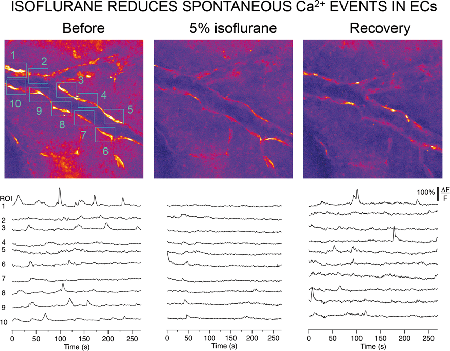
PB02-D06
Measurement of tissue PO2 in awake mouse cortex by using two-photon microscopy imaging
1Athinoula A. Martinos Center for Biomedical Imaging, Massachusetts General Hospital, Harvard Medical School, Charlestown, MA 02129, USA
2Departments of Biochemistry and Biophysics, University of Pennsylvania, Philadelphia, PA 19104, USA
3Department of Neurosciences, University of California San Diego, La Jolla, CA 09293, USA
4Department of Radiology, University of California San Diego, La Jolla, CA 09293, USA
5Departments of Biomedical and Electrical and Computer Engineering, Boston University, Boston MA 02215, USA
Abstract
Objectives
This study aims to characterize tissue pO2 in awake mice, free from confounding factors of anesthesia on the animal physiology including altering the blood flow, metabolism, and neuronal responses.
Methods
Two-photon phosphorescence lifetime microscopy was performed through a glass cranial window. The pO2 probe (Oxyphor2P, ∼3 µL, 30 µM) was delivered into the tissue by direct injection to the site via a pressured glass micropipette, using PicoSpitzer, through a silicone-filled port in the chronic glass cranial window (Fig. 1a). For imaging the microvasculature by the two-photon fluorescence microscopy, a dextran-conjugated Fluorescein isothiocyanate (FITC; ∼8.3 mg/mL, FD2000S, 2 MDa, Sigma Aldrich) was injected retro-orbitally to label the blood plasma. All injections were completed during brief isoflurane anesthesia, at least 1 hr before imaging. At each cortical depth, we measured pO2 over a grid of locations (500-1000 locations over 0.5 × 0.5 mm2 area) around arterioles, venules, and capillaries.
Results
We mapped the tissue pO2 distribution in the whisker barrel cortex of awake mice at rest across different cortical layers under normal physiological conditions, hypercapnia, hypoxia, and hyperoxia.
Conclusions
Our results will help better understanding the oxygen transport in cortical tissue at microvascular scales during normal and perturbed conditions, leading to improved understanding of the cerebral physiology, pathophysiology and more quantitative interpretation of the fMRI signals.
(a) Oxyphor2P injected with the glass micropipette through a silicone-filled port in the chronic sealed cranial window. The inset shows the bottom of the glass window that is partially inserted through the cranial window. Silicone-filled port is highlighted red for better visibility. (b) Tissue pO2 measurements in awake mouse. pO2 values around a diving arteriole are color-coded and overlaid over a bright field image of the pial surface. (c, d, and e) pO2 measurements around the same diving arteriole overlaid over two-photon images of the FITC dextran-labeled plasma at the pial surface (c), and at cortical depths of 100 µm (d) and 200 µm (e).
PB02-D07
The variability of cerebrovascular reactivity under different anaesthetics: relevance to preclinical fMRI
1Neuroimaging, IOPPN, King's College London
2Forensic and Neurodevelopmental Sciences, IOPPN, King's College London
Abstract
Objectives
Preclinical functional MRI (fMRI) in rodents is increasingly used to study brain physiology in combination with genetics, disease modelling or pharmacological manipulations. Because most fMRI paradigms utilise anaesthesia or sedation which affect brain activity, it is important to better understand the contribution of anaesthesia to the responses of interest1. Amongst most commonly used anaesthetics in rodent MRI are isoflurane and medetomidine – alone or in combination. A combination of low doses of medetomidine and isoflurane in particular has been hailed as an optimal regime for preserving the resting state brain activity in a mouse – a particularly challenging experimental paradigm2.
Here we aimed to test some of these anaesthetics for their effect on cerebrovascular reactivity (CVR). CVR is a change in cerebral blood flow (CBF) brought about by a vasoactive stimulus, such as vasodilation in response to hypercapnia (increase in partial pressure of CO2 in blood). Known to be affected by some diseases and drugs including anaesthetics, a normal CVR reflects cerebrovascular reserve and a homeostatically regulated vascular system3- prerequisites for the functional neurovascular coupling which underlies BOLD response in fMRI studies.
Methods
We measured changes in regional cerebral blood flow (rCBF) through intact skull over the parietal cortex in adult male C57Bl6 mice, induced through alternating air/oxygen (9:1, 1 L/min) and air/oxygen/CO2 (9:1:2, 1.2 l/min) delivered to animals via a facemask. A Laser Doppler flowmeter (Moore Instruments Ltd), interfaced with Biopac MP100, was used for data recording and analysis. All recordings comprised of 5 minutes baseline, 2 min of CO2 and 5 minutes post CO2 recordings. We tested 2-4 mice per group for the following conditions: urethane (1.4 mg/kg ip), isoflurane (1.5%), medetomidine plus isoflurane (0.5% isoflurane, 0.05 mg/kg sc bolus & 0.1 mg/kg/h sc infusion of med), medetomidine (0.1 mg/kg/h sc infusion, minimum 30 min after stopping isoflurane).
Results
We observed a robust and immediate (5-20 sec) ca. 100% increase from baseline of CBF under urethane (as expected based on existing in-house data), and a similar CVR under medetomidine in absence of isoflurane. Medetomidine plus isoflurane condition, however, resulted in an initial 20-30% decrease in CBF followed by an increase smaller than that of urethane or medetomidine alone (ca. 50%). CBF increase was also smaller (30-40%) and slower under isoflurane alone.
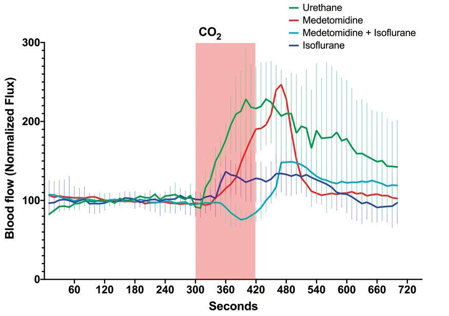
Conclusions
Striking differences in CVR were observed under four common anaesthetic regimes in mice. The CVR responses detected under urethane and medetomidine resembled those of neurotypical, non-anaesthetised brains. Isoflurane, on other hand, appeared to attenuate CVR which may be related to its known vasodilatory effect and impact on neurovascular de-coupling4. Experiments are underway to characterise CVR under additional regimes, and also to test the same protocols in rats. Such observed differences in CVR may reflect underlying differences in CBF regulation as well as in cerebrovascular coupling. Care should therefore be taken when choosing anaesthetic regimes for preclinical MRI, where effects on CVR should also be taken into account.
References
PB02-D08
Activation of smooth muscle TRPV1 channels in non-brain arteries contributes to cerebral blood flow maintenance during acute decreases in blood pressure
1Dept. of Pharmacology, University of Vermont, Larner College of Medicine, VT, USA
2Institute of Cardiovascular Sciences, University of Manchester, Manchester UK
Abstract
Objectives
Global cytosolic Ca2+ is a primary signal used by the contractile machinery of arterial smooth muscle (ASM) to translate humoral, metabolic, and neural signals into vascular responses that support the appropriate delivery of oxygen and nutrients throughout the body. In many vascular beds, including cerebral arteries, Ca2+ influx, and global cytosolic Ca2+ are primarily dictated by L-type voltage-dependent Ca2+ channel (VDCC) activity. We have found that a second major Ca2+ entry pathway—the TRPV1 channel— exists in ASM of non-brain arteries of the head. Here, we explored the hypothesis that TRPV1-induced constriction of non-brain head arteries tunes vascular resistance to shunt blood to brain arteries, which lack TRPV1 channels.
Methods
In vivo and ex vivo measurements of arterial diameter and ASM Ca2+ were obtained in cerebral arteries and non-brain arteries of the head from wild type (wt), TRPV1-KO and genetically-encoded Ca2+ indicator (TRPV1-GCaMP6f and SM-GCaMP6f) mice. Relative changes in CBF over a range of blood pressures were measured using laser Doppler flowmetry and estimates of actual CBF were obtained in separate animals using fluorescent-tagged polystyrene microspheres.
Results
Widespread functional expression of TRPV1 channels in ASM throughout non-brain arteries of the head was contrasted by a striking absence of these channels in the cerebral vasculature. Consistent with this differential expression, the TRPV1 agonist, capsaicin, constricted ex vivo pressurized middle meningeal arteries (MMAs) and facial arteries (FAs) from wild type (wt) mice (EC50 ≈ 30 nM), but did not constrict FAs from TRPV1-KO mice or cerebral arteries from either strain. Similarly, using two-photon in vivo imaging through a cranial window in anesthetized mice, we observed that capsaicin caused MMA constriction but did not directly impact cerebral artery diameter. Our data also demonstrate engagement of ASM TRPV1 channels by activation of α1-adrenergic receptors—the major vasoconstrictor pathway of the sympathetic nervous system. Importantly, we also found that pharmacological or genetic inhibition of TRPV1 channels causes a dramatic worsening of the maintenance of CBF during acute decreases in systemic blood pressure. To explore this pathway in a pathological setting, we examined CBF under conditions designed to mimic hemorrhagic shock. In this model, blood was withdrawn from mice to maintain a blood pressure of 40 mmHg. Remarkably, CBF dropped to a much greater extent in TRPV1-KO mice or wt mice treated with the TRPV1 antagonist capsazepine compared to un-treated wt mice.
Conclusions
Our findings suggest that activation of ASM TRPV1 channels constricts non-brain arteries of the head and promotes increased CBF. This mechanism is enabled by the selective expression of TRPV1 channels in extracerebral ASM and not within the brain vasculature. In response to acute decreases in blood pressure, engagement of ASM TRPV1 through activation of α1-adrenergic receptors may help shunt vital blood to the brain.
PB02-D09
No effect of the angiotensin receptor blocker candesartan on cerebrovascular autoregulation in rats during very high and low sodium intake
1Neurobiology Research Unit, Rigshspitalet, Copenhagen University Hospital, Denmark
2Department of Nephrology, Herlev Hospital, Copenhagen University Hospital, Herlev, Denmark
3Department of Intensive Care, Rigshspitalet, Copenhagen University Hospital, Denmark
4Institute of Molecular Medicine, University of Southern Denmark, Odense, Denmark
Abstract
Objectives
Autoregulation of cerebral blood flow (CBF) denotes that CBF is constant despite fluctuation of blood pressure within wide limits (figure). Inhibition of the renin-angiotensin system is known to decrease the lower and upper limits of CBF autoregulation. We have previously shown that this includes inhibition by the angiotensin receptor blocker (ARB) candesartan. In the present study we investigated the influence on the effect of the ARB candesartan on the lower limit CBF autoregulation in two groups of animals, one group with low and one with high renin levels induced by high and low salt intake respectively. It is hypothesized that a high salt intake/low renin activity would suppress or even abolish the effect of candesartan; while a low salt intake/high renin would conserve or even amplify the effect of candesartan blocking the effect of renin.
Methods
Two groups of Sprague Dawley rats were investigated, one on high (4.0% Na+) and one on low (0.004% Na+) sodium diet. Control animals were given the same diet, but no ARB. CBF was studied with the laser-Doppler method. Blood pressure was lowered by controlled bleeding.
Results
Both high and low sodium diet induced the expected changes in plasma renin, low and high levels respectively. The influence of candesartan on CBF autoregulation was blocked in both groups.
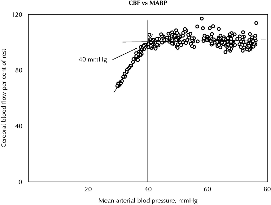
Conclusions
The findings were expected in rats on a high salt diet with a low renin level, but unexpected in rats with a low salt intake with a high renin level. The hypothesis could thus not be confirmed with regard to the low salt intake/high renin level since the lower limit of autoregulation did not decrease. One might speculate whether an activation of the sympathetic nervous system during low salt intake might contribute to the findings.
Reference
PB02-D10
Breakthrough threshold of intracranial pressure regulation
1Department of Neurosurgery, University of New Mexico
2Department of Pharmaceutical Sciences, University of New Mexico
Abstract
Introduction
Prevention of secondary injury after cerebrovascular accidents (CVA) or traumatic brain injury (TBI) is the only treatment option as there is no clinically effective neuroprotective strategy available. An important secondary injury process is high intracranial pressure (ICP) > 20 mmHg that could arrest perfusion to the entire brain. Early treatment paradigms beyond Tier 1 therapy (hyperventilation, osmotherapy, head up tilt, and CSF drainage) includes vasopressor induced increase in cerebral perfusion pressure (CPP) to maintain cerebral blood flow (CBF) which could reduce high ICP but could catastrophically increase ICP and lethal brain herniation. Precisely when the patient with high ICP is at this juncture in the injury process is unknown and clinicians are unable to predict it. Attempts have been made to identify this threshold in ICP by serum biomarkers inflammatory mediators TNF-a, IL-8, S100B and MMP-9. We
Methods
Male Long Evans rats 400-450 g were mechanically ventilated on 2% isoflurane/70% N2O/30%O2. Femoral artery and vein catheters were inserted. A catheter in the transverse sinus was used for measurement of blood gases and oxygen and glucose content for cerebral A-V oxygen and glucose content. ICP was increased from baseline of 10 mmHg by elevating a reservoir filled with mock CSF to achieve ICP values of 30, 40 or 60 mmHg with MRI T1, T2, DTI, BOLD and ASL CBF before and after a 10 min 5% CO2 challenge and measurement of cerebral venous and arterial blood gases and oxygen and glucose content with an IL GEM4000 (Bedford, MA). ADC, CBF and BOLD data were obtained and quantitated by voxel analysis.
Results
Induced ICP increase from a baseline of 10 to 60 mmHg resulted in a generalized reduction in CBF throughout the brain (Fig). Five % CO2 inhalation at ICP 10 mmHg increased CBF in 75% of the voxels and decreased it in 23% of voxels whereas at ICP 60 the increase was 37% and decrease 60% reflecting an attenuated cerebrovascular reserve at high ICP.
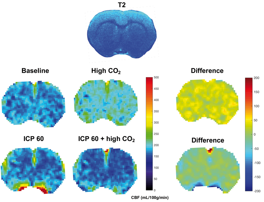
Conclusions
Increased ICP resulted in an increase in the volume of brain tissue with loss of CBF autoregulation. The relationship between ICP and volume of brain tissue with loss of CBF autoregulation may define the threshold where the application of ICP targeted therapy is indicated in the treatment of high ICP after brain injury.
PB02-D11
The long-term effects of systemic inflammation on hemodynamic response in the mouse cortex
1Center for Neuroscience Imaging Research, Republic of Korea
2Dept. of Biomedical Engineering, Sungkyunkwan University, Republic of Korea
3Samsung Advanced Institute for Health Science & Technology, Republic of Korea
4Biomedical Institute for Convergence at SKKU, Republic of Korea
Abstract
Objectives
Neuroinflammation is one of features in many neuronal disease. The persistence of neuroinflammation could lead to the damage of neuronal cell and eventually dysfunctional brain (1). Also, neuroinflammation contributed to the impairment of brain-blood barrier (BBB) and accelerated the pathological changes of neurovascular unit and its function (2). However, we barely know how the neuroinflammation actually alters hemodynamic response and neurovascular coupling (2, 3).
Method
Open-skull cranial window was implanted on the cortex of C57BL/6 mouse. At 4-weeks post cranial window surgery, lipopolysaccharide (LPS, 5 mg/kg, i.p.) was injected to the animal for the induction of whole-body systemic inflammation and therefore neuroninflammation. Intrinsic optical signal was recorded at before injection of LPS and at 4 hour, 2 weeks and 4 weeks after the injection of LPS with 570 nm band-pass filter following whole whisker stimulation by air-puffing (5 Hz, 4 s). The mice of control group were received saline. The other group was administered anti-inflammatory drug (Minocycline [22.5 mg/kg] + N-acetylcysteine [75mg/kg]) at 1 hour, 1 day and 2 day after the injection of LPS. Immunohistochemistry (IHC) were performed to confirm the expression of microglia and astrocyte.
Results
Differential hemodynamic response was observed before and after the injection of LPS within single animal. The hemodynamic responses after the LPS injection were largely different compared to the control group and anti-inflammatory treated group.
Conclusions
Systemic inflammation induced by LPS caused neuroinflammation in mouse brain and altered the hemodynamic response following sensory stimulation.
References
PB02-E01
A physiologically accurate model of cerebral autoregulation
1Institute of Biomedical Engineering, University of Oxford, United Kingdom
Abstract
Background
The metabolic demand of the tissue is one of the key drivers of cerebral blood flow (CBF) regulation. Indeed, a low oxygen level within the tissue will call for an increase in blood perfusion. The precise mechanism of demand and delivery has not clearly been defined, yet it is becoming clear that both capillaries and arterioles are involved. Recent research has underlined the importance of pericytes, isolated contractile cells found on capillary vessels, that induce capillary vasodilation at a rate faster than the arterioral response. Oxygen demand and pericyte activity can therefore be regarded as the driving agents of CBF regulation and can here be used to design a mathematical model of CBF regulation in the brain. Such a model can be used to establish the role of both oxygen and pericytes in CBF regulation.
Methods
To link the metabolic demand of the tissue to CBF, a microvasculature network of arterioles, capillaries and venules dependent on tissue oxygenation levels and pericyte cells activity was coupled with a macrovasculature lumped parameter model of large arteries and veins. Hence, the microvasculature network reacts to changes in oxygen partial tissue pressure, which is then reflected in the ability of the small arteries to contract or dilate, varying blood flow accordingly. Pericyte activity was introduced as an additional vessel compliance regulator, acting through changes in calcium concentration. Pericyte induced contractility was validated with in vivo data of rat capillary diameter change. For comparison purposes, we used the same model to evaluate the response time of blood flow driven arterial compliance.
Results
Both the oxygen feedback based model and the flow feedback based model were able to replicate the autoregulation curve. The dynamic analysis of the two models however highlighted their difference in time response. In response to a 10% increase in arterial blood pressure (ABP), the flow feedback model reacted in about 4 seconds time, increasing the CBF by about 20% before dropping to its baseline condition. The response of the oxygen based feedback model, proved to be much slower (10 seconds) and presented an increase in CBF of about 30% before settling back to baseline value. Complementing the O2 feedback model with the pericytes governed compliance reduced the overall response time. The relationships between arterial blood pressure, CBF and cerebral blood volume (CBV) in arterial, venous and capillary vessels were found to give very good agreement with available experimental data.
Conclusion
A model of CBF regulation was designed as being dependent solely on tissue oxygenation levels and pericyte activity. Under steady state conditions, the model replicates experiental data very well. The dynamic behaviour of the model suggests that the slow response time of the model indicates that oxygen and pericytes are not the only driving agents of CBF regulation. The next step for consideration will be glucose metabolism in order to assess the likely contribution of both oxygen and glucose to the autoregulation response.
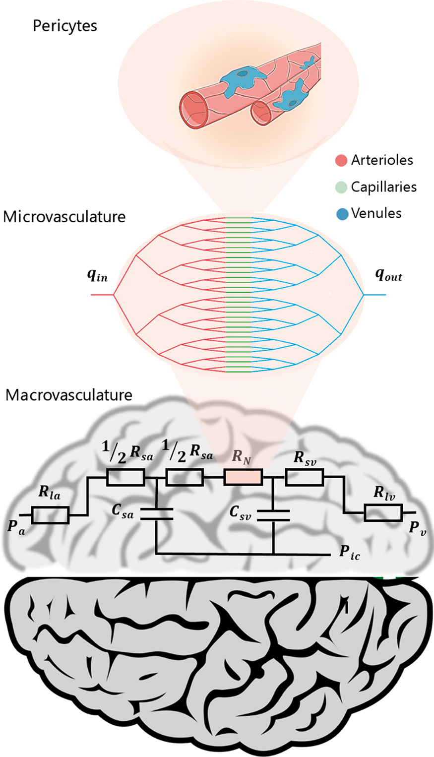
PB02-E02
The influence of cerebral blood flow autoregulation on the development of carotid siphon stenosis: Study of multi-scale coupling model
1School of Biological Science and Medical Engineering, Beihang University, Beijing 100083, China
2Beijing Advanced Innovation Center for Biomedical Engineering, Beihang University, Beijing, 100083, China
3Department of Radiology, China-Japan Friendship Hospital, Beijing, 100029, China
4State Key Laboratory of Virtual Reality Technology and Systems, Beihang University, Beijing, 100083, China
Abstract
Objectives
As a common cerebrovascular disease, internal carotid artery stenosis (ICAS) has seriously affected the supply of cerebral blood flow. On one hand, there is a close link between the occurrence and development of internal carotid artery stenosis and its hemodynamic factors. On the other hand, the circle of Willis and pial arteries exert an influence on hemodynamics of the internal carotid artery (ICA), which may further affect the development of ICAS. Therefore, it is of great significance to clarify the influence of cerebral blood flow (CBF) autoregulation on hemodynamics of the ICA, for guiding the clinical study, especially the treatment plan of ICAS.
Methods
A 3D model was reconstructed from the MR images of a patient with mild ICAS. The model was modified to get different degrees of stenosis, and form into 4 models with 0%, 20%, 50%, 70% stenosis. Two kinds of lumped parameter models of Willis circle with/without pial arteries, were coupled with the 3D models respectively. The multi-scale models can reflect the effects of CBF autoregulation on the level and distribution of hemodynamic factors in the ICA and the downstream. The wall shear stress (WSS) of ICA was calculated by the 3D hemodynamic model and the two kinds of multi-scale models, and were presented by the time-averaged WSS (TAWSS) and oscillatory shear index (OSI). The results were compared between the 3 models, to clarify the effect of CBF autoregulation.
Results
At a low degree of stenosis, the cerebral blood supply was kept stable through the compensation mechanism of the collateral circulation of the Willis circle. With the increase of the stenosis degree, the outlet blood flow and blood pressure of the internal carotid artery all decreased, and the region of low TAWSS and high OSI increased. When the stenosis degree increased to 70%, the CBF autoregulation regulated the blood flow through the expansion of the pial arteries, and the regulating effect caused by the stenosis was stronger in the ipsilateral pial arteries than the contralateral ones. Due to the regulation of pial arteries, the decrease and oscillation of WSS amplitude at the carotid siphon caused by the stenosis was inhibited.
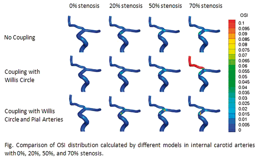
Conclusion
The CBF autoregulation has a non-negligible influence on the hemodynamic parameters of internal carotid artery, which may further affect the development trend of ICAS. The study of this effect may help us gain a better understand of the hemodynamic mechanism of ICAS and CBF autoregulation function, and provide theoretical basis for the clinical therapy and the prognosis of ICAS.
Keywords
Internal Carotid Artery, Cerebral Blood Flow Autoregulation, Hemodynamics, Multi-scale model
References
PB02-E03
The role of preoperative modeling during installing a vascular bypass graft
1Lavrentyev Institute of Hydrodynamics SB RAS
2Novosibirsk State University
3Federal Neurosurgery Center (Novosibirsk)
Abstract
One of the ways to treat cerebral aneurysms is to install a vascular anastomosis. Despite the fact that local quantitative criteria were recently obtained (Hidetoshi Matsukawa et al. J Neurosurg, 2018, DOI: 10.3171 / 2016.11.JNS161986.), Allowing to assess the need for anastomosis installation and its throughput, the task of installing anastomosis in full (taking into account redistribution of blood flow throughout the circle of Willis) is not fully investigated.
This study demonstrates the use of numerical simulations in the ANSYS 17.2 (CFX) program for a retrospective analysis of an EC-IC type vascular anastomosis clinical case that resulted in anterior cerebral artery thrombosis. As a result of a numerical calculation, a stagnant zone was discovered in which thrombus formation is possible.

This work was supported by a grant from the Government of the Russian Federation 14.W03.31.0002.
PB02-E04
Conceptualizing the conducted response as an electrical pliant process: Implications to cell signaling in the vessel wall
1Dept. of Neuroscience, University of Copenhagen, Denmark
2Robarts Research Institute, Dept of Physiology and Pharmacology, University of Western Ontario, London, ON, Canada
Abstract
Vasomotor responses conduct along and among resistance arteries to coordinate blood flow delivery with energetic demand. The distance they conduct is set by the electrical and mechanical properties of vascular cells, the former tied to charge movement via gap junctions and ion channels, elements subject to regulation. Using in silico approaches, this study conceptualized the idea of electrical pliancy, illustrating in clear terms how gap junctional and ion channel properties impacts conduction along a virtual artery or branching arteriolar network. Initial simulations reproduced known conducted behavior, highlighting the endothelium’s importance as an electrical conduit and a key charge source. Next, each set of gap junctions was shown to uniquely impact charge distribution, with diminished: 1) endothelial-to-endothelial coupling impairing longitudinal spread; and 2) myoendothelial coupling modestly enhanced conduction by limiting radial spread to the smooth muscle layer. A final set of simulations noted how subtle change in ionic conductance impact charge loss through membranes and consequently conduction. Of note was detailed work on inward rectifying- or voltage dependent- K+ channels showing how their voltage dependent properties tune conducted hyperpolarization and depolarization, respectively. This analysis led us to view conduction as an electrically pliant process, with subtle regulatory changes in ion channel activity, most likely to impact charge spread in network structures and consequently blood flow control.
PB02-E05
The conducted vasomotor response underlying cerebral blood vessel communication is a malleable process
1Department of Neuroscience, University of Copenhagen, Denmark
Abstract
Objectives
Neurovascular coupling links neuronal activity to capillary blood flow and is mediated by signaling from neurons to capillaries and signaling within and between blood vessels. Vascular signaling arise from fast and long-range conducted vasomotor responses (CVRs) i.e. coordinated diameter changes along blood vessels that arise from 1) electrical signaling within vessel walls and 2) electromechanical coupling that translates the electrical impulse into a vasomotor response. While the CVR is crucial for functional hyperemia, it remains unclear whether the CVR is a static, adaptable or regulated process. Our objective is to probe whether the CVR is malleable as a consequence of the highly regulated processes determining myosin phosphorylation.
Methods
We use an in silico approach to conceptualize the electromechanical translation of electrical signaling along a simple cerebral artery or a small reconstructed network comprising the contractile elements connected to a cortical penetrating arteriole and assess how electromechanical translation influence conduction efficiency. A simple model of cell-cell communication and vasomotor dynamics is implemented using realistic structural, electrophysiological, and electromechanical values and the CVR across the vascular network is simulated.
Results
Simulations of the CVR in cortical vasculature illustrate the dependency of the translation of electrical signaling into local vasomotor responses, i.e. electromechanical coupling. Translation, in turn, depends partly on 1) Ca2+-flux into the cytosol (notably L-type Ca2+-channels), 2) the dynamics of myosin dephosphorylation, and 3) on the organization of cytoskeletal/structural matrix elements. We show how the regulation of such factors enables local contraction control without influencing electrical spread and consequently facilitates the vascular network to direct the hyperemic response precisely to areas in need of additional perfusion.
Conclusions
The electromechanical state of vascular smooth muscle determines the translation of an electrical perturbation into a vasomotor response. The CVR is therefore heavily shaped by the overall electromechanical state of each brain blood vessel perturbed by the electrical spread. Functional hyperemia in the brain depends on graded dilation of contractile elements from capillaries along arterioles up to flow-controlling arteries. The ability of the vasculature to control tone electrically (non-local response) and non-electrically (local response) enables the cerebrovascular network to shape functional hyperemia to regions of neuronal activity.
PB02-E06
Simulating vasodilations and -constrictions to regulate blood flow during activation
1Institute of Fluid Dynamics, ETH Zurich, Zurich, Switzerland
2Institute of Pharmacology and Toxicology, University of Zurich, Zurich, Switzerland
Abstract
Objectives
The brain is capable of up-regulating cerebral blood flow in response to local changes in neural activity. However, the precise pattern of the underlying vasodynamics is still poorly understood. Our goal is to improve our understanding of the impact of diameter changes on blood flow regulation in the vasculature.
Methods
We present a numerical framework, which is based on a previously developed blood flow model [1] and on well-established optimization algorithms. The strength of our model is that we can compute diameter changes necessary to achieve desired flow distributions. This is done by adjusting the diameters of the network, in order to minimize a predefined cost function J. The required diameter changes are computed iteratively by using a gradient based optimization algorithm, where the sensitivity of J with regard to the diameters is calculated with the adjoint method. One challenge is that the solution for this inverse problem can be ambiguous, since multiple possible diameter distributions can minimize J. Therefore, the cost function is augmented with a regularization term that aims to find the most likely solution, e.g. the solution that minimizes the total change of all diameters. This method allows us to investigate different scenarios related to blood flow regulation. Furthermore, the same simulation framework can also be used to reduce uncertainties of diameters and lengths of blood vessels in microvascular networks based on sparse in vivo measurements and to tune model parameters such as boundary conditions of our blood flow model.
Results
We performed simulations in realistic microvascular networks [2] to compute diameter changes of individual blood vessels that are necessary to increase the mean blood flow rate in one specific barrel of the mouse cerebral cortex during activation. The simulations yield that vessels within the activated barrel mainly dilate in order to achieve the desired flow increase. However, outside of the barrel we observe a combination of constrictions and dilations. Furthermore, the required diameter changes are relatively small (approx. 3 % for a 30 % flow increase). Next, we investigated how the dilation and constriction of different vessel types affect the blood flow distribution in the vasculature. Our results reveal that a very localized increase of blood flow can only be achieved, if capillaries are allowed to change their diameters (Figure 1). For the case where only larger vessels can respond, the flow increase is much less local and also affects neighbouring barrels.
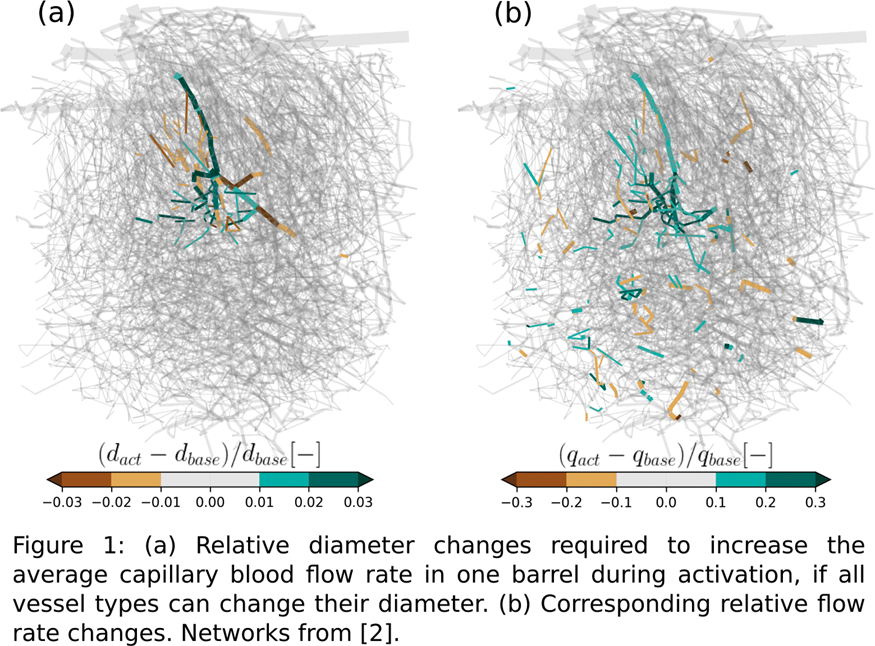
Conclusions
These findings suggest that not only arteriole-, but also capillary dilations play an important role in the local regulation of cerebral blood flow.
Funding for this research is provided by the Swiss National Science Foundation Grant No. 166707
References
PB02-E07
Modeling nitric oxide (NO) diffusion in the brain reproduces the post-stimulus undershoot in the hemodynamic response function (HRF) and vasomotion
1Medical Scientist Training Program (MSTP), Hershey College of Medicine
2The Pennsylvania State University, USA
3Department of Engineering Science and Mechanics
Abstract
Objectives
Active neurons release the vasodilator nitric oxide (NO), which plays an important role in neurovascular coupling1. Because NO is a diffusing small molecule that is rapidly degraded, the impact of NO release on vasodilation will depend on the spatial patterns of NO production and degradation. Due to the rate of NO degradation in hemoglobin being orders of magnitude higher than in brain tissue2, the size of the blood vessel could exert a dynamic effect on NO signaling. Additionally, high levels of NO will inhibit mitochondrial respiration, putting upper bounds on its physiological concentrations. We used computer simulations to investigate how the dynamics of NO production and degradation influence neurovascular coupling, the post-stimulus undershoot, and vasomotion.
Methods
NO production sources and sinks were organized in a Krogh model of a single penetrating artery using COMSOL. The impact of different locations of NO production was evaluated by calculating how much mitochondrial respiration3 was consequently inhibited in the surrounding tissue. The vessel response kernel was estimated from measurements in mouse cortex during somatosensory stimulation4. We created a dynamic model where NO degradation was coupled to vascular diameter which was controlled by the concentration of NO in the smooth muscle compartment.
Results
We found that production of NO adjacent to the vessel allows for maximal activation of the smooth muscle without inhibiting mitochondrial respiration. Because of the increased NO degradation rate in the blood, the diameter of the vessel played an important role in determining the concentration of NO. Larger vessels were less responsive to NO signaling, consistent with experimental measurements5. Following vasodilation, the increased degradation rate of NO due to the larger vessel diameter led to an overshoot in the return to baseline diameter, producing a post-stimulus undershoot in diameter. The model predicted that the degradation of NO by blood could drive arterial diameter oscillations from 0.1–0.2 Hz, similar to arterial vasomotion seen in vivo6,7.
Conclusions
Production of NO adjacent to the vessel allows for NO mediated signaling while minimizing mitochondrial inhibition. Vasodilation can decrease perivascular NO concentrations creating the post-stimulus undershoot found in the HRF. Increased NO degradation during dilation reproduces vessel oscillations at similar frequencies to arterial vasomotion seen in vivo.
References
PB02-E08
A coupled multi-compartment model of blood and oxygen transport in the human cerebral cortex
1Department of Biomedical Engineering, University of Oxford, UK
Abstract
Objectives
Developing full brain models of the vasculature in the brain is currently unfeasible due to the extremely large number of microvessels in the human brain. However, compartmental level modelling of the microvasculature, as previously adopted in the heart1 and liver2 and more recently demonstrated in the human brain capillary bed3, allows us to move towards full brain models of the vasculature. The aim of this work is thus to generate full human brain compartmental models of coupled oxygen and blood transport in the microvasculature as part of the INSIST (In-silico trials for treatment of acute ischaemic stroke) project. The broader aim of this project is to develop full human brain statistical computational models of blood and oxygen transport in order to simulate in-silico thrombectomy clinical trials.
Methods
Homogenization methods are used to upscale the penetrating arterioles (compartment 1), the capillary bed (compartment 2), and the tissue (compartment 3), as outlined in the figure. These upscaled equations have averaged parameters that capture the micro-scale variations in geometry that affect the flow and oxygen transport and that allow us to characterise the overall microvasculature as a continuum. These parameters are derived from statistical models of the capillary bed and penetrating arterioles. Once the micro-scale dependent parameters are determined, the macro-scale equations are then used to compartmentally model an idealised human cerebral cortex where a hexagonal pial geometry acts as a flow and oxygen source for the arteriolar compartment, which in turn acts as a source for the tissue and capillary compartments, and the capillary compartment acts as a further source for the tissue compartment which metabolises the oxygen. The pial vasculature is coupled via an ODE/PDE model to the microvascular compartments, with the microvascular compartments coupled via coupling coefficients derived from voxel-sized microvascular models.
Results
The homogenization of the flow in the capillary and arteriolar beds results in a Darcy flow problem with an isotropic permeability and an anisotropic permeability respectively. The oxygen transport problems result in an averaged mass transport problem with the parameter of interest being the surface area to volume ratio of the microvasculature. All parameters converge(the capillaries at a length scale of approximately 350 microns and the descending arterioles at a length scale of approximately a few mm), allowing us to model accurately full-brain simulations of coupled blood and oxygen transport in this framework.

Conclusions
The novel multi-compartment, multi-scale solver of coupled blood and oxygen transport in the human microvasculature developed here can be used on real brain mesh simulations of the human brain (derived from MRI or otherwise). The statistical nature of the microvascular model means that patient specific geometries of the large vasculature can then be superimposed over the model presented here allowing for the development of in-silico full brain modelling. This will then be used to develop computational thrombectomy trials on in-silico subjects.
References
PB02-E09
A new model for molecule exchange in the brain microvascular system: consequences of capillary occlusions in alzheimers disease
1Institut de Mécanique des Fluides de Toulouse, IMFT, Université de Toulouse, CNRS, Toulouse, France
2Meinig School of Biomedical Engineering, Cornell University, USA
Abstract
The brain microvascular system is a key actor in Alzheimer's disease (AD) development. Indeed, a significant decrease of cerebral blood flow is the earliest biomarker of AD [1].
In vivo TPLSM of cortical vasculature in APP/PS1 mice suggests the mechanism underlying the blood flow reduction is capillary occlusions. Leucocytes adhere to inflamed vessel walls and limit the flow.
The impact of capillary occlusions on blood flow has been quantified numerically in large (>10000 vessels) anatomical networks in humans and mice [2]. The regional blood flow has been found to depend linearly with no threshold effect on the fraction of capillary occlusions, so that a small fraction of stalls (2-4%) yields a significant decrease in blood flow (5-12%).
Such flow decrease has a strong impact on nutrient delivery and waste clearance. That is why we devised a new model to study the effect of capillary stalling on molecule transport. The geometry of anatomical networks is too complex to use classic numerical approaches like finite elements. Instead, our model, inspired by pore-network approaches, reduces computational costs while capturing most of the underlying physics.
To derive this model, we apply upscaling methods [3] to the 3D transport equations within each vessel to obtain 1D average equations along the axis. Contrary to previous models, this new formulation describes accurately radial concentration gradients, capturing effects like longitudinal dispersion.
We further use a Green's function formulation inpired by [4] to calculate the concentration fields inside the tissue where diffusion and reaction occur. The coupling between vessels and tissues is modelled using a membrane condition [5] representing the blood brain barrier.
This new molecule transport model is coupled with our previously validated blood flow model to examine the effects of capillary stalling [2] on molecular exchange in transient and stationary regimes in anatomical networks. In particular, in stationnary regimes, we demonstrate an increase of the extraction coefficient with the proportion of stalled capillaries, which does not compensate for the associated blood flow reduction.

Acknowledgements
ERC Brain Micro Flow (GA 616102). NIH-IMAT (Award Number R21CA214299).
References
PB02-E10
Modelling the effects of stages of hypoxia ischemia on plasticity of whisker barrel cortex
1Department of Biotechnology, Indian Institute of Technology Madras (IITM), Chennai, Tamil Nadu, India
2Department of Chemical Engineering, Purdue University, West Lafayette, USA
3Department of Chemical Engineering, Indian Institute of Technology Madras (IITM), Chennai, Tamil Nadu, India
Abstract
Classical
descriptions of neurovascular interaction treat it as a unidirectional influence arising from neurons and terminating in the cerebral vessels. However, the fact that neural activity depends on the energy substrates released by the vessels suggests the possibility of the vascular feedback actively influencing neural activity. Many modelling studies have captured the neuron to vessel influence underlying the hemodynamic response. However, there is a dearth of studies addressing the retrograde influence of vascular feedback on the neural activity at single neuron[1] and network[2] levels.
In this work, we model the effect of vascular feedback on neural activity instantiated in a model of whisker barrel maps under hypoxic and ischemic conditions. The proposed model combines a rate coded neural network model(LISSOM)[3] and a vascular network model derived from Boas et al[4]. The LISSOM network is trained to simulate the whisker response maps found in the rat whisker barrel cortex. Vascular feedback influences the threshold of firing of the neural units, which in turn influences the plasticity of the network. The LISSOM is retrained while a few of the whisker inputs are suppressed in order to simulate whisker lesion. The ratio of whisker barrel cortex representing intact whiskers to those representing lesioned whiskers is taken as a quantitative measure of plasticity. While retraining, vascular pathologies like hypoxia and ischemia are introduced by reducing the inlet oxygen saturation and vessel radius respectively. Plasticity is observed under (a) healthy vascular feedback (b) vascular feedback at different stages of hypoxia (c) vascular feedback at different stages of ischemia (d) vascular feedback during hypoxia-ischemia.
The model shows that when whiskers are intact, all whiskers have almost equal area of representation in barrel cortex making their ratio (D/C ratio) equal to one. When a whisker is lesioned, the D/C ratio increases indicating plasticity. The plasticity remains unaffected for a wide range of hypoxia. But the introduction of ischemia along with hypoxia in the vessel adversely affects the plasticity. The results of the models are compared with that of the experiment done by Ranasinghe et al[5](Fig1.a). The study also shows that even though short-term hypoxia does not compromise the neural functionality as compared to ischemia, a drop of inlet saturation below 65% does affect the plasticity (Fig 1.b). Similarly, there is a difference in how the plasticity is affected with reduction in arteriolar diameter under normal oxygen supply and hypoxic condition (Fig 1.c). The model also predicts that there is an intermediate stage where plasticity is not completely lost.

References
PB02-F01
Use of in vivo bioorthogonal chemical reporter technology to study brain sialoglycan changes after injury
1CNRS-UMR 5287, University of Bordeaux
2Basic Sciences Department, Loma Linda University, Loma Linda, CA 92354, USA
3Institut Européen de Chimie et Biologie, University of Bordeaux, 33607 Pessac, France
Abstract
Background
In order to improve the drug testing and classification of patients with brain disorders, it is urgent to find new biomarkers and develop novel neuroimaging tools for early diagnosis and long-term following of the disorder, including mild traumatic brain injury (mTBI) and neurodegenerative diseases. In most brain disorders, tissue generally presents neuroinflammation and remodeling associated with changes of expression of sialylated-glycoproteins. For example, immunohistochemistry of glycoproteins revealed that the expression of polysialic acid (PSA) attached to the N-glycans of neural cell adhesion molecule (NCAM) is increased in tissue remodeling early after injury. The recent emergence of in vivo bioorthogonal chemical reporter technology represents today the only existing method to the sialoglycans in vivo. Here, we are proposing to take advantage of this new technology to probe and track sialic acid metabolism for studying the changes in sialylated-glycoproteins after TBI.
Methods-results
Peracetylated analogues of N-acetylmannosamine (ManNAc) bearing chemical reporter groups such as the azide (i.e., peracetylated N-azidoacetylmannosamine, ManNAz) has been chemically prepared in our group. C57BL6 adult male mice and juvenile (postnatal 17d) with or without TBI (4 experimental groups, n = 48 mice) were injected i.p for 7 days either ManNAz (300 mg.kg-1/0.70 mmol/kg) or for control group ManNAc (not containing azide chemical group) (300 mg.kg-1) prepared in 30% (vol/vol) DMSO. ManNAz is enzymatically deacetylated in the cytosol and then metabolically converted to the corresponding N-azidoacetyl sialic acid (SiaNAz), which is subsequently incorporated into sialoconjugates. Once it is found on the cell surface, the azido-labeled sialylated glycans can be visualized via “click chemistry” employing a terminal alkyne-probe (along with copper reagents for CuI-catalyzed azide–alkyne cycloaddition (CuAAC)) or a strained cyclooctyne-probe in a copper-free fashion.
ManNAz and ManNAc injections did neither affect the weight of the mouse nor exhibited brain cytotoxic effect with clear absence of opening of BBB and neuronal cell death. The presence of SiaNAz was positively detected by western blots through click chemistry in the homogenates prepared from the heart, liver and various brain structures. The intensity of the ManNAz labeling varied between the brain regions with higher expression in the cerebellum compared to the cortex in both adult and juvenile mice. In primary cultures, ManNAz was incorporated in the endothelial cells, astrocytes and neurons, showing that the cells were able to metabolize and incorporate the ManNAz. The presence in the cells was after confirmed by analysis on the brain slices from animals treated with ManNAz. Seven days after TBI, the level of ManNAz staining varied between the brain structures with a significant increase in the hippocampus. It showed that the sialoglycan metabolism was changed after injury
Discussion
Here, we show for the first time that the bioorthogonal chemical reporter technology can be used in vivoto probe and track sialic acid metabolism in the brain, and study the changes of the level of expression after traumatic brain injury. As indicated PSA-NCAM is known to be a signature of tissue remodeling after injury, and this approach can be later use in neuroimaging, such as PET-scan.
PB02-F02
Investigating the mechanisms of neurovascular dysfunction after modelled traumatic brain injury and the neuroprotective potential of human umbilical cord perivascular cells on the injured brain
1Institute of Medical Science, University of Toronto, Canada
2Keenan Research Centre, St. Michael's Hospital, Canada
3Create Fertility Centre, Toronto, Canada
Abstract
Introduction
Methods and Results
Rats were subjected to a fluid percussion injury (FPI) and systemically infused with 1.5 × 10⁶ cells at 1.5 h post-injury and sacrificed at acute time points for analysis. Vascular leakage was assessed using an Evan’s blue assay and expressed in µg of dye per gram of tissue. At 24 h and 48 h post-FPI vascular leakage was 6.4 mg and 15.5 µg, respectively and 1.7 µg in sham rats. Vascular leakage was reduced in HUCPVC treated rats with 5.5 µg and 3.3 µg at 24 and 48 hours, respectively. Vascular density assessed by RECA-1 immunohistochemistry (IHC) was reduced by ∼40% in FPI animals relative to sham and HUCPVC-treated animals. Neurofilament expression (NF200) revealed aggregate formation in axons and reduced axonal length in FPI animals relative to sham and HUCPVC-treated animals. Cortical tissue was extracted at 24 h and 48 h for Western blot analysis. At 24 h and 48 h, NF200 expression was increased by ∼200% and ∼140%, respectively, in injured animals relative to sham animals. HUCPVC administration reduced NF200 expression to sham levels at 48 h. Expression of occludin was increased ∼400% and ∼200% in FPI and HUCPVC treated animals at 24 h respectively and was comparable to sham values by 48 hours, Co-immunoprecipitation of the Occludin-ZO1 complex indicated a 100% increase in complex formation at 24 h in FPI animals and a 650% increase in HUCPVC treated animals. Behavioral analysis of motor and emotional responses is in progress.
Conclusion
Modeled TBI demonstrated increased vascular leakage and axonal irregularities. The infusion of HUCPVCs following injury was associated with reduced vascular leakage with associated effects on tight junction expression. The preliminary results suggest a potential therapeutic strategy to address vascular disruption after TBI.
PB02-F03
Microglia specific histone deacetylases 2 deficiency promotes inflammation and aggravates long-term functional deficits after traumatic brain injury
1State Key Laboratory of Medical Neurobiology, Institute of Brain Science, Fudan University, China
Abstract
Objectives
Traumatic brain injury(TBI) causes death and neurological disability
Methods
Male and female 8-12-week-old mice with microglia specific HDAC2-conditional knockout (HDAC2-cKO) or their wild type littermates (HDAC2-WT) were subjected to controlled cortical impact (CCI) to model TBI. Neurological deficits were evaluated by rotarod, wire-hanging, grid-walking and body curl test 1-28 days after TBI. Tissue loss was evaluated by NeuN immunostaining at 35 days after TBI. The integrity of myelin and axon was assessed by myelin basic protein (MBP) and neurofilament (NF200) immunostaining at 35 days after TBI. Immunostaining and flow cytometry analysis were used to evaluate microglial activation and immune cell infiltration at 3 and 7 days after TBI.
Results
Our results showed that HDAC2-cKO aggravated short- and long-term neurological deficits up to 28 days after TBI, and enlarged tissue loss volume at 35 days after TBI. The corpus callosum (CC) width, as measured by MBP staining, significantly reduced in TBI mice compared to sham-operated controls. HDAC2-cKO and control mice exhibited comparable loss in CC width after TBI. Interestingly, the expression of NF200 after TBI was significantly reduced in the striatum of HDAC2-cKO mice, while MBP expression was unchanged, suggesting that HDAC2-cKO resulted in impaired white matter integrity through exacerbating axonal damage rather than increasing myelin loss. Furthermore, HDAC2-cKO inhibited microglial activation, reducing the expression of both pro-inflammatory and anti-inflammatory markers. The deficiency of HDAC2 in microglia also accelerated the infiltration of peripheral neutrophils, macrophages, and T cells into the brain at 3 days after TBI. The differences in peripheral immune cell infiltration between HDAC2-cKO and WT mice diminished at 7days after TBI.
Conclusion
HDAC2 expression in microglia enhances microglial activation and delays peripheral immune cell infiltration in acute phase after TBI. Specific HDAC2 induction may represent a therapeutic strategy to modulate post-TBI neuroinflammation, rescue axonal damage and improve neurological functions after TBI.
Keywords
Traumatic brain injury, microglia, HDAC2, immune cell infiltration
PB02-F04
Wnt/beta-catenin signaling modulates the response of the cerebral vasculature after traumatic brain injury
1Department of Pediatrics, University of California Irvine, Irvine, CA USA
2Department of Physiology and Pharmacology, Loma Linda University, Loma Linda, CA, USA
3Cell, Molecular and Developmental Biology Program, University of California Riverside, Riverside, CA, USA
4Center for Perinatal Biology, Loma Linda University School of Medicine, Loma Linda, CA, USA
5Departments of Anesthesiology, and Neurosurgery, Loma Linda University School of Medicine, Loma Linda, CA, USA
Abstract
Objectives
Traumatic brain injury (TBI) results in damage to the cerebral vasculature and is often associated with hemorrhage, edema, blood flow abnormalities, and cell death. An important and unexplored avenue is how blood vessels undergo repair and remodeling. At present, the temporal evolution of vascular repair is unknown with little knowledge of the molecular mechanism(s) underlying vascular repair after TBI. One possible molecular pathway that may be involved in vascular repair is the Wnt/b-catenin cascade. The Wnt/β-catenin pathway promotes blood vessel formation during vascular development, but its role in vascular repair after injury is unknown. We examined how the cerebral vessels respond over 7 days following a moderate TBI focusing on what role Wnt/β-catenin signaling plays in the vascular repair process.
Methods
A controlled cortical impact mouse model was used to induce a moderate TBI, which led to gross injury to the cerebral vessels. Using a novel vessel painting technique to label the cerebral vessels within the entire brain, we assessed vascular alterations at 1 and 7 days post injury (dpi). We assessed b-catenin inside blood vessels around the lesion and utilized a Wnt transgenic mouse line to evaluate Wnt gene expression. To assess the role of β-catenin in vascular repair, we utilized Lithium (GSK3 inhibitor) to enhance b-catenin expression and JW74 (tankyrase inhibitor) to inhibit b-catenin expression and evaluate their effects on vessel morphology and development of hemorrhage and edema. Lithium or JW74 were administered to mice for 6 consecutive days and then sacrificed at 7 dpi.
Results
We report that TBI results in vascular loss at 1 dpi followed by increases in vascular structures by 7 dpi. β-catenin expression was increased in perilesional vessels at 1 and 7 dpi. Similarly, we found increased number of Wnt-GFP-positive vessels after TBI. Lithium treatment after TBI significantly increased average vessel length compared to saline treatment. Conversely, JW74 treatment after TBI reduced vessel density, branch points, and average vessel length compared to vehicle treatment. Magnetic resonance imaging T2 and susceptibility weighted imaging of vessel painted brains at 7 dpi revealed a reduction in hemorrhage but an increase in edema volumes following Lithium treatment. Conversely, there was an increase in hemorrhage and edema volumes following JW74 treatment.
Conclusions
Our findings suggest that endogenous developmental programs, such as Wnt/β-catenin, become activated after TBI to initiate repair. Treatment regimens to enhance activation of Wnt/β-catenin appear to contribute to the vascular repair process after TBI and represents a potential target for future therapeutics.
PB02-F05
A novel class of lipid mediators, elovanoids, improve behavior and preserve brain tissue after traumatic brain injury in rats
1Neuroscience Center of Excellence, LSUHSC, USA
2Dept. of Pediatrics, University California Irvine, USA
3Dept. of Chemistry, University of Southern California, USA
Abstract
Objectives
Traumatic brain injury (TBI) often leads to substantial cognitive impairments and permanent disability. Many experimental drugs were not effective in clinical trials; therefore, the development of new therapeutic approaches for TBI represents a huge unmet need. Recently, we reported the discovery and characterization of a novel class of homeostatic lipid mediators, termed elovanoids (ELV), which are derivatives from very long chain polyunsaturated fatty acids (VLC-PUFAs,n-3) (Bazan NG, Mol. Asp. Med, 2018). ELV display neuroprotective bioactivities both in vitro neuronal injury models and in vivo experimental ischemic stroke. The purpose of this study was to determine whether treatment with ELV (ELV-34:6) would be beneficial in a rat model of TBI.
Methods
Male SD rats (450-400 g) were anesthetized with 3% isoflurane, mechanically ventilated, physiologically regulated, and subjected to moderate right parieto-occipital parasagittal fluid-percussion injury. ELV-34:6 (300 µg/per rat) or saline treatment was administered i.v. at 1 h after TBI (n = 5-6 per group). Behavior was evaluated on days 1, 2, 3, 7 and 14 after TBI; a grading scale of 0-12 was employed (normal score = 0, maximal deficit = 12). Ex vivo T2WI of the brains was conducted on 11.7 T on day 14 and volumes of preserved brain tissue in CA1, CA3, Dentate Gyrus (DG) and white matter connectivity (diffusion tensor imaging, DTI) was analyzed. After completion of MRI study, histopathology was conducted and the contusion areas and number of normal pyramidal neurons in the CA1 and CA3 regions were quantitated.
Results
The physiological variables were entirely comparable among the six groups. There were no adverse side effects after ELV administration. Treatment with ELV improved the neurological score compared to saline on days 1, 2, 3, 7 and day 14 (panel A). The hippocampus is a structure known to have reduced volumes after TBI. ELV treatment preserves hippocampal volume loss, significantly in the CA3 and DG regions (panel B). Panel C: Representative T2WI images from saline and ELV treated rats. Saline rats exhibited cortical, CA3 and reduced DG volumes, more edema (bright cortex) and small hemorrhages within the white matter of the external capsule (arrow) compared to ELV treated rats. Dotted line indicates the expanded area showing white matter (corpus callosum, and hippocampus. * = FPI site of injury.
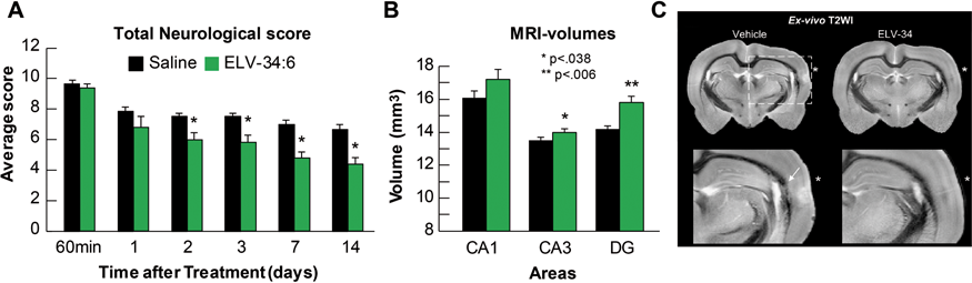
Conclusion
These results show that ELV are neuroprotective in rats following TBI and open avenues of exploration as a possible therapeutic approach for TBI.
PB02-F06
Identification of peripheral biomarkers following brain injury in mice
1Mario Negri institute for pharmacological research, Milan, Italy
2Istituto Clinico Humanitas, Milan, Italy
Abstract
Objectives
Acute brain injury, including stroke and traumatic brain injury (TBI) represent major public health issues that may lead to devastating neurological disability. They cause persistent and progressive symptoms and currently, reliable biomarkers allowing to inform on the underlying pathology are lacking. Animal models replicating the various aspects of human brain injury, have been developed to explore new therapeutic agents. Therefore, validation of biomarkers in mice represents an essential tool for the development and testing of new treatments that can be translated into clinic.
Methods
Here we evaluate the use of different innate immunity markers found in mouse plasma applied to a clinically relevant murine model of TBI, the controlled cortical impact (CCI) injury mouse model, that mimics several pathophysiological aspects of TBI found in humans.
Results
We found a time dependent increase of the complement components C4 and C3 fragments in TBI mouse plasma with a peak at 48 hours, indicating a persistent activation of the complement system (7.05 and 2.76–fold change, respectively). Other components of the classic and lectin pathway of complement such as C1q, MBL-A and MBL-C were also evaluated, allowing to differentiate the activation pathways involved. We found that MBL-C, responsible for the activation of the lectin pathway of complement, increased in mouse plasma 48 hours after TBI (2,48-fold change). Furthermore, we measured the protein plasma levels of the anti-inflammatory lectin-like shedded protein domain (sTM) of thrombomodulin (TM), founding a significant reduction persistently at 1 week after brain trauma (0,79-fold decrease). We also quantified the levels of Pentraxin-3 (PTX3), a member of the pentraxin superfamily proteins involved in the acute inflammatory response, finding an increase of PTX3 up to 3 weeks following TBI (1.93–fold change).
Conclusions
Finally, our results provide an outline on the use of different potential biomarkers in mice following traumatic brain injury, as indicators of assessment of brain damage and therapeutic treatment.
References
PB02-F07
Microcirculatory biomarkers of secondary cerebral ischemia at traumatic brain injury
1Department of Neurosurgery, Privolzhsky Research Medical University, Nizhny Novgorod, Russia
2Department of Neurosurgery, University of New Mexico School of Medicine, Albuquerque, USA
Abstract
Objectives
Traumatic brain injury (TBI) is a major public health and economic problem throughout the world. It is a leading cause of mortality and disability among young people. Approaches to TBI management focus mainly on preadmission emergency and intensive care management. Patients who suffered a moderate and severe traumatic brain injury (TBI) might have secondary cerebral ischemia (SCI). Perfusion computed tomography (PCT) is performed to identify these patients because the majority of non-contrast CT will be negative. Biomarkers are a key aspect of neuromonitoring. A broad definition of a biomarker is any observable feature that can be used to evaluate the state of the patient, e.g., a molecular species, a feature on a scan, or a monitoring characteristic, e.g., cerebrovascular resistance (CVR), cerebral arterial compliance (CAC), cerebrovascular time constant (CTC) and critical closing pressure (CCP). Biomarkers are usually quantitative measures, which can be utilized in diagnosis, prognosis, monitoring the evolution of injury and recovery, and response to treatments. They are thus crucial to the development of therapies in many pathological states. Here we studied microcirculatory parameters for their ability to predict the development of SCI in TBI patients.
Methods
A total of 220 moderate and severe TBI patients were recruited from two different Trauma Center Level 1, within 6 h after head injury with a GCS less than 12. All patients were subjected to PCT on 64-slice scanner. PCT was performed 1–12 days after TBI. The levels of cerebrovascular resistance, cerebral arterial compliance, cerebrovascular time constant and critical closing pressure were measured using a complex of the neuromonitoring. The patients were dichotomized into SCI-positive (n = 207) and SCI-negative (n = 13) groups for statistical analyses using Student’s t-criterion and multiple regression. P-values significant if <0.05.
Results
The level of such parameters of the microcirculation as CVR and CCP were significantly increased, while CAC and CTC were significantly decreased in SCI-positive patients (p < 0.05). Multiple regression analyses showed that critical closing pressure levels were not significantly associated with the development of secondary ischemia (p > 0.05). Regression analyses also revealed that CVR, CAC, and CTC were significantly associated with the performance of cerebral posttraumatic ischemia (p < 0.05).
Conclusions
The results indicate severe dysregulation of cerebral capillary blood flow in polytraumatized patients, which increases in the patients with TBI and traumatic intracranial hematomas. Moderate and severe TBI is associated with accelerated changes in parameters of the microcirculation over time, which may indicate remodeling of cerebral blood flow. Cerebrovascular resistance, cerebral arterial compliance and cerebrovascular time constant may be potential biomarkers that reflect the development of secondary cerebral ischemia in TBI patients. A further prospective multicenter study is warranted.
PB02-F08
Serum IL-6: A novel biomarker for outcome prediction in isolated severe traumatic brain injury
1Neurosurgery, All India Institute of Medical Sciences, New Delhi
Abstract
Background
Interleukin-6 (IL6) is one of the first cytokines produced after traumatic brain injury (TBI) and may have a role both in the pathogenesis of neuronal damage and in the recovery mechanisms of injured neurons.
Aims & Objectives
We investigated whether serum IL-6 was associated with in hospital mortality in isolated severe traumatic brain injury patients.
Methods
A prospective study was carried out in the neurosurgery intensive care unit (NICU) at the Jai Prakash Narayana Apex Trauma Center, AIIMS, New Delhi, India, over a period of one year (January 2013-Januray 2014). Isolated severe traumatic brain injury patients (GCS ≤ 8) aged between 18 and 60 years who were admitted to NICU were included in this study. Blood sample were collected within 24 hours of injury and serum level of IL-6 was measured by ELISA. Outcome was assessed using in-hospital mortality. The association of IL-6 level and in hospital mortality was analyzed by t-test and sensitivity-specificity analysis was also performed.
Results
A total of 80 patients were enrolled for the study. The median length of hospital stay was 9 days (1-76 days). In-hospital mortality was 15% (12 patients). The mean interleukin IL-6 (295.3 ± 38.8) was higher in survivor group as compared to non-survivor group (224.8 ± 95.8). Significantly higher levels of IL-6 were found in patients who survived compared with those who died (P = 0.014). A serum IL-6 level of > 283 pg/ml had an 86% sensitivity and specificity of predicting survival in isolated severe head injury patients.
Conclusions
Our study is the first of its kind which shows a cut-off value for IL6 with high specificity and sensitivity and can be a very useful tool to predict survival in isolated severe traumatic brain injured patients.
References
PB02-F09
Microthrombosis and oxidative stress reduction by novel resuscitation fluid for traumatic brain injury with hemorrhagic shock
1Department of Neurosurgery, University of New Mexico School of Medicine, USA
2McGowan Institute for Regenerative Medicine, University of Pittsburgh, USA
Abstract
Abstract
Cerebral microcirculation impairment plays a key role in the pathophysiology of traumatic brain injury (TBI). Hemorrhagic shock, a frequent complication in TBI due to polytrauma, significantly worsens neurologic outcome and doubles mortality rate. The main reason is microvascular cerebral blood flow (mvCBF) impairment and capillary microthrombosis leading to tissue hypoxia, neuronal death and a two-fold increase in contusion volume. Existing volume expansion approach with resuscitation fluids (RF) for combined TBI/HS is controversial as do not adequately alleviates impaired microvascular cerebral blood flow (mCBF). We previously reported that resuscitation fluid with drag reducing polymers (DRP-RF) improves mvCBF by rheological modulation of hemodynamics. Here, we evaluate the efficacy of DRP-RF, compared to lactated Ringers and Hetastarch resuscitation fluid (LR-RF and Ht-RF) in reducing cerebral microthrombosis and reperfusion mitochondrial oxidative stress after TBI complicated by HS.
Methods
Fluid percussion TBI (1.5 ATA, 50 ms) was induced in rats and followed by controlled HS to a mean arterial pressure (MAP) of 40 mmHg. DRP-RF, LR-RF or Ht-RF was infused to restore MAP to 60 mmHg for one hour (pre-hospital period), followed by blood re-infusion to a MAP = 70 mmHg (hospital period). In vivo 2-photon laser scanning microscopy over the parietal cortex was used to monitor microvascular blood flow, NADH (hypoxia) and mitochondrial oxidative stress (superoxide by i.v. hydroethidine [HEt], 1 mg/kg) for 4 hours after TBI/HS, followed by Dil vascular painting during perfusion-fixation. Brain and rectal temperatures, MAP, blood gases and electrolytes were monitored.
Results
TBI/HS decreased mCBF resulting in capillary microthrombosis and tissue hypoxia. Microvascular CBF and tissue oxygenation were significantly improved in the DRP-RF compared to the LR-RF and Ht-RF treated groups (p < 0.05). Reperfusion-induced oxidative stress, reflected by HEt fluorescence, was 32 ± 6% higher in LR-RF and 28 ± 5% vs. DRP-RF (p < 0.05). Post-mortem whole-brain visualization of DiI painted vessels revealed multiple microthromboses in both hemispheres that were 29 ± 3% and 27 ± 5% less in DRP-RF vs. LR-RF and Ht-RF groups, respectively (p < 0.05).
Conclusions
Resuscitation fluid with DRP additives effectively restores cerebral microcirculation, reduces hypoxia, microthrombosis formation, and mitochondrial oxidative stress compared to conventional volume expansion with lactated Ringer and Hetastarch. In addition, DRP-RF requires an infusion of a smaller volume to improve tissue perfusion and oxygen utilization which reduces brain edema formation due to hypervolemia, which often occurs with standard fluid resuscitation.
Supported by DOD DM160142.
PB02-F10
Leakage sign for acute subdural hematoma
1Dept. of neurosurgery, Kurume University, Japan
Abstract
Background
Acute subdural hematoma (ASDH) is a serious traumatic disease and there is some problems of surgical indication, prediction of outcome and so on. We have established a novel method to predict the expansion of contusional hematoma in traumatic brain injury (TBI) using the “leakage sign” detected by computed tomography angiography (CTA) previously. The purpose of this study was to evaluate the incidence of the leakage sign among patients with ASDH and to identify its prognostic value.
Methods
67 patients with ASDH and received complete our CTA protocol were analyzed (mean age 64.1 +/- 20.6, 24 males). After 2 serial scans of CTA phase and delayed phase (5 min after the CTA phase), we set a region of interest (ROI) with a 10 mm diameter and calculated the Hounsfield units (HU). We defined positive leakage sign as a >10% increase in HU in the ROI. Additionally, hematoma expansion was determined on plain CT at 24 h in patients who did not undergo emergent surgery.
Findings
Here were 67 ASDH patients carried into our department from 2012 to 2015. Among 67 cases, conservative therapy was performed in 35 cases, 9 out of 35 cases showed an expansion hematoma, 8 of 9 (88.9%) cases was positive leakage sign. The sensitivity of leakage sign to hematoma expansion in the no-surgery group was 88.8% and the specificity was 76.1%. Regarding the outcome, the leakage sign positive group had poor outcome in all cases and surgical cases.
Conclusion
The results indicate that the leakage sign is a useful and sensitive method to predict hematoma expansion and poor outcome in ASDH. In addition, if the hematoma is small but Leakage sigh is positive, strict observation is necessary and aggressive surgery might improve ASDH patients outcome.
PB02-F11
Bedside non-invasive assessment of the hemodynamic consequences of hyperventilation treatment in traumatic brain injury
1ICFO – Institut de Ci&amp;egrave;ncies Fot&amp;ograve;niques, The Barcelona Institute of Science and Technology, SPAIN
2Institute of Biocybernetics and Biomedical Engineering, POLAND
3Neurotraumatology and Neurosurgery Research Unit (UNINN), Vall d’Hebron University Research Institute (VHIR), Universidad Autònoma de Barcelona, SPAIN
4Department of Neurosurgery, Vall d’Hebron University Hospital, Universidad Autònoma de Barcelona, SPAIN
5Institució Catalana de Recerca i Estudis Avançats (ICREA), SPAIN
6HemoPhotonics S.L., SPAIN
Abstract
Objectives
Hyperventilation (HV) is a treatment employed to decrease raised intracranial pressure (ICP) resulting from traumatic brain injury (TBI). A side effect of HV is the possibility to cause vasoconstriction leading to a decrease of cerebral blood flow (CBF) which may provoke secondary ischemia1. Current invasive techniques are insufficient for clarifying when HV is deterious. Non-invasive diffuse-optics based techniques could fill in this lack by providing continuous measurements of microvascular CBF, blood oxygenation (StO2), cerebral metabolic rate of oxygen (CMRO2) and oxygen extraction fraction (OEF)2. This may allow the clinicians to establish thresholds to personalizing the therapy by avoiding the misery perfusion3.
Methods
Invasive monitors were used to measure ICP and tissue oxygen tension (PtiO2). Non-invasive measurements were carried out by diffuse correlation and time-resolved spectroscopies whose probes were bilaterally placed on the forehead. All signals were synchronized together for comparison during the protocol, which consisted in 15 minutes of baseline, 30 minutes of HV induced by a mechanical ventilator and 30 minutes of recovery. Eleven TBI patients were enrolled in the study so far (Glasgow coma scale ≤13, 3 females, age = 37.9 ± 10 y) for a total of 16 measurements (15 with ICP sensor, 9 with PtiO2 sensor).
Results
Median values from the population revealed that: 1) ICP significantly decreased during HV and then recovered afterwards, 2) PtiO2 decreased and then did not recover, 3) relative CBF increased and continued to increase, 4) StO2 decreased and afterwards increased, 5) relative OEF decreased and then lowered even more, 6) relative CMRO2 decreased but increased during recovery. These findings suggest that when HV is reducing ICP, vasocontriction is not moving CBF towards dangerous secondary effects. Next was to investigate each subject/measurement independently
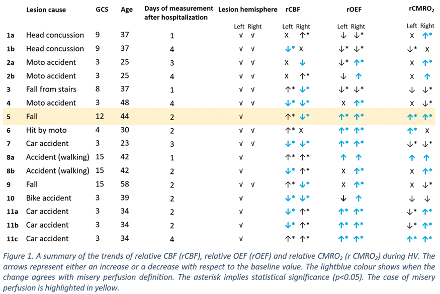
Conclusions
This on-going proof-of-principle study detected one significant misery perfusion during HV therapy. Further data is being added and we will further discuss the correlations between different variables and an evaluation of auto-regulation.
Acknowledgements & disclosures
Funding
H2020 ITN BitMAP (European Comission, No. 675332), Fundació CELLEX Barcelona, Obra social “laCaixa” (LlumMedBcn), KidsBrainIT (ERA-NET NEURON) and FIS/ERDF PI18/00468. We disclose the following. JF is an employee of HemoPhotonics SL his role has been defined by the BitMap project and was reviewed by the European Commission.
References
PB02-F12
Multimodal assessment to understand posterior cingulate gyrus connectivity in the chronic phase after severe traumatic brain injury caused by traffic accidents
1Dept. of Neurosurgery, Chubu Medical Center for Prolonged Traumatic Brain Dysfunction, Japan
2Dept. of Neurosurgery, Kizawa Memorial Hospital, Japan
3Dept. of Clinical Brain Sciences, Gifu University Graduate School of Medicine, Japan
Abstract
Objectives
The present study aimed to understand the pathology of connectivity in chronic brain injury patients following severe traumatic brain injury (TBI), which will be useful for the development of novel neurorehabilitation methods. In functional connectivity (FC) research, the default mode network (DMN), in which the posterior cingulate cortex (PCC) functions as the hub, is related to the level of consciousness. The cingulate gyrus is one of the most commonly damaged brain regions after severe TBI due to traffic accidents. Concussion studies have shown that amyloid levels may be increased in injured brains, and Alzheimer’s disease studies have shown that amyloid deposits can affect FC in the DMN. Here, we investigated FC by resting state functional magnetic resonance imaging (rsfMRI) and structural connectivity (SC) by diffusion tensor imaging (DTI) in TBI patients who underwent Pittsburgh compound-B positron emission tomography (PiB – PET) for amyloid deposition.
Methods
Nine patients were assessed. Although all patients were in a coma in the acute phase, at the time of examination three patients were in a vegetative state (VS), two patients were in a minimally conscious state (MCS), and four patients had no disorder of consciousness (DOC), but cognitive dysfunction (non-DOC) was present. At the time of scanning, the mean time post-injury was 23.3 months (range: 10–53) and the mean patient age was 49.3 years (range: 20–70).
All patients took part in the PiB – PET and DTI studies. Of these, five patients (1 non-DOC, 2 MCS, and 2 VS patients) also completed rsfMRI. Integrated PiB was analyzed by the 3DSRT1). In the DTI study, the mean fractional anisotropy value (FA) of the PCC (FA-PCC) was calculated using the dTV2). The rsfMRI data were preprocessed using SPM123). FC of the PCC to DMN-related ROIs (e.g., FC-PCC) was assessed using the CONN toolbox4).
Results
Amyloid deposition in PCC was not correlated with the arousal of consciousness or FA-PCC, but was inversely correlated with FC-PCC. With respect to SC, FA-PCC was higher in the later phase as well as non-VS (non-DOC + MCS) patients. With regard to FC, FC-PCC was also higher in later phase patients, and FC to the medial prefrontal cortex (MPFC) was significantly higher in non-VS patients than VS patients. FC to the MPFC also showed strong correlation with FA-PCC.
Conclusions
In the present study, SC showed good correlations with the level of consciousness and elapsed time after injury. Although FC does not generally require proof of anatomical connections, FC-PCC—especially in the cingulate gyrus—was supported by SC in patients after TBI. Amyloid deposition did not affect consciousness; however, it weakened FC more than SC. Differences in FC and amyloid findings between the present cases and Alzheimer’s disease might relate to inhomogeneous tissue damage in our patients, which is a challenge of neurotrauma studies.
References
PB02-G01
Protective effect of ISO-1 on advanced glycation end products aggravating of PC12 cell injury induced by Aβ1-40
1Department of neurology, affiliated hospital of jiangsu university
2Medical college of jiangsu university
Abstract
Advanced glycosylation end products (AGEs) are important pathogenic substances involved in diabetes mellitus (DM) and its complications, and AGEs play important roles in promoting the development of Alzheimer’s disease (AD). Macrophage migration inhibitory factor (MIF), another pathogenic factor involved in DM, was also shown to be present at significantly higher levels in the cerebrospinal fluid (CSF) of patients with AD and mild cognitive impairment (MCI) than in normal patients of the same age. By exploring the relationship between AGEs and MIF and the effect of neuroinflammation on AD, we will be able to further improve our understanding of the specific molecular mechanisms of diabetes and AD. PC12 cells were cultured in vitro. The levels of the MIF mRNA and protein in PC12 cells were measured by reverse transcription-PCR (RT-PCR) and Western blotting; Screening the optimum concentration of AGEs and the concentration of Aβ1-40 in the AD cell model by the CCK-8 assay and the MTT method. Cell numbers and morphological changes were observed under an inverted microscope following treatment of Aβ1-40 stimulated PC12 cells with AGEs and the MIF inhibitor ISO-1. The expressions of the IL-1β, IL-6 and TNF-α mRNAs were measured by RT-PCR. The results showed that the levels of the MIF mRNA and protein were significantly increased in cells treated with AGEs compared with the black control group. In the AD model group, Percentage of PC2 cell growth inhibition was significantly increased, and the expressions of the IL-1β, IL-6 and TNF-α mRNAs were increased in the AD model group. Compared with the AD model group treated with AGEs, combination AGEs with ISO-1 significantly improved the cell survival rate and downregulated the expression of inflammatory mediators in the AD cell model. Thus ISO-1 reduced the damage of AGEs to AD cell model, the mechanism may be that AGEs mediated MIF expression to promote the neuritis of AD cell model, while ISO-1 decreased the expression of neuroinflammatory mediators.

References
PB02-G02
Xingnaojing injection improves Aβ1-42-induced memory deficit in mice by altering of excitatory amino acid toxicity and synaptic plasticity
1Department of Neurology, Drum Tower Hospital of Nanjing University Medical School, PR China
Abstract
Objectives
Alzheimer’s disease (AD), the widespread type of dementia, is characterized by depositions of senile plaques composed of insoluble amyloid β (Aβ) peptides, and neurofibrillary tangles[1, 2]. It causes progressive degeneration of synaptic plasticity, especially in hippocampus and cortex, leads to memory loss and cognition impairment[3]. Abnormal changes in synaptic structure and excitatory amino acid toxicity occurred in early stage in AD and are associated with decreased cognitive function. Xingnaojing (XNJ), a well-known prescription in traditional Chinese medicine, has been used for treatment of stroke in China[4]. Intrahippocampal injection of Aβ1–42 mice induce memory impairment and is a reliable animal model of AD. In this research, we aim to investigate the effects of XNJ on synaptic plastic and excitatory amino acid toxicity of Aβ1-42-induced memory deficit in mice.
Methods
Mice were randomly divided into 4 groups: control, Aβ1-42 injected (Aβ) and Aβ1-42 injected with two doses of XNJ (low-dose, high-dose) administration groups. We performed bilateral intra-CA1 injection of Aβ1-42 except control group. 7 days later, the XNJ groups were given an intraperitoneal injection of different doses of XNJ and the same amount of saline was given for Aβ group for up to 15 days. Spatial reference and working memory were evaluated by using the Morris water maze (MWM), Open field test (OFT) and New object recognition (NOR). Immunofluorescence and Golgi staining were used to measure the synaptic morphology. Western blot was conducted to detect the protein expression of synaptic-related protein and excitatory amino acid receptors.
Results
Behavioral results showed that the mice treatment with high-dose of XNJ group has a longer exploration time for NOR and more crossing platform times for MWM compared with Aβ group. The expression of MAP-2 was increased by administration of XNJ in immunofluorescence results. Golgi staining showed that XNJ can improve dendritic spine density of damaged neurons. It also increased the protein levels of GAP-43, PSD-95, NR2b, p-AKT/AKT and p-mTOR/mTOR in the brain tissues compared with Aβ group.
Conclusions
Our results indicated that XNJ exhibited a protective effect against excitatory amino acid toxicity and synaptic plasticity via AKT/mTOR signal pathway in mice with Aβ1-42-induced memory deficit. These results provided evidences for the novel and potential application of XNJ for the treatment of AD.
References
PB02-G03
In vivo dynamics of amyloid β in glymphatic system observed with multiphoton microscopy
1Dept. of Neurology, Osaka City University Graduate School of Medicine, Japan
Abstract
Objectives
Amyloid β(Aβ), one of the causative proteins found in Alzheimer’s disease (AD), does not cross the blood-brain barrier besides transcytosis at the capillaries. Glymphatic pathway has been recently suggested as a possible lymphatic system in the brain and may be involved in the clearance of Aβ. To elucidate the pathomechanism of parenchymal Abaccumulation, perivascular passage and accumulation of Aβwas analyzed in vivo.
Methods
Tie2-GFP mice (Stock Tg [Tie2-GFP] 287Sato/J) were introduced to visualize the vascular endothelial cells. Under anesthesia with 2.2% isoflurane, the skin covering left parietal skull of male mice was incised and a cranial window (3.5 mm in diameter) was opened with a dental drill. After removing the dura mater, HiLyte648-Amyloid β1-40(Anaspec) or HiLyte648-Amyloid β1-42(Anaspec) was topically applied on the cortical surface and the window was closed with a cover glass. Three dimensional imaging of the parietal cortex down to 300 mm was repeatedly conducted with multi-photon microscope (A1RMP + 1080, Nikon).
Results
One-hour after the cortical application of Aβ, accumulated Aβ was observed along the penetrating vessels debranching from the pial vessels, as well as the small vessels in the deepest layer (300 mm). The Aβ accumulation was localized to the abluminal surface of endothelial cells. The penetrating arteries were involved more than veins. The average number of Aβ1-40 deposits around the artery were increased 66.2% per hour (0 h:8.3, 1 h:13.8), and around the vein were decreased 32.9%(0 h:9.7, 1 h:7.3). The deposition of Aβ 1-42 was increased either (artery 0 h:10.3, 1 h 12.3, vein 0 h:10, 1 h: 12.3). Aβ1-42 deposit moved to deeper than Aβ1-40, and Aβ deposit were found more around the artery than vein in deep layer. This might be because the difference of diffusion and convection in the glymphatic system.
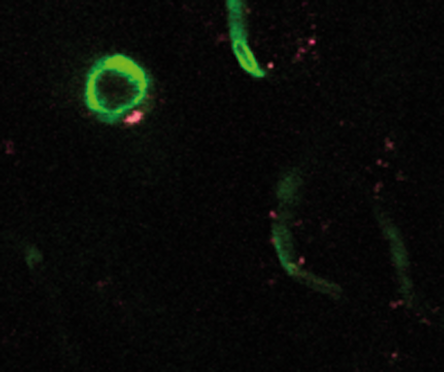
Conclusion
Passage and accumulation of Aβwas repeatedly demonstrated along the penetrating arteries and capillaries in the deep cortical layer. Disturbance of the clearance system may be involved in the pathogenesis of AD.
PB02-G04
Non-invasive volumetric mapping of amyloid pathology across the whole mouse brain by photoacoustic tomography
1Institute for Biomedical Engineering, ETH Zurich and University of Zurich; Switzerland
2Neuropathology, University Hospital Zurich, Switzerland
3Brain Research Institute, University of Zurich, Switzerland
4Martinos Center for Biomedical Imaging, Harvard Medical School, USA
5Institute for Chemical and Bioengineering, ETH Zurich, Switzerland
Abstract
Objectives
The deposition of fibrillar beta-amyloid (Aβ) deposits in the brain is one major histopathological hallmark of Alzheimer’s disease (AD); a feature that has also been recapitulated in transgenic mouse models of the disease. Non-invasive imaging techniques that can map Aβ deposition across the whole mouse brain with high spatial and temporal resolution would be highly desirable to assess the region-specific accumulation and spread of Aβ and to assist the development of Aβ-targeted therapies. Here, we describe a novel 3D high-resolution whole brain photoacoustic tomography (PAT) method to visualize and quantify brain Aβ fibril accumulation in a transgenic mouse model of cerebral amyloidosis using the Aβ-binding probe CRANAD-2.
Methods
In vitro binding of CRANAD-2 to recombinant Aβ42 fibrils from E. coli was assessed with fluorescence spectrometry. Transgenic arcAβ mice and non-transgenic littermates of 18-24 months of age underwent whole brain imaging by using PAT after intravenous injection of CRANAD-2. T2-weighted magnetic resonance imaging (MRI) of the same animal was performed on a 7 T small animal MRI scanner to derive anatomical reference data. PAT images were reconstructed using a model linear algorithm with a non-negativity constraint imposed during inversion. Two set of images were analyzed: 1) reconstructed images at single wavelength 680 nm and 850 nm; 2) a unmixed CRANAD-2 and background images. Resulting PAT images were co-registered with structural MR images using landmark selection and affine transformation [Ni et al. 2018]. Light sheet microscopy and immunohistochemistry were performed to investigate CRANAD-2 distribution and binding.
Results
CRANAD-2 is a difluoroboron-derivated curcumin and has a high binding affinity to Aβ [Ran et al. 2009]. We have demonstrated in vitro that the probe binds to recombinant Aβ42 fibrils in a quantitative way. In vivo PAT (axial resolution 100 µm) successfully detected fibrillar Aβ deposits in the cortical brain regions of arcAβ mice compared to non-transgenic littermates. Wash-out kinetics demonstrated higher retention of CRANAD-2 in the brain of arcAβ mice. Ex vivo selective plane illumination microscopy validated the distribution of Aβ deposits in the brain parenchyma and cerebral amyloid angiopathy in cleared whole brain from arcAβ mice. Fluorescent microscopy showed co-localization of Thioflavin-S and CRANAD-2 signals in brain tissue sections, thus verifying the specificity of the probe.
Conclusion
In conclusion, we demonstrate a new high-resolution in vivo imaging platform for detecting Aβ deposits across the murine brain in animal models of AD pathology, which facilitates mechanistic studies and the monitoring of putative treatments targeting Aβ deposits.
References
PB02-G05
Functional MRI reveals mitigation of cerebrovascular dysfunction by bradykinin receptor 1 and 2 antagonism in a mouse model of cerebral amyloidosis
1Institute for Biomedical Engineering, ETH Zurich and University of Zurich; Switzerland
2Center for Molecular Cardiology, University of Zurich, Schlieren, Switzerland
3Department of Internal Medicine, University of Genoa, Genoa, Italy
4Institute for Regenerative Medicine – IREM, University of Zurich, Schlieren, Switzerland
Abstract
Objectives
The kallikrein-kinin system (KKS) has been shown in animal models of Alzheimer`s disease to mediate cerebrovascular impairment, neuroinflammation and amyloid-β pathology, and thus constitutes a potential therapeutic target in AD [Nokkari et al. 2018]. Kinins exert vasoactive and pro-inflammatory actions through the activation of two G protein-coupled bradykinin receptors B1 and B2. We aimed to use functional magnetic resonance imaging (fMRI) techniques to assess the effect of the B1 and B2 receptor antagonist noscapine in the arcAβ mouse model of cerebral amyloidosis. In addition, we also investigated the effects of noscapine treatment on amyloid deposition and neuroinflammation.
Methods
Bradykinin levels were measured in plasma samples with an enzyme immunoassay. Transgenic arcAβ and wild-type littermates of 14 months-of-age were treated with noscapine or control for 3 months (n = 8-11 per group). After treatment, mice underwent fMRI examination using a high-field 7T MRI system. Cerebral blood flow was assessed using an arterial spin labeling technique and vascular reactivity was examined by measuring changes in cerebral blood volume upon pharmacological stimulation using vessel dilator acetazolamide. After imaging, animals were euthanized and brain tissue sections were stained for amyloid-β (6E10) and microglia (Iba1).
Results
Increased bradykinin plasma levels were found in arcAβ mice as compared to non-transgenic littermates. Perfusion MRI using arterial spin labeling showed regional hypoperfusion in arcAβ compared to non-transgenic controls, which was alleviated by noscapine treatment. Similarly, measuring cerebral blood volume changes upon acetazolamide challenge revealed recovery of regional impairment of cerebral vascular reactivity in arcAβ mice after noscapine treatment. In contrast, equivalent amyloid-β and microglia immunoreactivity was found in the cortex and hippocampus of noscapine-treated and untreated arcAβ mice.
Conclusion
Bradykinin receptors blockage mitigated cerebral vascular dysfunction, but did not alter microgliosis and amyloid-β levels. fMRI method revealed the functional deficit of the cerebral vasculature in cerebral amyloidosis and was a useful tool to monitor the treatment response.
Reference
PB02-G06
High-resolutionpost-mortem MRI reveals progressive cerebromorphological changes in the APP23 mousemodel of alzheimer’s disease
1Biomedical MR Imaging and Spectroscopy Group, Center for Image Sciences, University Medical Center Utrecht and Utrecht University, Utrecht, the Netherlands
2Department of Neurology, UniversitätsMedizin Mannheim, University of Heidelberg, Mannheim, Germany
3Neurovascular Research Laboratory, Vall d'Hebron Research Institute, Universitat Autonoma de Barcelona, Barcelona, Spain
Abstract
Objectives
Alzheimer’s Disease (AD) is characterized by amyloid β (Aβ) depositions that trigger neuronal dysfunction and cell death. Amyloid precursor protein (APP) is cleaved into Aβ, which accumulates inside neuronal cells as well as extracellularly, where it aggregates into plaques. Plaques can be found in the brain parenchyma and in cerebral blood vessels, where it is known as cerebral amyloid angiopathy, which causes vascular degeneration and neuroinflammation1,2. Despite increasing insights in the pathophysiology of AD, the spatiotemporal development of parenchymal and vascular pathology in AD remains incompletely characterized. Therefore, we applied whole-brain ultrahigh-resolution MRI to assess development of microbleeds and plaques as well as alterations in brain morphology in APP23 mice, a widely used mouse model of AD, at different ages.
Methods
Brains of male and female APP23 mice and wild-type controls were extracted at 3, 7, 15, 18, or 21 months of age. Post-mortem ultrahigh-resolution MRI included anatomical MRI (balanced steady-state free precession (bSSFP) sequence with 4 phase cycling angles with 6 averages per phase cycle, repetition time (TR)/echo time (TE) = 15.4/7.708 ms, flip angle = 40°, field-of-view (FOV) = 19.7x18x19.7 mm3, matrix size = 262x240x263 voxels), and T2* mapping (3D multi-echo gradient echo acquisition. TR/TE1/TE2 = 100/3.2/4.2 ms, 22 echoes, flip angle = 25°, 8 averages, FOV = 19.7x18x19.7 mm3, matrix size = 262x240x263 voxels), resulting in an isotropic spatial resolution of 75 µm.
Results
As compared to wild-type control brains, APP23 brains appeared more granular-like on the anatomical bSSFP scans starting at ∼15 months of age; focal hypointensities were detected in APP23 brains from an age of 15 months onwards (see figure). A lower degree of contrast in the hippocampus of APP23 mice and smaller white matter volumes were already observed at 3 months of age. T2* maps revealed microbleeds in APP23 mice but not in wild-type brains, starting at 7 months of age.

Conclusions
Our data reveal that morphological changes in the hippocampus and thinning of white matter may occur at a relatively early stage in APP23 mice, before the formation of microbleeds and plaques. Quantitative analysis of high-resolution MRI data may provide important further insights into the progression of markers that inform on the development of AD.
Funded by the EU Joint Programme-Neurodegenerative Disease Research through the Netherlands Organisation for Health Research and Development (733051067; SNOWBALL).
References
PB02-G07
A role of gut dysbiosis in non-coding RNA-histone acetylation regulatory profile associates with cognitive impairment in alzheimer’s disease
1University of Louisville
2Dept. of Physiology
Abstract
Objectives
Aging is the top risk factor for most chronic illnesses of old age, including Alzheimer disease (AD), and neurodegenerative disorder with currently no therapies that prevent, slow, or stop disease progress1, 2. However, the contribution of the aging process to AD pathology remains unclear. Increasing evidence suggests a role for the gut microbiome in central nervous system disorders3. However, whether age-related disruption of gut-blood brain barrier integrity induced by microbiota dysbiosis may mediate or affect AD pathogenesis and neurodegenerative disorder progression is not clear. In this study, we are also investigating the effect of garlic extract “Ally Sulfide” on cognitive impairment mediated through gut eubiosis.
Methods
To test the hypothesis, we used 1. 4-months age-matched mice as a control group (Young), 2: 22-months old age-matched mice as an aging group (AG), 3. 22-months (AG) + allyl sulfide (AS) at the dosage of 2 mg/kg/b.wt/per day through oral gavage (AG + AS).
Results
The results show that lower behavioral proficiency and decreased long-term and short-term memory was observed in AG mice and AS administration in AG mice (AG + AS) prevents the aging effects. Gut microbiota is significantly altered in AG group in compared with Young and AG + AS group, as assessed by the denaturing gradient gel electrophoresis (DGGE) analysis. The mechanistic study shows that AG mice display an increased plasma level of circulating non-coding RNA-Hotair (lncRNA-Hotair) and loss gut-barrier integrity as assessed by RT2 lncRNA qPCR Assay and FITC-Dextran assay. Further, we investigate that hippocampal histone acetylation pattern is significantly altered in the AG group and more specifically cause a deregulation of histone H3 lysine 27 (H3K27) acetylation (ac) at the neuronal derived natriuretic factor (NDNF) promoter and fail to initiate a hippocampal associated with memory consolidation. However, administration of AS in the AG group could ameliorate gut dysbiosis and memory functions. The data found that age-related microbiota changes and memory impairment is prevented in a genetic model of ncRNA-Hotair-deficient mice. Furthermore, administration of recombinant NDNF-therapy reinstates to the recovery of cognitive abilities in AG mice.
Conclusion
We report for the first time an association between altered non-coding RNA profile, and H3K27ac regulatory activity implicated in AD and cognitive changes in AG mouse brain. These findings warrant further investigation of gut dysbiosis and the possible role of gut-brain axis in the pathogenesis of AD.
Acknowledgment
This work is financially supported by the National Institute of Health grant AR-067667-NT is greatly acknowledged.
References
PB02-G08
Tau-induced astrocyte senescence as a driver of neuroinflammation and neuronal dysfunction in alzheimer’s disease
1Barshop Institute for Longevity and Aging Studies, University of Texas Health San Antonio, USA
2Dept. of Cellular and Integrative Physiology, University of Texas Health San Antonio, USA
3South Texas Veterans Health Care System, USA
4Dept. of Neurology, University of Texas Medical Branch at Galveston, USA
Abstract
Objectives
Chronic sterile inflammation is a pathological feature of Alzheimer’s disease (AD) and other neurodegenerative diseases. The mechanisms that drive neuroinflammation and its impact on AD progression are still incompletely understood. The accumulation of molecular damage in somatic cells can trigger cellular senescence, an irreversible state of cell cycle arrest accompanied by the expression of proinflammatory mediators known collectively as the “senescence-associated secretory phenotype” (SASP). During the pathogenesis of AD and other tauopathies, the microtubule-stabilizing factor tau is phosphorylated, becomes misfolded, and detaches from microtubules, destabilizing the microtubule cytoskeleton. Misfolded tau forms pathogenic soluble aggregates that are released extracellularly and are transmitted trans-neuronally, promoting native tau phosphorylation and aggregation in target cells. We recently showed that, in addition to neurons, pathogenic soluble extracellular tau aggregates propagate to brain microvascular endothelial cells, where microtubule destabilization triggers senescence/SASP. Because astrocytes have a critical role in the regulation of both synaptic function and cerebral blood flow and are directly exposed to tau at its site of release, the tripartite synapse, we conducted studies to define whether soluble aggregated tau propagates to astrocytes, inducing astrocyte senescence/SASP and neuronal dysfunction/damage.
Methods
To test our hypothesis, we used in vitro and in vivo models, including mice overexpressing a non-mutant form of human tau (hTau mice) that model Alzheimer’s tauopathy, and cocultures of primary human astrocytes with primary mouse or non-human primate (marmoset) neurons in transwell systems, in combination with fluorescently-labeled or unlabeled recombinant purified soluble tau aggregates and biochemical and imaging approaches such as confocal imaging, image analysis with Imaris 3D, multiparametric high content image analysis, FACs sorting, quantitative rtPCR, immunohistochemistry, and Wes automated western blot.
Results
Our studies showed that pathogenic soluble tau aggregates trigger cellular senescence measured as increased levels of cell cycle arrest markers and pro-inflammatory cytokines and chemokines of the SASP in primary human astrocytes. Exposure to astrocytes undergoing tau-induced senescence dramatically reduced dendritic spine density and dendritic area in mature primary mouse or non-human primate marmoset neurons. Consistent with these observations, we found increased cellular senescence/SASP in brains of hTau mice and identified senescent astrocytes as a driver of SASP in hTau brains.
Conclusions
Our studies indicate that, similar to neurons, tau can be propagated transcellularly to astrocytes, triggering cellular senescence/SASP. Our studies suggest that astrocyte senescence is detrimental to dendritic and synaptic structure and density, suggesting that pathogenic soluble tau-induced astrocyte senescence may contribute to synaptic dysfunction and loss in AD. Drugs that eliminate senescent cells are FDA-approved and antibody-based approaches to remove tau from brain are already in clinical trials. Our studies suggest that these interventions could be effective in the treatment of AD and other tauopathies.
PB02-G09
Novel marker of disease progression in a transgenic rat model of alzheimer’s disease
1Sunnybrook Research Institute
Abstract
Objectives
To date, the detection of AD pathology can be confirmed only with histopathology in ex vivo preparations1, which precludes longitudinal assessments within a subject in preclinical models. Here we describe a novel, non-invasive hippocampal neuronal activity-based marker for the detection of AD in the presymptomatic stage of the disease.
Methods
We employed the TgF344-AD rat model that presents amyloid deposition, tau hyperphosphorylation, progressive cognitive decline and neuronal loss 2. Local Field Potentials (LFPs) were recorded from the hippocampus using laminar multi electrode arrays followed by post-mortem pathological assays of neuronal and interneuronal integrity in 36 Tg and nTg littermate spanning 2-13 months of age. Resting state LFPs were decomposed into theta, alpha, beta and gamma bands and analysed to extract the phase-amplitude coupling (PAC), i.e. a composite signal carrying information about the theta band phase modulation of the amplitude of gamma band 3.
Results
Resting power and theta to low- or high-gamma PAC were not different between the genotypes in young animals (2-4 months of age, all p > 0.9). In the presymptomatic stage (start of amyloidosis in the absence of cognitive deficits, 8–9 months), low-gamma power was attenuated by 43 ± 4% (p = 0.0088) in the TgF344-AD animals when compared to the nTg. Moreover, theta to high-gamma (but not theta to low-gamma) hippocampal TgF344-AD PAC was reduced by 79 ± 1% (p = 0.005) relative to that of the nTg. These functional deficits were accompanied by interneuronal, but not total neuronal cell loss in the hippocampus of TgF344-AD rats. By 13-14 months, TgF344-AD animals showed amyloidosis and tau hyperphosphorylation as well as cognitive deficits. Electrophysiological analysis revealed reduced theta to low-gamma (27 ± 0%, p = 0.04) and theta to high-gamma (33 ± 0%, p = 0.02) PAC as well as reduced neuronal power across all frequency bands (p < 0.001). This pronounced dysfunction of the neuronal network was associated with 11 ± 4% total neuronal cell loss in the granular layer of the hippocampus.
Conclusions
Reduced theta to high-gamma PAC in the hippocampus of TgF344-AD is a predictor of latent AD in the presymptomatic stage of the disease. With subsequent disease progression, TgF344-AD hippocampal neuronal network shows attenuation of theta to high-gamma and theta to low-gamma PACs. Furthermore, advanced disease brings about neuronal power reduction across all frequency bands as well as total neuronal loss in the hippocampus.
References
PB02-G10
Cortical-region-dependent dynamic alterations of Neuronal activity and Vascular response in Alzheimer's disease model mice
1Department of Bio and Brain Engineering, KAIST, Daejeon, Republic of Korea
2KI for Health Science and Technology, KAIST, Daejeon, Republic of Korea
Abstract
Objectives
Alzheimer’s disease is the most common cause of dementia, which shows the loss of cognitive functioning and behavior abilities. In Alzheimer’s disease, the accumulation of amyloid beta (Aβ) plaques is the major pathological property, which induces neuronal cell death. In this study, we investigated the change of cortex-wide neuronal activity and vascular responses as progression of amyloid pathology in Alzheimer's disease model mice.
Methods
Multimodal in vivo imaging techniques [1,2] which utilize Alzheimer’s disease model mice (APPswe/PSEN1dE9, MMRRC stock #34829) [3] with GCaMP6 calcium indicator [4]
Main
According to previous studies, the default mode network (DMN) is disrupted in Alzheimer’s disease, positively correlated with the amount of amyloid beta plaques. Because of the methodological limitations, previous studies observed neuronal activity by brain regions through vascular responses, such as intrinsic optical signal imaging (IOSI), functional magnetic resonance imaging (fMRI), based on the concept of neurovascular coupling. However, a growing number of studies have suggested that neurovascular coupling is damaged in Alzheimer’s disease [5,6]. In this study, we observed cortex-wide neuronal activity and vascular responses in Alzheimer’s disease model mice with GCaMP6 calcium indicator by using multimodal imaging technique. This study may provide a further understanding about neurovascular coupling in multiple cortical regions in Alzheimer’s disease.
References
PB02-G11
Effect of exercise on capillary red blood cell dynamics in transgenic ad mice
1Dept. Electrical Engineering, Polytechnique Montreal, Canada
2Research Center, Montreal Heart Institute, Canada
3Athinoula A. Martinos Ctr. for Biomedical Imaging, Massachusetts General Hospital, USA
4Amirkabir University of Technology, Tehran, Iran
5Université de Montréal, Department of Surgery and Pharmacology, Faculty of Medicine, Montreal, Canada
Abstract
Objectives
Neuro-degenerative diseases such as Alzheimer’s disease (AD) are associated with neurovascular changes that can lead to local changes in cerebral blood flow [1]. This study aimed to investigate the effects of AD on microvascular red blood cell (RBC) flux properties in awake mice and the modulation effects of voluntary exercise on these alterations.
Methods
Wild type (WT) and AD (APP-PS1) mice were measured at 6 months, with or without a voluntary exercise intervention started at 3 months. A custom titanium bar was implanted on the skull and a three-layer stack cover glass window was created over the left barrel cortex. Awake imaging was done following fixation training on a treadmill wheel which allowed free movement of the limbs while the head was restrained. Cerebral capillary blood flow properties were measured using two-photon microscopy. 3D angiogram measurements up to 550μm deep were used to guide the measure of capillaries properties and assess and capillary branch orders from arterioles and venules, respectively. Flux, hematocrit, speed and their heterogeneity (quantified by standard deviation/mean) were measured.
Results
We found that capillary flow properties (including RBC diameter, speed and hematocrit) changed significantly with AD and were modulated by exercise, except for the RBC flux. Further exploration of the data using the angiograms suggests that the capillary flow properties decreased with branch order (characterized by segment distance to arteries or veins), an effect more apparent for the RBC speed. We observed that for the initial three orders (from A1 to A3), the RBC speed decreased by 65% for all groups. Studying the temporal dynamics of RBCs, we observed more frequent high fluctuations capillaries, characterized by high temporal heterogeneity of flux and velocity, in AD groups. This was further associated with a decreased brain perfusion and mean transit time. Although exercise decreased the heterogeneity of RBC capillary properties in AD groups, the opposite effect was observed in WT animals.
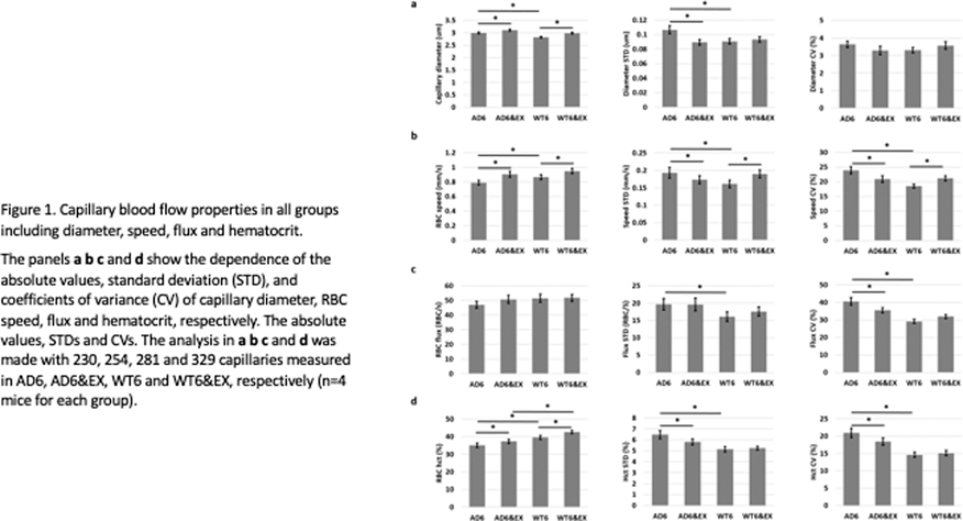
Conclusions
We observed decreased brain perfusion and higher heterogeneity of capillary flow properties in AD mice. We further quantified these changes as a function of branch order. Voluntary exercise was shown to modulate heterogeneity properties. Given the hypothesized role of capillary transit time heterogeneity (CTTH) on oxygenation, this data will help better characterize the origin of CTTH changes occurring with AD.
Reference
PB02-G12
Hemorheologic enhancement of cerebral perfusion by drag-reducing polymers for the treatment of the alzheimer’s disease
1University of New Mexico, USA
2University of Pittsburgh, USA
Abstract
Alzheimer’s disease (AD) dementia is a consequence of complex interactions of age-related neurodegeneration and vascular-associated pathologies which presently affects more than 44 million people worldwide. For the last decade, it has been suggested that chronic brain hypoperfusion and consequent hypoxia play a direct role in the pathogenesis of AD. However, current treatments of AD have not focused on restoring or improving microvascular perfusion. We propose a new treatment approach using modulation of hemorheology by drag reducing polymers (DRP). DRP are linear, soluble macromolecules that reduce flow separations at blood vessel bifurcations leading to a reduction of pressure gradients across the arterial system and an increase in the precapillary blood pressure enhancing capillary perfusion. In a previous study, we showed that DRP enhance cerebral blood flow and tissue oxygenation. Here we tested the efficacyof hemorheologic enhancement of cerebral perfusion by drag-reducing polymers for the treatment of the Alzheimer’s disease.
Methods
To test the efficiency of DRP we used double transgenic B6C3-Tg(APPswe, PSEN1dE9) 85Dbo/Mmjax AD mice. DRP or vehicle (saline) were i.v. injected every week starting at one month of age till 12 months of age (10 mice/group). In-vivo 2-photon laser scanning microscopy was used to evaluate amyloid plaques development by autofluorescence (second harmonic generation) and by i.v. labeling with 0.001% of thioflavine S. For microcirculation evaluation, blood plasma was labeled with tetramethylrhodamine isothiocyanate dextran. The imaging sessions were repeated once a month till 12 months of age when animals were undergo transcardiac perfusion for further histochemical analysis. Differences between groups were determined using two-way analysis of variance (ANOVA) for multiple comparisons and post hoc testing using the Mann-Whitney U test.
Results
In vehicle group, numerous plaques completely formed in the cortex by 9 months of age. The development of plaques accumulation was accompanied with the progress of cerebral microcirculation disturbances and metabolic impairment. DRP mitigated microcirculation reduction (microvascular perfusion was 29.5 ± 5 % higher than in vehicle group, P < 0.05). In DRP group, amyloid plaques deposition was substantially less than in vehicle group (P < 0.01).
Conclusions
Rheological enhancement of blood flow by DRP effectively ameliorates the amyloid plaques development and cognitive decline in AD mice.
PB02-G13
Neuroprotective and anti-radical potential of butanolic extract of Myristica fragrans against scopolamine-induced cognitive deficit in the experimental model of Alzheimer’s disease
1Department of Pharmaceutical Sciences, SHUATS, Allahabad
Abstract
Back ground
Neurodegenerative disorders such as dementia and Alzheimer’s disease are widespread clinical problem which is associated with advanced dysfunction and structural abolition of functional neurons leads to neuronal cell death. Myristica fragrans is a commonly available medicinal plant which is used as antimicrobial, anti-inflammatory, anxiolytic, digestive tonic, headache and in cosmetic purpose. However, It is well accounted in the literature about its memory enhancing activity and anti-oxidant activity.
Objective
This study was design to examine the reversal of cognitive deficits and anti-radical potential of butanolic extract of Myristica fragrans (MFBu) scopolamine induced memory impairment in mice.
Materials and method
Butanolic extract (MFBu) at a dose level of 100 and 200 mg/kg, p.o was administered to mice daily for 21 days. On the last day (21 day), scopolamine (0.6 mg/kg, i.p.) was injected to induce memory impairment before performing the behavioral test. The phytoconstituents of the butanolic extract was determined by HPLC/DAD analysis. Two behavioural paradigms i.e. Hebbs William maze (HWM) and Elevated plus maze were (EPM) were employed to assess the memory. Brain homogenate was used to measure the acetylcholinesterase (AChE) activity. Antioxidant profile i.e. Superoxide dismutase,glutathione peroxidase,catalase and TBARS were also measured in the hippocampus of the mice brain.
Results
Major compounds kaempferol, gallic acid, chlorogenic acid, caffeic acid and ellagic acid were quantiifed in the plant extarct (MFBu). Furthermore, butanolic extract reversed the scopolamine-induced retention transfer latency (RTL) in EPM model and time taken to reach reward chamber (TRC) in HWM model. Treatment with MFBu also recovered scopolamine-induced alteration of the antioxidant parameter (enhanced the level of Catalase, SOD and Glutathione peroxidase) and acetylcholinesterase activity in the hippocampus of the mice brain.
Conclusion
These outcome demonstrate that MFBu could be potent neuropharmacological and antioxidant agent against Alzheimer’s disease.
PB02-G14
Pharmacological stimulation of soluble guanylate cyclase improves memory function and reduces plaque load in a mouse model of Alzheimer’s disease
1Laboratory of Experimental Stroke Research, Institute for Stroke and Dementia Research (ISD), University of Munich Medical Center
2Munich Cluster of Systems Neurology (Synergy), Munich, Germany
3Bayer AG, Research & Development, Pharmaceuticals, Wuppertal, Germany
Abstract
Objectives
Nitric oxide/soluble guanylate cyclase/cyclic guanosine monophosphate (NO-sGC-cGMP) signaling is important for modulating synaptic transmission and plasticity. This is supported by the fact that reduced levels of cGMP were reported in CSF from patients suffering from Alzheimer’s Disease (AD) and correlated with memory decline1. Therefore, we hypothesize that pharmacological stimulation of soluble guanylate cyclase (sGC), the enzyme which produces cGMP, using a novel class of sGC stimulators2 may improve memory function in a mouse model of AD.
Methods
5XFAD mice or wild-type littermates (n = 11/group) were fed between 2 and 12 months of age with chow containing 45 mg/kg BAY 41-2272, a specific sGC stimulator, or placebo in a strictly randomized and blinded fashion. The overall condition of the animals was recorded regularly and learning and memory were assessed monthly by Barnes maze and analyzed with an automated tracking software package (Noldus). At the end of the observation period Aβ plaque load was quantified by Thioflavine-S and Congo Red staining. The study was unblinded after all data were assessed and analyzed.
Results
Untreated 5XFAD mice started to develop progressive memory loss at an age of 5 months as compared to untreated WT mice (p < 0.001). Animals receiving BAY 41-2272 showed no obvious adverse effects, but displayed a significantly decreased escape latency, less total errors, and spend more time in the goal area at 6 (p < 0.05), 7 (p < 0.01) and 8 (p < 0.05) months of age. Histochemical analysis at the end of the observation time (12 months) revealed no plaques in WT mice and massive accumulation of Aβ plaques in untreated 5xFAD mice. In treated 5xFAD mice, however, the plaque area was significantly (p < 0.05) reduced 30% in all investigated brain regions (cerebral cortex, hippocampus, thalamus and midbrain).
Conclusion
Our results demonstrate that long-term pharmacological stimulation of sGC by oral application of BAY 41-2272 is well tolerated and has beneficial effects on memory function in a mouse model of AD. This finding supports our hypothesis that increasing/normalizing cGMP levels in the brain may serve as a novel therapeutic concept in AD. Unexpectedly, treatment with BAY 41-2272 also reduced plaque load. Although this finding further explains the positive effect of BAY 41-2272 on memory function, the underlying mechanisms remain unclear and warrant further investigations. Over all, our study indicates that activation of sGC may serve as a novel therapeutic target for AD.
References
PB02-G15
The neural substrate of memory, attention, and executive function; A study of early stage AD using MRI and SPECT
1Department of Neurology, Tokyo Women's Medical University
2Department of Diagnostic Imaging and Nuclear Medicine, Tokyo Women's Medical University
Abstract
Objectives
To investigate the neural substrates of neuropsychological domains in early stage Alzheimer’s disease (AD) who underwent an extensive neuropsychological examination, high resolution MRI, and brain perfusion SPECT with voxel-based morphometry (VBM) and Statistical Parametric Mapping 12 (SPM12).
Methods
We included 83 patients with early stage AD presenting with Clinical Dementia Rating ≤ 1. The mean age of the patients was 75.4 ± 7.8, the mean years of education was 13.3 ± 2.5, and the mean MMSE score was 24.1 ± 3.1. The neuropsychological testing included the Mini-Mental State Examination (MMSE), the Rey’s Auditory Verbal Learning Test (RAVLT), the Rey-Osterrieth Complex Figure Test (ROCFT), verbal fluency tests of initial letter and category, Trail Making Test A and B, Symbol Digit Modalities Test (SDMT), Frontal Assessment Battery (FAB), and the Digit Span subtest from the Wechsler Adult Intelligence Scale Revised. To identify the underlying factor structure, a factor analysis was performed on 14 neuropsychological variables using maximum likelihood method with Promax rotation. Factor extraction was based on the Kaiser-Guttman rule of retaining components with eigenvalues >1. Factor loading of each test >0.45 was considered as significant contributor to the factor. 3D-T1WI MRI data and 99mTc-ECD SPECT data were preprocessed using VBM and SPM12. The volume of white matter hyperintensity (WMH) was calculated using 3D slicer based on 3D-FLAIR image.
Conclusions
The difference of neuropsychological profile in early stage AD reflects the difference of atrophy of neuroanatomical structure and regional hypo-perfusion.
PB02-G16
Theragnostic approach for early diagnosis of Alzheimer's Disease: PANA Project
1Clinical Neurosciences Research Laboratory, Health Research Institute of Santiago de Compostela (IDIS), Santiago de Compostela, Spain
2Centro Singular de Investigación en Química Biolóxica e Materiais Moleculares (CIQUS), and Departamento de Física de Partículas, Universidade de Santiago de Compostela, Santiago de Compostela, Spain
3Molecular Imaging Group, Health Research Institute of Santiago de Compostela (IDIS), Santiago de Compostela, Spain
4Institute of Biomedical Research of Salamanca, University Hospital of Salamanca, University of Salamanca, Consejo Superior de Investigaciones Científicas (CSIC), Salamanca, Spain
Abstract
Objective
Alzheimer’s disease (AD) is the leading cause of dementia and loss of autonomy in the elderly, implying a progressive cognitive decline and limitation of social activities. Progressive aging of population will increase the magnitude of this problem in the next decades. Currently, there is not an effective method for the early diagnosis of AD. Therefore, there is an urgent need to develop new effective early diagnostic and therapeutic strategies to help in delaying the appearance of the most adverse symptoms of this disease. To defeat this challenge, this study bases its approach on the importance of tau in the early pathophysiological processes of AD. Our strategy was based on two fundamental pillars; on one hand, efforts were focused on multimodal PET/MRI imaging which is gaining relevance as the best solution for diagnostic purposes due to the complementary advantages of both technologies, combining the high structural characterization of tissue provided by MRI with the enhanced sensitivity of PET imaging. On the other hand, the challenging development of a theragnostic nanostructure was focused on tau detection, which has to deliver theragnostic agents into the brain to provide in situ diagnostic and therapeutic effects.
Methods/Results
Our design was based on iron oxide nanoparticles (NPs) doped with zinc and manganese (1), stabilized with oleic acid/oleylamine in organic solvents, which were transferred to the aqueous phase by coating with an amphiphilic polymer (dodecyl-grafted-poly(isobutylene-alt-maleicanhydride, in the followign referred to as PMA) (2, 3). Modified-PMA conferred colloidal stability to the NPs in high ionic strength media and provides several chemical groups (e.g., carboxyl, dibenzocyclooctyne, furfuryl, etc.) for functionalization with other macromolecules, including homing antibodies (anti-tau and anti-B_amyloid oligomers), polyethylene glycol, PET radiotracers (Zirconium-89) and fluorescence markers. The designed nanostructures worked as good contrast agents for MRI in T2 and T2* sequences (r2 = 409.1 mM-1 s-1). Furthermore, non-toxic effects (up to concentrations of [Fe] greater than 50 ug/mL) were observed in vitro in both endothelial (bEnd.3) and primary neurons cell cultures by LDH, MTT, and IP/Annexin. Effective biofunctionalization of NPs with monoclonal antibodies of tau and B-amyloid was obtained by the click-chemistry cu-free method. Radiolabelling of NPs for PET studies was performed with 89Zr.
Conclusions
This study shows a novel theragnostic nanostructures that specifically recognize very-early molecular markers of AD, and can be detected by means of non-invasive imaging methodologies (MRI and/or PET, which are already common techniques accessible in most hospitals), and eventually provide a therapeutic action if needed.
PB02-H01
Arterial stiffness, cerebral blood flow and white matter integrity in the elderly: an arterial spin labelling and Neurite orientation dispersion and density study
1NeuroPoly Lab, Institute of Biomedical Engineering, Polytechnique Montreal, Montr&amp;amp;eacute;al, QC, Canada
2Centre de recherche de l’Institut Universitaire de Gériatrie de Montréal (CRIUGM), Montreal, QC, Canada
3Department of Neurosciences, Faculty of Medicine, Université de Montréal, QC, Canada
4Department of biomedical Sciences, Faculty of Medicine, Université de Montréal, QC, Canada
5Institut de Recherches Cliniques de Montréal, Université de Montréal, Montréal, QC, Canada
6Montreal Heart Institute, Montreal, QC, Canada
7PERFORM Centre, Concordia University, Montréal, QC, Canada
8Department of Pharmacology and Physiology, Faculty of Medicine, Université de Montréal, QC, Canada
9Department of Medicine, Faculty of Medicine, Université de Montréal, QC, Canada
10Physics Department, Concordia University, Montréal, QC, Canada
Abstract
Objectives
Arterial stiffness impacts the brain’s white and grey matter (WM and GM) and have been consistently associated with cognitive dysfunction and dementia 1–3. The European Society of Hypertension established a threshold for carotid-femoral pulse wave velocity (cfPWV) of 10 m/s, above which cardiovascular risks are likely to occur. However the threshold at which arterial stiffness alters brain integrity has never been established. Our aim is to better understand the impact of cfPWV on brain integrity and to estimate the cfPWV threshold at which there is an impact on brain integrity in elderly without dementia.
Methods
54 healthy individuals (age = 69.91 ± 3.31years) were scanned on a 3T (MAGNETOM Prisma Fit, Siemens) to compute Neurite orientation dispersion and density (NODDI) metrics from diffusion-weighted images (DWI). CBF in the GM was measured using pseudo-continuous arterial spin labelling (pcASL). DWIs were processed using TOAD4 to compute the intracellular volume fraction (vic) and the isotropic volume fraction (v iso) corresponding respectively to the neurite density index and the orientation dispersion index5. pcASL data were processed using Neurolens6. MRI analyses were done in global GM and WM. Additional ROI analysis included 4 regions denoted vulnerable to cfPWV1: the corpus callosum, internal capsule, corona radiata and thalamus 1,4,7. cfPWV was measured following the Van Bortel protocol8. The relationship between cfPWV, CBF and NODDI metrics was assessed using a linear regression model in global WM and GM and in ROIs using age, sex and 24 h systolic blood pressure as covariates. cfPWV cut-off value was estimated using maximally selected rank statistics. Correction for multiple comparisons was performed using a false discovery rate (FDR) procedure.
Results
cfPWV was a significant predictor of CBF in the global GM (p = 0.014). However cfPWV was not a significant predictor for alterations in the global WM (p = 0.241, p = 0.532 for v iso and vic respectively). When looking at the effect of cfPWV regionally, cfPWV was found to be a significant predictor of vic and viso in the corpus callosum (p = 0.023, p = 0.049 respectively), but the results did not remain significant for viso after FDR. cfPWV was not found to be a predictor of CBF in the thalamus (p = 0.125).
Figure 1 shows that the cfPWV cut-off is 10 m/s when taking into account CBF in the GM as a ground truth for comparison. Interestingly, the cfPWV cut-off was found to be around 8.5 m/s when taking into account vic and viso in the corpus callosum (figure 1).
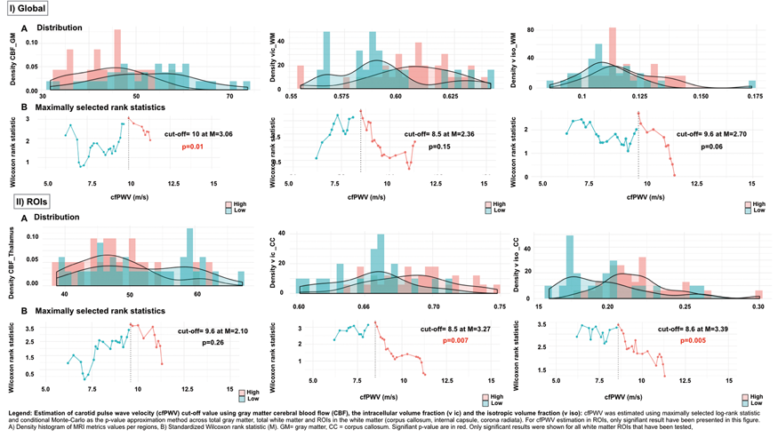
Conclusion
We provide a first evidence that arterial stiffness impacts the neurite density of the corpus callosum (vic). Our findings also suggest that the 10 m/s cfPWV cut-off in the literature may have to be lowered to around 8.5 m/s to make a more accurate distinction between lower and higher risk groups of individuals for brain structural changes. This may help preserve brain health and prevent cognitive decline.
References
PB02-H02
Cerebral perfusion in middle-aged masters athletes: associations with age and cardiorespratory fitness
1Human Informatics Research Institute, National Institute of Advanced Industrial Science and Technology, Tsukuba, Ibaraki, Japan
2University of Texas Southwestern Medical Center, Dallas, Texas, USA
3Texas Health Presbyterian Hospital Dallas, Dallas, Texas, USA
Abstract
Objectives
Cerebral perfusion starts decreasing during middle age which may contribute to the risk of developing stroke and dementia in later life (Tarumi et al., 2018). In contrast, regular aerobic exercise has been shown to improve cardiovascular health and neurocognitive function (Tarumi et al., 2015); however, the long-term effects of aerobic exercise training on cerebral perfusion in middle-aged adults remains unknown. Therefore, the purpose of this study was to determine whether middle-aged endurance Masters athletes (MA) have the attenuated age-related reductions of cerebral perfusion.
Methods
Thirty middle-aged MA (54 ± 4 years, 15 women) who have participated at least 10 years of endurance training were compared with 30 age-matched sedentary adults (MS, 54 ± 4 years, 15 women) and 30 young sedentary adults (YS, 32 ± 6 years, 15 women). Using a 3T MRI system, each participant underwent T1-weighted magnetization-prepared rapid acquisition of gradient echo (MPRAGE) imaging and pseudo-continuous arterial spin labeling (pCASL) imaging. The pCASL data were analyzed in both individual native T1 space and standard MNI template for the region-of-interest and voxelwise statistics.
Results
By comparing between the YS and MS groups, age-related reductions of cerebral perfusion were evident across the whole brain, including the cerebellum (all P < 0.05). Conversely, MA demonstrated increased perfusion in the cerebellar, inferior parietal, lateral occipital, medial orbitofrontal, and frontal pole cortical areas when compared with the MS group (all P < 0.05). The age-related reduction of cerebellar perfusion was significantly attenuated in the MA group (P < 0.05). Voxelwise statistics further demonstrated that higher cardiorespiratory fitness, as measured by maximal oxygen uptake (VO2max), is positively correlated with higher perfusion in the cerebellar cortex, lateral occipital area, and middle temporal gyrus among the middle-aged participants (Figure).
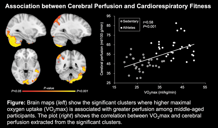
Conclusions
Long-term endurance training attenuated the age-related reductions of regional cerebral perfusion in the cerebellar cortex, prefrontal cortex, and lateral occipital areas in middle-aged adults.
References
PB02-H03
Baseline aerobic fitness and cortical volume atrophy across 5 years in older adults from the general population
1Department of neuromedicine and movement science, NTNU, Norway
2Department of Circulation and Medical Imaging, NTNU, Norway
Abstract
Objective
Aerobic fitness is considered an important cause of late onset dementia. Cross-sectional (and interventional) MRI studies show a relationship between different measures of physical fitness and brain volumes. In this study, we tested the hypothesis that baseline aerobic fitness reduces brain volume atrophy over a five-year period.
Methods
The participants, born between 1936-1942, were a random selection of 99 participants (53 women) from the Generation 100 (G100) study. The G100 study is a randomized clinical trial on the effect of fitness training on cardiovascular morbidity and mortality (https://clinicaltrials.gov/ct2/show/NCT01666340). The participants underwent MRI at baseline and at end of intervention, i.e. after 5 years. All MRI scanning was performed with the same protocol on the same 3T Skyra scanner with a 32-channel head coil (Siemens). The T1-weighted MPRAGE (TR = 1900; TE = 3.16; FOV = 256x256; slice thickness = 1 mm; gap = 0 mm) and T2-weighted volumes (TR = 5000; TE = 388; FOV = 256x256; slice thickness = 1 mm; gap = 0 mm) were used in this study. The MPRAGE volumes were analyzed in the Freesurfer suite v. 6.0 (http://surfer.nmr.mgh.harvard.edu/). The following volumes were selected for analysis: cortex, hippocampus, caudate, total cerebral white matter. Right and left hemisphere volumes were combined, and the volume differences between baseline and 5 years used in the regression analysis. Intracranial volume (ICV) were estimated from the T1 and T2 weighted volumes in SPM8 (http://www.fil.ion.ucl.ac.uk/spm) with the automatic reverse brain mask method.
VO2 was assessed using graded maximal exercise testing on a motor-driven treadmill or exercise bike. VO2max was used in all but 34% of the participants who did not achieve VO2max. For those VO2peak were used. VO2peak reflects the highest value of oxygen uptake attained in a maximal exercise test where no plateau in oxygen consumption is observed.
The registered VO2max/peak values were normalized to weight measured with an eight-polar bioelectrical impedance analyzer (Inbody 720).
Hierarchical linear regression was performed in RStudio version 3.3.3 to study if baseline VO2max/peak predicts volume loss across 5 years. Since both age and brain volume are associated with the atrophy rate, we added cortical volumes at baseline and age as a second step. Brain volumes were corrected for ICV using the residuals method. Threshold for statistical significance was set at p < 0.05.
Results
In step 1 the atrophy rate was associated with VO2max/peak. The second step significantly improved the fit of the model. Including these covariates led to a non-significant association between VO2max/peak and atrophy rate.
Conclusions
The association between VO2max/peak and cortical atrophy rate is complex. Cortical atrophy and cortical volumes are each significantly associated with VO2max/peak, but when both covariates are added in the model they are not statistically significant.
The present results demonstrate that multiple confounders are present and more complex/advanced statistical modelling is needed.
PB02-H04
The effect of physical fitness on hippocampal volume and cognition in older adults from the Generation100 study
1Department of Neuromedicine and Movement Science, Norwegian University of Science and Technology (NTNU), Trondheim, Norway
2Department of Radiology and Nuclear Medicine St Olavs Hospital, Trondheim University Hospital, Trondheim, Norway
3Department of Circulation and Medical Imaging, Norwegian University of Science and Technology (NTNU), Trondheim, Norway
Abstract
Objectives
Cardiorespiratory fitness has emerged as important determinant for healthy brain aging. The aim of this study was to examine the relationship between VO2, which is a gold standard for assessing cardiorespiratory fitness, hippocampal volume and performance on cognitive tests associated with hippocampus in older adults from the general population.
Methods
A random selection of older adults (n = 98), born between 1936-42 from the randomized clinical trial Generation100 (https://clinicaltrials.gov/ct2/show/NCT01666340), underwent structural MRI on the same 3T Skyra scanner with a 32 channel head coil (Siemens). The T1-weighted MPRAGE (TR = 1900; TE = 3.16; FOV = 256x256; slice thickness = 1 mm; gap = 0 mm) and T2-weighted volumes (TR = 5000; TE = 388; FOV = 256x256; slice thickness = 1 mm; gap = 0 mm) were used in this study. Hippocampal volumes were obtained from the T1 weighted scans using Freesurfer (v. 6.0) (http://surfer.nmr.mgh.harvard.edu/). Right and left hippocampal volume was combined. Intracranial volume was estimated from the 3D T1 and T2 weighted scans in SPM. Hippocampal volumes were adjusted for intracranial volume using residual method. Cognitive testing of pattern separation, verbal memory and spatial memory was performed. Aerobic physical fitness was assessed using graded maximal exercise testing on a motor-driven treadmill or exercise bike. Since 34% of the participants did not achieve VO2max, VO2peak was used. VO2peak reflects the highest value of oxygen uptake attained in a maximal exercise test where no plateau in oxygen consumption is observed. The registered VO2peak values were normalized to weight measured with an In-Body Machine. Statistical analyses were performed in SPSS. Associations between VO2peak and hippocampal volume and cognitive test scores were assessed adjusting for sex and education level. Threshold for statistical significance was set at p < 0.05.
Results
There was a significant positive association between VO2peak and hippocampal volume. Those with higher VO2peak were likely to have higher hippocampal volumes (p < 0.01, partial η2 = 0.10). Effects of sex and education were not significant.
There was no significant association between VO2peak and any of the cognitive test scores. Pattern separation approached significance (p = 0.068). Effects of education were not significant. Women were significantly better in spatial memory (p = 0.044, partial η2 = 0.04) and verbal memory tests (p = 0.41, partial η2 = 0.05). Pattern separation test scores approached significance (p = 0.06).
There was a significant association between hippocampal volume and spatial memory (p = 0.03, partial η2 = 0.05). The rest of the cognitive tests were not significantly associated. Effects of sex and education were not significant.
Conclusions
In a cohort of older adults from general population, cardiorespiratory fitness was positively associated with hippocampal volume, but not memory performance. Hippocampal volume was however, related to spatial memory. This implies that while there is a relationship between physical activity and the brain structure, it does not translate directly into improving cognitive functions of older adults.
PB02-H05
Alterations in brain phospholipidome of dementia with lewy bodies and their associations with neuropathological parameters
1Dept of Pharmacology, Yong Loo Lin School of Medicine, National University of Singapore, Singapore
2Memory, Aging and Cognition Centre, National University Health System, Singapore
3Department of Biochemistry, Yong Loo Lin School of Medicine, National University of Singapore, Singapore
4Singapore Lipidomics Incubator (SLING), Life Sciences Institute, National University of Singapore, Singapore
5Wolfson Centre for Age-Related Diseases, King’s College London, London, UK
6Department of Neurobiology, Ward Sciences and Society, Karolinska Institute, Stockholm, Sweden
7Institute of Neuroscience, Newcastle University, CAV, Newcastle upon Tyne, UK
Abstract
Objectives
Previous studies have demonstrated alterations of specific phospholipid species, including those that are docosahexaenoic acid (DHA) containing, in the brains of Alzheimer’s disease (AD) or mixed dementia (AD and vascular dementia) patients (1, 2). DHA is involved in neuronal survival, synaptic plasticity, and cognitive processes (3). Deregulation of DHA is suggested to underlie cognitive impairments observed in AD patients, and increasing evidence showed that DHA dysregulation or deficit may contribute to disease pathogenesis (1, 3, 4). However, there is a lack of research into brain phospholipid-DHA changes in dementia with Lewy bodies (DLB), another major cause of neurodegenerative dementia characterized by Lewy bodies (LBs) containing aggregated α-synuclein as well as variable degree of AD pathology such as amyloid-β (Aβ) plaques and hyperphosphorylated tau in the cortex (5). In our study, we reported alterations in the DHA-containing phospholipid species in DLB neocortex, and the potential associations between such alterations and a range of neuropathological parameters.
Methods
Post-mortem samples from parietal cortex (BA40) of 28 DLB patients and 17 age-matched controls were studied. Liquid chromatography coupled to mass spectrometry was used to analyze the lipidomes of the subjects. Phospholipid species with a fatty acid sum composition containing at least 38 carbons and 6 double bonds were considered DHA-containing (6). Soluble Aβ42 and phosphorylated tau levels were measured using enzyme-linked immunosorbent assay kits. A P < 0.05 was considered to be statistically significant.
Results
Among the 214 phospholipid species identified, only 28 species were significantly altered in DLB. Of which, eight of them were DHA-containing, with seven being significantly decreased in DLB, the majority of which were derived from the phosphatidylcholine (PC) subclass. There was no significant difference in total levels of DHA derived from each subclass between controls and DLB. Correlation analysis revealed that in the DLB patients group, PC-DHA was negatively correlated with soluble Aβ42 levels, while phosphatidylethanolamine (PE)-DHA showed similar correlation with NFTs and LBs scores, and phosphorylated Tau levels. Focusing on individual significantly altered DHA-containing phospholipid species, only PC 40:6 and PC 40:8/PC 39:1/PC O-40:1/PC P-40:0 correlated negatively with soluble Aβ42 levels.
Conclusion
In this study, we described novel alterations of DHA-containing phospholipids in DLB. Of particular interest is PC40:6, a major DHA-containing phospholipid species, which was consistently decreased in DLB, similar to that reported for AD brain and plasma (2) (7). It has been postulated that the decrease in PC40:6 in the plasma may be associated with its decrease in the brain. Besides, this DHA-containing phospholipid species correlated with soluble Aβ42 levels, the more neurotoxic form of Aβ (8). The findings from the current study provide a basis to better understand the putative pathogenic roles of phospholipid dysregulation in neurodegenerative dementias and to advance current diagnostic and therapeutic strategies.
References
PB02-H06
Post-mortem assessment of apoptotic factors in Lewy body dementia
1Dept. of Pharmacology, National University of Singapore, Singapore
2Memory, Ageing and Cognition Centre, National University Health System, Singapore
3Wolfson Centre for Age-Related Diseases, King’s College London, London, UK
Abstract
Objectives
After Alzheimer’s disease (AD), Lewy body dementia (LBD) is the most common forms of neurodegenerative dementias. LBD is a collective term for two major clinical diagnoses – dementia with Lewy bodies (DLB) and Parkinson’s disease dementia (PDD), distinguished on the basis of the temporal manifestation of cognitive impairments relative to parkinsonism. The typical pathological feature of DLB and PDD is the accumulation of Lewy bodies (LBs) containing aggregated α-synuclein. Interestingly, DLB frequently shows concomitant AD pathology of variable severity, while PDD manifests AD pathology at a lower frequency and and severity compared to DLB. It has been suggested that the presence of these abnormal protein aggregates in the cortex may underlie the neuronal loss and cognitive symptoms observed in the patients. However, there is little evidence to suggest an alteration in the levels of apoptotic factors in the brains of PDD and DLB patients, and the association with AD neuropathological burden.
Methods
Post-mortem tissues from parietal cortex (BA40) of 17 PDD and 31 DLB patients, and 15 age-matched controls were studied. Apoptotic factors such as Bcl-2, Bcl-xl, BAX and cleaved caspase 3 were measured using Western blot. Soluble Aβ42 and phosphorylated tau levels were measured using Aβ42 enzyme-linked immunosorbent assay (ELISA) kits. p < 0.05 were considered statistically significant.
Results
Compared with control brain, there were significant increases in the mean protein level of anti-apoptotic factor Bcl-2 in PDD and DLB. In contrast, the level of pro-apoptotic factor BAX was marginally decreased in DLB. In a smaller cohort, there was an increasing trend of cleaved caspase 3, another pro-apoptotic factor, in the dementia groups. No significant correlation was observed between the individual apoptotic markers and the neuropathological burden.
Conclusion
Our preliminary results demonstrated a dysregulation in apoptosis in the brains of the LBD patients. Interestingly, we have observed an increase in the anti-apoptotic factor Bcl-2 in the dementia groups, possibly suggesting a compensatory response in the parietal cortex.
PB02-H07
The optimal cut-off points on 11C-PIB-PET-MR based on visual assessments of amyloid positivity in Singapore
1Department of Pharmacology, National University of Singapore, Singapore
2Memory Aging & Cognition Centre, National University Health System, Singapore, Singapore
3Department of Neurology, National Cerebral and Cardiovascular Center, Japan
4Clinical Imaging Research Centre, National University of Singapore, Singapore
5Department of Diagnostic Imaging, National University of Singapore, Singapore
Abstract
Objectives
To assess amyloid PET, dichotomous approaches to define amyloid positivity are required in certain situations such as determining eligibility in clinical trials. There are several SUVr cut-off points calculated using various methods, however, there are few cut-off points reported for Asian populations and cut-off points in Caucasian might not be suitable due to genetic and other differences. Hence, we aim to explore the optimal cut-off point on 11C-PiB-PET-MR in Singapore.
Methods
All subjects underwent physical, clinical and neuropsychological assessments and neuroimaging at National University Hospital in Singapore. Assessments of participants were performed from April 2016 to September 2018. Participants were classified into 4 diagnostic groups: no cognitive impairment (NCI), cognitive impairment no dementia (CIND), vascular dementia (VaD) and Alzheimer’s disease (AD). 11C-PIB-PET-MR imaging was performed on a mMR synchronous PET/MR scanner (Siemens Healthcare GmbH). PET images were independently interpreted visually by one expert rater. By using the two quantitative pipelines [conventional standard uptake ratio (SUVr), amyloid load utilising a new method (AβL, explained in another presentation)], we took two approaches to determine the cut-off points. The first approach was based on the distribution of the NCI group only. In Gaussian mixture, hierarchical cluster, and k-means cluster models, which are unsupervised learning algorithms, we divided the results into 2 clusters (high and low cluster) and defined the cut-off point as the upper 95th percentile of the low cluster. The second approach was based on the receiver operating characteristic (ROC) curve analysis with amyloid status on visual reading assessment of 11C-PIB-PET-MR. Finally, sensitivities and specificities were computed for each of the 4 cut-off points using the visual reading assessments as the gold standard for comparison.
Results
After excluding subjects with incomplete data acquisition, 174 subjects (mean age 75.7 years, 94 females) were included in this study. Subjects were in the following diagnostic categories: NCI (n = 27), CIND mild (n = 54), CIND moderate (n = 43), VaD (n = 17), AD (n = 33). The allele frequencies of APOE ɛ4 gene in NCI group was 22.2%. Of 174 subjects, 68 (39.1%) subjects were positive on the visual reading assessment after consensus discussion. The mean values of SUVr and AβL of all subjects were 1.40 (SD, 0.42) and 28.8 (SD, 29.9). The cut-off points obtained with the 3 unsupervised methods (1st approach) were respectively1.23, 1.35 and 1.35 for SUVr and 21.5 for the 3 techniques for AβL. The second approach led to a 1.38 cut-off point for SUVr and 19.9 for AβL. After comparing the cut-off points, we defined the optimal cut-off points as 1.38 for SUVr and 20 for AβL (SUVr: sensitivity 88.2%, specificity 98.1%, accuracy 94.3%, AUC 0.97, AβL: sensitivity 98.5%, specificity 96.2%, accuracy 97.1%, AUC 0.99).
Conclusions
Using a Singaporean cohort, we found that the optimal cut-off points on 11C-PiB scans for classification as PiB negative or positive was 1.38 using SUVr and 20 if AβL is employed.
PB02-H08
Optimisation of 11C-PiB scan data analysis leads to increased correlations between image derived biomarker and cognitive function scores
1Clinical Imaging Research Centre, National University of Singapore
2Department of Pharmacology, National University of Singapore
3Memory Aging & Cognition Centre, National University Health System, Singapore
4ASTAR, Singapore
5SBIC, ASTAR, Singapore
Abstract
Objectives
To optimise the analysis pipeline for 11C-PiB scans and determine optimal biomarkers for cognition in a memory clinic cohort from Singapore.
Methods
186 subjects consisting of No Cognitive Impairment n = 30; Cognitive Impairment No Dementia (mild n = 56; moderate n = 45); Vascular Dementia n = 19; and Alzheimer’s Disease (AD) n = 36 underwent a 30-min 11C-PiB PET scan and a structural T1 magnetic resonance imaging.
Motion correction (MOCO)
Each 11C-PiB listmode was reconstructed without and with motion correction using an in-house developed rebinner1.
Subject specific volumes of interest (VOIs) versus population-based VOIs in MNI-152 space:
Two different pipelines were implemented and compared for the processing of the 11C-PiB scans.
a) The first approach involves parcellation of T1-weighted images (Freesurfer version 6.0.0; surfer.nmr.mgh.harvard.edu) which were aligned with the PET image (ANTS version 1.9) and thus were used to define cortical regions of interest and reference regions in the subject’s native space. Standard Uptake Value Ratio (SUVr) volumes were generated using the cerebellar gray matter as the reference region. Subject’s global SUVr (
b) The second pipeline for SUVr measurements does not require the parcellation of the subject MRI. After linear and non-linear normalisation in MNI-152 space, SUVr values (
SUVr biomarker versus AβL biomarker
The AβL biomarker is obtained by modeling the subject SUVr_AβL volume in MNI-152 space as a linear combination of 2 canonical volumes: NS representing the Non-Specific binding component and K, the carrying capacity which represents the regional maximum possible concentration of Aβ.
SUVr_AβL = ns x NS + AβL x K
Where ns and
Pipeline assessment
We assessed the performance of each pipeline & biomarker using Pearson’s correlation analysis between the resulting biomarker measurements with cognitive scores.
Results
6 scans with extreme motion were identified: mean motion magnitude over the scan duration [5.21 – 12.07] mm and max motion magnitude [11.90 – 39.55] mm led to SUVr underestimations by 40%. Without motion correction, those scans would have been excluded from the analysis. Motion correction led to an increased correlation between the SUVr and the cognitive function scores (Table).

Automated MRI parcellation failed for 7 subjects. In addition, segmentation quality for 4 additional subjects was suboptimal leading to a total of 11 missing SUVr_mri values. The table shows that SUVr_ AβL values better correlate with cognitive function scores than SUVr_mri. Finally, the biomarker AβL better correlates with cognitive scores than SUVr_AβL.
Conclusions
This optimization effort led to improved correlations between computed SUVr and the cognitive function scores which increased from 17% to 40%. In addition, the use of the AβL in place of the traditional SUVr_AβL led to additional increases by up to 18%.
Reference
PB02-H09
Role of semi-quantitative assessment using FP-CIT SPECT in the differential diagnosis of degenerative dementia
1Department of Dementia Research, Akita Prefectural Center for Rehabilitation and Psychiatric Medicine, Japan
2Department of Radiology, Akita Prefectural Center for Rehabilitation and Psychiatric Medicine, Japan
Abstract
Introduction
The differential diagnosis of degenerative dementias (e.g., Alzheimer disease (AD), dementia with Lewy bodies (DLB), frontotemporal dementia (FTD), corticobasal syndrome (CBS)) has been challenging in view of overlapping clinical features. Variants of AD often show similar MRI findings and cerebral perfusion patterns on SPECT to non-AD dementias. The role of FP-CIT SPECT in FTD and CBS is rather unclear, whereas FP-CIT SPECT is well-established in the differentiation of DLB and AD. The present study aimed to explore a potential of semi-quantitative assessment using FP-CIT SPECT in the differential diagnosis of FTD, CBS, and DLB from AD.
Methods
We recruited 215 consecutive patients with clinical diagnosis of FTD (n = 24; 8 behavioral variant, 16 semantic dementia), CBS (n = 6), DLB (n = 149), and AD (n = 36) undergoing MRI and both IMP and FP-CIT SPECT/CT. These patients fulfilled the previously mentioned diagnostic clinical criteria and presented typical findings on MRI and IMP SPECT. MRI and IMP SPECT/CT were also used to exclude patients with stroke including vascular pathology, idiopathic normal pressure hydrocephalus, and chronic subdural hematoma. The specific binding ratio (SBR) and the asymmetry index (AI) for caudate nucleus, putamen, and whole striatum were calculated using a new striatal analysis software (Scenium®). Those for the whole striatum were also compared to the striatal SBR and AI calculated using DaTView® software excluding the CSF fraction by the threshold method. The AI of the SBR was automatically calculated as follows: AI =
Results
Patients with FTD showed a mild reduction of all SBRs using Scenium® and significant reduction of striatal SBR using DaTView® compared to AD patients (P < 0.01). FTD patients revealed a more homogenous uptake of FP-CIT than patients with CBS and DLB. 8.3% (2/24) of FTD patients presented with parkinsonism, which was probably related to slight decrease in striatal SBR. The AIs of the SBR for striatum and putamen using Scenium® and the SBR using DaTView® in FTD patients were significantly higher than those of AD patients (P < 0.05, P < 0.01, respectively). Patients with CBS and DLB exhibited significant reduction of all SBRs using Scenium® and DaTView® (P < 0.05, P < 0.01, respectively), compared to AD patients. Patients with CBS and DLB typically demonstrated a more severely reduced uptake of FP-CIT in the putamen compared to the caudate nucleus, based on the striatal analysis using Scenium®. Patients with CBS and DLB had more documented signs of parkinsonism (83.3% (5/6), 73.8% (110/149), respectively).
The AI of the SBR using DaTView® in CBS patients was significantly higher than that of AD patients (P < 0.01). The AIs of the SBR for putamen using Scenium® and the SBR using DaTView® in DLB patients were significantly higher than those of AD patients (P < 0.05, P < 0.01, respectively).
Conclusions
CBS patients showed a more pronounced dopaminergic dysfunction in the putamen than in the caudate nucleus, whereas FTD patients had a slight and uniform decrease in the dopamine uptake sites. Semi-quantitative assessment using FP-CIT SPECT may provide important information on the differential diagnosis of not only DLB but also FTD and CBS from AD.
PB02-O01
Elucidating the diagnostic, therapeutic and mechanistic implications of stroke in Alzheimer’s disease
1Department of Anatomy, Radboud University medical center, Donders Institute for Brain, Cognition & Behaviour, Donders Center for Medical Neuroscience, Preclinical Imaging Centre PRIME, Radboud Alzheimer Center, Nijmegen, The Netherlands
2Department of Radiology and Nuclear Medicine, Radboud University medical center, Nijmegen, The Netherlands
Abstract
By 2040 Alzheimer’s disease (AD) will affect approximately 107 million people worldwide (PMID: 19595937). Studies show that hypertension, diabetes, atherosclerosis and obesity are risk factors for both vascular disorders such as stroke and AD (PMID:25096624). Risk of stroke increases with aging and AD is more common in elderly (PMID:23303851). Emerging evidence shows that stroke increases the risk of developing AD and in return, AD is a risk factor for stroke (PMID:25096624). But exact mechanisms behind this correlation are unknown and remain to be investigated. To understand underlying mechanisms of stroke on AD pathophysiology and sex-specific differences, we investigated the effect of ischemic stroke on female and male double transgenic APPSWE/PS1ΔE9 (AD) and C57BI/6 wild type (wt) mice from 4 months until 12 months of age. Mice were subjected to transient occlusion of the right middle cerebral artery (tMCAo) to induce an ischemic stroke. Before the stroke induction baseline measurement of general health parameters (e.g, body weight, blood pressure) and motor skills (e.g. activity, strength, coordination) were measured. After stroke induction, these measurements are being repeated at several time points along with MRI measurements (e,g, rsfMRI, DTI, MRS, FAIR-ASL) to assess the effect of stroke on brain structure, function and connectivity. Results from the behavioral and imaging experiments will be presented and are currently being analyzed. Digital ventilated cages (DVC, Tecniplast) system was used to study individual locomotion via calculation of DVC metric measures (activity, walked distance, walked velocity, total turnings, laterality index) 24/7 before and after surgery. Underlying vascular mechanism of stroke on AD will be elucidated. This study will help to elucidate mechanisms of stroke on AD pathophysiology and sex-specific differences to stimulate the development of tailor-based treatment strategies.
PB02-O02
The effect of carbonic anhydrase inhibitors in brain microcirculation of the Tg-SwDI model of Alzheimer's disease
1CFIN, Dept. of Clinical Medicine, Aarhus University Hospital, Aarhus University, Denmark
2Institute of Biomedicine, Aarhus University, Denmark
3Dept. of Neurology, NYU School of Medicine, USA
4Athinoula Martinos Center for Biomedical Imaging, Massachusetts General Hospital, Harvard Medical School, USA
5Dept. of Neuroradiology, Aarhus University Hospital, Denmark
Abstract
Background
Alzheimer’s disease (AD) is thought to involve a cerebrovascular component and AD shares many risk factors with cardiovascular diseases. Amyloid beta (Aβ) is neurotoxic and damages brain microcirculation. Aβ production is up-regulated by hypoxia. Oxygen availability and extraction not only depends on the increase in cerebral blood flow (CBF) but also on the decrease of capillary transit-time heterogeneity (CTH). Structural or functional damage caused to the brain microcirculation will reduce the oxygen availability by a reduction in CBF and increased CTH (capillary dysfunction). Carbonic anhydrase inhibitors (CAIs) have shown to reduce the Aβ neurovascular mitochondrial toxicity, but the effect of this treatment in brain hemodynamics has not been evaluated. The present work aims to investigate the effect of long-term treatment with carbonic anhydrase inhibitors (CAIs) on the neurovascular response in Tg-SweDI mice.
Methods
All experiments were approved by the Danish Animal Inspectorate. Tg-SwDI mice (4 months old) were fed for 140 – 150 days with a diet supplemented with acetazolamide (G1), methazolamide (G2) or no medication (G3). The groups were randomized and researchers were blind during the data acquisition. A control group of wild-type (WT; G4) mice was included. We performed imaging in awake head-restrained TgSwDI and WT mice through a cranial window placed onto the somatosensory area of the barrel cortex. We estimated relative changes in brain hemodynamics during whiskers stimulation (10 s). We estimated CBF and cerebral blood volume (rCBV) using laser Doppler flowmetry and optical intrinsic signal imaging, respectively. Two-photon imaging was employed to estimate intravascular oxygen partial pressure (ptO2), mean transit-time (MTT), CTH and capillary hemodynamics.
Results
Our preliminary analysis in awake WT mice (G4; N = 7) shows that functional activation produced an increase in regional CBF and CBV of 9.41 ± 3.7% and 1.49 ± 0.35%, respectively. During activation, MTT and CTH respectively decreased 14.06 ± 5.92% and 23.28 ± 9.60%. Arterial and venous ptO2 increased by 1.33 ± 0.33% and 2.60 ± 0.56%. The estimated OEF showed a decrease of 12.4% ± 2.9%. A blinded analysis is currently assessed to evaluate the effect of CAIs on the brain hemodynamic response to functional activation.
Conclusions
Our work describes the signature of capillary dysfunction in an AD mouse model. Our results also enable us to elucidate the effect of the carbonic anhydrase inhibitors in capillary dysfunction in AD.
PB02-O03
Altered cerebrovascular integrity and cellular metabolites in a co-morbid mouse model of VCID
1Dept. of Neurology, University of Kentucky
2Sanders-Brown Center on Aging, University of Kentucky
3Dept of Neuroscience, University of Kentucky
4Dept of Neurosurgery, University of Kentucky
5Dept of Radiology, University of Kentucky
Abstract
Introduction.
Vascular contributions to cognitive impairment and dementia (VCID) is a broad term that encompasses a spectrum of initial asymptomatic cerebrovascular changes (seen in small vessel disease and cerebral amyloid angiopathy) to the profound symptomatic damage following acute stroke. In addition to sex and aging, metabolic disorders (e.g., type 2 diabetes) further complicate our mechanistic understanding of vascular alterations associated with dementia. Importantly, MRI spectroscopy (MRS), which non-invasively measures changes in cellular metabolites, can be used to help decipher the role metabolism may have in the development of dementia or decreased survival.
Objective.
Our aim was to determine whether alterations in metabolite levels would correlate with the vascular abnormalities observed in a co-morbid mouse model of VCID (diabetic APP/PS1 knock in (db/AD)), which suffer from metabolic dysfunction, hemorrhages, aneurysms, Aβ deposition, cognitive decline and increased mortality.
Methods.
To determine the diabetic contribution to VCID, changes in vascular integrity proteins such as PECAM, Collagen IV, claudin-5, and perlecan, were analyzed by immunohistochemistry in 9-11 month old female and male db/AD mice, along with wild-type controls. In addition, a 7T BrukerClinscan horizontal bore system (7.0T, 30 cm, 300 Hz) using a 14 K quadrature cryocoil was used to perform MRS.
Results.
Cognitive changes observed at 9-12 months were associated with a sex-dependent increase in PECAM and decrease in claudin-5, collagen IV, and perlecan proteins, all of which correlate with the aberrant vasculature as measured in previous studies by two-photon imaging as well as observed microhemorrhages. Metabolically, the db/AD mice exhibit decreases in phosphocreatine, inositol, N-acetylasparylglutamate (NAAG) and increases in creatine, N-acetyl Aspartate (NAA), and taurine. Interestingly, male db/AD, compared to female db/AD mice, showed decreases in the antioxidant, glutathione, and the energy metabolite, creatine methylene, which may account for their increased mortality.
Conclusion.
These data show sex dependent vascular changes that correlate to cognitive decline and altered metabolic function in a co-morbid model of VCID, giving insight to mechanisms contributing to memory loss and increased mortality.
PB02-O04
mTOR-driven alterations in the brain microvascular proteome of mice modeling Alzheimer’s disease
1The Barshop Institute for Longevity and Aging Studies, University of Texas Health San Antonio
2Department of Cellular and Integrative Physiology
Abstract
Objectives
Effective therapeutic interventions for Alzheimer’s disease (AD) remain limited due to an incomplete understanding of the molecular mechanisms of its onset and progression. Cerebrovascular dysfunction occurs early during disease development. Attenuation of mTOR, a key regulator of aging, attenuates and reverses cerebrovascular deficits by restoring cerebral blood flow, brain vascular density, neurovascular coupling, and vascular amyloid-β clearance in several mouse models of AD, including hAPP(J20) and other AD model mice. The mechanisms by which mTOR drives AD-like deficits, however, are poorly understood. The present study mapped out changes in the microvascular proteome of AD hAPP(J20) mice that result from mTOR attenuation to further our understanding of mechanisms of mTOR-mediated cognitive and vascular dysfunction in AD.
Methods
Starting after onset of cognitive deficits at 7 months of age, hAPP(J20) mice were fed a vehicle- or rapamycin-supplemented diet (2.24 mg/kg/day) for 4 months. Highly enriched purified brain microvascular fractions were collected by dextran gradient centrifugation and were analyzed by mass spectrometry to measure abundance of individual proteins. Changes in protein levels identified were validated by automated, quantitative immunoblot analyses using Wes (ProteinSimple).
Results
AD-like changes in hAPP(J20) brain led to significant changes in 840 out of 3361 brain microvascular proteins identified by mass spectrometry (p < 0.05). In addition to changes in proteins involved in mTOR signaling, mTOR attenuation led to significant changes in 26 brain microvascular proteins in hAPP(J20) mice, some of which are functionally involved in the regulation of cerebrovascular function, including tight junction maintenance, calcium signaling, and actin cytoskeleton regulation. We independently validated changes in several members of the heterogeneous nuclear ribonucleoprotein (hnRNP) family of RNA binding proteins, nucleoporin 54, and vacuolar ATPase assembly factor, that were all strongly downregulated in microvasculature of hAPP(J20) mice but were restored to levels indistinguishable from those of WT animals as a result of mTOR attenuation with rapamycin.
Conclusions
We have identified strong candidate mediators of mTOR-driven microvascular and neuronal dysfunction in AD by singling out individual proteins aberrantly altered in hAPP(J20) microvasculature that were normalized by rapamycin treatment. The proteins identified included members of the hnRNP family (hnRNPA/B and hnRNPD, which regulate the stabilization of genes related to cellular senescence and inflammatory cytokines as well as localization of proteins critically involved in nitric oxide signaling); and nucleoporin 54 and vacuolar ATPase assembly factor, which are reduced in aging and neurodegeneration. Thus, our data suggest that mTOR-driven activation of senescence, inhibition of nitric oxide signaling, and downregulation of nuclear transport and lysosomal proteins in microvasculature have a role in brain vascular dysfunction in a model of AD. Subsequent studies, however, are required to clearly define the role of brain microvascular proteins identified in our proteomic studies in mTOR-driven cerebrovascular dysfunction in AD.
PB02-O05
mTOR promotes BBB breakdown and dysregulation of tight junction proteins in an alzheimer’s disease model
1Barshop, University of Texas Health Center at San Antonio
Abstract
Cerebral amyloid angiopathy (CAA) is characterized by fibrillar amyloid β (Aβ) association with cerebrovasculature, which leads to impaired brain vascular function, and is present in 87% of people with Alzheimer’s disease (AD). We previously showed that inhibition of mTOR by rapamycin prevented BBB breakdown and reduced vascular fibrillar Aβ in 18-19 month old Tg2576 mice that model AD-associated CAA. This finding suggests that mTOR attenuation restores integrity of the blood brain barrier (BBB) and concomitantly reduces vascular Aβ accumulation in this mouse model.
Objective
To determine the mechanisms by which mTOR drives BBB breakdown we measured the abundance of tight junction proteins zonula occludens 1 (ZO-1), occludin, and claudin-5.
Methods
We used immunofluorescent confocal microscopy on frozen brain tissue sections of the same Tg2576 mice used in the previous study.
Results
We confirm BBB breakdown in Tg2576 mouse brains and showed that some, but not all tight junction proteins measured were decreased in cerebrovasculature of Tg2576 mice. Attenuation of mTOR by rapamycin preserved BBB integrity, decreased vascular Aβ accumulation, and increased levels of tight junction protein abundance in Tg2576 mice, which also showed a reduced number of cerebral microhemorrhages.
Conclusions
Taken together, these data suggest that mTOR promotes brain vascular Aβ deposition, BBB breakdown and vascular damage in the Tg2576 mouse model. Thus, mTOR inhibitors such as rapamycin – an FDA approved drug – may have promise in the treatment of AD and other dementias with related cerebrovascular dysfunction.
PB02-O06
mTOR-driven neurovascular uncoupling precedes cognitive deficits in a mouse model of alzheimer’s disease
1Barshop Institute of Longevity and Aging Studies, University of Texas Health San Antonio, San Antonio, Texas, United States
Abstract
Objectives
The brain consumes a large amount of energy but lacks energy reservoirs. Energy substrates, primarily oxygen and glucose, need to be delivered to active areas of the brain via the brain vasculature during times of increased neural activity to maintain proper brain function. Failure of neurovascular coupling (NVC) occurs early in the etiology of Alzheimer’s disease (AD) and is recapitulated in AD mouse models. Nitric oxide (NO) is a key regulator of vascular function, and neuronal nitric oxide synthase derived NO is central to NVC. Our laboratory has identified the mammalian/mechanistic target of rapamycin (mTOR) as a major driver of cerebrovascular dysfunction in AD mouse models. Thus, the objectives of the present study were to 1) establish the role of mTOR in the etiology of neurovascular uncoupling in AD mice, and 2) define the mechanisms of mTOR-driven neurovascular uncoupling in AD using in vivo and in vitro approaches.
Methods
Male hAPP(J20) or wildtype littermate mice were fed vehicle- or rapamycin-supplemented chow starting at 4 months of age (early disease) for 8 or 32 weeks to determine mediation of neurovascular uncoupling by mTOR, or for 8 weeks starting at 10 months of age (late disease) to determine the translational potential of mTOR attenuation. Hippocampal-dependent contextual memory was measured with fear conditioning, followed by assessment of changes in CBF in the barrel cortex as a response to whisker stimulation, a measure of NVC. In vitro studies were performed in neuronal N2a cells.
Results
NVC deficits were prominent in hAPP(J20) mice at 6 and 12 moths of age, and were largely driven by deficiencies in the nNOS-mediated component of NVC. Remarkably, NVC was restored to WT levels by attenuation of mTOR activity for at least 8 weeks. Like in human AD, NVC deficits occurred prior to the onset of cognitive impairments. Additionally, mTOR inhibition increased nNOS activation in mouse N2a cells.
Conclusions
These results establish that mTOR drives nNOS deficits during NVC in an AD mouse model, and suggest that mTOR attenuation may have potential to treat cerebrovascular dysfunction in AD.
PB02-O07
Cerebral vasomotor reactivity in patients with amnestic mild cognitive impairment
1Institute for Exercise and Environmental Medicine, Texas Health Presbyterian Hospital Dallas, USA
2Dept. of Neurology and Neurotherapeutics, University of Texas Southwestern Medical Center, USA
3Dept. of Internal Medicine, University of Texas Southwestern Medical Center, USA
4Human Informatics Research Institute, National Institute of Advanced Industrial Science and Technology, JAPAN
Abstract
Objectives
Impaired cerebral blood flow (CBF) regulation increases the risk of cognitive impairment1. However, it remains unclear whether CBF response to dynamic change in end-tidal carbon dioxide (EtCO2) is altered in patients with mild cognitive impairment (MCI). This study assessed cerebral vasomotor reactivity (CVMR) to acute hypocapnia and progressive hypercapnia in patients with amnestic MCI, a prodromal stage of Alzheimer disease.
Methods
Fifty-three amnestic MCI patients and 22 cognitively normal subjects (control group) underwent the measurements of both cerebral and systemic hemodynamics at rest and during hyperventilation and rebreathing protocols2. CVMR was assessed by measuring changes in CBF velocity (CBFV) using transcranial Doppler, mean arterial pressure (MAP) via plethysmograph, and EtCO2 via capnography. To account for the effect of changes in arterial pressure on CVMR, cerebrovascular conductance index (CVCi) was calculated from the ratio of CBFV and MAP. CVMR indices were calculated as the slope of CBFV and CVCi responses to changes in EtCO2.
Results
At rest, cerebral hemodynamic parameters did not differ between amnestic MCI and control groups. During hypocapnia, MCI patients showed reduced CVMR indicating decreased cerebral vasoconstriction. In contrast, during hypercapnia, CVMR was increased in MCI patients, indicating an increased vasodilatory response. Furthermore, we observed a negative correlation between hypo- and hypercapnic CVMRs across all subjects (CBFV%, r = -0.490; CVCi% r = -0.502, P < 0.001 for both, Figure).
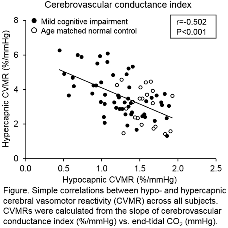
Conclusions
Amnestic MCI exhibited diminished cerebral vasoconstrictor responses during hypocapnia whereas their hypercapnic vasodilatory responses were enhanced when compared with cognitively normal older adults. These findings suggest that the operating point of CVMR curve is shifted closer to ischemic threshold in MCI patients.
References
PB02-O08
The presence of cerebral microbleeds is associated with cognitive impairment in Parkinson’s disease
1Division of Neurology, Department of Medicine, Jichi Medical University, Japan
2Department of Neurology, Juntendo University, Japan
Abstract
Objectives
Cerebral microbleeds (CMBs) are well-known markers of small vessel disease and are often observed in Parkinson’s disease (PD). We previously reported that CMBs were frequently observed in Parkinson’s disease (17.4%),1) however, their association with cognitive decline has been unclear.
Methods
We performed a retrospective analysis of 124 cases of clinically diagnosed PD admitted to Juntendo University Hospital for diagnostic assessment, drug adjustment, or evaluation for deep-brain stimulation between January 2014 and July 2016. The cognitive function was assessed using the Mini-Mental State Examination (MMSE). We enrolled PD with dementia patients but not dementia with Lewy bodies (DLB) patients based on the “one-year rule”, and the diagnosis of Parkinson’s disease with dementia (PDD) was based on the diagnostic criteria and procedures from the Movement Disorder Society Task Force. Based on these criteria, we used the cut-off of MMSE for cognitive decline as <26 and notion that cognitive decline should be sufficient to impair daily life and not attributed to motor or autonomic symptoms. We compared the clinical characteristics and cognitive decline base on the presence of CMBs or not, and did the statistical analysis.
Results
Of the 124 participants, 21 (16.9%) was diagnosed as PDD in this cohort. CMBs were observed significantly more frequently in the PDD than in the PD (47.6% vs 7.8%, P < 0.001). The presence of both deep/infratentorial (40% vs 14.9%, P < 0.05) and strictly lobar (75% vs 12.9%, P < 0.001) CMBs were associated with PDD. The values of MMSE were significantly lower in the presence of both deep/infratentrial (23.9 ± 7.6 vs 27.0 ± 4.1, P < 0.05) and strictly lobar CMBs (22.3 ± 6.8 vs 27.1 ± 4.2, P < 0.01) than PD without CMBs. A multivariable logistic regression analysis showed the presence of strictly lobar CMBs as well as a male gender, orthostatic hypotension, periventricular hyperintensity on magnetic resonance imaging were significantly associated with PDD in this cohort.
Conclusion
This study showed the presence of CMBs, especially strictly lobar type, was strongly associated with PDD.2) We suspect that the burden of small vessel disease and cerebral amyloid angiopathy may be related to the development of cognitive decline in PD, and a prospective study enrolling more cases is warranted.
References
PB02-O09
MR findings and long-term cognitive change in patients with atrial fibrillation (AF) after ablation
1Dept. of Neurology, Graduate School of Medicine, Mie University, Japan
2Dept. of Dementia Prevention and Therapeutics, Graduate School of Medicine, Mie University, Japan
3Dept. of Cardiology, and Nephrology, Graduate School of Medicine, Mie University, Japan
4Dept. of Radiology, Graduate School of Medicine, Mie University, Japan
5Dept. of Advanced Diagnostic Imaging, Graduate School of Medicine, Mie University, Japan
6Dept. of Neurologic Science, Graduate School of Medicine, Nippon Medical School, Japan
Abstract
Objectives
Atrial fibrillation (AF) is an important risk factor for stroke and dementia (Ref1). It is suggested that dementia in AF patients is associated with cortical microinfarction (CMI), microbleeds (MBs) and chronic cerebral hypoperfusion (Ref 2). Ablation of AF is effective in stroke prevention, but long-term effect of ablation on cognitive function remains unclear (Ref 3). We aimed to delineate the incidence and distribution of CMI after AF ablation using 3 tesla MRI, 3D-double inversion recovery (DIR) method, as well as the long-term effect of ablation on cognitive function.
Methods
This study included 56 patients who underwent AF ablation in Department of Cardiology, Mie University Hospital (41 male, 15 female, 32–84 years old, mean ± SD: 66.8 ± 9.8 years old). 23 patients had paroxysmal AF, and 33 patients had persistent AF. Brain MRI and mini-mental state examination (MMSE) were administered at 1-3 days and 6 months after the ablation. MRI included diffusion weighted imaging (DWI), 3-dimentional fluid-attenuated inversion recovery (3D-FLAIR), 3-dimentional DIR (3D-DIR), T1WI and susceptibility-weighted imaging (SWI).
Results
None of the patients had neurological abnormality after the ablation procedure. At 1-3 days after the ablation, DWI and 3D-DIR detected CMI in 50 patients (89%). SWI detected MBs in 36 patients (64%). At 6 months after the ablation, CMIs detected by DIR disappeared in 33 out of 50 patients (66%). Number of MBs remained unchanged in 16 patients, disappeared in 1 patient, and increased in 29 patients. 21 of the 29 patients had new MBs exactly at the same location where the CMIs occurred 6 months ago. Mean MMSE score significantly improved from 27.8 ± 2.4 at 1-3 days to 28.7 ± 1.7 at 6 month after the ablation (p = 0.03).
Conclusions
Six months after ablation, asymptomatic de novo CMIs disappeared in 66% of patients, but MBs appeared at the same location in most cases. MMSE score improved in spite of these transient increase of CMIs and MBs, and this improvement was more evident in the patients who had lower baseline MMSE score. Because cerebral blood flow has been shown to increase after ablation, ablation may improve cognitive function possibly by improving chronic cerebral hypoperfusion and preventive effect of CMI in a long interval.
References
PB02-O10
Changes in dynamics of cerebrovascular reactivity responses to breath-hold in APOE e4 carriers across the adult lifespan
1Center of Functionally Integrative Neuroscience, Clinical Institute, Aarhus University, Aarhus, Denmark
2Department of Physiology and Pharmacology, Karolinska Institutet, Stockholm, Sweden
3Department for Congenital Disorders, Statens Serum Institut, Copenhagen, Denmark
Abstract
Objectives
The strongest known risk factors for sporadic Alzheimer’s disease (AD) are older age, a family history of AD, and the e4 variant of the apolipoprotein E (APOE) gene allele. Altered hemodynamic responses has been observed in APOE e4 carriers and in demented patients [1-3]. Our objective was to characterize blood oxygenation level dependent (BOLD) responses to functional activation and reactivity responses to breath-hold in non-demented APOE e3 and e4 carriers and to study response changes across age groups. A further objective was to investigate whether these responses were modulated by elevated nitrate intake [4].
Methods
Our study was designed as a randomized, double-blinded, placebo-controlled crossover study. The participants (n = 81 included, 30–70 years of age at enrollment) underwent two scanning sessions. Prior to scanning sessions, the participants ingested either sodium chloride (NaCl) or sodium nitrate (NaNO3) over a three-day ingestion period. Functional magnetic resonance imaging (fMRI) sessions lasted ten minutes and involved two concurrent tasks – a visual stimulation task and a breath-hold task. Three hemodynamic response features were extracted to characterize the hemodynamic (BOLD) responses: peak time, peak magnitude, and peak width. Participants were partitioned into three age groups (<45, 45–60, >60), and the experimental data was analyzed according to a factorial structure AGExAPOExNaCl/NaNO3.
Results
For the reactivity responses (breath-hold task) we observed significant changes (p < 0.05) in peak time, peak magnitude, and peak width across age and APOE sub-groups. Specifically, APOE e4 carriers had a significant increased peak time than non-carriers within the 45-60 age group, and e4 carriers in the 45-60 age group also had an increased peak time relative to e4 carriers in the <45 age group. Peak magnitude significantly decreased with age when comparing the <45 and 45-60 age groups and the <45 and >60 age groups, whereas APOE e4 carriers showed a tendency (p < 0.1) for a reduced peak magnitude. APOE e4 carriers had a significantly wider peak than non-carriers. For the functional activation responses (visual stimulation task) we observed significant changes in peak magnitude across the age groups. Specifically, peak magnitude significantly decreased between the <45 and 45-60 age groups, and a tendency towards decrease between the <45 and >60 age groups. We observed no significant modulatory effect of nitrate ingestion on the hemodynamic responses.
Conclusions
Vascular dysfunction has been implicated in the pathogenesis of AD [5-7], and previous studies has demonstrated altered hemodynamics in APOE e4 carriers [1-2]. Our present results support the notion of altered hemodynamics in APOE e4 carriers and adds novel experimental evidence, that not only the hemodynamic response magnitudes but also the temporal characteristics of the responses are altered in asymptomatic APOE e4 carriers.
References
PB02-O11
Apolipoprotein E4 worsens cerebrovascular and cognitive dysfunction induced by cerebral hypoperfusion via perivascular macrophages
1Feil Family Brain and Mind Research Institute, Weill Cornell Medicine, USA
2Department of Neurology, National Cerebral and Cardiovascular Center, Japan
Abstract
Objective
The apolipoprotein E4 (ApoE4) allele is a major risk factor for Alzheimer’s disease (AD) and white matter (WM) ischemic lesions. In mice, human ApoE4 induces cerebrovascular dysfunction through oxidative stress, and exacerbates WM damage and cognitive deficits in cerebral hypoperfusion produced by bilateral common carotid artery stenosis (BCAS) (1). However, the cellular mechanisms of these effects remain unexplored. Perivascular macrophages (PVM), innate immune cells closely apposed to the outer wall of cerebral vessels, are a major source of oxidative stress and mediate cerebrovascular dysfunction in models of hypertension and Alzheimer’s disease (AD) (2, 3). We used ApoE4 targeted replacement (TR) mice to test the hypothesis that PVM are also involved in the neurovascular dysfunction and increased susceptibility to cerebral hypoperfusion and cognitive dysfunction associated with ApoE4.
Methods
Male wild-type (WT) mice (age 2 months) were transplanted with bone marrow (BM) of ApoE4-TR mice (ApoE4→WT) to repopulate their perivascular space with ApoE4+ PVM. WT mice receiving WT BM (WT→WT) served as controls. Alternatively, PVM were depleted by intracerebroventricular injection of liposome-encapsulated clodronate (CLO). Cerebral blood flow (CBF) was assessed by laser-Doppler flowmetry in the somatosensory cortex of mice anesthetized with urethane-chloralose (n = 5/group), 5 weeks after BM transplantation or 7 days after CLO administration. In some experiments, BCAS was induced and CBF was monitored by laser-speckle flowmetry (n = 8/group). WM integrity (n = 5/group) and cognitive function (n = 12/group) were evaluated 1 month later.
Results
In ApoE4-TR mice, the increase in CBF induced by whisker stimulation or topical application of the endothelium-dependent vasodilator acetylcholine (10 µM; ACh) were attenuated (−47 ± 8% and −52 ± 6%, respectively; p < 0.05 from WT). CLO reduced PVM by 60 ± 4% and prevented the cerebrovascular dysfunction in ApoE4-TR mice (p < 0.05 from vehicle). To examine the involvement of ApoE4 in PVM, we investigated WT mice transplanted with ApoE-TR BM (ApoE4→WT). We found that the CBF increase induced by whisker stimulation or by ACh were attenuated (−45 ± 4% and −35 ± 3%, respectively; p < 0.05 from WT→WT). The CBF increase evoked by the smooth muscle relaxant adenosine (400 µM) was not affected (p > 0.05), attesting to the integrity of the vasomotor apparatus. After BCAS, the CBF reduction was greater in ApoE4→WT (−25 ± 1%) compared to WT→WT mice (−15% ± 3%;p < 0.05), an effect associated with increased WM damage assessed by the MBP/SMI312 ratio (+42 ± 3%) and Klüver-Barrera stain (+66 ± 1%). In addition, BCAS produced more severe cognitive deficits in ApoE4→WT mice at the Y-maze (+52 ± 2%;p < 0.05), novel object recognition test (+55 ± 2%;p < 0.05), and Banes maze probe test (+56 ± 0.03%;p < 0.05).
Conclusions
ApoE4 in PVM mediates cerebrovascular dysfunction and exacerbates hypoperfusion-induced WM damage, resulting in more severe cognitive deficits. The data elucidate the cellular bases of the CBF alterations observed in ApoE4-TR mice and raise the possibility that PVM may be involved in the increased susceptibility to WM damage observed in ApoE4 carriers and in patients with AD. Therefore, targeting ApoE4 in PVM may be a putative therapeutic strategy to counteract the deleterious cerebrovascular effects of ApoE4.
References
PB02-O12
Local and global microvascular disturbances in the brains of APOE-ɛ4 carriers: Vascular and cognitive responses to nitrate supplementation
1Center of Functionally Integrative Neuroscience, Clinical Institute, Aarhus University, Aarhus, Denmark
2Department of Physiology and Pharmacology, Karolinska Institutet, 17177 Stockholm, Sweden
3Department for Congenital Disorders, Statens Serum Institut, Copenhagen
Abstract
Objectives
The ɛ4 allele of the apolipoprotein E (APOE) gene is a major genetic risk factor for sporadic Alzheimer’s Disease (AD)1. Reports of abnormally high regional cerebral blood flows (CBF) in young asymptomatic APOE-ɛ4 carriers2may indicate that their oxygen extraction efficacy is impaired by capillary dysfunction3. In particular, apoE4 protein could impair capillary function by reducing endothelial nitric oxide (NO) production4, which is obligatory for the dilation of cerebral microvessels5.
Methods
To examine, whether the APOE-e4 allele is associated with disturbed cerebral capillary hemodynamics, we performed dynamic susceptibility contrast magnetic (DSC) resonance imaging in 38 APOE-e4 carriers (29 e4/e3 heterozygotes, 9e4 homozygotes – hereinafter “carriers”) and 38 e3 homozygotes (hereinafter “non-carriers”) aged between 30 and 70 years. Neuropsychological testing was conducted to study the cognitive effects of the APOE-e4 allele. Concomitantly, nitrate supplementation was conducted in a randomized, double-blinded, placebo-controlled crossover design to study the effects of physiological NO augmentation on both these measures.
Results
We show that microvascular flow distributions are profoundly altered in the brains of APOE-ɛ4 carriers compared to non-carriers, particularly in the hippocampal areas of younger (≤52 years) APOE-ɛ4 carriers. We show that nitrate, given to augment endothelial NO availability, reduce these differences and improve delayed recall, except in older (>52 years) APOE-ɛ4 carriers.
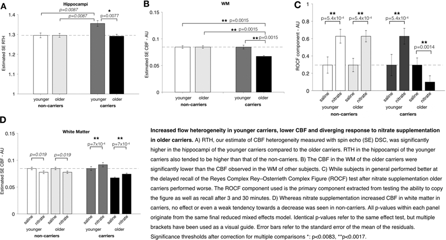
Conclusions
Our data suggest that APOE-ɛ4 genotype affects brain microvascular function, decades before typical AD onset, and that early dietary nitrate supplementation may modify microvascular and cognitive functions.
References
PB02-P01
Cortical photothrombotic stroke induces cognitive deficits and is associated with an increased levels of neurotoxic proteins accumulation
1School of Biomedical Sciences and Pharmacy and the Priority Research Centre for Stroke and Brain Injury, University of Newcastle, Callaghan, NSW, Australia
2Hunter Medical Research Institute, Newcastle, NSW, Australia
3NHMRC Centre of Research Excellence Stroke Rehabilitation and Brain Recovery, Australia
4Centre for Rehab Innovations, University of Newcastle, Callaghan, NSW, Australia
Abstract
Objectives
Numerous clinical studies have documented the high incidence of cognitive impairment after stroke. Stroke survivors have reported experiencing poorer performance in learning, memory and executive functions. However, the underlying mechanisms behind these deficits are yet to be determined. Our interest is to better understand the neuropathological characteristics leading to this impairment using a preclinical stroke model. We investigated whether there is a relationship between these cognitive deficits and an increased accumulation of neurotoxic proteins (amyloid-β and α-synuclein). We then assessed whether a failure of the brain clearance mechanisms might be the responsible of this protein accumulation.
Methods
We induced phototrombotic occlusion at the motor and somatosensory cortices in mice (male C57BL/6, 10 – 12 weeks old) or sham surgery. We used a mouse touchscreen testing system for the assessment of cognition starting from 1 week to 1 month post-stroke. Motor function was evaluated using the cylinder and grid walk task. Brains were collected for further proteins and histology analyses.
Results
We observed that the visual discrimination performance of stroked mice were significantly lower than the sham mice at 2 weeks post-stroke. Further immunohistochemical and protein analyses of the brain revealed an increased accumulation and aggregation of both amyloid-β and α-synuclein within the peri-infarct and secondary neurodegeneration regions. We also observed significant disturbances in the aquaporin-4 protein expression and polarisation at the astrocytic end-feet.
Conclusion
Together, our results suggest that the cognitive impairment seen after stroke might be due to an increased accumulation of neurotoxic proteins in different regions of the brain, which is caused by a dysregulation in the brain clearance pathways. This avenue may represent a therapeutic target for brain recovery.
PB02-P02
Longitudinal investigation of spatiotemporal dynamics of blood cell stagnation in cerebral capillaries using optical coherence tomography angiography during subcortical vascular dementia development
1Dept. of Bio and Brain Engineering, Korea Advanced Institute of Science and Technology, Republic of Korea
2KI for Health Science and Technology, Korea Advanced Institute of Science and Technology, Republic of Korea
3Dept. of Mechanical Engineering, Korea Advanced Institute of Science and Technology, Republic of Korea
Abstract
Objectives
Subcortical vascular dementia (SVaD) is the most common subtype of vascular dementia that exhibits white matter lesions and lacunes in the brain along with cognitive impairments typically including memory impairment, and frontal executive dysfunctions. In SVaD patients, it has been proposed that microcirculation disruption such as blood-brain barrier breakdown proceeds to leukoaraiosis and other pathological outcomes. However, due to lack of proper SVaD animal models and high spatiotemporal in vivo imaging technology, the detailed mechanisms of microcirculation disruption has not been elucidated well. We hypothesized that blood cell stagnation increases during initial SVaD development and relates with the histopathological features of SVaD.
Methods
We performed longitudinal volumetric imaging using optical coherence tomography angiography (OCTA) to reveal detailed microcirculation disruption. We utilized SVaD mouse models which were recently developed. Longitudinal volumetric imaging was taken for 9 weeks including baseline. After imaging, we performed immunohistochemistry (IHC) for astrogliosis, neuron density, vessel density, and blood-brain barrier. Behavioral tests such as Y-maze, open field test, and novel object recognition, were conducted for measuring cognitive dysfunction. The relationship between parameters for measuring microcirculation by OCTA and histopathological features were calculated.
Results
We observed altered microcirculation and its spatiotemporal dynamics. Global capillary speed gradually decreased and became heterogeneous. The ratio of stagnated capillary segments distinctly increased during the development of SVaD with steep increase in late period of SVaD development. We also found that the parameters for microcirculation from OCTA imaging, such as ratio of stagnated capillary segments, showed correlation with histopathological features such as astrogliosis.
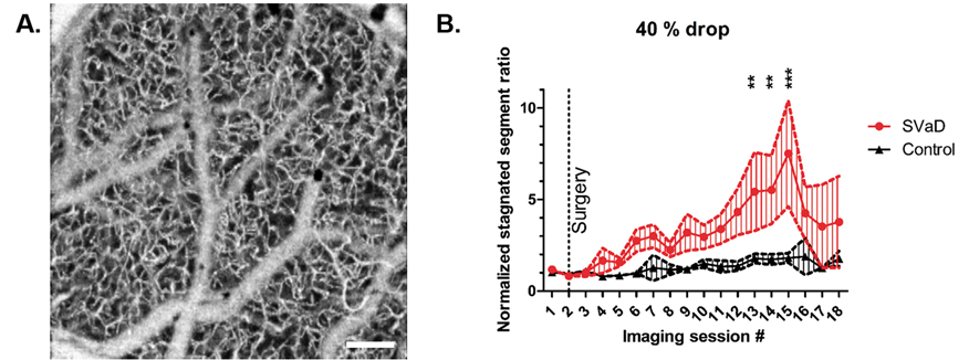
Conclusions
We reveal spatiotemporal dynamics of blood cell stagnation and its mechanism during the development of SVaD, and this study may herald possible therapeutic target mechanisms for future drug developments.
References
PB02-P03
Role of inflammasome activation in a chronic cerebral hypoperfusion model of vascular dementia
1Dept. of Physiology, National University of Singapore, Singapore
Abstract
Objectives
Inflammasome, a multi-protein complex initiating the innate immune response, is known to play a critical role in multiple diseases such as Alzheimer’s disease, stroke and artheriolosclerosis (Guo, Callaway, & Ting, 2015). Upon activation, it displays its signature characteristics: elevated Cleaved Caspase-1 activity and production of downstream inflammatory cytokine mature IL-1β and IL-18. This leads to overactivation of microglia and astrocytes, and dysfunction of endothelial cells eventually resulting in neuronal loss and disintegration of the neurovascular unit (Lénárt, Brough, & Dénes, 2016). In this study, we investigate the role of inflammasome activation in a chronic cerebral hypoperfusion model of vascular dementia by looking into the biochemical mechanisms, structural integrity and cognitive aspects of this disease.
Methods
The chronic hypoperfusion model that we use is the bilateral common carotid artery stenosis (BCAS) model. This model involves two micro-coils that are placed around both common carotid arteries of C57BL/6 mouse to restrict blood flow to the brain. First, they were left inside the animal for different durations of 1 to 60 days to investigate the effects of chronic cerebral blood flow reduction. Animals were sacrificed at each timepoint to study the protein expression levels and degree of white matter lesions. Next, we used a Caspase-1 inhibitor, to study the role of inflammasome activation when the blood flow is reduced for 7, 15 and 30 days. Upon obtaining the brain samples, the role of inflammasome was assessed. In addition, blood flow was closely monitored throughout the duration of the study using a Laser Speckle Contrasting Imager.
Results
With the BCAS surgery, there was an immediate average blood flow reduction of 36.42% across different groups. Although the blood flow slowly increased over time, the reduction was maintained at 20-30% from 1 to 30 days, which was eventually restored at 60 days.
We isolated different brain regions and characterised the expression levels of proteins in the inflammasome pathway. In the cerebral cortex, we observed an upregulation of NLRP1, NLRP3, NLRC4 and AIM2 inflammasome receptors across the different timepoints with a corresponding increased cleaved caspase-1 level. The downstream cytokine, mature IL-1β, apoptotic marker, cleaved caspase-3, and pyroptotic marker, N-terminal GSDMD were all upregulated with chronic cerebral hypoperfusion, indicating the involvement of inflammasome activation in the cortical region. However, in the hippocampus and striatum, we observed a decrease in cleaved caspase-1 expression levels while cleaved caspase-3 expression levels increased over time.
Conclusion
Currently, our data indicate the involvement of inflammasome activation during chronic cerebral hypoperfusion. However, the exact role will be determined by the study via pharmacological inhibition of Caspase-1.
References
PB02-P04
Acceleration of NLRP3 inflammasome by chronic cerebral hypoperfusion in Alzheimer’s disease model mouse
1Department of Neurology, Graduate School of Medicine, Dentistry and Pharmaceutical Sciences, Okayama University
Abstract
Cerebral neuroinflammation defines a novel pathway for progressing Alzheimer’s disease (AD) pathology. We investigated immunohistological changes of neuroinflammation with nucleotide-binding domain and leucine-rich repeat (NLR)-protein 3 (NLRP3), activated caspase-1 and interleukin-1 beta (IL-1β) in a novel AD (APP23) mice with chronic cerebral hypoperfusion (CCH) model from 4 months (M) of age, moreover, examined protective effect of galantamine. CCH strongly enhanced NLRP3, activated caspase-1 and IL-1β expressions in hippocampus and thalamus at age 12 M of AD mice. CCH also exaggerated amyloid-beta (Aβ) 40 depositions in cerebral cortex. Furthermore, CCH exacerbated a marked dissociation of neurovascular unit (NVU). These pathological changes were ameliorated by galantamine treatment. The present study demonstrated that CCH strongly enhanced primary AD pathology including neuroinflammation, Aβ accumulations and NVU dissociation in AD mice, which was greatly protected by an allosterically potentiating ligand galantamine.
PB02-P05
Chronic cerebral hypoperfusion accelerates Alzheimer's disease pathology with the change of mitochondrial fission and fusion dynamics in a novel mouse model
1Department of Neurology, Okayama University, Japan
Abstract
Mitochondrial dynamically undergo massive fusion and fission events to continuously maintain their function in cells. Although an impaired balance of mitochondrial fission and fusion was reported in in-vitro and in-vivo Alzheimer's disease (AD) model, changes of mitochondrial fission and fusion proteins have not been reported in AD with chronic cerebral hypoperfusion (HP) as an etiological factor related to the development of elder AD. To clarify the impacts of HP on mitochondrial fission and fusion, related oxidative stress in the pathogenesis of AD, and protective effect of galantamine, the novel AD with HP mouse model (APP23 + HP) was applied in this project. Compared with APP23 mice, APP23 + HP mice greatly enhanced the number of Aβ oligomer-positive/phosphorylated tau (pTau) cells, the expression of mitochondrial fission proteins (Drp1 and Fis1), and decreased the expression of mitochondrial fusion proteins (Opa1 and Mfn1) in the cerebral cortex (CTX) and thalamus (TH) at 12month (M) of age. Moreover, the expression of peroxidation products (4-HNE and 8-OHdG) showed a significant increase in CTX and TH of APP23 + HP mice at 12M. However, above neuropathological characteristics were retrieved by galantamine (Gal) treatment, detected through immunohistochemical analyses. The present study demonstrates that cerebral HP shifted the balance in mitochondrial morphology from fusion to fission with increasing Aβ oligomer/pTau accumulations in APP23 mice, and such neuropathologic processes were strongly attenuated by Gal treatment.
PB02-P06
The effect of intermittent fasting on the brain vasculature in vascular dementia using a mouse model of chronic cerebral hypoperfusion
1Dept. of Physiology, National University of Singapore, Singapore
Abstract
Vascular dementia (VaD) is one of the common causes of dementia after Alzheimer’s disease. Dementias are a type of neurodegenerative disease that results in progressive neuronal death and their interconnections. This neurodegeneration in VaD commonly originates from vascular dysfunction causing disruption of the cerebral blood flow (CBF). Chronic cerebral hypoperfusion (CCH) resulting from vascular damages is invariantly the prominent feature of this disease. VaD has been shown to have vascular changes including decreased blood-brain barrier (BBB) integrity [1] and blood vessel density [2]. BBB breakdown is a well-known feature in dementia but there are no mechanisms established to elucidate this process, yet this mechanism may be crucial in discovering novel therapeutic opportunities and can be adapted into understanding or treating other diseases outside the dementia faculty. Although many aspects CCH effects have been examined, there are no licensed treatments against VaD that have been reported. Studies have shown that intermittent fasting (IF) pretreatment activates neuroprotective signaling pathways such as neurogenesis and ameliorate neuroinflammation [3]. Whilst IF has proven to be neuroprotective, whether IF can protect against VaD remains to be confirmed. Ultimately, this project aims to understand whether IF can function as a therapy in VaD patients to alleviate late-onset cognitive impairments and improve the detrimental effect on the brain vasculature during CCH in VaD, and if so, their underlying mechanisms.
Hence, we seek to investigate whether long term IF (4 months IF with 16 h fasting per day) can alleviate the vascular damage in the brain by experimentally reproducing CCH conditions and cognitive impairments observed in VaD patients via the bilateral carotid artery stenosis (BCAS) model in mice. BBB integrity and blood vessel density will be investigated using Evans Blue staining and lipophilic carbocyanine dye DiI respectively. White matter lesions (WML) that are used as a diagnostic criterion in VaD patients are examined using the Luxol Fast Blue staining. Finally, the behavioral outcomes are tested by performing behavioral tests.
Currently, we have established that CBF is restored in the mouse following CCH for 30 days, but studies have shown that vasculature density was attenuated in the brain [2], an irony that we wish to uncover through this project. The BCAS model has indeed shown BBB and vasculature disruption and displays WMLs. Investigation into whether IF is able to improve not only the molecular basis of the disease, but also create significant changes in the brain so that it translates into positive behavioral outcomes in the mice is still ongoing.
References
PB02-P07
Tauopathy in a rat model for the post-stroke dementia combining chronic cerebral hypoperfusion and acute ischemic stroke
1Department of Neurology, Konkuk University School of Medicine, Seoul, Korea
Abstract
Background and purpose
Post-stroke dementia (PSD) is one of major consequences after stroke. Tauopathy has been reported in diverse neurodegenerative diseases. We investigated cognitive impairments and pathomechanism of tauopathy in a rat model for PSD combining acute ischemic stroke and underlying chronic cerebral hypoperfusion (CCH).
Methods
We performed middle cerebral artery occlusion (MCAO) surgery in rats mimicking acute ischemic stroke, which was followed by bilateral common carotid artery occlusion (BCCAo) surgery mimicking CCH. We performed behavioral tests and histological investigations focused on the characterization of tauopathy. Parenchymal infiltration of the cerebrospinal fluid (CSF) tracers after the intracisternal injection was observed to evaluate glymphatic function.
Results
Cognitive impairment was aggravated when BCCAo was superimposed on MCAO. Hyperphosphorylation of tau was more synergistically accentuated in the ipsilateral corpus callosum than the cortex. Parenchymal infiltration of CSF tracers was decreased by CCH.
Conclusions
Our experimental results suggest that CCH may aggravate tauopathy-related cognitive impairment in a rat model for PSD by interfering tau clearance through glymphatic pathway. Therapeutic strategy to improve clearance of brain metabolic waste including tau may be a promising approach to prevent PSD after stroke.
PB02-P08
The role of age and neuroinflammation in the mechanism of cognitive and neurobehavioral deficits in sickle cell disease
1Neuroscience Program, Graduate Division of Biological and Biomedical Sciences, Laney Graduate School, Emory University
2Neuroscience and Behavioral Biology Program, Emory University, Atlanta GA, USA
3Aflac Cancer and Blood Disorder Center of Children’s Healthcare of Atlanta and Emory University Department of Pediatrics, Atlanta, GA, USA
Abstract
Objectives
Cognitive and neurobehavioral abnormalities are the most common and complex complications of sickle cell disease (SCD). Known risk factors are stroke and silent cerebral infarcts, but a majority of cases do not have overt cerebral injury and the underlying mechanism is not well understood. The study’s aim was to determine whether age, neuro-inflammation and neuroplasticity influenced cognition in SCD.
Methods
Using a longitudinal, cross-sectional design, we evaluate cognition and neurobehavioral deficits in 13 months old Townes humanized sickle cell (SS) and matched control (AA) mice. We used the combination of novel object recognition (NOR) and fear conditioning tests to measure anxiety/depression, learning and memory. Immunohistochemistry was performed to quantify bone marrow-derived microglia (CD45+) and activated microglia (Iba1+) in the dentate and peri-dentate gyrus while Golgi-Cox staining was used to assess dendritic spine density and morphology, as well as dendrite arbors.
Results
Compared to matched AA controls, 13 months old SS mice showed significant evidence of anxiety/depression by the shorter distance traveled in the NOR test (p£0.01) as well as thigmotaxis (p < 0.05). Additionally, SS mice were significantly less likely to recognize the novel object (p < 0.001). There were no significant differences between 6 months old SS and AA mice (See Figures i to iv). But the difference reappeared after the same mice were aged to 13 months (Figures v to viii). Aged mice exhibited more anxiety/depressive behaviors (p < 0.05) and thigmotaxis (p < 0.05) and were less likely to identify the novel object (p < 0.05) when compared to aged AA mice (Figure 1(v-viii)). Treatment of aged SS mice with minocycline resulted in significant improvement of cognitive (p < 0.01) and neurobehavioral function (p < 0.05) compared to matched non-treated SS mice. Immunohistochemical and histological analysis showed treated SS mice had significant reduction in number of CD45+ cells (p < 0.05) and activated microglia (p < 0.05) in the dentate and peri-dentate gyrus area. There was also significant improvement in dendritic spine density as well as dendritic spine maturation (p < 0.001).
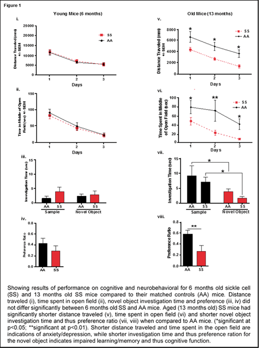
Conclusion
Taken together these results indicate that age, neuro-inflammation and neuroplasticity, and specifically spine maturation and density, are possible mechanisms underlying cognitive deficits in sickle cell disease.
PB02-P09
BDNF improves cognitive function after stroke in aged mice
1Department of Anatomy, Brain Health Research Center and Brain Research New Zealand, University of Otago, Dunedin, New Zealand
Abstract
Background
Stroke remains a leading cause of adult death and disability worldwide. Recently, we have established an animal model of stroke that results in delayed impairment in spatial memory, allowing for us to better investigate cognitive deficits. Young and aged brains show different recovery profiles after stroke.
Aim
Assess aged-related differences in post-stroke cognition. Methods: Young (3-month old) and aged (16-month old) mice were trained in operant touchscreen chambers to complete a pairwise discrimination (PD) task. Stroke or sham surgery was induced using a photochemical model in the prefrontal cortex. Following which animals underwent a reversal task to identify stroke-induced cognitive deficits 3-4 weeks post-insult.
Result(s)
Assessment of sham animals using the Kaplan-Meier survival curves demonstrated that aged mice are more impaired on PD reversal learning compared to young controls. Stroke in the young mice revealed that they were slightly, albeit not significantly, impaired on the reversal task. In contrast, stroke in aged mice facilitated a significant improvement in reversal learning. We propose this mechanism may be mediated by brain-derived neurotrophic factor (BDNF), a neurotrophin critical for post-stroke recovery of motor function, as local delivery of the either the BDNF decoy, TrkB-Fc, from a biopolymer hydrogel blocked the recovery. No differences in motor function or infarct volume were observed between treatment groups.
Conclusion
Our findings support age-related differences in recovery of cognitive function after stroke. Interestingly, aged stroke animals outperformed their sham counterparts, suggesting reopening of a critical window for recovery, which is occurring in a BDNF-dependent manner.
PB02-P10
The altered reconfiguration pattern of brain modular architecture regulates cognitive function in cerebral small vessel disease
1Department of Neurology, Drum Tower Hospital of Nanjing University Medical School, PR China
Abstract
Background
Cerebral small vessel disease (SVD) is a common cause of cognitive dysfunction. However, little is known whether the altered reconfiguration pattern of brain modular architecture regulates cognitive dysfunction in cerebral small vessel disease.
Methods
We recruited 25 cases of SVD without cognitive impairment (SVD-NCI) and 24 cases of SVD with mild cognitive impairment (SVD-MCI). According to the Framingham Stroke Risk Profile, healthy controls (HC) were divided into 17 subjects (HC-low risk) and 19 subjects (HC-high risk). All individuals underwent resting-state functional magnetic resonance imaging and cognitive assessments. Graph-theoretical analysis was used to explore alterations in the modular organization of functional brain networks. Multiple regression and mediation analyses were performed to investigate the relationship between MRI markers, network metrics and cognitive performance.
Results
We identified four modules that corresponded to the default mode network (DMN), executive control network (ECN), sensorimotor network and visual network. With increasing vascular risk factors, the inter- and intranetwork compensation of the ECN and a relatively reserved DMN itself were observed in individuals at high risk for SVD. With declining cognitive ability, those with SVD-MCI showed a disrupted ECN intra-network and evoked DMN connection. Furthermore, the intermodule connectivity of the right inferior frontal gyrus of the ECN mediated the relationship between periventricular white matter hyperintensities and visuospatial processing in SVD-MCI.
Conclusions
The reconfiguration pattern of the modular architecture within/between the DMN and ECN advances our understanding of the neural underpinning in response to vascular risk and SVD burden. These observations may provide novel insight into the underlying neural mechanism of SVD-related cognitive impairment and may serve as a potential non-invasive biomarker to predict and monitor disease progression.
PB02-Q01
Stroke comorbidities impair the dynamic recruitment of cortical collateral perfusion
1Stoke and Brain Imaging, Institute of Neuroscience and Psychology, University of Glasgow, UK
2Center for Advanced Translational Stroke Science, Sanders-Brown Center of Aging, Department of Neurology, University of Kentucky, USA
Abstract
Objectives
Chronic hypertension and acute post stroke-hyperglycaemia are stroke comorbidities which exacerbates stroke outcome in humans, and accelerates cerebral ischaemic lesion in experimental stroke. Studies have suggested reduction in cerebral blood flow (CBF) as a potential mechanism, using imaging tools with poor temporal resolution. The extent of leptomeningeal anastomoses determines how much penumbra can be salvaged. This study determined, using Laser speckle contrast imaging (LSCI) with excellent temporal resolution, how stroke comorbidities influence the dynamic cortical collateral perfusion during the first 4 hours following permanent middle cerebral artery occlusion (pMCAO) experimental stroke.
Methods
28 male Wistar rats, 300–360 g, were fasted overnight (15-17 hours) and randomly allocated to 3 groups: normoglycaemic non-stroke control group, glucose group which received 10 mg/kg of 15% D-glucose i.p., and vehicle group which received an equivalent volume of saline i.p. 10 min before pMCAO by intraluminal filament. pMCAO was induced in 9 male stroke prone spontaneously hypertensive rats (SHRSP, 320–360 g). Under alpha chloralose anaesthesia (80 mg/kg bolus i.v. and 30 mg/kg/h continuous infusion i.v.) and following pMCAO induction under 2-2.5% isoflurane anaesthesia, laser speckle contrast imaging (LSCI; 1 image every 5 s) enabled real time assessment of cortical collateral perfusion from 30 min until 4 hours post-MCAO. Regions of interest (ROIs) were determined at 30 min post-MCAO based on perfusion thresholds viz: Ischaemic core (0-43% of mean contralateral hemisphere), hypoperfused tissue (CBF between 43-75% of mean contralateral hemisphere) and corresponding contralateral ROIs. Blood glucose was measured prior to the administration of glucose/ vehicle, 30 min post-MCAO and every hour thereafter.
Results
A gradual recovery of perfusion via the collateral circulation was observed in the ROIs of the vehicle group over the time course of ischaemia. The mean perfusion increase by 240 min post-MCAO was 358 ± 156% within the ischaemic core and 152 ± 58% within the hypoperfused tissue. However, the recruitment of perfusion was slower in the SHRSP and the glucose group (Figure 1). By 240 min post-MCAO, the mean perfusion increase was 40 ± 28% and 70 ± 43% within the ischaemic core; 55 ± 15% and 74 ± 49% within the hypoperfused tissue, for SHRSP and glucose group respectively. For normoglycaemic non-stroke controls, there was no change in cortical perfusion. During the 4 hour period, blood glucose values were normal for hyperglycaemia (glucose group; 6.8–10 mmol/L) and normoglycaemia (vehicle group; < 6 mmol/L). Physiological variables (body temperature, blood pressure, pH, PaCO2 and PaO2) were stable in all groups.

Conclusions
Chronic hypertension and acute post-stroke hyperglycaemia impairs the dynamic recruitment of cortical collateral blood flow following pMCAO in clinically relevant rat models. The findings of this study set the pace for the investigation of potential collateral flow enhancers in the setting of ischaemic stroke.
PB02-Q02
Dynamic capillary stalls in ischemic penumbra: persistent traffic jams become therapeutic candidates
1Boston University, Department of Biomedical Engineering, Boston, MA, USA
2Hacettepe University, Institute of Neurological Sciences and Psychiatry, Ankara, Turkey
3Meinig School of Biomedical Engineering, Cornell University, Ithaca, NY, USA
4Martinos Center for Biomedical Imaging, Massachusetts General Hospital, Harvard Medical School, Charlestown, MA, USA
Abstract
Ever since the first introduction of thrombolytic strategies and further expansion of endovascular treatment for acute ischemic stroke, it still remains a mystery why actual clinical outcomes are less than expected upon early restoration of blood flow. In this work, taking advantage of novel optical technologies for high spatio-temporal resolution imaging of cerebral capillary circulation in mice, we introduce the pathological phenomenon of dynamic stalls in capillaries, as blood cells temporarily but repeatedly get stuck in the capillary lumen. Optical coherence tomography angiography served as an excellent tool to quantify and analyze those events as it is highly sensitive to erythrocyte flow and can image hundreds of capillaries simultaneously. Interestingly, the dynamic cessations of blood flow in individual capillaries, were occurring extensively in the salvageable ischemic penumbra and persisting after recanalization, in a transient distal middle cerebral artery occlusion model. These stalls, which could be considered distinct from permanent capillary occlusions, were lasting from seconds to minutes and resolving spontaneously before they recur, that could potentially serve as a pharmacological target. Indeed, these dynamic events resembling temporary traffic jams, could be modulated significantly by improving leukocyte passage through capillary lumen by in vivoadministration of anti-Ly6G monoclonal antibody against neutrophils immediately after recanalization. Besides improving stall parameters and cerebral blood flow in the penumbra, this modulation also provided an improved capillary oxygenation, less tissue damage and significantly better functional outcome at 24 hours following ischemia. We conclude that this dynamic stall phenomenon contributes significantly to the ongoing tissue injury even with recanalization and is a potential therapeutic target that can be relevant for improving endovascular recanalization. These findings can be applicable not only for acute ischemic stroke but also for other neurological disorders with microcirculatory issues.
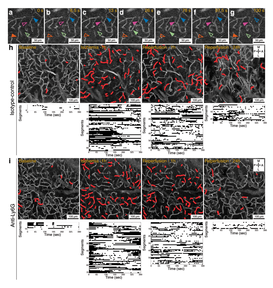
PB02-Q03
The role of systemic inflammation on cortical blood flow during the acute phase after experimental stroke
1Division of Neuroscience and Experimental Psychology, School of Biological Sciences, Faculty of Biology, Medicine and Health, University of Manchester, United Kingdom
Abstract
Objectives
Restoring cerebral blood flow after ischaemic stroke is critical for improving patient outcome and reducing neuronal injury. Clinically this can be achieved through pharmacological recombinant tissue plasminogen activator (tPA). Reperfusion therapy is only suitable for a small percentage of stroke patients and there is often incomplete reperfusion. Studying the dynamics of cerebral blood flow is essential in order to improve our understanding about the mechanisms underlying the no-reflow phenomenon that prevents the brain from receiving adequate blood perfusion. Recent evidence has demonstrated that systemic inflammation is an important contributing factor in the no-reflow phenomenon and interleukin (IL)-1 is a key cytokine known to induce systemic inflammation. Hence, we hypothesise that systemic inflammation induced by IL-1 will impair blood flow and we will investigate the early change of blood flow after experimental stroke.
Methods
We will use the middle cerebral artery occlusion (MCAO) model in C57 mice using a doccol filament to induce stroke. We then image blood flow changes on the cortical surface using laser speckle spectroscopy. We intraperitoneally (i.p.) inject 20 ng/kg of mouse recombinant IL-1β 30 min prior to the MCAO to investigate the effect of systemic inflammation on early changes of cerebral blood flow after stroke. Post-mortem tissue damage will be assessed using immunohistochemistry including cresyl violet (nissl) staining for neuronal cell damage, IgG for blood-brain barrier breakdown, Iba-1-positive activated microglia identification, GFAP-positive activated astrocyte staining, CD41-positive platelet cell identification and staining for IL-1α. These findings will further our understanding of the role of acute systemic inflammation in the early stages of blood reperfusion following ischaemic injury.
We demonstrate that laser speckle contrast imaging (LSCI) is an excellent tool to investigate the cerebral blood flow changes before, during, and after ischaemic stroke.
Results
Blood flow flux average performed on the two groups A and B. No significant difference was observed between groups within the same time points and both groups behave the same against the stroke insult, the blood flow in both group was very highly significantly reduced at the time of occlusion P < 0.0001 with no significant change after 20 min P < 0.0001. The brain starts to restore blood after 4 h with P = 0.0116 but not enough to reach the baseline value confirming the (no-reflow) phenomenon
Conclusion
Our findings show a significant reperfusion deficits at later time points (20 min and 4 h) confirm the no-reflow phenomenon of the cerebral microvasculature. There was no significance difference of cerebral blood flow between systemic IL-1 administration groups when compared to vehicle treated animals. Further work will be carried out to identify the difference between early biomarkers in both groups
Reference
PB02-Q04
Longitudinal in vivo imaging reveals structural and functional resiliency of dis-inhibitory cortical interneurons after stroke
1Division of Medical Sciences, University of Victoria, Victoria, BC, Canada
Abstract
Although inhibitory cortical interneurons play a critical role in regulating brain excitability and function, the effects of stroke on these neurons is poorly understood. In particular, interneurons expressing vasoactive intestinal peptide (VIP) specialize in inhibiting other classes of inhibitory neurons, and thus serve to modulate cortical sensory processing. To understand how stroke affects this circuit, we imaged VIP neuron structure and function (using GCaMP6s) before and after focal stroke in forelimb somatosensory cortex. Stroke led to a significant loss of peri-infarct pre-synaptic boutons and dendritic spines that was followed by a wave of bouton/spine production. Larger-scale changes, such as pruning/growth of axons or dendritic branches was observed, albeit on a limited scale. Functionally, the fraction of forelimb responsive VIP interneurons and their response fidelity (defined as the % of forelimb responsive trials) was significantly reduced in the first week after stroke. The loss of responsiveness was most evident in highly active VIP neurons (defined by their level of responsiveness before stroke), whereas less active neurons were minimally affected. Of note, a small fraction of VIP neurons that were minimally active before stroke, became responsive afterwards suggesting that stroke may unmask sensory responses in some neurons. Although VIP responses to forepaw stimulation generally improved although not fully from 2-5 weeks recovery, the variance in response fidelity after stroke was comparatively high and therefore less predictable than that observed before stroke. Lastly, stroke related changes to synaptic structure and response properties were both restricted to within 400 µm of the infarct border. These findings reveal the dynamic and resilient nature of VIP neurons and suggest that a sub-population of these cells are more apt to lose sensory responsiveness during the initial phase of stroke, whereas some minimally responsive cells are progressively recruited into the forelimb sensory circuit. Furthermore, stroke appears to disrupt the predictability of sensory evoked responses in these cortical interneurons which could have important consequences for sensory perception.
PB02-Q05
Functional impariment after stroke is exacerbated by vitamin D deficiency but is mitigated by supplementation
1Vascular Biology and Immunopharmacology Group, Department of Physiology, Anatomy and Microbiology, La Trobe University, Melbourne, Australia
2Cardiovascular Disease Program, Biomedicine Discovery Institute and Department of Pharmacology, Monash University, Melbourne, Australia
3Department of Physiology, National University of Singapore, Singapore
4Department of Anatomy, University of Otago, Dunedin, New Zealand
5School of Medicine, University of Tasmania, Hobart, Australia
Abstract
Vitamin D (VitD) deficiency is endemic in humans and increases the risk for cardiovascular events, including stroke. VitD exerts a protective effect on the cardiovascular system. However, the effectiveness of VitD as a therapy for ischemic stroke is unclear. We aimed to determine whether VitD deficiency and/or post-stroke supplementation with VitD affect stroke outcomes. Young adult (4 months; n = 72) or middle-aged (12-14 months; n = 11) male C57Bl6 mice were placed on either a VitD sufficient (2200 IU/day) or VitD deficient (0 IU) diet. After 8 weeks on the diet, thrombotic stroke was induced in the primary motor cortex. Young adult mice received 1,25-OH2 VitD3 (active form; 1.4 ug/kg per dose i.p.) or vehicle at 30 min and days 1, 3, 5 and 7 post-stroke. Motor function, infarct volume, markers of inflammation and neurogenesis were assessed at day 7. VitD deficiency exacerbated motor dysfunction (p < 0.05), with the magnitude of impairment being greater in middle-aged VitD deficient mice (p < 0.05). Infarct volume did not differ between the two diet groups in both young adult and middle-aged mice. VitD deficiency was associated with increased Iba-1-positive microglia, loss of infarct containment by reactive astrocytes and a reduction in doublecortin-positive staining. Post-stroke supplementation with VitD3 mitigated the motor impairment by ∼50% (p < 0.05) in VitD deficient mice but did not affect infarct volume in young adult mice. These data suggest that VitD deficiency worsens post-stroke functional outcomes via increasing brain inflammation and reducing recovery mechanisms. Administration of exogenous VitD post-stroke can attenuate poorer functional outcomes and thus, may represent a direction for acute stroke therapy.

PB02-Q06
Reactive oxygen species generation, neuronal degeneration and neurologic dysfunction after ischemic stroke in mice
1Department of Animal Bioscience, Division of Bioscience, Nagahama Institute of Bio-Science & Technology, Nagahama, Japan
2School of Pharmaceutical Sciences, Ohu University, Koriyama, Japan
3Department of Pharmacology, Hamamatsu University School of Medicine, Hamamatsu, Japan
Abstract
Objectives
In ischemic stroke, neurologic dysfunction is caused by neuronal damage via interruption of the blood supply associated with thrombosis in cerebral vessels. This interruption also causes the increase in blood-brain barrier (BBB) permeability. It is thought that cerebral ischemia and subsequent blood reperfusion induce reactive oxygen species (ROS) which exacerbates stroke outcome. Recently, it has been reported that the facilitation of vascular permeability at the spinal cord induced ROS generation via microglial activation, which was resulted in spinal axon injury in murine multiple sclerosis model (Ref. 1). Here, we investigated ROS generation, neurodegeneration and subsequent neurologic dysfunction on ischemic stroke.
Methods
Ischemic brain injury was induced by photochemically-induced thrombotic brain damage (PIT-BD) model, in which both location and size of damage are highly reproducible (Ref. 2).
Results
At 8 hours and 24 hours after damage induction, BBB permeability increased at the surrounding region of the damage, which was concurrent with fibrinogen leakage and ROS generation. This ROS generation was suppressed by treatment of a CD11b/CD18 antagonist. Neuronal degeneration was also found at the same region where vascular permeability increased, which was accompanied by neurological dysfunction. In addition, both dendritic branch length and spine number were significantly improved by treatment of Edaravone (EDV), a radical scavenger. Both the neuronal degeneration and neurological dysfunction were also improved by EDV treatment.
Conclusions
These findings indicated that the increase in the BBB permeability induced ROS generation via activation of microglia, which resulted in the induction of neuronal degeneration and accompanied neurological dysfunction in acute phase of ischemic stroke.
References
PB02-Q07
Post-stroke remote limb conditioning attenuates transneuronal degeneration in substantia nigra and improves functional recovery in chronic stroke
1Burke Neurological Institute, USA
2The Rockefeller University, USA
3Weill Cornell Medicine, USA
Abstract
Objective
Stroke-induced inflammatory response causes a massive infiltration of peripheral immune cells into the ischemic area, a process that contributes to the brain injury. Infiltrationof immune cells, particularly monocytes/macrophages,in the injured brain suggests the possibility that the manipulation of peripheral immune cells could influence stroke outcome. One such manipulation is remote
Methods
C57 mice (M/F, 3 m-old) were subjected to transient MCAO and received sham conditioning (Sham) or LC (5 cycles of inflation and deflation of 200 mmHg, 5 min x 5 min interval between cycles) using a small blood pressure cuff at 2 h post-stroke. Hemisphere, striatum, and SN volume were determined at acute and recovery stages. Transgenic mice that express GFP in dopaminergic TH+ neurons were subjected to whole-brain-clearing and imaging techniques and immunohistochemistry of TH and Iba1. The number of dopaminergic neurons and Iba1 staining were determined in SN at 3 d and 2 m. Longitudinal measurements of motor and gait function were performed during acute and recovery phases.
Results
Stroke induced significant and continuous atrophy until 4 m in the ipsilateral hemisphere and striatum as early as 7 d. Volume of SN were decreased by 26.3% at 7 d and 37.1% at 2 m (p < 0.05, n = 5). Stroke caused ∼55% loss of dopaminergic neurons in the SN at 2 m (n = 6). This was accompanied by increased Iba-1 expression, a maker for mononuclear phagocytes in the SN region. LC significantly attenuated stroke-induced neuronal loss (P < 0.01, n = 4-6, Fig). LC significantly improved motor performance by 3w and the enhancement was sustained up to 4 m (p < 0.01, n = 20/group).
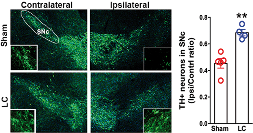
Conclusion
The temporal and spatial pattern analyses showed early atrophy in the primary injured area (striatum) and subsequent atrophy in the remote brain region (SN). The effect of LC on attenuating transneuronal degeneration and behavior improvement suggests that post-ischemic remote LC is a potential non-invasive therapeutic strategy to improve stroke outcomes.
PB02-Q08
Post-stroke physical exercise reduces ischemic brain damage and improves cognition in reproductively senescent female rats
1Cerebral Vascular Disease Research Laboratories, Department of Neurology, Leonard M. Miller School of Medicine, Miami, Florida 33136
2Department of Biomedical Engineering, University of Miami, Coral Gables, Florida 33146
Abstract
Stroke disproportionately kills more women than men and even a mild stroke causes disability in post-menopausal women [1]. The overall process of menopause lasts for years and during that period, disruption of multiple estrogen-regulated systems (including thermoregulation, sleep, circadian rhythms and sensory processing) and domains of cognitive function can be affected [2]. On the other hand, cognitive decline is a significant consequence of stroke survivors and almost two-thirds of stroke survivors experience cognitive deficits that last at least up to 6 years post-stroke [3, 4]. Our earlier study demonstrated that physical exercise (PE) reduced post-stroke brain injury and improved cognitive functions in male rats [5]. However, efficacy of PE in female counterparts remains elusive and the focus of our current study is to evaluate the improvement of post-stroke cognitive function in female rats. Reproductively senescent Sprague–Dawley female rats were exposed to transient middle cerebral artery occlusion (tMCAO; 90 min) and randomly assigned to either PE or sham-PE groups. Additionally, since ER-β agonist is shown to reduce ischemic damage [6], animals were treated with estrogen receptor beta (ER-β) agonist or vehicle to determine the synergistic effect with physical exercise. After three to five days, rats underwent sham-PE (0 m/min speed) or PE (15 m/min speed) for 30 mins either every day or alternate day for five times on treadmill. The rats that underwent alternate day paradigm were treated with ER-β agonist (diarylpropionitrile; 1 mg/kg) or vehicle-DMSO immediately following PE/sham-PE session. Seven days after the last PE/sham-PE, rats were tested for hippocampal-dependent contextual fear conditioning and freeze time was measured. Following behavioral testing, rats’ brains were processed for histology and infarcted area was measured using MCID software. Results demonstrated that post-tMCAO continuous PE did not reduce ischemic damage. However, alternate PE regimen with or without ER-β agonist reduced infract volume by 20% and 23%, respectively. Similarly, alternate PE regimen showed increased freezing on the second day of fear conditioning by 15%, indicating improved spatial memory. Overall, alternate PE paradigm and ER-β activation improves post-stroke cognition in reproductively senescent female rats. The study suggests that an alternate day PE paradigm and ER-β activation improves post-stroke cognition and future studies delineating underlying mechanism could help identify therapies to prevent/reduce stroke related cognitive decline in menopausal women stroke patients.
References
PB02-Q09
Functional MRI of reduced perilesional interhemispheric functional connectivity is not associated with changes in vascular reactivity after experimental stroke
1Biomedical MR Imaging and Spectroscopy Group, Center for Image Sciences, University Medical Center Utrecht and Utrecht University, Utrecht, the Netherlands
Abstract
Objectives
Resting-state functional MRI (rs-fMRI) studies in animal models and patients have shown that loss and recovery of motor function after unilateral stroke are associated with changes in interhemispheric functional connectivity within the sensorimotor network1. However, the vascular response to neuronal signaling – the foundation of rs-fMRI – may be compromised after stroke, which could affect measurements of functional connectivity2. Therefore, we assessed whether the observed reduction in interhemispheric functional connectivity after stroke is coupled with changes in vascular reactivity.
Methods
Unilateral photothrombotic stroke was induced in the sensorimotor cortex of adult male Sprague-Dawley rats. Serial MRI (at 9.4 T) was conducted from 1 week before to 26 weeks after stroke (n = 18). During MRI, rats were anesthetized by mechanical ventilation with 1.5 – 2.0% isoflurane in air/O2. MRI consisted of anatomical imaging, rs-fMRI (GE-EPI with TR/TE 0.7308s/15 ms, FOV 32*32*16.8 mm3, isotropic resolution of 600 μm) and BOLD MRI (multi-slice GE, 25 transversal slices with 0.5 mm thickness, TR/TE 500/15 ms, in plane resolution of 250 μm) during a vascular challenge with carbogen (5% CO2 in O2).
Results
Interhemispheric functional connectivity between left and right sensorimotor areas was significantly reduced 1 week after stroke (F(4,68) = 25.14, p < .0001) (Fig. 1A). Despite gradual recovery, interhemispheric functional connectivity remained significantly lower 26 weeks after stroke compared to baseline (t = 7.280, p < .0001). Vascular reactivity in the perilesional sensorimotor areas, however, remained largely intact over time after stroke (Fig. 1B).
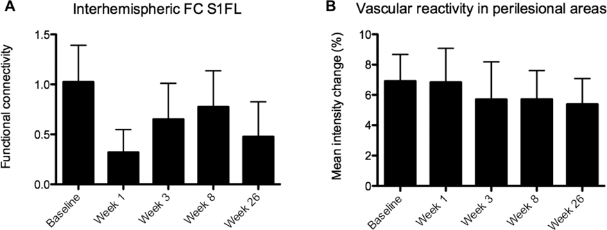
Conclusions
Our study shows that focal photothrombotic stroke to the ipsilateral sensorimotor cortex results in loss of interhemispheric functional connectivity of perilesional sensorimotor areas, which gradually recovers over time. The vascular reactivity, however, remains intact in the ipsilateral sensorimotor areas. Therefore, reduced cerebrovascular responsiveness does not explain the disturbed interhemispheric coherence of rs-fMRI signals after experimental focal unilateral stroke.
Funded by QNRF grant no. NPRP 5-381-3-101 and the NWO Graduate Programme (022.006.001).
References
PB02-Q10
Developing a method to assess functional recovery following stroke in a large animal model: a biomechanical and neurological outcome approach
1Adelaide Medical School and Adelaide Centre for Neuroscience Research, The University of Adelaide, Adelaide, SA, Australia
2Sansom Institute for Health Research, University of South Australia, Adelaide, SA, Australia
3Adelaide Spinal Research Group, Centre for Orthopaedics & Trauma Research, Adelaide Medical School, The University of Adelaide, SA, Australia
Abstract
Objectives
Stroke is a leading cause of mortality and morbidity worldwide, affecting over 17 million people annually. Much of this death and disability is attributable to the development of cerebral oedema, which precedes an elevation in intracranial pressure (ICP) and secondary neurological deterioration. Despite this, current treatments are limited and fail to address the underlying mechanisms of oedema, highlighting a need for the development of targeted treatments. When screening promising novel therapeutics, it is also essential to use clinically relevant large animal models to improve the likelihood of successful clinical translation. However, when screening such therapeutics in large animal models, assessment of neurological and functional outcome remains a significant hurdle. As such, the current study sought to develop a tailored post-stroke neurological outcome assessment scale and a biomechanical assessment of motor function for a clinically-relevant ovine stroke model.
Methods
Merino-sheep (6M;6F;18-36mths) were anaesthetised and subjected to 2 hrs middle cerebral artery occlusion with reperfusion. Animals underwent baseline functional testing and neurological assessment 8, 5 and 3 days pre-operatively, and 1, 3, 5, 7, 14, 21 and 28 days following stroke. A neurological assessment was carried out to evaluate changes in animal neurological status post-stroke. Functional outcome was determined using a 12 criteria neurological examination that quantified changes in animal demeanour, appetite, circling behaviour and deficits of motor function in the form of postural reaction tests. A score of 0 was considered normal with a possible total score of 40 indicating severe deficit. A motion capture system (Vicon Motion Systems Ltd. Oxford, UK) and reflective markers placed on the limbs and back, were used to record the animals’ motion during standardised walking tasks. Joint angles and symmetry of motion will be assessed to determine changes in gait post-stroke. All assessment and analysis was performed in a blinded fashion.
Results
The neurological assessment scale reliably detected differences in animal recovery following injury. A significant decline (p < 0.0001) in functional outcome was observed at 1day post-stroke with gradual resolution to 28days. Abnormalities in demeanour (p < 0.0001), appetite (p < 0.0001) and circling behaviour (p < 0.001) were evident 1day post-stroke and returned to normal by 14days. Contralateral functional deficits were significant at 1day post-stroke (p < 0.0001) whilst no ipsilateral deficits were observed (p > 0.05). Processing of the biomechanical data is ongoing.
Conclusions
We have developed a robust and reproducible ovine functional assessment scale that can be used to determine differences animal demeanor and neurological function following stroke. Furthermore, our novel biomechanical method has the potential to objectively quantify changes in gait and motion post-stroke. These methods will be used in our future studies to determine the efficacy of the novel therapies targeting cerebral oedema, and therefore outcome, following ovine stroke. They also have the potential be applied to other large animal models of acute central nervous system injury to reliably predict changes in functional recovery.
PB02-Q11
Detection of ischemic changes in the rat cortex after common carotid artery occlusion using intravoxel incoherent motion diffusion-weighted magnetic resonance imaging at 11.7 T
1Department of Neurosurgery, Iwate Medical University
2Institute for Open and Transdisciplinary Research Initiative, Osaka University
3Center for Translational Neuromedicine, University of Copenhagen
Abstract
Objectives
This study aimed to investigate whether intravoxel incoherent motion1 (IVIM) parameters can identify cerebral ischemic changes in the rat cortex after common carotid artery occlusion using an ultra-high-field preclinical 11.7 Tesla magnetic resonance imaging scanner (11.7 T MRI).
Methods
All experimental procedures involving animals and their care were carried out in accordance with the Guidelines of Osaka University for Animal Experimentation and the National Institutes of Health Guide for the Care and Use of Laboratory Animals. All experimental protocols were approved by the Research Ethics Committee of Osaka University. This manuscript follows the ARRIVE guidelines (Animal Research: Reporting in Vivo Experiments). At first, a permanent occlusion to the right common carotid artery with a 4-0 surgical thread ligation was performed in each rat, then, a left common carotid artery was occluded six days after the previous unilateral occlusion2. All surgical procedures were performed under general anesthesia with 2.0–2.54% isoflurane (Abbott Laboratories, Abbott Park, IL, USA) mixture with room air with a flow rate of 2 L/min. All rats were housed in cages under controlled temperature (20-22°C) and humidity (50-55%) during the surgical procedures. Rats were carefully observed during this study to avoid malnutrition due to anosmia and blindness immediately after the bilateral common carotid artery occlusion. The rats after the ligations that survived for 3 weeks were assigned to a long survival (LS) group and the others to a non-LS group. As the diffusion function, the following three different signal models were used for apparent diffusion coefficient (ADC) estimation; mono-exponential, kurtosis and bi-exponential models. And then, volume fraction of perfusion compartment (F), pseudo diffusion coefficient (D*) on the cortex were estimated with the same mono-exponential model as the perfusion function for three different diffusion parameter estimation functions.
Results
Finally, in nine female Wister rats (eight week-old), diffusion-weighted imaging for IVIM was successfully performed before (Pre), after the bilateral common carotid artery occlusion. As the results, the kurtosis model could provide more physiologically representative picture of the ischemic features than do other models. In quantitative assessments, rats in non-LS group showed significantly lower F, F × D* and ADC at the bilateral common carotid artery occlusion phase than at the Pre-phase. Conversely, in rats of the LS group, F significantly increased while F × D* and ADC decreased significantly.
Conclusions
Intravoxel incoherent motion perfusion MRI at 11.7T may aid in assessing the subtle difference between before and after cerebral ischemic states.
References
PB02-Q12
Leptomeningeal anastomosis and early ischemic lesions on diffusion-weighted imaging in murine focal cerebral ischemia
1Dept. of Neurology, Tokyo Women's Medical University, Japan
Abstract
Background
Leptomeningeal anastomosis is a key factor for determining early ischemic lesions on diffusion-weighted imaging (DWI) in human stroke. However, few studies have validated this relationship in an experimental model. This study sought to clarify the involvement of leptomeningeal anastomosis in early ischemic lesions using a murine model.
Methods
Adult C57BL/6 mice were subjected to unilateral common carotid artery (CCA) occlusion or sham surgery. Seven or fourteen days later, the middle cerebral artery (MCA) was occluded for 45 minutes. In the first experiment, the leptomeningeal collaterals were visualized using magnetic resonance imaging (MRI) DWI. In the second experiment, DWI was performed immediately after MCA reperfusion, and the infarct sizes were determined 24 hours after recirculation. During the operation, rectal temperature was monitored with a rectal thermometer and maintained at 37°C with a heat lamp.
Results
In the first experiment, the ADC volumes were 71.2 ± 8.5 mm3, 27.1 ± 13.5 mm3, 9.2 ± 6.5 mm3 and 68.9 ± 12.0 mm3 in the control, CCAO-D7, CCAO-D14 and sham-D14 groups, respectively. The ADC volume was robustly smaller in the CCAO-D7 and CCAO-D14 groups than in the control and sham-D14 group (P < 0.01). The diameters on the ischemic sides of the leptomeningeal collateral vessels on the dorsolateral surface were 24.7 ± 0.8 μm, 31.1 ± 2.0 μm, 38.0 ± 7.1 μm and 24.8 ± 2.0 μm in the control, CCAO-D7, CCAO-D14 and sham-D14 groups, respectively. The diameter of the leptomeningeal anastomoses was significantly larger on the ischemic side in the CCAO-D7 and CCAO-D14 groups than in the control and sham-D14 groups (P < 0.01). The vessel diameter of the leptomeningeal anastomoses was closely correlated to the degree of ADC volume (r = 0.84, P < 0.01). In the second experiment, the ADC volume were 62.8 ± 9.6 mm3, 16.4 ± 5.3 mm3, 6.1 ± 2.3 mm3 and 59.1 ± 10.4 mm3 in the control, CCAO-D7, CCAO-D14 and sham-D14 groups, respectively. Infarct sizes were 59.5 ± 8.7 mm3, 13.3 ± 7.9 mm3, 4.4 ± 1.9 mm3 and 53.6 ± 9.5 mm3 in the control, CCAO-D7, CCAO-D14 and sham-D14 groups, respectively. The infarct volumes were significantly smaller in the CCAO-D7 and CCAO-D14 groups than in the control and sham-D14 groups (P < 0.01). Also, the infarct size of the CCAO-D14 group was smaller than that of the CCAO-D7 group (P < 0.01). The ADC volume immediately after reperfusion was closely correlated to the degree of infarct volume (r = 0.96, P < 0.01).
Conclusion
In conclusion, involvement of the collateral circulation in early ischemic lesions was evident in the murine model. Both MRI and evaluation of leptomeningeal anastomosis could be used to develop a novel strategy targeting enhancement of the collateral circulation.
PB02-R01
Autophagosome dynamics is disrupted by dysregulation of Sec22b and Ykt6 expression in neurons during Cerebral ischemia-reperfusion injury
1Department of Neurosurgery & Brain and Nerve Research Laboratory, The First Affiliated Hospital of Soochow University, China
Abstract
Objectives
Ischemia-reperfusion (I/R) injury is accountable for poor outcome of ischemic stroke patients. Emerging evidence points to the conflict of neuroprotective versus harmful effects of autophagy activation in ischemic stroke. However, the potential mechanism switching the role of autophagy from protection to deterioration is delusive.
Methods
A murine middle cerebral artery occlusion/reperfusion (MCAO/R) model was established. Cultured neurons and brain microvascular endothelial cells (BMVECs) were exposed to oxygen-glucose deprivation/reoxygenation (OGD/R) to mimic I/R injury in vitro. Chemical modulators of autophagy and central nervous system-specific Atg7 conditional knockout mice were employed to assess the physiological and pathological functions of autophagy in brain. Proteins in neurons under OGD stress and OGD/R-stress were compared and analyzed by two-dimensional gel electrophoresis (2-DE) and identified by mass spectrometry.
Results
Here we show that autophagy induction by rapamycin and lithium carbonate performed before ischemia reduced neurological deficits and infarct volume but had an opposite effect once I/R has occurred in the murine MCAO/R model, both which were eliminated in mice lacking Atg7 specifically in central nervous system (Atg7flox/flox; nestin-Cre). Reperfusion led to a blockage of autophagosome retrograde trafficking in neurons, which then induce autophagic flux damage, mitochondrial damage and neuronal death. Studies using 2-DE of proteins form neurons exposed to OGD or OGD/R combined with mass spectrometry identified Sec22b as an increased factor and Ykt6 as a decreased factor during reperfusion, which were validated both in vivo and in vitro. Sec22b knockdown and Ykt6 overexpression rescued the autophagosome retrograde trafficking and autophagic flux in OGD/R neurons. In addition, Sec22b knockdown and Ykt6 overexpression reduced infarction and improved neurological function and spatial learning in murine MCAO/R model in an autophagy-dependent manner. Furthermore, Sec22b knockdown and Ykt6 overexpression improved the outcome of rapamycin post-treatment in MCAO/R rats.
Conclusions
These results unveil a previously unappreciated role of Sec22b and Ykt6 in autophagosome trafficking in neurons. Reperfusion-induced excess Sec22b and loss of Ykt6 in neurons lead to autophagosome retrograde trafficking failure, autophagic flux damage, and finally neuronal injury.
PB02-R02
Differential effect of Ischemia and Thrombin mediated toxicity on cerebral vascular unit
1Department of Neurology, Cedars-Sinai Medical Center
Abstract
Objectives
Brain is a complex structure during ischemia along with neurons; components of vascular unit astrocytes, pericytes and endothelial cells play a major role in hypoxic and blood brain barrier leakage mediated injury. Thrombin acts via protease activated receptor-1 (PAR-1) and leads to neuronal survival or death, PAR-1 knockout cells survive ischemia. We hypothesize that different elements of the Brain vascular unit play different roles in responding to injury. The present study aimed to establish a correlation between astrocytes, pericytes and endothelial cells vulnerability via OGD model of ischemia and Thrombin mediated toxicity.
Methods
Using highly reproducible oxygen glucose deprivation (OGD) model Primary cultured cells were exposed to OGD for various durations and Thombin doses to determine the optimum time point for 80 % cell death or cell viability measured using lactate dehydrogenase (LDH) release assay and 3-(4,5-dimethylthiazol-2-yl)-2,5-diphenyltetrazolium bromide (MTT) assay. LD50 dose of OGD and Thrombin was used to determine the effect of various drugs mediating PAR-1 receptor pathway
Results
The viability assay using MTT showed that the three elements of vascular unit respond differentially to longer durations of ischemia, with pericytes being the most vulnerable, followed by endothelial cells, followed by astrocytes. Similar vulnerability was observed with LDH release assay. Our thrombin dose effect shows that Pericytes and endothelial respond to thrombin similarly whereas, astrocytes on lower concentration proliferate, but don’t survive the higher concentrations.
Conclusion
An exciting, novel finding in our data is that different elements of the vascular unit—astrocytes, pericytes and endothelial cells—all exhibit effect of ischemia and reperfusion at different time scales. Up until recently, studies of neuroprotection addressed the brain as a homogenous unit, with the tacit assumption that all elements responded similarly. Our data on astrocytes, endothelial cells and pericytes—allows more focused investigation.
PB02-R03
Stress aggravates cerebral ischemia/reperfusion injury: role of mitochondrial quality control
1Department of anesthesiology, Renji Hospital, School of Medicine, Shanghai Jiaotong Univesity, China
Abstract
Objectives
Perioperative stroke is the most unwanted complication for patients. Before operations, patients usually undergo anxiety and depression, since operations are stressors. However, few studies have directly addressed the impact of stress on the severity of stroke. Mitochondria are key regulators of cell fate. Neurons have mitochondrial quality control mechanisms to retain functional mitochondria. The processes of mitochondrial fission and fusion allow for damaged mitochondria to be segregated and facilitate the equilibration of mitochondrial components. Therefore, it is essential to have stringent quality control mechanisms to ensure a healthy mitochondrial network. Multiple studies have reported that mitochondria malfunction is involved in the psychological stress-induced diseases, however, mitochondrial responses to acute and chronic stressors, especially mitochondrial quality control responses, are likely not linear. Hence, to assess the effect of acute and chronic stress on the severity of cerebral ischemia/reperfusion injury and the underlying mechanism of the involvement of mitochondria quality control, we investigated the effects of acute restraint stress and chronic sleep deprivation stress on cerebral ischemia/reperfusion.
Methods
Sixty-four C57/B6L male mouse were subjected into eight groups: sham, acute restraint stress, chronic sleep deprivation stress, MCAO, acute restraint stress +MCAO, mdivi-1 +acute restraint stress +MCAO, chronic sleep deprivation stress +MCAO, mdivi-1 +chronic sleep deprivation stress +MCAO. Acute restraint stress was induced by putting the mouse into a centrifuge tube for 2 hours. Chronic sleep deprivation stress was performed by water platform from 10 am to 4 pm for seven consecutive days. Middle cerebral artery occlusion (MCAO) model was induced by transient occlusion of proximal branches of the middle cerebral artery for 60 minutes, just after stress model. Mdivi-1 is an inhibitor of the fission-promoting molecule dynamin-related protein (drp1), a protein that mediated the mitochondrial fission with which tubular mitochondria is divided into mitochondria fragmentation, and mdivi-1 was injected into caudal vein before stress model. Sham group were subjected to sham surgery.
Results
The infarct volume increased significantly in the acute restraint stress +MCAO group and chronic sleep deprivation stress +MCAO group compared to MCAO group (p < 0.05), however, the acute and chronic stress+MCAO induced brain damages were attenuated by mdivi-1 pretreatment. The brain damages were consistent with the results of Morris water maze test, that mouse in both acute stress and chronic stress +MCAO group exhibited greater deterioration in cognitive functions compared to MCAO group (p < 0.05), and the reduced cognitive functions were eliminated by mdivi-1. Meanwhile, compared to sham group, the p-drp1/drp1 ratios were significantly decreased, and the drp1 and ATPB (a mitochondrial marker) co-localizations were significantly increased in acute and chronic stress group (p < 0.05). The reactive oxygen species (ROS) and mitochondrial membrane potential (MMP) in acute stress group, chronic stress group, acute stress + MCAO group and chronic stress +MCAO group were increased compared to sham group (p < 0.05), which effects were attenuated by mdivi-1.
Conclusions
Stress may aggravate cerebral ischemia/reperfusion injury, which may be due to stress-induced mitochondrial malfunction, particularly mitochondria excessive fission.
PB02-R04
HIV infection contributionto pathology and outcome of ischemic stroke
1Department of Biochemistry and Molecular Biology, University of Miami Miller School of Medicine
Abstract
Objectives
Several epidemiological studies evaluated the mortality causes of HIV patients, and cerebrovascular disease (CVD) is among the most prevalent. In stroke, the interruption of cerebral blood flow deprives the brain tissue of essential nutrients and oxygen. This process results in a decrease in neuronal viability and triggers a pro-inflammatory response. This affects the microvasculature, causing loss of the integrity of the neurovascular unit (NVU), characterized by an increase in blood-brain barrier (BBB) permeability and disruption of both the basal lamina and tight junctions. Several steps of this process have been shown to be viable targets to reduce stroke burden, such as inhibiting adhesion molecule expression and immune cell activation, or increasing anti-inflammatory response. Interestingly, HIV infected patients with CVD are often younger and less likely to have predisposing conditions such as high-blood pressure and elevated cholesterol. In the era of antiretroviral therapy (ART), HIV replication is typically repressed, however HIV-related co-morbidities, including cerebrovascular events, remain highly prevalent. We hypothesize that low-level HIV replication and inflammation that endure despite ART contribute to stroke severity.
Methods
We employed 12 week-old male C57BL/6 J mice (Jackson Laboratories) that were subjected to EcoHIV infection and stroke induction by the middle cerebral artery occlusion (MCAO) technique 3 weeks post-infection. Mice were evaluated for HIV viral load, stroke volume, BBB permeability, and induction of several inflammatory markers. Both early and long-term indices of stroke were analyzed.
Results
We present the first experimental analysis of the relationship between HIV and stroke outcome. We demonstrated that brain infection by HIV significantly increases infarct size and negatively impacts injury recovery. Stroke also resulted in increased EcoHIV presence in affected brain regions, which led to amplified pro-inflammatory status. Importantly, we established that stroke leads to HIV reactivation from brain reservoirs. ART with a high CNS penetration effectiveness (CPE) score was beneficial as compared to low CPE treatment in limiting tissue injury and accelerating post-stroke recovery.
Conclusions
Our results clearly demonstrate that HIV infection a has deleterious impact on stroke outcome by inducing structural and functional alterations of the BBB that sensitize brain tissue to ischemic injury. Both more pronounced tissue injury immediately after stroke, and the delay in post-stroke recovery were correlated with enhanced HIV viral load and more robust pro-inflammatory responses. Importantly, ART expressing a high CNS penetration effectiveness score exhibited beneficial impact on post-stroke recovery in HIV-infected brains. Overall, these results provide the first mechanistic link between HIV infection and ischemic stroke outcome, and also demonstrate that efficient control of HIV infection in the brain can diminish cerebrovascular comorbidities. These results provide potential guidelines for treatment of HIV-infected patients that are at risk of developing cerebrovascular episodes.
This work was supported by the NIH (HL126559, DA039576, MH098891, MH072567, DA040537, and DA044579), and by the Miami Center for AIDS Research funded by a grant (P30AI073961). LB was supported in part by the AHA postdoctoral fellowship (16POST31170002).
PB02-R05
Inflammatory cascade induces intracranial fusiform and dolichoectatic aneurysm formation
1Department of Neurosurgery, University of Tokyo
2Department of molecular cell biology, University of Tokyo
3Radioisotope center, University of Tokyo
Abstract
Background
Intracranial fusiform and dolichoectatic aneurysms (IFDA) are associated with high morbidity and mortality due to ischemic stroke, hemorrhagic stroke and mass effectdespite recent diagnostic and therapeutic advancements, and its pathophysiology remains poorly understood. This is because reproducible murine IFDA model has never been established. Recent studies about abdominal aortic aneurysms (AAA) strongly suggest the active participation of inflammatory cells, such as neutrophils and,macrophages, matrix metalloproteinases (MMPs) which -mediated chronic inflammatory response, and depletion and impairment of medial smooth muscle cell
Methods
A murine IFDA model was developed by applying calcium chloride periarterially. Three mice were randomly selected at the end of the day4, first, second, third and six weeks postoperatively, and their vessel diameters were measured. At the early formational sage, we examined transcriptome analysis looking for the molecular markers. Then Immunohistochemically analysis for intracellular signaling,
Results
IFDA formation was observed at 2 weeks after treatment showed an average 52% increase in basilar artery diameter in all mice. This IFDA formation lasted over 42 days. During the first 6 to 72 hours of IFDA formation, induction of NF-KB within SMC with upregulated expression of downstream genes
Conclusion
Our data indicate that NF-kB plays a crucial role as a key regulator in the initiation of IFDA development by inducing inflammatory genes related to neutrophils and microglia recruitment. Subsequently,SMC-and microglia-derived MMP--9 recruited and work to progress IFDA. Finally
PB02-R06
Reduction of intracerebral hemorrhage by rivaroxaban after tPA thrombolysis is associated with downregulation of PAR-1 and PAR-2
1Department of Neurology, Okayama University Graduate School of Medicine, Dentistry and Pharmaceutical Sciences
Abstract
This study aimed to assess the risk of intracerebral hemorrhage (ICH) after tissue-type plasminogen activator (tPA) treatment in rivaroxaban compared with warfarin-pretreated male Wistar rat brain after ischemia in relation to activation profiles of protease-activated receptor-1, -2, -3, and -4 (PAR-1, -2, -3, and -4). After pretreatment with warfarin (0.2 mg/kg/day), low-dose rivaroxaban (60 mg/kg/day), high-dose rivaroxaban (120 mg/kg/day), or vehicle for 14 days, transient middle cerebral artery occlusion was induced for 90 min, followed by reperfusion with tPA (10 mg/kg/10 ml). Infarct volume, hemorrhagic volume, immunoglobulin G leakage, and blood parameters were examined. Twenty-four hours after reperfusion, immunohistochemistry for PARs was performed in brain sections. ICH volume was increased in the warfarin-pretreated group compared with the rivaroxaban-treated group. PAR-1, -2, -3, and -4 were widely expressed in the normal brain, and their levels were increased in the ischemic brain, especially in the peri-ischemic lesion. Warfarin pretreatment enhanced the expression of PAR-1 and PAR-2 in the peri-ischemic lesion, whereas rivaroxaban pretreatment did not. The present study shows a lower risk of brain hemorrhage in rivaroxaban-pretreated compared with warfarin-pretreated rats following tPA administration to the ischemic brain. It is suggested that the relative downregulation of PAR-1 and PAR-2 by rivaroxaban compared with warfarin pretreatment might be partly involved in the mechanism of reduced hemorrhagic complications in patients receiving rivaroxaban in clinical trials.
PB02-R07
β arrestin-2 in PAR-1 Biased Signaling has a crucial role in endothelial function via PDGF-β in stroke
1Department of Neurology, Graduate School of Medicine, Osaka University, Japan
2Laboratory of Nutrition Chemistry, Division of Food Science and Biotechnology, Graduate School of Agriculture, Kyoto University, Japan
3School of Pharmacy, Aichi Gakuin University, Japan
4Department of Health Development and Medicine, Osaka University Graduate School of Medicine, Japan
5School of Dentistry, Aichi Gakuin University, Japan
Abstract
Objectives
Anti-thrombin drugs are used for treating acute ischemic stroke and for preventing recurrence. However, these anti-thrombin drugs rarely induce hemorrhage. The development of new drugs with less side effects of hemorrhage are required. Thrombin aggravates ischemic stroke and activated protein C (APC) has a neuroprotective effect. Both proteases interact with protease-activated receptor 1, which exhibits functional selectivity and leads to G protein- and β arrestin-mediated biased signal transduction. We focused on the effect of β arrestin in PAR-1 biased signaling on endothelial function after stroke or high fat diet (HFD).
Methods
Bovine brain microvasculature endothelial cells (BBMCs) and human umbilical vein endothelial cells (HUVECs) were used for evaluating endothelial function. We performed tube formation assay, migration assay, transepithelial electrical resistance assay, permeability assay. BBMCs and HUVECs were treated with thrombin or APC and these cells were subjected to OGD (oxygen glucose deprivation; in vitro ischemia). We knocked down β arrestin-2 (using the microRNA) or overexpressed β arrestin-2 using adenoviral-mediated gene transduction. To analyze the cleavage rate of PAR-1 on the surface of living cells, we applied real-time bioluminescence imaging to visualize secreted and cell surface-bound proteins. GLase is a suitable reporter protein to analyze proteins on the outside of the cell surface, because it exhibited high luminescence activity when expressed in the ER-Golgi pathway of mammalian cells using codon-optimized GLase genes and we used a fusion protein of GLase and human PAR-1. We also performed a mouse angiogenesis array. HFD-fed rats showed elevated endogenous thrombin potential and we examined endogenous thrombin and APC in high fat diet (HFD) fed mice and normal chow diet (NCD) fed mice using ELISA. We also performed transient middle cerebral artery occlusion (MCAO) at HFD mice and NCD mice.
Results
Thrombin had a rapid disruptive effect on endothelial function, but APC had a slow protective effect. Paralleled by prolonged MAPK 42/44 signaling activation by APC via β arrestin-2, a lower cleavage rate of PAR-1 for APC than thrombin was quantitatively visualized by bioluminescence video imaging. PDGF-β was upregulated under treatment of OGD and this upregulation was mitigated at β arrestin-2 knock down cells. HFD-fed mice showed more thrombin and less APC levels compared with NCD mice. HFD-fed mice showed more infarct volume and more severe neurological deficit due to enhanced vascular permeability. HFD-fed mice showed lower β arrestin-2 levels. The expression of β arrestin-2 in capillaries and PDGF-β secretion in HFD-fed mice were reduced in penumbra lesions.
Conclusions
These results suggested that β arrestin-2-MAPK-PDGF-β signaling enhanced protection of endothelial function and barrier integrity after stroke.
PB02-R08
The role of astrocytic NAMPT in brain protection and reactive astrogliosis after ischemic stroke
1University of Miussouri-Columbia, USA
Abstract
We previously demonstrated that nicotinamide phosphoribosyltransferase (NAMPT), a rate limiting enzyme in the salvage pathway of NAD+ biosynthesis, is primarily expressed in neurons under normal conditions and is brain protective after ischemic stroke in a mouse model of photothrombosis (PT). In the present study, we showed that NAMPT is largely upregulated in reactive astrocytes at peri-infarct region (PIR) after PT, suggesting astrocytic Nampt is an stress responsive gene. To test the role of astrocytic NAMPT in neuronal and brain protection, we generated astrocyte specific NAMPT conditional knockout (cKO) mice by crossing GFAP-Cre with floxed Nampt (Namptf/f) mice, i.e., GFAP-Nampt+/- and GFAP-Nampt-/- cKO mice. When subject to PT, Nampt cKO mice exhibit significant increases in infarct volume and neuronal death in the PIR as compared with wild type (WT) mice. Using immunostaining, we further found that the deletion of Nampt in reactive astrocytes also reduces cell proliferation and reactive astrogliosis in the PIR. Overexpression of NAMPT in astrocytes using AAV vector reduce brain infarction after PT and increase Sox2+ neuronal progenitor cells. Our study thus demonstrate that astrocytic NAMPT is brain protective and contributes to reactive gliosis and cell proliferation in focal ischemic stroke.
PB02-R09
Endothelial NOX4 oxidase exacerbates motor dysfunction after ischemic stroke
1Vascular Biology and Immunopharmacology Group, Department of Physiology, Anatomy and Microbiology, La Trobe University, Melbourne, Australia
Abstract
Oxidative stress plays a fundamental role in the pathogenesis of ischemic stroke. NOX1 and NOX2 oxidases have been shown to negatively impact stroke outcome; however, the role of NOX4 oxidase is less clear. We tested the hypothesis that overexpression of NOX4 oxidase in endothelial cells would worsen outcomes after ischemic stroke.
We studied male C57Bl6 wild type (WT; n = 30) mice and mice overexpressing NOX4 oxidase in endothelial cells (NOX4endo-Tg; n = 25) aged 3-4 months (young adult) or 12-14 months (middle-age). Thrombotic stroke was targeted to the primary motor cortex. We assessed motor function, infarct volume, blood-brain barrier (BBB) permeability, cerebral blood flow (Laser Speckle Contrast Imaging) and NOX4 mRNA expression at 7 days post-stroke.
In cerebral arteries from naïve animals, NOX4 mRNA in NOX4endo-Tg mice was 17-fold greater than in WT animals (P < 0.05). Following stroke, NOX4 mRNA expression in cerebral arteries was reduced by ∼50% in both strains. Forelimb asymmetry was increased in young adult NOX4endo-Tg mice, indicative of poorer outcome (WT, 20.0 ± 3.1% vs. NOX4endo-Tg, 32.4 ± 4.9%; P < 0.05). Similar to young adult mice, middle-aged mice displayed worsened forelimb asymmetry; however, the magnitude of impairment was greater (WT, 24.7 ± 7.3% vs. NOX4endo-Tg, 54.11 ± 8.7%; P < 0.05). Poorer functional outcomes were independent of infarct volume at both ages. Baseline cerebral blood flow was significantly reduced in middle-aged NOX4endo-Tg mice (396.6 ± 29.2 vs. 333.2 ± 17.4 perfusion units; P < 0.05). Following stroke, cerebral blood flow was reduced in the infarct area, but this did not differ between genotypes. BBB permeability was increased in middle middle-aged NOX4endo-Tg mice after stroke.
The findings of the present study suggest that endothelial NOX4 oxidase plays a detrimental role in functional outcomes after ischemic stroke. As functional recovery is of utmost clinical importance, selective blockers that target endothelial NOX4 oxidase may be a novel therapeutic strategy.
PB02-R10
Mechanism of synpatic plasticity impairments and cognitive dysfunction following global cerebral ischemcia
1Department of Anesthesiology, University of Colorado Denver, Aurora, CO, USA
2Department of Pediatrics, University of Colorado Denver, Aurora, CO, USA
Abstract
Introduction
Cardiac arrest (CA) and the consequent global ischemia occurs in approximately 600,000 people each year in the United States alone and is a major cause of mortality and morbidity. Global cerebral ischemia often leads to poor neurological outcomes, including learning and memory disturbances. The memory disorders commonly observed following CA are readily explained by the selective vulnerability and dysfunction of CA1 pyramidal cells of the hippocampus. Using our novel model of mouse cardiac arrest/cardiopulmonary resuscitation (CA/CPR) we investigated the mechanism of ischemia-induced cognitive deficits. We assessed the role of the calcium-permeable ion channel TRPM2 and downstream signaling to the calcium-activated phosphatase calcineurin (CaN) and glycogen synthase-3β (GSK-3β) in ischemia-induced synaptic dysfunction.
Methods
Male and female C57Bl/6 mice (20-25 g) were subjected to 8 min cardiac arrest and cardiopulmonary resuscitation (CA/CPR). Hippocampal CA1 function and synaptic plasticity were evaluated using acute brain slices 7 days after CA/CPR or sham controls. Synaptic plasticity was measured by long term potentiation (LTP) of synaptic signals following theta-burst stimulation (TBS). Increase in field excitatory post-synaptic potential (fEPSP) slope 60 min after TBS was analyzed as a measure LTP. Slices or mice were treated with our newly developed peptide inhibitor of TRPM2, termed tatM2NX and inhibitors of calcineurin (CaN) and glycogen synthase-3β (GSK-3β).
Results
Consistent with our previous reports, recordings obtained in brain slices from male mice 7 days after CA/CPR exhibited a near complete loss of LTP when cells were exposed to the same TBS stimulation that stimulates robust LTP in sham control mice (161 ± 9%, n = 6 in sham compared to 109 ± 4% on day 7 after CA/CPR). Remarkably, bath application of the TRPM2 channel inhibitor tatM2NX (1 µM) for 2 hours reversed the CA/CPR-induced loss of LTP, recovering to 149 ± 7% (P < 0.05 compared to paired 7 day CA/CPR slices recorded from the same animal on the same day). To test the hypothesis that TRPM2 activates calcineurin-GSK3β signaling we applied inhibitors of CaN (cyclosporin A and FK506) or GSK-3β (CT99027 and TDG) to slices obtained 7 days after CA/CPR. We observed reversal of CA/CPR-induced LTP deficits after inhibition of CaN (increasing LTP to 159 ± 9% and 169 ± 6% in cyclsoporinA and CT99021 treated slices respectively). Similarly, the acute inhibition ofGSK-3β restored LTP in slices obtained 7 days after recovery from CA/CPR (increasing LTP to 156 ± 11% and 167 ± 8% in FK506 and TDG treated slices respectively). Finally, to confirm the TRPM2-CaN-GSK3β signaling we co-administered tat-M2NX and TDG and did not observe additive effect (173 ± 9%).
Conclusions
These data indicate that following global cerebral ischemia synaptic TRPM2 channels are chronically activated, contributing to long-lasting impairments of the remaining hippocampal network. Further, we observed that TRPM2 activation causes increased GSK-3β activity, likely via increase CaN signaling, to oppose hippocampal LTP. Therefore, inhibition of TRPM2 channels at chronic timepoints following ischemia may represent a novel strategy to improve functional recovery following cerebral ischemia.
PB02-R11
Preserved integrity of microtubule associated protein 2 in DWI-FLAIR mismatch area in acute murine cerebral ischemia
1Dept. of Neurology, Tokyo Women's Medical University
Abstract
Background and purposes
Diffusion weighted imaging (DWI)-FLAIR mismatch is generally known as predictive of symptom onset within 4.5 hours before imaging. Few studies have reported about immunohistochemical findings within the DWI-FLAIR mismatch time window. In this study, we investigated temporal change of DWI, Apparent Diffuse Coefficient (ADC) and T2 weighted imaging (T2WI) findings in the ischemic lesion and corresponding immunohistochemical change, particularly in the DWI-T2WI mismatch region, using mouse transient ischemia model.
Methods
We examined time-dependent DWI, ADC and T2WI changes after transient middle cerebral artery occlusion (tMCAO) of different durations and compared with immunohistochemical change for microtubule associated protein 2 (MAP2) and fibrinogen, using C57BL/6 strain mice.
Result
DWI and T2WI hyperintensity and loss of MAP2 staining were first observed in the striatum at 24 hours after reperfusion in tMCAO of 15 minutes group. In tMCAO of 30 minutes model, DWI hyperintensity first appeared in striatum immediately after reperfusion and T2WI appeared in the similar region at 180 minutes after reperfusion. Loss of MAP2 and fibrinogen leakage were observed in a similar region at 24 hours after reperfusion. In tMCAO of 45 minutes model, DWI hyperintensity first appeared in cortex and striatum immediately after reperfusion and T2WI appeared at 180 minutes after reperfusion. Loss of MAP2 observed in the similar region at 180 minutes after reperfusion and fibrinogen leakage occurred 24 hours after reperfusion.
Conclusions
We demonstrated that loss of MAP2 was not observed within DWI-T2WI mismatch time window. Furthermore, ischemic volume and time course of DWI, ADC and T2WI signal intensity changes and immunohistochemical changes were dependent on tMCAO time before reperfusion.
PB02-R12
Which pathologic staining method can visualize the hyperacute infarction lesion identified by diffusion MRI?: A comparative study
1Dept. of Radiology, Chungbuk National University Hospital, Republic of Korea
2National Primate Research Center, Korea Research Institute of Bioscience and Biotechnology, Republic of Korea
3College of Medicine and Medical Research Institute, Chungbuk National University, Republic of Korea
Abstract
Purpose
Rarely reported in rats were 3-dimensional assessment of hyperacute infarction on Diffusion MRI with corresponding pathology. This is to correlate 3T Diffusion MRI of hyperacute infarction in 1 hour after middle cerebral artery occlusion (MCAO) with digitally processed pathology.
Methods
Immediately after MCAO, 5 consecutive apparent diffusion coefficient (ADC) maps showed diffusion restriction areas on prospective DWI (every 10 minutes) in thirteen rats. Last axial ADC images were followed by coronal DWIs for the pathologic correlation at 1 hour: TTC, H&E, MAP2, Nissl, and TUNEL stainings. Lesion boundary was evaluated using calculation of Dice coefficient index for comparing the volume similarity among the images. Single cell damage and cell death was validated by high magnification light microscopy (x400).
Results
H&E staining could delineate the boundaries of hyperacute infarction for 1 hour and was most correlated with diffusion MRI (Dice index = 0.83, p < 0.001) compared to other pathologic stains. High magnification microscopy (x 400) revealed the pathological evidences of ischemic brain damages in the areas of hyperacute infarction on H&E, MAP2, and Nissl stains. TTC and TUNEL stains failed to diagnose focal ischemic lesions on both gross and microscopic pathology.
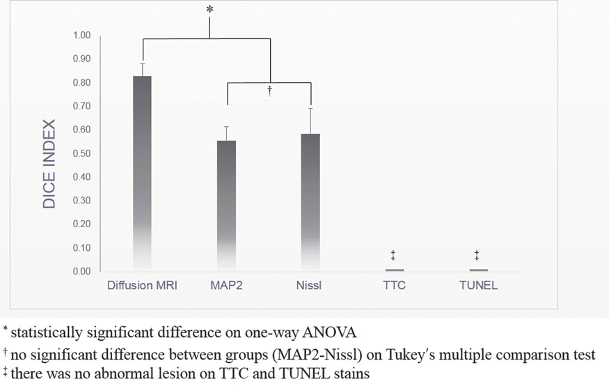
Conclusion
In 1 hour after MCAO in rats, hyperacute infarction were identifiable by digitally processed H&E pathology and they were well correlated with diffusion MRI.
PB02-S01
Multi-targets mechanism of MiR-122 to improve stroke outcomes
1Dept. of Neurology, University of California at Davis
Abstract
Our previous results demonstrated that intravenous (i.v.) miR-122 mimic, has 6 hr therapeutic window, significantly improves multiple pathological and behavioral outcome measurements after middle cerebral artery occlusion (MCAO) in rats. Using Taqman PCR arrays, we identified six genes in blood(e.g., Nos2, Pla2g2a, Vcam1, Olig1, Rhbdf1, Nrep) form hundreds of miR-122 target genes are responsible for the efficacy of miR-122 mimic to improve stroke outcomes.
Our data also demonstrated that miR-122 mimic decreased the two responsive target genes (Nos2, Pla2g2a) in
The combined use of i.v. miR-122 mimic and PEG-liposome to treat stroke is novel, and can be translated to treat human stroke, as miR mimic drugs and PEG-liposome have both been approved by FDA for clinical trials, with the latter even advanced for human use.
Acknowledgements
This study was supported by NIH grant RO1NS089901 and RO1NS101718. There were no conflicts of interest.
PB02-S02
Therapeutic effects of a novel RANKL-based peptide, MHP1-AcN, in stroke models in mice
1Dept. of Neurology, Osaka University, Japan
2Dept. of Health Development and Medicine, Osaka University, Japan
Abstract
Objectives
Although the regulation of post-ischemic inflammation is an important strategy to treat ischemic stroke, all clinical trials have failed to show its efficacy. Based on recent findings of receptor activator of nuclear factor-кB ligand (RANKL)/receptor activator of nuclear factor-кB (RANK) as a novel inhibitory system for post-ischemic inflammation through a Toll-like receptor (TLR) signal1, we previously developed a partial peptide of RANKL, microglial healing peptide 1 (MHP1)2. MHP1 is an anti-inflammatory and anti-osteoclastogenic peptide, which could reduce ischemic injury3; however, modification of the peptide was necessary to increase the stability and efficacies for clinical use. In the present study, we newly designed a series of modified MHP1 peptides and examined its effects in stroke models.
Methods
According to information gathered through HPLC/MS in serum, we designed MHP1 with an amino acid replacement by D-amino acid (MHP1-DN1, MHP1-DN2, MHP1-DN3, MHP1-DN4) or MHP1 acetylated in the N-terminus (MHP1-Ac) or amidated in the C-terminus (MHP1-N), with both modifications added (MHP1-AcN). The activity of these peptides was checked in LPS-stimulated MG6 cells. The effectiveness was examined in several stroke models.
Results
We found that N-terminal acetylation and C-terminal amidation in MHP1 (MHP1-AcN), can strengthen its anti-inflammatory effects and increase its stability without losing anti-osteoclastogenic effects. Interestingly, some peptides with substituted D-amino acids in the N-terminus lost the MPH1 anti-inflammatory effects and increased production of pro-inflammatory cytokines. Anti-TLR activity was reported to be reduced in MHP1 when incubated at 37°C for 24 hrs, but MHP1-AcN could keep the activity under the same condition. The therapeutic effect of MHP1-AcN was observed at lower dose than MHP1 in a transient middle cerebral aertery occlusion (tMCAo) model. Importantly, tPA-induced hemorrhagic transformation was attenuated by co-treatment with MHP1-AcN in tMCAo model without affecting hemolytic effects of tPA in FeCl3-induced thormbosis model.
Conclusions
Although further assessment on safety is necessary, MHP1-AcN could be a promising agent, which could be administered with tPA, for treating ischemic stroke patients.
References
PB02-S03
Anti-oxidant nanomedicine as neurovascular unit protection therapy for cerebral-ischemia-repefusion injury
1Department of Neurosurgery, Faculty of Medicine, University of Tsukuba
2Department of Neurosurgery, Graduate School of Comprehensive Human Science, University of Tsukuba
3Graduate School of Pure and Applied Sciences, University of Tsukuba
4Center for Integrative Medicine, Tsukuba University of Technology
5Department of Gastroenterology, Faculty of Medicine, University of Tsukuba
6Graduate School of Systems and Information Engineering, University of Tsukuba
7Department of Neurosurgery, Saitama Medical Center, Dokkyo Medical University
Abstract
BACKGROUND
In this research, we examined if anti-oxidant nanomedicine (RNPs; nitroxide radical-containing nanoparticles) could prevent neurovascular unit impairment caused by reactive oxygen species after cerebral ischemia-reperfusion.
METHODS
C57BL/6J mice (male, 6-7 weeks, body weight 20-25grams) underwent transient middle cerebral artery occlusion. After 60 minutes of occlusion, mice were randomly divided in three groups and received intra-arterial injection of RNPs (7μM/kg), EDARAVONE (17μM/kg) or PBS (Control) through the carotid artery. Survival rate and neurological score were evaluated 24 hours after injection. Infarction size was evaluated using Cresyl Violet staining and distribution of RNP was evaluated using different Immunofluorescence staining.
RESULTS
RNPs-treated group showed a significantly higher survival rate and lower neurological score than EDARAVONE-treated group 24h after tMCAO and intra-arterial injection. Cerebral infarct size measured using survived mice showed no significant difference between these groups. Immunofluorescence analysis used to evaluate distribution of injected RNP indicated that RNP could be distributed on the endothelial cells and in the cytoplasm of neuronal cells, rather than astrocytes and microglia.
CONCLUSION
Intra-arterial injection of RNPs reduced a mortality rate and improved neurological deficit, that was achieved by protection of neurovascular unit by scavenging reactive oxygen species specially in endothelial cells and neuronal cells. RNPs could be a promising anti-oxidant and neuroprotective nanomedicine for cerebral ischemia-reperfusion injury after mechanical thrombectomy in acute ischemic stroke.
Keywords
tMCAO model, RNPs, EDARAVONE, NEUROPROTECTION
PB02-S04
Acidosis-responsive nanoparticles release nimodpine in cerebral ischaemia and restrain cortical spreading depolarization in rats
1University of Szeged, Faculty of Medicine, Department of Medical Physics and Informatics, Szeged, Hungary
2University of Szeged, Faculty of Medicine, Department of Medical Chemistry, Szeged, Hungary
Abstract
Objectives
Nimodipine, an L-type Ca2+-channel blocker achieves neuroprotection, and vasodilation, concerning the cerebral vasculature in particular (1). Nanoparticles are emerging carriers of drug delivery, which can be constructed to release drugs in response to specific signals. We propose that, in the ischaemic tissue acidosis can be a relevant signal that could be utilized for the initiation of drug release from nanoparticles. Our aim was to design and test a novel treatment strategy for cerebral ischaemia resting on pH-sensitive nanoparticles containing nimodipine, in our in vivo global cerebral ischaemia model.
Methods
Anesthetized Sprague-Dawley rats (n = 18) were used. After washing a suspension of chitosan nanoparticles (d < 20 nm) with or without nimodipine (prepared according to Janovák et al) (2) on the exposed brain surface, both common carotid arteries were permanently occluded to create global forebrain ischaemia. Spreading depolarizations (SDs) were elicited by 1M KCl to aggravate the ischaemic insult. Local field potential, cerebral blood flow (CBF) and tissue pH-variations were recorded from the cerebral cortex.
Results
Ischaemia-induced tissue acidosis initiated nimodipine-release from nanoparticles, which was confirmed by the significant elevation of baseline CBF (47.8 ± 23.7 vs. 29.3 ± 6.9 %; nimodipine associated to nanoparticle vs. nanoparticle only) (Fig. 1). Nimodipine significantly shortened the duration of both SD itself (48.1 ± 23.3 vs. 76.2 ± 17.2 s), and the associated tissue acidosis (65.5 ± 20.2 vs. 138.3 ± 66.1 s), moreover it enhanced the SD-related hyperaemia (4604.4 ± 2572.3 vs. 2368.0 ± 1324.7 %*s).
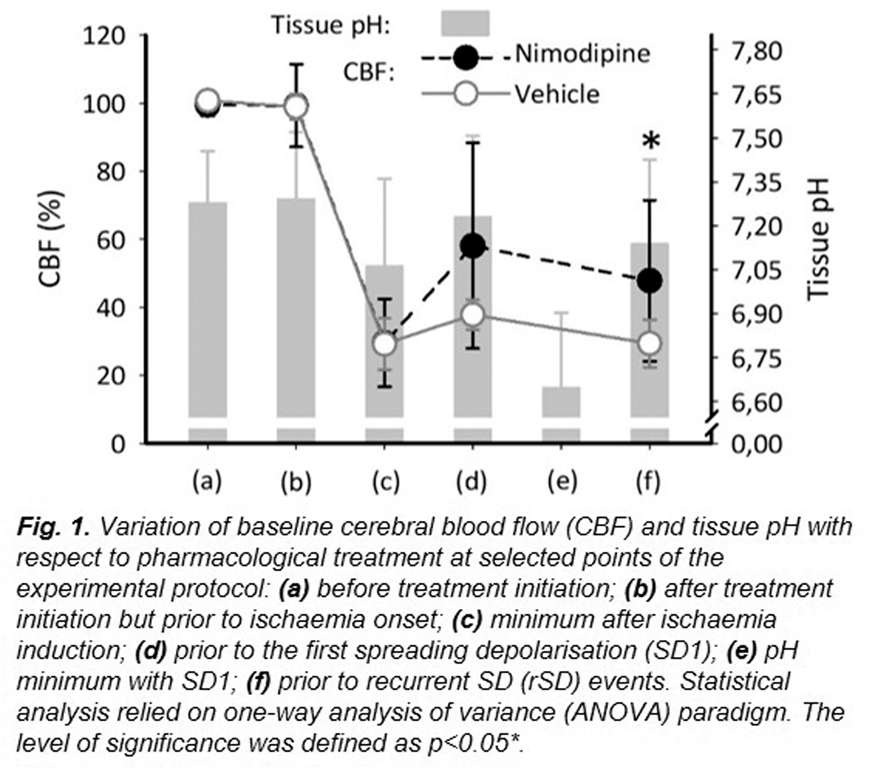
Conclusions
Our results show that the delivery of nimodipine targeted to the ischaemic nervous tissue with pH sensitive nanoparticles is a feasible approach to attenuate secondary brain injury mechanisms such as SD.
Funding
NKFIH (K120358, K111923); Ministry of Human Capacities of Hungary Nr. 34232-3/2016/INTFIN, to DH; EFOP-3.6.1–16-2016-00008., GINOP-2.3.2–15-2016-000060; UNKP-18-3-I-SZTE-26; EFOP-3.6.3-VEKOP-16-2017-00009.
References
PB02-S05
Encapsulation of recombinant tissue plasminogen activator (t-PA) in sono-sensitive structures
1Clinical Neurosciences Research Laboratory, Clinical University Hospital, Health Research Institute of Santiago de Compostela (IDIS), Santiago de Compostela (Spain)
2Centro Singular de Investigación en Química Biolóxica e Materiais Moleculares (CiQUS), Departamento de Física de Partículas, Universidade de Santiago de Compostela, 15782 Santiago de Compostela (Spain
3UMR-S UMR-S U1237 “Physiopathology and Imaging of Neurological Disorders" PhIND. INSERM / EFS Caen-Normandie University
4Caen University Medical Center (CHU Côte de Nacre) – Department of Clinical Research, Caen, France Withdrawn
PB02-S06
Effect of erythropoietin on inflammatory response and ischemic brain damage after carotid artery clamp in rat
1Students Research Committee, Tabriz University of Medical Sciences, Tabriz, Iran
2Department of General and Vascular Surgery, Tabriz University of Medical Sciences, Tabriz, Iran
3Neurosciences Research Center, Tabriz University of Medical Sciences, Tabriz, Iran
4Department of Nuclear Medicine, Odense University Hospital, Odense, Denmark
5Department of Emergency Medicine, Zanjan University of Medical Sciences, Zanjan, Iran
Abstract
Objective
Cerebrovascular diseases are considered a major healthcare-related condition, mostly resulting in ischemic events like stroke which is the third leading cause of death worldwide. Peripheral Artery Diseases (PADs) such as Carotid atherosclerosis and Carotid stenosis can lead to stroke and ischemia-induced brain damages either. Many studies suggest that erythropoietin (EPO) has a minimizing effect on the ischemic condition, whilst indirectly suppresses the inflammatory responses but the detailed association between EPO and the ischemic upshot is still not clearly understood. The aim of current study was to evaluate the effect of EPO on inflammatory response and ischemic brain damage after carotid artery clamp in rats.
Materials and Methods
In a trial study, 50 adult male Wistar rats with (250 to 300 grams) were randomly allocated to intervention and control groups. The intervention group was administered with an intraperitoneal injection of EPO (5000 U/kg) and the control group an intraperitoneal injection of normal saline equal in volume. Both groups had both common carotid arteries clamped for 20 minutes. By using the Nissl staining technique, the slides of brain ischemic areas were observed and the rate of ischemic injury in both groups was determined. The blood level of inflammatory cytokines was also measured.
Results
Levels of inflammatory markers including CPK, IL-6, IL-1B and TNF-α in the intervention group were significantly lower than the control group. Mean percentage of the ischemic area in the intervention group with an amount of 4.30 ± 2.15%, was significantly lower than the control group (11.20 ± 2.35%, P = 0.023).
Conclusion
Based on the findings of this study showed that the injection of EPO before carotid stenosis surgery is strongly effective in preventing cerebral ischemic injury.
Keywords
Erythropoietin, Ischemic Brain Damage, Carotid Artery Clamp, Rat
PB02-S07
Modulating the counter regulatory renin angiotensin system axis in the stroke prone spontaneously hypertensive rat in ischaemic stroke
1Institute of Cardiovascular and Medical Sciences, University of Glasgow, UK
2International Centre for Genetic Engineering and Biotechnology, Trieste, Italy
Abstract
Introduction
Several studies have assessed the potential of targeting the classical renin angiotensin system (RAS) with therapeutics for ischaemic stroke. Our group have previously demonstrated that the counter regulatory RAS peptide, angiotensin-(1-9), acts via the angiotensin II type 2 receptor (AT2R) to oppose detrimental effects of perturbations in RAS in cardiovascular pathologies. We hypothesise that Ang-(1-9) may have a beneficial effect on stroke outcome in the stroke prone spontaneously hypertensive rat (SHRSP), which possesses clinically relevant comorbidities. Initial studies investigated expression of the AT2R mRNA (AGTR2) in the SHRSP brain after experimental stroke, and a proof of concept investigation into the therapeutic potential of Ang-(1-9), delivered stereotactically using an adeno-associated serotype 9 viral vector (AAV9) with an expression cassette secreting a fusion protein that releases Ang-(1-9), in experimental stroke induced by transient middle cerebral artery (tMCAO) in SHRSP.
Methods
Male SHRSP (17-23 week old, 270–310 g) were used. For gene expression studies, after undergoing sham surgery (n = 7) or 35 min tMCAO followed by varying reperfusion times: no reperfusion, 2 hr and 24 hr (n = 4 each), RNA was isolated from ipsilateral brain regions. Expression of AGTR2 was calculated relative to a housekeeping gene (β2 microglobulin; B2M). For a proof of concept study with AAV9-Ang-(1-9) in ischaemic stroke, randomly allocated animals received a stereotactic injection of 5 × 10¹⁰ AAV9-Ang-(1-9) (n = 5) or AAV9-eGFP (control virus, enhanced green fluorescent protein) (n = 5) in 2 µL sterile saline, at striatal and cortical sites in each hemisphere, or cranial burrholes with durotomy for the non-viral control group (n = 5). After 4 days, animals underwent tMCAO with recovery for 10 days. Functional outcome was measured using a 30-point neurological score, tapered beam and sticky label test and infarct volume measured using histology. All measurements and analysis were performed blinded.
Results
Following stroke, AGTR2 showed a 4 fold increase in expression immediately after 35 min occlusion in infarcted tissue (RQ + RQmax: sham 1.0 + 0.3; 35 min MCAO 4.2 + 0.2
In the pilot therapeutic study (n = 5/group), AAV9-Ang-(1-9) delivery showed promising improved functional outcome compared to burrhole only and AAV-eGFP control groups (Table 1) although this failed to reach a statistically significant level. Specifically, neurological score (where greater score = better outcome) at day 3 showed a trend towards improvement (22/30, vs 17/30 (AAV-eGFP) and 19/30 (burrhole)), while %footfaults on the affected side during the tapered beam test trended towards improvement at day 7 (5.5 ± 1.9% vs 25.8 ± 14.0% (AAV-eGFP) and 19.5 ± 1.4% (burrhole)). Despite a tendency to improve functional outcome, so far, infarct analysis has shown no statistically significant differences between groups (n = 4/group) (Table 1).
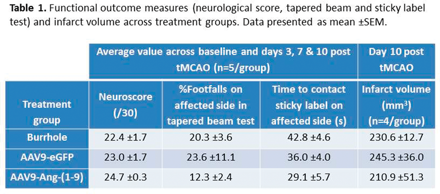
Conclusion
Taken together, these results, of alterations in AT2R receptor expression in the SHRSP brain acutely after stroke and the trends in functional outcome with delivery of AAV9-Ang-(1-9) prior to tMCAO, demonstrate a promising potential for targeting the counter-regulatory RAS in ischaemic stroke.
PB02-S08
Role of posttreatment with renal denervation against experimental acute stroke in rats
1Department of Pharmacology and Molecular Therapeutics, Kumamoto University Graduate School of Medical Sciences, Japan
2Department of Neurosurgery, Kumamoto University, Japan
Abstract
Objectives
Renal denervation (RD) was shown to have several organ protective effects in addition to the antihypertensive effect against resistant hypertension. Previously, we reported RD inhibited stroke onset in hypertensive rats. In this study, we further evaluated whether posttreatment with RD had protective effects against experimental acute stroke in rats.
Methods
In experiment 1, we evaluated the effect of RD against cerebral infarction (CI) in hypertensive rats (SHRSP). An CI model of SHRSP was prepared by 90-minute middle cerebral artery occlusion (MCAO) followed by reperfusion. RD was performed at 30 minutes after the start of MCAO. Twenty-four hours later, we examined the effect of RD on blood pressure (BP), neurological findings (NF), cerebral blood flow (CBF), infarct volume, and cytotoxic changes. In experiment 2, we evaluated the effect of RD against subarachnoid hemorrhage (SAH) in SD rats. SAH rats were produced by endovascular perforation model. RD was performed at 60 minutes after SAH induction. Twenty-four hours later, we examined the effect of RD on BP, NF, CBF, cerebral arterial diameter (cerebral vasospasm), and cytotoxic changes.
Results
In experiment 1, RD ameliorated NF and infarct volume. RD decreased elevated BP and the values of CBF. RD also significantly decreased elevated cerebral oxidative stress as estimated by dihydroethidium staining and gp91phox expression. In experiment 2, RD decreased elevated BP and inhibited cerebral arterial narrowing, although NF and CBF were not changed. In basilar artery, phosphorylated Extracellular Signal-regulated Kinase (ERK)-positive cells, the expression of Cyclooxygenase-2, and infiltration of macrophages were upregulated by SAH, but RD significantly reduced them.
Conclusions
The present study demonstrated that posttreatment with RD was benefit against experimental acute stroke both CI and SAH, and the protective effects were associated with suppression of elevated BP, oxidative stress, and inflammatory response.
PB02-S09
The extra virgine olive oil phenol hydroxytyrosol as acute therapeutic strategy after ischemic stroke
1Department of Anatomy, Radboud university medical center, Donders Institute Nijmegen, The Netherlands
2Department of Experimental Biology, University of Jaén, Jaén, Spain
3Peltarion AB Stockholm Sweden
Abstract
Stroke is one of the leading causes of adult disability worldwide. After ischemic stroke, damaged tissue (penumbra) surrounding the irreversibly damaged core of the infarct is still salvageable and therefore a target for acute therapeutic strategies. Mediterranean-style diet (MD) is characterized by intake of extra-virgin olive oil, of which hydroxytyrosol (HT) is the foremost phenolic component. HT has been shown to possess antioxidant effects, protecting the blood vessel wall against oxidative stress, and even improving cognitive performance in Alzheimer’s disease mice. This study investigated the acute effect of a HT-enriched diet after stroke on the regain of motor and cognitive functioning, and MRI parameters. Experimental transient (30 min) middle cerebral artery occlusion (MCAO) will be induced in male C57Bl/6 mice. After stroke, mice will be put on an HT-enriched diet or on a control diet. MRI will be performed at 7 and 35 days post-stroke. Additionally, multiple behavioral tests will be performed pre- and post-stroke to assess motor and cognitive recovery. Stroke mice on HT diet showed improved CBF and neurogenesis in the lesioned hippocampus. Additionally, HT-fed mice showed increased strength in the forepaws, as well as improved short-term recognition memory accompanied by an enhanced postsynaptic density. Digital ventilated cages (DVC, Tecniplast) system was used to study individual locomotion via calculation of DVC metric measures (activity, walked distance, walked velocity, total turnings, laterality index) 24/7 before and after surgery. Here, only stroke mice on control diet showed a decreased walked distance at 3 weeks after surgery compared to prior the surgeries. These results suggest that a HT-enriched diet may attenuate the damage after ischemic stroke and therefore HT may be an interesting compound to further test its beneficial role as a therapeutic approach in the functional and structural recovery after ischemic stroke.
PB02-S10
Whole body vibration therapy after ischemia reduces brain damage in reproductively senescent female rats
1Cerebral Vascular Disease Research Laboratories, Department of Neurology, Leonard M. Miller School of Medicine, University of Miami, Miami, Florida, USA
2Department of Neurological Surgery, Leonard M. Miller School of Medicine, University of Miami, Miami, Florida, USA
3Bruce W. Carter Department of Veterans Affairs Medical Center, Miami, and Florida, USA
Abstract
Background
Stroke disproportionately kills more women than men and the risk of stroke remains high even at a young age among women smokers. Smoking prior to stroke is associated with increased post-stroke frailty. Frailty is characterized by an increased vulnerability to acute stressors and the reduced capacity of various bodily systems due to age-associated physiological deterioration. Such age related physiological deterioration of bone in laboratory animals and humans has shown to reverse after therapeutic intervention of whole body vibration (WBV) (1). Our recently published study shows that post-stroke WBV intervention reduces ischemic brain damage in reproductively senescent female rats (2), suggesting WBV may be a potential therapy to reduce post-ischemic frailty and improve functional and cognitive outcomes after stroke. In the current study we aim to test the efficacy of WBV in reducing post-ischemic frailty and improving physical activity and cognition using a rat model of smoking attributed nicotine.
Methods
Nicotine or saline exposed adult female rats underwent transient middle cerebral artery occlusion (tMCAO; 90 min) / sham-surgery and randomly assigned (n = 6-8 per group) to either WBV or control groups. Animals placed in the WBV (40 Hz) group underwent 30 days of WBV treatment performed twice daily for 15 min each session for 5 days each week. We monitored the frailty index (FI) prior to and 1 month after tMCAO alone or in combination with WBV. The FI was composed of the following criteria: 1) activity levels, 2) blood pressure (BP), 3) basic metabolic status, and 4) cognitive performance of rats. Animals were sacrificed on the 30th day of WBV treatment, and brain tissue was harvested for histopathological analysis.
Results
Post-tMCAO WBV did not change activity levels or BP in nicotine or saline treated rats. Post-tMCAO WBV cognitive performance improved in saline group as compared to nicotine exposed rats. Sensorimotor function was also improved in tMCAO WBV saline group compared to nicotine-exposed rats. We observed 56% reduction in infarct volume of WBV treated rats as compared to control (p < 0.05). This difference was not seen in nicotine treated groups.
Conclusions
The post-ischemic WBV intervention had no detrimental effects on the frailty parameters, decreased brain damage, and reduced frailty in control female rats, but not in the nicotine-exposed group. This suggests that WBV may be a potential therapy for non-smokers to reduce post-ischemic frailty and improve functional and cognitive outcomes after stroke. Recently more tobacco users have switched to the electronic nicotine delivery systems (e-Cigarettes) as an aid for smoking cessation and the safety of e-Cigarettes remains questionable therefore, current research is timely.
References
PB03-A01
A novel, injectable carbon nanoparticle demonstrates electron shuttling capabilities: a potential therapeutic “nanozyme” for mitochondrial protection in ischemic injury
1Texas A and M Health Science Center – Institute of Biosciences and Technology, Houston, TX, USA
2Department of Chemistry, Rice University, Houston, TX, USA
3Department of Neurology, Houston Methodist Hospital and Research Institute, Houston, TX, USA
Abstract
Objectives
We previously showed that highly oxidized, poly(ethylene glycol)-ated carbon nanomaterials, hydrophilic carbon clusters (PEG-HCCs), are high capacity, catalytic superoxide dismutase mimetics also active at quenching hydroxyl radical
Methods
Employing solution-based UV-vis spectroscopy, we examined the reactions of PEG-HCCs between NAD(P)H and ascorbic acid to cytochrome C (CytC) and resazurin, a surrogate marker of mitochondrial viability. Cellular uptake and distribution in cultured SHSY-5Y neuroblastoma cells and b. End3 brain endothelial cells were assessed using deconvolution microscopy after 5 minute incubation, using GFP containing a CytC oxidase subunit IV targeting sequence. In-vitro protection against the mitochondrial Complex IV toxin, sodium cyanide (NaCN) 1uM (approximate LD50 in humans) was performed by adding PEG-HCCs immediately and delay of 30 minutes. A Seahorse (Agilent) mitochondrial “stress test” to measure oxygen consumption rate (OCR) was performed after addition of oligomycin to inhibit ATP synthase, trifluoromethoxy carbonylcyanide phenylhydrazone (FCCP), an ionophore to maximize respiration, and rotenone/antimycin to inhibit Complexes I/III.
Results
PEG-HCCs rapidly catalyzed the reduction of both resazurin and CytC by NAD(P)H and ascorbic acid. Kinetic analysis indicated a ternary complex, consistent with a transient structure containing two substrates to facilitate electron transfer. PEG-HCCs were frequently co-localized with the CytC subunit, both in strands and punctate morphology. PEG-HCCs were fully protective against 1uM NaCN in culture even after a 30 minute delay (p = .003 vs no treatment). PEG-HCCs increased the basal OCR in bEnd.3 cells and increased the maximal respiration when subjected to the Seahorse assay (Figure).

Conclusions
Cell-free experiments indicate electron shuttling between key mitochondrial constituents. The ternary complex kinetics are consistent with a “nanozyme”, a nanoparticle that catalyzes a reaction. Increasing maximal respiration achievable under stress conditions and protecting cells from NaCN suggests this action may be occurring in cells. Prior cyclic voltammetry showed that PEG-HCCs, unlike other graphene-based nanoparticles that were not effective in ischemia/reperfusion, have a very broad redox potential spanning the range of the mitochondrial constituents, potentially explaining their ability to shuttle from low reduction to high reduction species. Work is ongoing to identify the functional groups that facilitate this shuttling effect in order to optimize this activity. Our results indicate that supporting mitochondrial electron transport may contribute to their effectiveness in-vivo and suggests further investigation to address mitochondrial disruption following stroke.
Supported by
1R01NS094535
PB03-A02
Temporal therapeutic window for calcium release-activated calcium channel inhibition against experimental stroke
1Dept. Neurology,University of California, San Francisco & San Francisco Veterans Affairs Medical Center, USA
2Dept. Neurology, Tokai University school of medicine, Japan
3CalciMedica, USA
Abstract
Objectives
Inflammatory responses after ischemic stroke contribute to the worsening of brain. Calcium release-activated calcium (CRAC) channel, found on microlia, activates immune responses. We previously reported that the novel CRAC channel inhibitor (CM-EX-137) improved outcomes in protected against experimental brain injury and stroke.. Less clear, but clinically relevant is how long after stroke onset this approach is still effective. The purpose of this study is to clarify the therapeutic time window of CM-EX-137 in experimental stroke.
Methods
C57/BL6 male mice, aged 2 months were subjected to distal middle cerebral artery occlusion (dMCAO) and treated with CRAC channel inhibitor (CM-EX-137, 5 mg/kg IP) or vehicle daily (maximum 7days). CM-EX-137 treatment was delayed: 1 h, 4 h, and 6 h following dMCAO. These groups were compared to the dMCAO-vehicle group. Brains were extracted 14 days after brain injury. Neurological function was evaluated by modified Bederson score, elevated body swing test, and corner test before surgery and 1, 3, 7, 14 days after dMCAO. Lesion size was evaluated from hematoxylin & eosin staining. We also evaluated the microglia & monocytes activation,and immune markers.
Results
dMCAO mice survived for 14 days (n = 6–7/ group) with no mortality. Neurological functions were significantly improved when treatment was delayed1h and 4 h (p < 0.001). This was also associated with reduced infarct volume 14 days after dMCAO (p < 0.05). Microglial & monocyte/macrophage activation was also suppressed 14 days in the 1 h and 4 h delay groups(p < 0.01), but not 6 h
Conclusions
CRAC channel inhibition by CM-EX-137 was neuroprotective when given within 4 h of dMCAO, but not as late as 6 h. These results indicate that CM-EX-137 has a wide therapeutic time window and it may be useful in the treatment of acute ischemic stroke.
PB03-A03
Female mice benefit from calcium release-activated calcium channel inhibition following focal cerebral ischemia
1Dept. Neurology,University of California, San Francisco & San Francisco Veterans Affairs Medical Center, USAt. Neurology,University of California, San Francisco & San Francisco Veterans Affairs Medical Center, USAt
2Dept. Neurology, Tokai University school of medicine, JapanDept. Neurology, Tokai University school of medicine, Japan
3CalciMedica, USACalciMedica, USA
Abstract
Objectives
Calcium release-activated calcium (CRAC) channel inhibition suppresses the inflammatory response after brain ischemia in association with store-operated Ca2+entry. We previously reported that CRAC channel inhibitor (CM-EX-137) had neuroprotective effect to brain ischemia due to suppression of microglial activation in experimental stroke male mice. However, whether females similarly benefit from CRAC channel inhibition in brain ischemia is still unclear. The purpose of this study is to evaluated potential neuroprotective effects CRAC channel inhibition in a female stroke model.
Methods
C57/BL6 female mice, aged 2 months underwent distal middle cerebral artery occlusion (dMCAO) and were treated with the specific CRAC channel inhibitor CM-EX-137 (5 mg/kg IP; dMCAO-CM group, CalciMedica, Inc.) or vehicle (dMCAO group) daily (maximum 3days). Mice underwent dMCAO during the same phase in the estrus cycle(diestrus phase). These groups were also compared to sham (sham-operated) groups. CM-EX-137 was injected immediately after occlusion and at 24, 48 hours. Brains were extracted 3 days after ischemia. Neurological functions were evaluated by modified Bederson score, elevated body swing test, adhesive removal test, and corner test before surgery, 24 hours and 72 hours after brain ischemia in each group. Lesion size was evaluated from cresyl violet staining. We also evaluated microglia & monocyte/macrophage activation in association with CRAC channel activation from histochemistry and fluorescent microscopy.
Results
Neurological function 24 hours and 72 hours after ischemia were significantly improved in the dMCAO-CM group (p < 0.001), compared with the dMCAO-vehicle group. CM-EX-137 also significantly reduced infarct volume (p < 0.001). Activation of microglia & monocytes/macrphages was suppressed by CM-EX-137 3 days after ischemia (p < 0.001; isolectin B4, CD68). Further, iNOS expression was induced after brain ischemia and was significantly reduced by CRAC channel inhibition (p < 0.01). Finally, CM-EX-137 suppressed microglial activation (CD68 + ) as evidenced by reduction in CRAC channel components STIM1 and Orai1by (p < 0.001).
Conclusions
CRAC channel inhibition can reduce inflammatory responses after brain ischemia female mice, similar to males. Microglial activation was also suppressed via CRAC channel inhibition in females.
PB03-A04
Neuroprotective effects of SMTP-44D in mice stroke model in relation to neurovascular unit and trophic coupling
1Department of Neurology, Graduate School of Medicine, Dentistry and Pharmaceutical Sciences, Okayama University, Okayama, Japan
2Department of Applied Biological Science, Tokyo Noko University, Fuchu, Japan
Abstract
Objectives
Stachybotrys microspora triprenyl phenol (SMTP)-44D has both anti-oxidative and anti-inflammatory activities, but its efficacy has not been proved in relation to the pathological changes of neurovascular unit (NVU) and neurovascular trophic coupling (NVTC) in ischemic stroke. Here, the present study was designed to assess the efficacies of SMTP-44D, moreover, compared with the standard neuroprotective reagent edaravone in ischemic brains.
Methods
ICR mice were subjected to transient middle cerebral artery occlusion (tMCAO) for 60 min, SMTP-44D(10 mg/kg) or edaravone (3 mg/kg) was intravenously administrated through subclavian vein just after the reperfusion, and these mice were examined at 1, 3, and 7 d after reperfusion.
Results
Compared with the vehicle group, SMTP-44D treatment revealed obvious ameliorations in clinical scores and infarct volume, meanwhile, markedly suppressed the accumulations of 4-HNE, 8-OHdG, nitrotyrosine, RAGE, TNF-α, Iba-1, and cleaved caspase-3 after tMCAO. In addition, SMTP-44D significantly prevented the dissociation of NVU and improved the intensity of NAGO/BDNF and the number of BDNF/TrkB and BDNF/NeuN double positive cells. These effects of SMTP-44D in reducing oxidative and inflammatory stresses were similar to or stronger than those of edaravone.

Conclusions
The present study demonstrated that SMTP-44D showed strong anti-oxidative, anti-inflammatory, and anti-apoptotic effects, moreover, the drug also significantly improved the NVU damage and NVTC in the ischemic brain.
References
PB03-A05
Farnesiod X receptor knockout protects brain against ischemia-reperfusion injury through reducing neuronal apoptosis in mice
1School of Biomedical Engineering, Shanghai Jiao Tong University, China
2Department of Cardiology, Renji Hospital, School of Medicine, Shanghai Jiao Tong University, China
Abstract
Objectives
Farnesiod X Receptor (FXR) is a nuclear receptor that plays a critical role in controlling cell apoptosis in diverse diseases(1). Previous studies showed that knockout of FXR improves cardiac function by reducing cardiomyocytes apoptosis in myocardial ischemic mice(2). However, the role of FXR after cerebral ischemia remains unknown. In this study we explored the effect of FXR on functional recovery of mice after cerebral ischemia and potential mechanisms.
Methods
Adult male C57BL/6 mice (n = 72) and FXR knockout mice (n = 30) were subjected to 90-min middle cerebral artery occlusion (MCAO). FXR expression was examined using immunohistochemistry and Western blot, brain infarct volume and neurobehavioral tests were performed at 1, 3, 7, 14 days after ischemic stroke to evaluate the outcomes of FXR knockout mice. Protein levels of related apoptosis factors were also examined.
Results
The expression of FXR started to increase at 6 h after stroke (p < 0.05), reached a peak at 24 h (p < 0.01), and returned to the same level of sham group at 72 h after reperfusion. Cresyl violet staining showed that knockout of FXR reduced infarct volume at day 1 after stroke, compared to the control (p < 0.05). Behavioral tests including NSS and hanging wire test showed that knockout of FXR improved functional recovery at 14 days after stroke. Immunostaining result showed that the number of TUNEL+ cells in infarct core region was decreased in FXR knockout mice compared to the control group (p < 0.05), and the expression of cleaved caspase-3 was attenuated in FXR knockout mice compared to the control mice (p < 0.05).

Conclusion
FXR knockout reduced brain infarct volume, apoptosis and promoted neurobehavioral recovery after ischemic stroke, suggesting that FXR is a potential target for stroke therapy.
References
PB03-A06
L-glutamine protects mouse brain from ischemic injury via up-regulating of heat shock protein 70
1School of Biomedical Engineering, Shanghai Jiao Tong University
2Shanghai Ruijin Hospital, School of Medicine, Shanghai Jiao Tong University
Abstract
Introduction
L-glutamine (L-GLN) is an antioxidative1 drug approved by FDA for the treatment of sickle cell disease.2 However, the effect of L-GLN on ischemic stroke is unknown. In this study, we investigated the effect and mechanism of L-GLN in ischemic brain injury.
Methods
Adult male ICR mice (n = 135) were subjected to 90-minute middle cerebral artery occlusion (MCAO), and divided into four groups: 1) Sham, 2) Saline, 3) L-GLN treatment, and 4) L-GLN plus Apoptozole (AZ) treatment, a HSP70 ATPase inhibitor. Treatments were delivered intraperitoneally once daily for the first three days after MCAO. Neurobehavioral tests were performed before and 1, 3, 7 and 14 days after MCAO. Infarction volume was assessed by cresyl violet staining. The expression levels of heat shock proteins and their signaling pathway related proteins were evaluated by Western blot analysis and real-time PCR. The level of oxidative stress was evaluated by malondialdehyde, superoxide dismutase, and glutathione assays. Astrocyte, BEND.3 cell and neuron cultures and oxygen glucose deprivation model were used to further examine the molecular pathways involved in the protective effects by L-GLN.
Results
We found that L-GLN treatment reduced brain infarct volume and promoted neurobehavioral recovery in mice after ischemic injury. Such beneficial effect was abolished by the coadministration of AZ. L-GLN induced the up-regulation of HSP70 and Nuclear factor erythroid 2-related factor 2, reduced oxidative stress level and neuronal apoptosis (p < 0.05). Superoxide dismutase and glutathione levels were increased in the L-GLN group compared to the saline group (p < 0.05). Our cell culture study further revealed that the conditioned medium from astrocytes cultured with L-GLN effectively reduced the apoptosis of neurons after oxygen glucose deprivation (p < 0.05).

Conclusion
L-GLN attenuated ischemic brain injury and promoted functional recovery, suggesting its potential for the ischemic stroke therapy.
References
PB03-A07
Troxerutin exerts neuroprotection at middle cerebral artery occlusion/reperfusion model in rats
1China-America Institute of Neuroscience, Xuanwu Hospital, Capital Medical University, Beijing, China
2Department of Neurosurgery, Wayne State University School of Medicine, Detroit, MI, USA
Abstract
Objectives
Troxerutin, as a commonly used drug in treatment of chronic venous insufficiency (CVI) disease, is natural bioflavonoid derived from rutin. It has antioxidant and anti-inflammatory properties. It also has been found to have protective effects on myocardial ischemia/reperfusion (I/R) injury. In this study, we aim to evaluate its possible neuroprotective effect in experimental ischemic stroke in rats.
Methods
The experimental ischemic stroke model was established with middle cerebral artery (MCA) occlusion by an intraluminal filament for 2 hours in rats. Troxerutin was given after reperfusion at a dose of 270 mg/kg/day. The extent of brain injury was determined by infarct volume and neurological deficit. Infarct volume was evaluated at 24 h after reperfusion by the TTC staining, and neurological deficits were assessed according to Zea-Longa 5-point scoring and 12-scoring scale. The brain edema was measured. Matrix metalloproteinase (MMP)-9 and aquaporin (AQP)-4 were evaluated by Western blotting. Expressions of apoptotic proteins (Bcl-2, Caspase-3) were also determined.
Results
Troxerutin significantly reduced brain infarct volume and improved neurological deficits. Brain edema was reduced, and MMP-9 and AQP-4 were also decreased at 24 h after reperfusion. In addition, anti-apoptosis protein Bcl-2 was increased and pro-apoptosis protein caspase-3 was decreased.
Conclusion
These findings indicate neuroprotective potential of troxerutin against ischemia/reperfusion injury in experimental ischemic model.
Key words
Troxerutin, neuroptrotection, ischemic stroke, edema
PB03-A08
Neuroprotective effects of IQ-1S in the model of global cerebral ischemia/reperfusion
1Cardiovascular Research Center, Cardiology Division, Massachusetts General Hospital, Harvard Medical School
2Kizhner Research Center, Tomsk Polytechnic University, Tomsk, Russia
3Goldberg Research Institute of Pharmacology and Regenerative Medicine, Tomsk NRMC, Tomsk, Russia
4Siberian State Medical University, Tomsk, Russia
5Research Institute of Biological Medicine, Altai State University, Barnaul, Russia
6Cardiology Research Institute, Tomsk NRMC, Tomsk, Russia
7Department of Microbiology and Immunology, Montana State University, Bozeman, USA
Abstract
Objectives
A new specific inhibitor of c-Jun N-terminal kinases (JNK), 11H-indeno[1,2-b]quinoxalin-11-one oxime sodium salt (IQ-1S), has a high affinity toward JNK3 compared to JNK1/JNK2. We demonstrated previously neuroprotective role of IQ-1S against stroke injury in mouse middle cerebral artery occlusion model with filament [1]. The aim of this study was to characterize the neuroprotective potential of IQ-1S in rat model of global cerebral ischemia (GCI).
Methods
Twenty-nine male Wistar rats were used in the experiments. GCI was reproduced by temporary blocking blood flow by occlusion of three vessels supplying the brain: the brachiocephalic artery, the left subclavian artery, and the left common carotid artery for 7 min as described [2]. Animals of experimental group received five intraperitoneal injections of IQ-1S at dose of 25 mg/kg for four days after GCI. The control group was treated with vehicle: 2 mL of physiological saline solution containing 20 µl of Tween 80. Neurological deficit was assessed by 14-point scale as described [2]. Histoquantitative study of hippocampal neurons was done by calculating number of neurons per 1 mm2 of the CA1 area and by determining percentage of morphologically unchanged neurons and neurons with irreversible injury. Statistical analysis was performed with Fisher's exact test and Mann–Whitney U–test for neurological deficit and with Student t-test for histological investigation.
Results
In control group 24 h after GCI, all rats (n = 7) had severe neurological deficit (7.0). At day 3, mean score of neurological deficit was 6.9; severe degree of neurological deficit persisted in 71.4% of rats. At day 5, 28.6% of rats had severe degree of neurological deficit. In experimental group (n = 8) at days 3 and 5 after GCI, neurological deficit scores were 3.5 and 3.1, respectively, and were lower than control value (p < 0.05). At days 1 and 3 after GCI, 37.5% and 12.5% of rats had severe neurological deficit (p < 0.05 compared with control). No severe neurological disorders were found in animals at day 5. IQ-1S significantly increased the numeric density of the pyramidal neurons in the hippocampal CA1 area (from 2034 ± 140 in control to 3771 ± 239 in treated mice, p < 0.05), increased the number of unaltered neurons (from 25 ± 2 in control to 52 ± 2 in treated mice, p < 0.05), and decreased the number of neurons with irreversible injury (from 42 ± 3 in control to 16 ± 2, p < 0.05).
Conclusions
IQ-1S demonstrated pronounced neuroprotective properties in rat model of GCI.
Acknowledgements
The study was supported by the Ministry of Education and Science of the Russian Federation (grant No. 4.8192.2017/8.9); the histoquantitative study was supported by the Russian Science Foundation (No. 17–15-01111).
References
PB03-A09
Neuroprotective effects of a novel carnosine-hydrazide derivative on hippocampal CA1 damage after transient cerebral ischemia
1Dept. of Neurosurgery, Kurume University, Japan
2Dept. of Bioorganic and Medicinal Chemistry, Kumamoto University
Abstract
Ischemia-reperfusion injuries produce reactive oxygen species that promote the peroxide lipid oxidation process resulting in the production of an endogenic lipid peroxide, 4-hydroxy-trans-2-nonenal (4-HNE), a highly cytotoxic aldehyde that induces cell death. We synthesized a novel 4-HNE scavenger – a carnosine-hydrazide derivative, L-carnosine hydrazide (CNN) – and examined its neuroprotective effect in a model of transient ischemia.
PC-12 cells were pre-incubated with various doses (0–50 mmol/L) of CNN for 30 min, followed by incubation with 4-HNE (250 mM). An MTT assay was performed 24 h later to examine cell survival. Transient ischemia was induced by bilateral common carotid artery occlusion (BCCO) in the Mongolian gerbil. Animals were assigned to sham-operated (n = 6), placebo-treated (n = 12), CNN pre-treated (20 mg/kg; n = 12), CNN post-treated (100 mg/kg; n = 11), and histidyl hydrazide (a previously known 4-HNE scavenger) post-treated (100 mg/kg; n = 7) groups. Heat shock protein 70 immunoreactivity in the hippocampal CA1 region was evaluated 24 h later, while delayed neuronal death using 4-HNE staining was evaluated 7 days later.
Pre-incubation with 30 mmol/L CNN completely inhibited 4-HNE-induced cell toxicity. CNN prevented delayed neuronal death by >60% in the pre-treated group (p < 0.001) and by >40% in the post-treated group (p < 0.01). Histidyl hydrazide post-treatment elicited no protective effect. CNN pre-treatment resulted in high heat shock protein 70 and low 4-HNE immunoreactivity in CA1 pyramidal neurons. Higher 4-HNE immunoreactivity was also found in the placebo-treated animals than in the CNN pre-treated animals.
Our novel compound, CNN, elicited highly effective 4-HNE scavenging activity in vitro. Furthermore, CNN administration both pre- and post-BCCO remarkably reduced delayed neuronal death in the hippocampal CA1 region via its induction of heat shock protein 70 and scavenging of 4-HNE.
PB03-A10
Drp-1, a potential therapeutic target for brain ischaemic stroke
1Peking Union Medical College Hospital, Chinese Academy of Medical Sciences & Peking Union Medical College
Abstract
Objectives
The resistance of CA3 neurons to ischaemia and the ischaemic tolerance conferred by ischaemic preconditioning (IPC) are two well-established endogenous neuroprotective mechanisms. Elucidating the molecules involved may help us find new therapeutic targets. Thus, we determined whether dynamin-related protein 1 (Drp-1) is involved in these processes.
Methods
In vivo, we subjected rats to either 10 min severe global ischaemia using a four-vessel occlusion (4-VO) model or 2 min IPC before the onset of 4-VO. In vitro, we performed oxygen glucose deprivation (OGD) studies in rat hippocampal neurons. Drp-1 was silenced or inhibited by siRNA or pharmacological inhibitor Mdivi1. To assess whether mitochondrial Drp-1 alters neuronal vul- nerability to ischaemic injury, various approaches were used including western blot, immunohistochemistry, immunofluorescence staining and electron microscopy. Hippocampal function was assessed using an open-field test.
Results
Mitochondrial dynamin-related protein 1 (mtDrp-1) was selectively induced by ischaemia in hippocampal CA3 neurons. In hippocampal CA1 neurons, mtDrp-1 was not affected by ischaemia but significantly up regulated by IPC. Suppression of Drp-1 increased the vulnerability of cells to OGD and global ischaemia. Inhibition of Drp-1 in vivo resulted in loss of acquisition and encoding of spatial information, and also prevented ischaemia-induced mitophagy in CA3. Thus mitochondrial mediated injury was amplified and resistance to ischaemic injury lost.
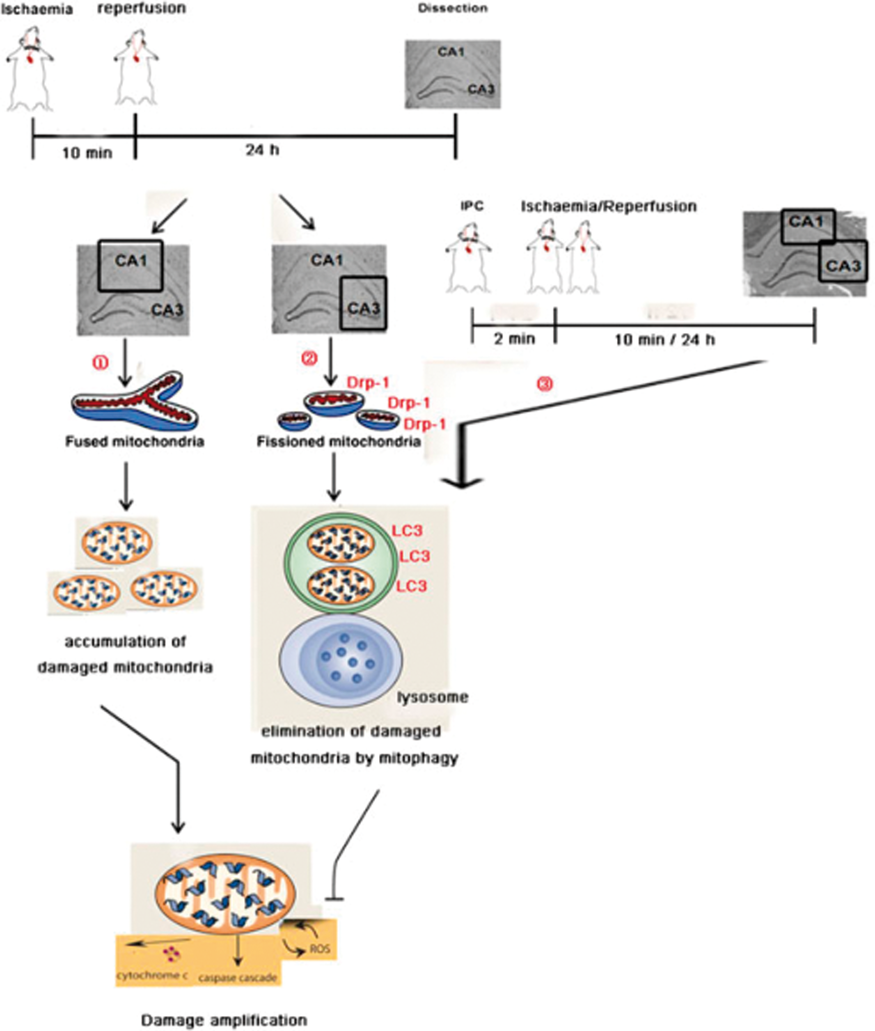
Conclusions
Our findings that Drp-1 increases the resistance of neurons of hippocampal CA3 affected by global ischaemia and contributes to the tolerance conferred by IPC highlight Drp-1 as a potential therapeutic target for brain ischaemic stroke.
PB03-A11
Contribution of astrocytes related to neuroprotection against delayed neuronal cell death in hippocampus
1Department of Innovative Medical Photonics, Preeminent Medical Photonics Education & Research Center, Hamamatsu University School of Medicine, Japan
2Department of Neurosurgery, First Affiliated Hospital of China Medical University, China
Abstract
Objective
In the hippocampus, delayed neuronal death is observed in neurons of the CA1 region but not in those of the CA3 region. Astrocytes have been reported to play multiple supporting roles in neuronal functions. We hypothesized that the astrocyte contributes in the process of the delayed neuronal death, that is, loss of astrocyte might induce the neuronal cell death following transient forebrain ischemia in CA3. In this study, we elucidate the roles of astrocyte in the process of delayed neuronal death.
Methods
Anesthetized (Isoflurane) male SD rats (approximately 300 g-body weight) were used for the experiment. Astrocyte-specific gliotoxin, L-α-aminoadipic acid (L-α-AAA), was injected into the CA3 region of the rat hippocampus. Three days after the injection, transient forebrain (10 min) ischemia was induced by 4-vessel occlusion method. The morphology of the neurons and the astrocytes, and the number of the cells were evaluated in CA1 and CA3 regions 7 days after the ischemia. In rats with ischemia and reperfusion, regional cerebral blood flow (rCBF) was measured by laser-Doppler flowmetry, and change in intracellular Ca2+ concentration ([Ca2+]i) were separately measured in CA1 and CA3 regions using intravital fluorescence imaging.
Results
1. It was confirmed that L-α-AAA injection decreased the number of astrocytes in CA3 region. L-α-AAA injection did not affect the pattern of rCBF changes upon ischemia/reperfusion in CA1 and CA3 regions.
2. In rats without L-α-AAA injection, CA1 neuron decreased 7 days after the transient forebrain ischemia, but CA3 neuron did not. In rats received with L-α-AAA injection, CA3 neuron decreased following the transient forebrain ischemia. This indicates that the loss of astrocyte may induce the neuronal cell death in CA3 region.
3. Persistent increase in [Ca2+]i was observed in CA1 region after the transient forebrain ischemia but not in CA3 region in rats without L-α-AAA injection. However, in rats received with L-α-AAA injection, persistent increase in [Ca2+]i in CA3 was observed after the transient forebrain ischemia, that is comparable to that observed in CA1 region.
4. Numbers of pyramidal neurons per 0.1 mm2 in the CA1 was significantly more than those in the CA3. The previous paper reported that the number of astrocyte in CA1 is little more than CA3 (M. Jahanshahi, Y. Sadeghi and A. Hosseini, Estimation of Astrocyte Number in Different Subfield of Rat Hippocampus. Pakistan Journal of Biological Sciences, 9: 1595–1597, 2006). Taken together, the neurons/astrocyte ratio of CA1 may less than that of CA3.
Conclusion
After the forebrain ischemia, loss of astrocyte induces the delayed neuronal cell death in CA3 region, that is originally not vulnerable. Astrocyte may protect neuron by suppressing the persistent increase in [Ca2+]i. Astrocytes in CA1 region are not enough to protect neurons against transient forebrain ischemia.
References
PB03-A12
By the demonstration of its anti-ischemic potential, the spermidine analog GC7 identifies a new targetable pathway against stroke
1Université Côte d’Azur, CNRS
2Institut de Pharmacologie Moléculaire et Cellulaire, IPMC, UMR7275
3LP2M, UMR7370
Abstract
Objectives
Stroke is a leading cause of mortality and disablement worldwide. There is a growing number of stroke survivors requiring assistance in daily living, due to severe residual motor and/or cognitive deficits. Apart from reperfusion therapies – for which most patients are still ineligible, stroke patients are left with an extremely limited repertoire of therapeutic options.
Pharmacological evidence in model organisms like drosophila and in vitro models of cardiomyoblasts and neuronal ischemia suggested that the inhibitory targeting of polyamines synthesis could be protective. Our recent work proved this concept rationality by demonstrating that the inhibition of the polyamines synthesis by the spermidine analog N1-guanyl-1,7-diaminoheptane (GC7) protects against transient renal artery occlusion1. In search of new therapeutic against stroke, we have investigated the therapeutic potential of GC7 against transient focal cerebral ischemia.
Methods
To evaluate the anti-ischemic potential of GC7, we used an appropriately designed combination of in vitroapproaches (cell culture, oxygen glucose deprivation model – OGD…), and in vivoanimal model of stroke (transient focal cerebral ischemia was induced by intraluminal occlusion of the right middle cerebral artery (MCAo) in mice). In this model playing with two durations of tMCAo (60 min or 30 min) we assessed the therapeutic effect of GC7 on the infarct size and long-term functional recovery. Motor and cognitive deficits were assessed using rotarod test and Morris water maze test, respectively.
Results
Our results show that GC7 by preserving mitochondrial function affordsin vitroneuronal protection against 60 min of OGD. In addition, an in vivoGC7-treatment similar to the GC7-treatment affording protection against transient unilateral renal artery ischemia1reduces stroke-induced infarct size and improves functional recovery after transient focal cerebral ischemia.
Conclusions
Extending to brain ischemia, our work in renal ischemia showing that the spermidine analog GC7 displays anti-ischemic properties opens new therapeutic perspectives against stroke. While we are still working on deciphering the molecular mechanisms involved in such protection, our work suggested that drug candidates targeting polyamines synthesis could be designed to specifically protect different organs from hypoxic/ischemic challenges, and specifically protect the brain from stroke.
Reference
PB03-B01
RBM3 acts a molecular marker of hypothermia associated with good outcome in ischemic stroke
1Clinical Neurosciences Research Laboratory, Health Research Institute of Santiago de Compostela (IDIS), Hospital Clínico Universitario, Santiago de Compostela, Spain
2Stroke Unit, Department of Neurology, Hospital Clínico Universitario, Santiago de Compostela, Spain
3Cerebrovascular Pathology Research Group, Department of Neurology, Girona Biomedical Research Institute (IDIBGI). Girona, Spain
Abstract
Objective
RBM3 is a molecular marker of hypothermia that has shown neuroprotective effects in vitro and in animal models of neurodegenerative diseases; however, its relation with ischemic stroke (IS) has not been studied yet.
Methods
Here, we studied the cerebral expression of RBM3 is ischemic animal models submitted to systemic and focal brain hypothermia. The expression of RBM3 after the application of systemic and focal brain hypothermia protocols that previously demonstrated to produce therapeutic effect in an animal model of cerebral ischemia. RBM3 expression was analyzed using quantitative polymerase chain reaction (qPCR) to check RBM3 mRNA (Rbm3) levels, western blot (WB) to quantify protein expression, and immunofluorescence to study the spatial distribution. Then the correlation between the changes in the expression of RBM3 and the body temperature was tested in ischemic stroke patients. Two independent cohorts of 301 IS patients were included in a prospective observational study in two independent hospitals. The main clinical outcomes were hyperthermia and good clinical outcome at 3 months. RBM3 levels were measured by ELISA on admission and after 24 hours for all patients included in the study.
Results
In animals models, RBM3 is overexpressed in response to protocols of systemic and focal hypothermia in healthy and ischemic animals. While systemic hypothermia induces an increase of RBM3 expression in whole brain, focal brain hypothermia induces local expression of RBM3. In patients, RBM3 levels both on admission and after 24 hours did not show significant differences between the two centers included in the study. Patients with normothermia had higher values of RBM3 at 24 hours. We observed a clear relationship between RBM3 levels at 24 hours and the functional situation of the patient at three months. We found that the increase of deltla_RBM310% was associated independently with a 27 times higher probability of presenting a good outcome at 3 months.
Conclusion
RBM3 is a molecular marker associated with hypothermia and with good outcome in IS patients. Our results suggest that RBM3 could represent a potential target for neuroprotection in IS as an alternative to hypothermia.
PB03-B02
Nicotine mediates neuroprotection in normal weight and obesity ischemic rats: involvement of AMPK, endoplasmic reticulum stress and autophagy pathways
1Clinical Neurosciences Research Laboratory, Health Research Institute of Santiago de Compostela (IDIS), Santiago de Compostela, Spain
2Department of Physiology, CIMUS, Universidade de Santiago de Compostela – Heath Research Institute of Santiago de compostela (IDIS), Santiago de Compostela, Spain
Abstract
Objective
Obesity is a risk factor for stroke. However, the obesity paradox suggests that obese patients may have higher survival rates after stroke as compared to normal weight patients. Therefore, our aim was to study the effect of obesity in diet-induced obese and normal-weight male rats subjected to transient occlusion of middle cerebral artery (tMCAO). Furthermore, because obesity affects energy homeostasis, we also studied the potential role of AMPK, ER stress and autophagy pathways in cerebral ischemia associated to obesity. Finally, we also tested the effects of nicotine and nicotine+ AMPK activator AICAR on energy balance and neuroprotection.
Methods
We used SD rats (n = 60) subjected to tMCAO. Rats were firstly divided into 2 diet groups 1) control group (normal weight); and 2) obesity group (a diet-induced obese model); and these were randomized into 3 treatment groups (each of n = 10): a) control group (saline); 2) nicotine group (2 mg/kg each 12 hours); and 32) nicotine + AICAR group (2 mg/kg + 0.5 g/kg each 12 & 24 hours, respectively). Lesion volumes were measured by DWI after occlusion and T2-weighted magnetic resonance images (MRI) at 24 and 72 hours, and days 7, 14 and 28 after MCAO. Neurological and functional recovery was assessed by Bederson and cylinder tests, respectively, at baseline and days 7, 14 and 28. On the other hand, we studied the AMPK, ER stress and autophagy pathways as well as inflammation (IL-6) and apoptosis (caspase-3) in ipsilateral brain tissue by western blot.
Results
We observed that obese rats showed a reduction of lesion volume at day 14 and edema at 72 hours (both p < 0.05) compared to normal-weight group. On the other hand, obese rats showed attenuated neurological deficit at day 7 (p < 0.01). Moreover, obese rats had increased expression of AMPK-α2, and reduced activation on pULK1 (autophagy) and ER stress (<peIF2α) (all p < 0.05). Furthermore, obese rats showed reduced inflammation (<IL-6 levels) and apoptosis (cleaved Caspase-3) (both p < 0.01). Nicotine treatment induced a reduction in infarct and edema volumes and an improvement of functional recovery in both (normal and obesity) groups (all p < 0.05). The neuroprotective effect of nicotine was reversed by the AMPK activator AICAR.
Conclusions
Obesity is associated with reduced lesion volume and improved neurological recovery in an animal model of cerebral ischemia, probably through AMPK, ER stress and autophagy pathways. Nicotine induces neuroprotection in both (normal weight and obesity) groups through AMPK.
PB03-B03
Nicotine alters brain energy metabolism and exacerbates ischemic injury in female rats
1Department of Neurology, Leonard M. Miller School of Medicine, University of Miami, Miami, Florida, USA
2Cerebral Vascular Disease Research Laboratories, Department of Neurology, Leonard M. Miller School of Medicine, University of Miami, Miami, Florida, USA
Abstract
Background
Smoking-derived nicotine (N) and oral contraceptives (OC) are known to synergistically magnify the risk and severity of cerebral ischemia in females. The underlying pathological mechanism remains elusive. Our studies have shown that N toxicity is exacerbated by OC via altered mitochondrial function, which involves a defect in the activity of cytochrome c oxidase, the terminal enzyme of the electron transport chain [1]. To understand the impact on brain metabolism, we investigated the global metabolomic profile of brains of adolescent and adult female rats exposed to N+/-OC. We also determined if pharmacological stimulation of mitochondrial biogenesis (using bezafibrate) abrogates N+OC-induced mitochondrial dysfunction in brain. Bezafibrate is clinically approved for the treatment of hyperlipidemias [2] and is under investigation to treat mitochondrial diseases.
Methods
Adolescent (6 weeks old) and adult (12 weeks old) Sprague-Dawley female rats were randomly (n = 8/group) exposed to either saline, N (4.5 mg/kg) +/- OC for 16–21 days. At the end of the treatment, brain tissue was harvested to obtain an unbiased global metabolomic profile (performed by Metabolon Inc.) The metabolomic study was complemented with western blot analysis and enzyme activity measurements of key altered pathways. To determine the effects of bezafibrate along with N+OC on mitochondrial biogenesis, adolescent and adult rats exposed to N+/-OC were fed ad libitum with either standard chow or 0.15% (w/w) bezafibrate-supplemented chow. Mitochondrial biogenesis was assessed by determining the mtDNA levels compared to nuclear DNA using real time PCR, by western blots of oxidative phosphorylation system subunits and by enzymatic determination of complex IV activity and citrate synthase [1, 3].
Results
Pathway enrichment analysis showed significant changes in energy metabolism (glycolysis and TCA cycle) and neurotransmitters in both adolescent and adult rats exposed to N+/-OC in relation to saline treatment. The changes were more pronounced in adolescent rats with a significant decrease in glucose, glucose 6-phosphate and fructose-6-phosphate, along with a significant increase in pyruvate in N and N+OC exposed groups when compared to saline (p < 0.05), suggesting alterations in the glycolytic pathway. In addition, there were significant differences on neuropeptides such as GABA. Western blot analyses of glycolytic enzymes support the observed metabolic changes.
Conclusion
Nicotine and N + OC exposure increased brain glycolysis in an age-dependent manner. Since glucose metabolism is critical for brain physiology, altered glycolysis deteriorates neural function, thus exacerbating ischemic brain damage. Moreover, a significant decrease in the neuroactive peptide GABA was observed in young female rats treated with N + OC when compared to saline group. Discerning the exact effects of N+/-OC on overall brain metabolism and the molecular mechanisms affecting mitochondrial function at different ages will open a new window for future therapeutic intervention. Stimulation of mitochondrial biogenesis could boost the residual mitochondrial function and restore normal brain metabolism and neurotransmission.
Funding
American Heart Association grant # 16GRNT31300011.
References
PB03-B04
The PERK branch of the unfolded protein response confers neuroprotection in ischemic stroke through modulation of protein synthesis and autophagy
1Dept. of Anesthesiology, Duke University Medical Center, USA
Abstract
Objectives
Ischemic stroke impairs endoplasmic reticulum (ER) function and consequently activates the unfolded protein response (UPR). The UPR has 3 branches controlled by ER stress sensor proteins: activating transcription factor-6 (ATF6), inositol-requiring enzyme-1 (IRE1), and protein kinase RNA-like ER kinase (PERK). We have shown that both the ATF6 and IRE1 UPR branches are neuroprotective response pathways in ischemic stroke [1, 2]. The current study aimed to determine the role of the PERK UPR branch in ischemic stroke. It is known that upon activation, PERK phosphorylates the alpha subunit of the eukaryotic initiation factor 2 (eIF2a), which leads to suppression of protein synthesis.
Methods
Neuron-specific and tamoxifen-inducible PERK knockout (PERK-cKO) mice were generated by cross breeding Camk2a-Cre-ERT2 mice with PERKf/f mice. Mice were subjected to an intraluminal filament stroke model of transient middle cerebral artery occlusion (tMCAO). Short- (3 days) and long-term (4 weeks) stroke outcomes were assessed. Functional outcomes were evaluated by a battery of behavior tests, including neurologic scoring, open field test, rotarod and tight rope test.
Results
To induce PERK deletion in forebrain neurons, PERK-cKO mice were treated with tamoxifen for 5 days, and 3 weeks later, PERK deletion was confirmed by Western blot and PCR analyses, which validated our PERK knockout mouse model. Then, we subjected PERK-cKO and control mice to tMCAO. The data from both short- and long-term studies showed that PERK-cKO mice exhibited significantly larger infarct volumes and worse neurologic deficits, compared to control mice, suggesting that the PERK UPR branch is a neuroprotective pathway. Next, to determine whether restoring the levels of phosphorylation of eIF2a impaired by PERK deletion can rescue this worse phenotype, we treated PERK-cKO mice with salubrinal, an inhibitor of eIF2a dephosphorylation, after tMCAO. Post-treatment with salubrinal markedly reversed the detrimental effects of PERK deletion on stroke outcome, indicating that PERK-mediated suppression of protein synthesis in the acute phase is an endogenous pro-survival response. Finally, we found that ischemia-induced activation of autophagy was impaired in PERK-cKO mouse brains. Post-treatment with rapamycin, an autophagy inducer, significantly improved stroke outcome in PERK-cKO mice.
Conclusions
Here we presented, for the first time, evidence that activation of the PERK UPR branch after ischemic stroke is crucial to recovery of neurologic functions. Further, our results strongly suggested that the mechanisms underlying PERK-mediated neuroprotection are associated with suppression of protein synthesis and preservation of autophagy activation after ischemic stroke. The data from this study, together with our previous findings on the ATF6 and IRE1 UPR branches [1, 2], constitute strong evidence to support that UPR is a promising therapeutic target for neuroprotection in ischemic stroke.
References
PB03-B05
New peptide PAR1-agonist demonstrates neuroprotective effect on ischemia injury
1Department of Animal and Human Physiology, Lomonosov Moscow State University
2Department of Physiology, Pirogov Russian National Research Medical University
3Koltzov Institute of Developmental Biology of Russian Academy of Sciences
Abstract
Introduction
Ischemic damage of brain tissue is associated with impaired cerebral circulation. This process leads to activation of hemostasis proteases, such as APC and thrombin. Activated protein C is known as serine protease with neuroprotective and anti-inflammatory effects on cells through activation of PAR1. Recent studies have shown that peptide-analogs of the "tethered ligand" released by APС can also have protective properties. The aim of this study – to investigate the possible neuroprotective role of the new peptide, AP9, in an ischemia in vivo and in vitro models.
Methods
Experiments were performed on model of ischemia in vitro by the incubation of astrocytes of rat cortex at the glucose deprivation and 5% O2 and on models of ischemia in vivo by photothrombosis of cortex vessels on rat and mice. Injection AP9 (i/v) was carried out in two ways: once – 10 min before thrombosis and twice –10 min before and 1 h after the induction of ischemia. Вrain damage was assessed by the volume of the lesion (MRI), the BBB permeability, the number of damaged neurons and the number of GFAP + and Iba-1 + cells in the penumbra, and also by the level of motor deficiency in animals.
Results
Ischemia led to an increase in the level of necrosis of cultured astrocytes on 19% via 24 hours after damage. Thrombin (10 nM), APC and a new synthetic peptide-agonist PAR1 (pept9) reduced the necrosis level of astrocytes on 18%, 10% and 17%, respectively. The ischemia resulted in a redistribution of actin from the membrane region to the nucleus region. APC and pept9 prevent this effect of ischemia. Thrombin during ischemia caused the formation of stress fibrils of astrocytes.
At the rat model in vivo the intraventricular injections of APC and pept9 reduced the level of apoptosis caused by ischemia by 35% and 40%, respectively. Photothrombosis also led to activation of astrocytes in the sensorimotor cortex and GFAP+-cells rose to 44.5%. The intraventricular injections of APC and pept9 decreased the GFAP+-cells in penumbra region to 25% and 22%.
It was found that the peptide 20 mg/kg significant decreased the lesion volume (30.1 ± 8.2% compared to the control group) at 24 h after thrombosis on mice. At the same time, AP9 did not influence either the cell death or the sensorimotor dysfunction of mice. The repeated injection of AP9 (20 mg/kg) provided an even more pronounced protective effect in the form of a decrease in the damage volume by 44.2 ± 9.5%, which persisted after 96 h. We also observed a significant improvement in the neurological status of the animals in the “Gridwalk” and the “Cylinder” tests.
Conclusion
Thus, at the first time we demonstrated the neuroprotective effect of AP9 in vivo and in vitro.
The reported study was funded by RFBR according to the research project №18-34-00977.
PB03-B06
Neuropeptide Y-mediated neuroprotection in cerebral ischemia
1Dept. of Neurology, Louisiana State University Health Sciences Center, USA
2Dept. of Cellular Biology and Anatomy, Louisiana State University Health Sciences Center, USA
Abstract
Cardiopulmonary arrest (CA) is a major cause of serious long-term disability or death world-wide1,2. Although multi-faceted treatment strategies against CA have improved, the survival rate and prognosis of CA remain poor. Therefore, it is important to understand the mechanism(s) underlying cardiopulmonary arrest-induced brain injury. We recently reported that attenuation of the sympathetic nervous system (SNS) via decentralization (interruption of preganglionic fibers) of the superior cervical ganglion (SCG) can decrease asphyxial cardiac arrest (ACA)-induced hypoperfusion, afford neuroprotection, and reverse CA-induced working memory deficits3. Hypoperfusion (a decrease in cerebral blood flow, CBF) following CA results in subsequent brain damage causing neurological deficits. It is important to note that neuropeptide Y (NPY, a 36-amino acid neuropeptide) is released upon SNS activation to induce a long-lasting and potent vasoconstriction (100-fold more potent than other sympathetic neurotransmitters, such as norepinephrine), as a result of reduced blood supply to the brain. We sought to inhibit NPY release from pre-synaptic sympathetic nerves via peptide YY (PYY)3–36 (a pre-synaptic NPY2 receptor agonist) to study the role of NPY in CA-induced hypoperfusion and brain injury via a rat model of global cerebral ischemia (6 mins ACA). Results from intra-vital two-photon laser scanning microscopy and laser speckle contrast imaging suggest that post-treatment with PYY3–36 (56.87 ± 4.48%; p < 0.05 evaluated by Student’s t-test) attenuated cortical hypoperfusion (-33.20 ± 2.65%) 24 hrs after ACA. Interestingly, post-treatment with PYY3–36 inhibited neuronal cell death (via hematoxylin/eosin and Fluoro-Jade C stain), while maintaining synaptic transmission (via hippocampal slice recording) in the CA1 region of the hippocampus. Finally, we assessed cognitive/behavioral function (via Y-maze and novel object recognition test) to evaluate the rats’ functional learning/memory after ACA. Rats post-treated with PYY3–36 (0.5 ± 0.17) presented with better neurological outcomes than untreated rats (0.26 ± 0.05) after ACA. In conclusion, NPY is detrimental to CA-induced brain injury. Attenuation of NPY release via PYY3–36 affords neuroprotection against CA-induced hypoperfusion, neuronal cell death, and neurological deficits. Future studies are needed to delineate the specific NPY receptor subtype(s) involved to ultimately prevent NPY release to promote favorable functional outcomes.
References
PB03-B07
Stearic acid methyl ester affords neuroprotection and improves functional outcomes after cardiac arrest
1Dept. of Neurology, Louisiana State University Health Science Center-Shreveport, USA
2Center for Brain Health, Louisiana State University Health Science Center-Shreveport, USA
3Dept. of Cellular Biology and Anatomy, Louisiana State University Health Science Center-Shreveport, USA
4CardioVascular and Metabolomics Research Center, Dept. of Medical Research, Hualien Tzu Chi Hospital; Dept. of Pharmacology and Toxicology, Tzu Chi University. Taiwan
5Dept. of Biomedical Science, West Virginia University School of Osteopathic Medicine, USA
Abstract
Cardiac arrest (global cerebral ischemia) can cause neuronal damage and functional impairments. The hippocampus is a region of interest due to its’ importance in learning/memory, and is also a vulnerable area susceptible to damage after ischemia.
We previously discovered that PAME, the saturated fatty acid (palmitic acid methyl ester), has been shown to increase cerebral blood flow in vivo and reduce neuronal cell death in the presence of cerebral ischemia, highlighting its’ neuroprotective properties. Additionally, stearic acid methyl ester (SAME, C:18, saturated fatty acid) was co-released with PAME from the superior cervical ganglion (SCG, sympathetic ganglion) that possesses neuroprotective, but not vasoactive properties in models of focal and global cerebral ischemia.
Here, we show that pre and post-treatment with SAME (0.02 mg/kg, iv) can combat the detrimental effects of global cerebral ischemia. Our results suggest,
References
PB03-B08
Elovanoids are a novel class of homeostatic lipid mediators that induce neuroprotection in experimental ischemic stroke
1Neuroscience Center of Excellence, LSUHSC, USA
2Dept. of Pediatrics, University California Irvine, USA
3Dept. of Chemistry, University of Southern California, USA
Abstract
Objectives
Ischemic stroke triggers a pattern of cellular and molecular disturbances that include lipid peroxidation, uncompensated oxidative stress, and neuronal injury. Recently, we have uncovered and characterized a novel neuroprotective signaling mechanism, which involves the activation of the biosynthesis of a family of lipid mediators in the brain made from omega-3 very-long-chain polyunsaturated fatty acids, that we named Elovanoids (ELVs) (Bazan NG, Mol. Asp. Med, 2018). We defined structures and stereochemistry, determining the complete R/S configuration as well as the Z/E geometry of their double bonds. The present study evaluated ELVs, made of 32 and 34 C atoms in length (ELV-N32 and ELV-N34) their potential mechanisms of action in cerebral ischemia.
Methods
Male Sprague-Dawley rats were subjected to 2 h of middle cerebral artery occlusion (MCAo). As sodium salts (Na) or methyl esters (Me), ELVs were dissolved in artificial CSF and administered into right lateral ventricle at 3 h after onset of stroke. There were five groups: ELV-N32-Na, ELV-N32-Me, ELV-N34-Na, ELV-N34-Me (5 µg/50µl), and CSF (50 µl). Neurological function was evaluated on days 1, 3, and 7 after MCAo. Ex vivo MRI and immunohistochemistry were conducted on day 7.
Results
All ELV treatments greatly improved neurologic scores in a sustained fashion up to the 7-day survival period. Ischemic core and penumbra volumes (computed from T2WI) were significantly reduced by all ELV treatments, and total lesion volumes were significantly reduced by ELV-N32-Na, ELV-N32-Me, ELV-N34-Na, and ELV-N34-Me compared to CSF-treated group (by 60%, 56%, 99%, and 91%, respectively). ELV-treated rats showed less infarction with an increased number of NeuN- and GFAP-positive cells as well as SMI-71-positive vessels in the cortex and less IgG staining in the cortex. ELV-mediated protection was extensive in the frontal-parietal cortex (tissue was salvaged by 57–96%), subcortex (73–75%), and total infarct volume, correction for brain swelling was dramatically reduced in all ELV-treated groups by 55–91%.
Conclusion
We have shown that the administration of ELVs provides high-grade neurobehavioral recovery, decreases ischemic core and penumbra volumes, as well as attenuates cellular damage, blood vessel integrity, and BBB disruption. These results open avenues for future therapeutics explorations for ischemic stroke patients.
PB03-B09
Peripheral administration of IL-13 modulates inflammation and protect against ischemic stroke
1A. I. V. Institute for Molecular Sciences, University of Eastern Finland, Finland
2HiLIFE Neuroscience Center, University of Helsinki, Finland
Abstract
Objectives
Ischemic brain damage initiates inflammatory processes, which are mediated by neutrophils, microglia, and resident macrophages to restore tissue homeostasis. However, these early inflammatory processes are largely detrimental and can contribute to aggravated brain damage. Proinflammatory microglial phenotype (M1) predominates the early phase after ischemia while anti-inflammatory phenotype (M2) becomes most abundant in the inflamed brain at a later stage to promote tissue repair (1). Thus, modulating the microglial phenotype from pro-inflammatory to anti-inflammatory phenotype during the early phase of ischemia might be beneficial for ischemic stroke. In this study, we investigated the anti-inflammatory effect of IL-13 and its ability to shift the phenotype of macrophages/microglia from M1 to M2 state.
Methods
BALB/cOlaHsd mice were subjected to permanent cerebral artery occlusion (pMCAO) (2). Mice received single intravenous injection of recombinant IL-13 (1,2 or 5 µg) or PBS as a vehicle right after surgery. The effect of IL-13 on the cortical infarct size was assessed using magnetic resonance imaging at day 3 post ischemia and behavioural improvement at day 14 using adhesive removal test. In addition, phenotypic modulation, and brain and plasma cytokine profiles were analyzed at day 3 post ischemia.
Results
IL-13 treatment significantly reduced the size of the ischemic lesion when compared to mice that received vehicle independent of the dose at 3 days post ischemia. In addition, IL-13 treated mice exhibited significant behavioural improvement over vehicle treated mice. Immunohistochemical analysis of arginase-1 (Arg-1) demonstrated a major shift of microglial phenotypes towards M2. Similarly, microglia/macrophage activation was significantly dampened by IL-13. IL-13 treated mice showed significant upregulation in plasma levels of IL-10 and IL-6. Moreover, RT-PCR analysis of peri-ischemic brain area at day 3 post ischemia revealed significant increase in mRNA levels of Arg-1, Ym1 and IL-6 in IL-13 treated mice.
Conclusion
Post-stroke inflammation triggers neuronal damage due to infiltrated leukocytes and resident microglia into the site of ischemia. Thus, dampening and modulating inflammation can help prevent delayed brain damage. In this study, our result indicates that IL-13 promotes neuroprotection in a preclinical stroke model through alternative activation of microglial phenotypes and secretion of anti-inflammatory cytokines.
References
PB03-B10
Role of phagocytosis in murine stroke: first experience with registered reports and group sequential designs in preclinical cerebrovascular research
1Charité – Universitätsmedizin Berlin, corporate member of Freie Universität Berlin, Humboldt Universität zu Berlin, and Berlin Institute of Health, Department of Neurology and Department of Experimental Neurology, Neurocure Cluster of Excellence, Berlin, Germany
2Center for Stroke Research, Charité – Universitätsmedizin Berlin, Berlin, Germany
3QUEST – Center for Transforming Biomedical Research, Berlin Institute of Health (BIH)
4Berlin Institute of Health (BIH), Berlin, Germany
5Department of Cellular Neurology, Hertie Institute for Clinical Brain Research, University of Tübingen, Germany
6German Center for Neurodegenerative Diseases (DZNE), Tübingen, Germany
7Charité Core Facility 7 T Experimental MRIs, Charité – Universitätsmedizin Berlin, Berlin, Germany
8German Center for Neurodegenerative Diseases (DZNE), Berlin, Germany
9German Center for Cardiovascular Research (DZHK), Berlin, Germany
Abstract
Objectives
Despite its apparent relevance for stroke pathophysiology, our understanding of the cross talk between the ischemic brain and the immune system is still limited. Recently, we demonstrated that following focal ischemia, death of mature viable neurons can be executed through phagocytosis by microglial cells or recruited macrophages, i.e. through phagoptosis1 (figure 1). Following endothelin-1 induced focal cerebral ischemia, inhibition of phagocytic signaling pathways lead to increased neuronal survival and neurological recovery1, suggesting that inhibition of specific phagocytic pathways may prevent neuronal death during cerebral ischemia. To increase external validity of this potential therapeutic target, we are assessing the role of phagocytosis in an established model of temporary (45min) middle cerebral artery occlusion (tMCAo) using mice with genetically or pharmacologically induced deficiency of phagocytosis2.
Methods
The protocol for this study has been peer-reviewed and pre-registered prior to research being conducted2. We subsequently published two modifications of the initial protocol: i) an additional control group will be injected with peptide c(RADfV), which is inactive at the vitronectin receptor, constituting a more appropriate control than PBS; ii) the longest survival time of animals will be limited to 21 days2. In addition, the Rotarod protocol was modified to allow for individual testing sessions lasting up to 500 sec and up to 60 rpm to avoid a possible ceiling effect. Two different phagocytosis-deficient mouse knockout strains are used (n = 80), and phagocytosis inhibitors are applied in a novel group sequential design (n = 122)3.
Results
Primary outcome reported is forelimb function (staircase test), secondary outcomes are Rotarod performance, infarct volume (MR imaging or brain sections), diffusion tensor imaging followed by connectome mapping, histology including neuronal and microglial densities, as well as phagocytic activity.

Conclusions
To the best of our knowledge this is the first registered report (i.e. reviewed before onset of the experiments) in experimental cerebrovascular research, and the first mouse study harnessing the power of an adaptive group sequential design. Besides the pathobiological implications of our study we will discuss possible confounders, methods to prevent bias, and our experiences with the novel publication format of a registered report as well as group sequential designs in preclinical research.
References
PB03-B11
Neuroprotective effects of combination therapy of regional cold perfusion and hemoglobin-based oxygen carrier administration on rat transient cerebral ischemia
1Department of Neurosurgery, Hokkaido University Graduate School of Medicine,Japan
2Department of Applied Chemistry, Faculty of Science and Engineering, Chuo University
Abstract
Background
In previous studies, we demonstrated that regional cold perfusion and hemoglobin-based oxygen carrier (HBOC) administration intra-arterially, respectively, had neuroprotective effects. In the present study, we aimed to investigate whether stronger neuroprotective effects can be obtained by combining the two therapies.
Materials and mehods: A combination therapy of regional cold perfusion and HBOC administration was performed with a core-shell structured hemoglobin-albumin cluster “HemoAct” in rat transient middle cerebral artery occlusion model. Neuroprotective effects of the combination therapy (10°CHemoAct) started at the onset of reperfusion were evaluated in comparison with those of a monotherapy of regional cold perfusion (10°C saline) or HemoAct administration (37°C HemoAct) and an untreated control in rats with 2-hour ischemia/24-hour reperfusion. The long-term effects of the combination therapy were evaluated in rats with 100-minute ischemia/7-day reperfusion. Finally, therapeutic time window of the combination therapy was evaluated in rats with 4-hour, 5-hour, or 6-hour ischemia/24-hour reperfusion.
Result
In analysis of rats with 2-hour ischemia/24-hour reperfusion, neurological disabilities, relative cerebral blood flow (rCBF) reduction, and infarction volume were less in the mono- and combination therapy groups than in the control group. Comparing between the mono- and combination therapy groups, they did not differ in neurological disabilities, but combination therapy tended to be less in rCBF reduction and infarct volume. Significant therapeutic effects of the combination therapy were also confirmed in rats with 100-minute ischemia/7-day reperfusion. Furthermore, the combination therapy ameliorated neurological disabilities in rats with 5-hour ischemia/24-hour reperfusion, but not in rats with 6-hour ischemia/24-hour reperfusion.
Conclusion
The combination therapy exerted robust neuroprotection in transient cerebral ischemia. Because the therapeutic effects can be expected until 5 hours of ischemia and reperfusion, this therapy is a promising neuroprotective strategy in treatment of acute ischemic stroke.
PB03-B12
Unravelling the (sub) cellular mechanisms of low frequency electromagnetic stimulation as ischemic stroke therapy
1Biomedical Research Institute (BIOMED), UHasselt, Diepenbeek, Belgium
2Universidad de Oriente, Centro Nacional de Electromagnetismo Aplicado, Santiago de Cuba, Cuba
3Laboratory of Neurobiology, Center for Brain and Disease Research, VIB, KU Leuven, Leuven, Belgium
Abstract
Objectives
Neuroprotection for the treatment of acute ischemic stroke has been unsuccessful in clinical practice. We explored low frequency electromagnetic stimulation (LF-EMS) as an emerging safe and non-invasive neuroprotective therapy for stroke. Previous data demonstrated that LF-EMS ameliorates neurological outcome in rats subjected to global cerebral ischemia which was likely mediated by nitric oxide (NO). However, the mechanism by which NO production is induced remains unknown. Here we studied whether LF-EMS enhances NO production via activation of the endothelial nitric oxide synthase (eNOS) pathway in endothelial cells (EC). In addition, the therapeutic response of LF-EMS in a focal experimental stroke mouse model was investigated.
Methods
EC were stimulated with LF-EMS (13.5 mT, 60 Hz) for 20 min or left unstimulated. cGMP levels were evaluated as indirect measurement for NO production via ELISA. eNOS phosphorylation (peNOS), phosphorylation of Akt (pAkt) and β-catenin levels were assessed by Western Blot. Distal middle cerebral artery occlusion (dMCAO) was induced in C57BL/6 J mice to determine the effect of LF-EMS on infarct volume. dMCAO operated mice were subjected to sham treatment or LF-EMS for 20 min during 4 days. After 7 days, animals were sacrificed and brain slices were stained with TTC.
Results
eNOS activation is regulated by phosphorylation on multiple amino acid residues. peNOS at Thr495 attenuates eNOS function, while phosphorylation at Ser1177 enhances eNOS activity. LF-EMS significantly increased peNOS at Ser1177 (p = 0.049, n = 7) and decreased Thr495 phosphorylation (p = 0.005, n = 10) in EC, suggesting enhanced eNOS activation. Additionally, increased cGMP levels were observed in LF-EMS treated EC (n = 2). pAkt and β-Catenin were investigated as possible upstream eNOS targets. pAkt (Ser473) was significantly induced by LF-EMS (p = 0.026, n = 12), while β-catenin levels were only moderately increased (p = 0.076, n = 11). In the experimental stroke model, LF-EMS stimulated dMCAO mice showed a trend towards a reduction of 25% in infarct size compared to sham treated dMCAO mice (p = 0.078, n = 9).
Conclusions
Our findings indicate that LF-EMS enhances eNOS activation by modulating its phosphorylation status. Furthermore, the phosphorylation of the possible upstream kinase Akt was increased in response to LF-EMS. Future experiments using pathway inhibitors and eNOS siRNA will be performed to elucidate the pathway(s) activated by LF-EMS. Data obtained from the dMCAO model suggest that LF-EMS results in reduced infarct volumes compared to sham treated mice. In conclusion, this study provides more insight into the subcellular mechanisms of LF-EMS, which aids its clinical translation as a new effective therapy for ischemic stroke.
PB03-C01
Thermal mapping during brain cooling for therapeutic hypothermia
1Department of Biomedical Engineering, Yale University, USA
2Department of Radiology and Biomedical Imaging, Yale University, USA
3CoolSpine LLC, Woodbury, Connecticut, USA
4Department of Comparative Medicine, Yale University, USA
5Department of Neurosurgery, Yale University, USA
6Inova Fairfax Hospital, Fairfax, Virginia, USA
7Department of Cardiothoracic Surgery, Yale University, USA
Abstract
Objectives
Therapeutic hypothermia has been demonstrated to be neuroprotective for brain injuries in animals, yet its translation to humans has been problematic. Adverse events during systemic blood cooling have been reported. An innovative intraventricular cooling catheter (CoolSpine LLC,Woodbury,CT;
Methods
Thermal imaging of healthy sheep brain (n = 4) was done at 7.0 T using Biosensor Imaging of Redundant Deviation in Shifts (BIRDS) with TmDOTMA-. The MR images were acquired using a 128x128 gradient-echo sequence with an FOV of 10cmx10cm, 17 slices of 5 mm thickness with a TR of 200 ms and an echo-time of 5 ms. The 17x17x17 3D chemical shift imaging datasets were acquired with spherical encoding of k-space
T = 34.45 + 1.46(δCH3 + 103) + 0.0152(δCH3 + 103)2
The rate of temperature change k was calculated from the change in temperature ΔT divided by the time interval Δt. The brain was divided into 17 separate regions (Fig.1C) for regional temperature analysis (Fig.1D).
Results
Under baseline conditions, the global brain temperature was 38.5 ± 0.8°C (
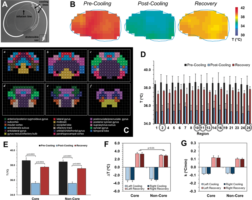
Conclusions
Intraventricular cooling reliably and reproducibly reduces global brain temperatures, with significant and uniform temperature decreases in all brain regions to levels known to be neuroprotective while maintaining systemic normothermia. Cooling cessation allows for rapid rewarming of both core and non-core regions. Dynamic thermal mapping of sheep brain during cooling can aid in optimizing selective neuroprotective protocols for translation to human use.
Acknowledgements
Supported by R01 EB-023366, R01 EB-011968, R01 CA-140102, P30 NS-052519, NSF-0923928, and T32 GM007205.
Reference
PB03-C02
Activation of GPR55induces neuroprotection of hippocampal neurogenesis and immune responses of neural stem cells following chronic systemic inflammation
1Dept. of Pathology Laboratory Medicine, Lewis Katz School of Medicine Temple University, USA
2Center for Substance Abuse Research, Lewis Katz School of Medicine, Temple University
Abstract
Objective
The cannabinoid system exerts functional regulation of neural stem cell (NSC) proliferation and adult neurogenesis. The recently de-orphaned GPR55 has been shown to be activated by numerous cannabinoid ligands suggesting that GPR55 is a third cannabinoid receptor. We examined the role of GPR55 activation in NSC proliferation and early adult neurogenesis.
Methods
The effects of GPR55 agonists (LPI, O-1602, ML184) on human NSC proliferation in vitro were assessed by flow cytometry and expression of cytokine receptors was assessed by RT-PCR. hNSC differentiation was determined by flow cytometry, qPCR, and immunohistochemistry. Early adult neurogenesis in the hippocampus of C57BL/6 and GPR55-/- mice was evaluated by immunohistochemistry. To determine inflammatory responses in vivo, we treated C57BL/6 and GPR55-/- mice with LPS continuously for 14 days via osmotic mini-pump.
Results
Activation of GPR55 significantly increased proliferation rates of hNSCs in vitro. These effects were attenuated by ML193, a selective GPR55 antagonist. ML184 significantly promoted neuronal differentiation in vitro while ML193 reduced differentiation rates from vehicle treatment. Continuous administration into the hippocampus of O-1602 via cannula connected to osmotic pump resulted in increased Ki67+ cells within the dentate gyrus. O-1602 increased neurogenesis as assessed by DCX+ and BrdU+ cells as compared to vehicle treated animals. GPR55-/- animals displayed reduced rates of proliferation and neurogenesis within the hippocampus while O-1602 had no effect when compared to vehicle controls.
Next, we examined NSCs exposed to IL-1b in vitro to assess inflammation-caused effects on NSC differentiation and the ability of GPR55 agonists to attenuate NSC injury. GPR55 agonist treatment protected against IL-1b induced reductions in neurogenesis rates. Moreover, inflammatory cytokine receptor mRNA expression was down regulated by GPR55 activation in a neuroprotective manner. To determine inflammatory responses in vivo, we treated C57BL/6 and GPR55-/- mice with LPS continuously for 14 days via osmotic mini-pump. Reduced NSC survival, immature neurons, and neuroblast formation due to LPS were attenuated by concurrent direct intrahippocampal administration of the GPR55 agonist O-1602. Molecular analysis of the hippocampal region showed a suppressed ability to regulate immune responses by GPR55-/- animals manifesting in a prolonged inflammatory response (IL-1β, IL-6, TNFα) after chronic, systemic inflammation as compared to C57BL/6 animals.
Conclusions
These results suggest a neuroprotective role of GPR55 activation on NSCs in vitro and in vivo and that GPR55 poses a novel therapeutic target against negative regulation of hippocampal neurogenesis by inflammatory insults.
PB03-C03
Neuroprotective potential of curcumin against copper-induced astroglial, neuronal and behavioral impairments in rats
1Department of Biology, Cadi Ayyad University/Faculty of sciences semlalia Marrakech, Morocco
Abstract
Copper (Cu) is a transition metal and an essential trace element, but excessive levels of Cu might disturb vital functions and systems including the Central Nervous System (CNS). Curcumin has numerous beneficial effects including protective potential on the CNS toxicity. Previous studies have revealed solid evidence showing metal elements implication in the physiopathology of neurodegenerative diseases. The aim of the present study was to evaluate the influence of acute Cu intoxication (10 mg/Kg B.W. i.p) for 3 days, then, subchronic Cu-intoxication (0.125%) for 6 weeks on the dopaminergic, serotonergic and astroglial systems together with behavioral impairments. In addition, test the therapeutic efficacy of curcumin (30 mg/kg B.W.). In Cu-exposed rats, we noted a significant increased innervation of 5HT in Dorsal raphe nucleus (DRN) and Basolateral Amygdala (BLA) outputs; decreased innervation of TH within Substantia nigra (SNc), Ventral Tegmental Area (VTA), and in striatum dorsal. This was correlated with decreased astroglial plasticity in hippocampus, motor cortex, striatum, DRN, SNc and VTA. Such effects were associated with decreased locomotor performance and anxiogenic-like effects but did not alter learning and memory. Curcumin co-treatment prevented Cu-induced behavioral impairments and reversed 5-HT, TH and astroglial alterations and main spatial learning and memory performance was remarked in treated rats with curcumin in Morris water maze. Results demonstrated that Cu intoxication induced an evident impaired neuroplasticity that was alleviated with curcumin treatment. Therefore, curcumin may be valuable in the treatment of metals-induced neurobehavioral deficits. The impairment of monoamine neurotransmitters may be one of the major mechanism implicated.
PB03-C04
Identification of novel neuroprotective drug combinations for the treatment of ischemic stroke through a systems biology-based drug repositioning approach
1Neurovascular Research Laboratory, Vall d'Hebron Institute of Research (VHIR), Universitat Autònoma de Barcelona (UAB), Barcelona, Spain
2Anaxomics Biotech, S.L, Barcelona, Spain
Abstract
Objectives
Ischemic stroke is a leading cause of disability and mortality worldwide. Beyond the standard thrombolytic therapies, there is still no effective treatment to mitigate or even reverse the progression of stroke disease. Since numerous candidates emerged from pre-clinical studies have failed to translate into clinical success, effective neuroprotective drug discovery strategies are still urgently needed. Combinational treatment approaches are emerging as powerful strategies to synergistically and simultaneously target more than one disease-response mechanism underlying complex pathologies such as stroke. In this line, we hypothesize that the individual modulation of a single pathological mechanism might not be sufficient to attenuate the progression of stroke, so we aimed to identify combinations of FDA-approved drugs with potential neuroprotective effect for stroke treatment.
Methods
We used a systems biology-based technique relying on artificial intelligence and pattern recognition models to integrate available biological, pharmacological and medical knowledge into mathematical models that simulate in silico the complex behavior of stroke disease. Novel brain proteomics and transcriptomics data obtained from deceased ischemic stroke patients were used to enrich and curate the emergent mathematical models. Drug repositioning solutions were acquired by perturbing the stroke-mimicking models with multiple sets of stimuli, which corresponded to two-by-two combinations of drugs from the DrugBank Database. We experimentally tested in vivo the neuroprotective effect of two resulting drug combinations in a mouse model of transient cerebral ischemia. To that end, male mice were subjected to intraluminal transient cerebral ischemia by 90-min Middle Cerebral Arterial Occlusion (MCAO) and intravenously treated with drug combinations (CA and CB), individual drug treatments or their respective vehicles during occlusion. Infarct volume was assessed 24 h after MCAO using 2,3,5-triphenyltetrazolium chloride (TTC) staining.
Results
More than 5 million drug combinations were evaluated for their neuroprotective effect in our mathematical models. Approximations to the best treatment solution for ischemic stroke were obtained establishing a threshold of minimum predicted probability to exert neuroprotection in the ischemic brain of 80%. This cut-off point corresponded to the maximum predictive capacity obtained from screening in our mathematical models a set of 29 previous neuroprotective treatments tested in clinical trials for ischemic stroke without success. Based on the predicted probability of success in our stroke models, two drug combinations were selected and evaluated for their safety and synergic neuroprotective efficacy in vivo. Compared to the control groups, both drug combinations showed a significant reduction of 39.54% (CA, p = 0.018) and 39.42% (CB, p = 0.022) in the infarct volume, whereas no reduction in the ischemic lesion was observed when drugs were given alone.
Conclusions
By using a systems biology-based simulation of the complex behavior of stroke, we have identified two potential neuroprotective combinations of FDA-approved drugs. Despite further studies are needed to corroborate the therapeutic use of these drug combinations, the simultaneous modulation of different motives and pathways altered after ischemic stroke seems a promising strategy to acutely mitigate the progression of stroke disease.
PB03-C05
Exercise increases circulating levels of the neuroprotective LG3 peptide
1Tissue Repair and Translational Physiology Program, Institute of Health and Biomedical Innovation, Queensland University of Technology, Brisbane, QLD, Australia
2School of Biomedical Science, Faculty of Health, Queensland University of Technology, Brisbane, QLD, Australia
3School of Nursing, Faculty of Health, Queensland University of Technology, Brisbane, QLD, Australia
4Dept. of Neurology, University of Kentucky, Lexington, KY, USA
5Dept. of Neurosurgery, Sanders Brown Centre on Aging, University of Kentucky, Lexington, KY, USA
6Dept of Anatomy & Neurobiology, University of Kentucky, Lexington, KY, USA
Abstract
Background and Aims
Stroke is a leading cause of death and disability globally. Interestingly, physical activity and exercise is thought to reduce stroke risk; enhance rehabilitation following stroke; and in particular induce remodelling of cerebral blood vessels and their basement membranes, although the mechanisms underpinning these effects remain unclear. Perlecan is a major ECM protein of vascular basement membranes, neuromuscular junctions and cartilage, which is known to be proteolytically processed to release a C-terminal bioactive fragment: the LG3 peptide. Importantly, the LG3 peptide is naturally released in the stroke-injured brain and is profoundly neuroprotective / neuroreparative when administered systemically in animals. We have previously found that the LG3 peptide was increased in the urine of physically active mining workers compared to sedentary controls; however, no previous studies have investigated the relationship between exercise and LG3 abundance in human serum where it might have therapeutic benefit.
Method
Venous blood was collected at pre- and post-exercise from six male volunteers following an intermittent, high-intensity exercise and a continuous, moderate-intensity exercise. Serum LG3 levels were measured by quantitative mass spectrometry.
Results
We found that circulating levels of the neuroprotective / neuroreparative LG3 peptide was significantly increased under normal physiological conditions in healthy individuals following moderate exercise.
Conclusions
We are currently undertaking a pilot clinical study to determine if patients with mild stroke can increase circulating levels of LG3 peptide with moderate to low levels of exercise. These studies may help establish a baseline level of physical activity required to elevate LG3 peptide to potentially beneficial levels in the blood.
PB03-D01
Characterizing the role of B cell-derived brain-derived neurotrophic factor (BDNF) following ischemic injury
1Department of Neurology & Neurotherapeutics, UT Southwestern Medical Center, USA
2Department of Future Basic Medicine, Nara Medical University, Japan
3Department of Neurology, University of Kentucky, USA
Abstract
Objective
Neuronal networks require significant neurotrophic support for recovery after stroke[1,2], but the delivery of neurotrophins (NTs) failed thus far in clinical trials[3–5]. B cells possess the ability to produce NTs[6,7], including brain-derived neurotrophic factor (BDNF), and infiltrate the post-stroke brain. Studies in our lab show that systemic B cell depletion impedes neurogenesis, increases anxiety, and exacerbates memory deficits in mice after stroke. Thus, we hypothesize that BDNF+ B cells could enhance neuroplasticity after stroke.
Methods
To assess endogenous BDNF expression in leukocytes, bone marrow, spleen, cervical lymph node and brain tissues were isolated from naïve 12–16-week old C57Bl/6 J mice, or 24 h or 48 h after 15-min photothrombotic stroke and analyzed by flow cytometry. To induce B cell-derived BDNF, a CMV-BDNF-p2A-mCherry plasmid was transfected into LPS-stimulated splenic B cells isolated from 6–10-week old C57Bl/6 J mice. Transfected B cells were co-cultured with supporting splenocytes for 24, 48, or 72 h. Flow cytometry assessed mCherry+ B cells and BDNF secretion measured via ELISA. Cortical and hippocampal brain cells were isolated from postnatal day 0–2 pups and cultured until confluent (minimum 7 days). Oxygen glucose deprivation (OGD) was induced in cortical cell cultures for 2 hours and allowed to recover with naïve, un-transfected, or transfected B cells at increasing B cell-to-cortical cell concentrations (0:1, 1:10, 1:1) for 4 days. Cortical cells were stained with MAP2, imaged via confocal microscopy, and quantified using the counter tool in Photoshop. All analyses were blinded and Graphpad Prism analyzed statistical differences. Significance was p < 0.05.
Results
Assessment of BDNF expression in CD45+ leukocytes identified distinct BDNF+ populations in naïve primary and secondary lymphoid organs, and brain. B220+ B cells were the most abundant CD45+BDNFhi population of each organ (bone marrow: 87.8%, spleen: 93.9%, cervical lymph node: 90%, and brain: 78.1%). After stroke, BDNFhi B cells decreased in the periphery at 24 h (bone marrow; p<0.01 vs. no-stroke control) and returned to baseline by 48 h. Increased BDNFhi B cells were observed in the ipsilesional hemisphere. Transfected splenic B cells showed highest mCherry expression at 24 h (12.6% of CD45+CD19+ cells) that was reduced by 48 h (8.77%; p < 0.01 vs. 24 h) and 72 h (6.03%; p < 0.01 vs. 24 h). BDNF secretion was consistently detected over 72 h (24 h-303.3 pg/mL, 48 h-453 pg/mL, 72 h-317.6 pg/mL). A 1:1 ratio of CMV-BDNF-p2A-mCherry+ B cells:cortical cells increased the number (p < 0.0001) and dendritic arborization (p < 0.01) of uninjured MAP2+ neurons and fully protected against loss of neurons and neuronal dendrites following OGD injury (p < 0.05).
Conclusions
Our studies identified that B cells comprise the majority of BDNFhi leukocytes in both the periphery and brain, which decrease in bone marrow within the first 24 h after stroke. Successful overexpression of BDNF in primary B cells demonstrate their neurotrophic effect on neurons in both homeostatic conditions and an in vitro stroke injury model. Future studies will characterize the phenotype and function of BDNFhi B cells and dissect the mechanisms by which B cell-derived BDNF can modulate neuroplasticity following stroke injury.
References
PB03-D02
Brain pericytes after ischemic stroke: a possible role as multipotent stem cells to differentiate into functional neurons
1Institute for Advanced Medical Sciences
2Department of Therapeutic Progress in Brain Diseases
3Department of Neurosurgery
4Department of Ophthalmology
5Department of Anatomy and Cell Biology
6Department of Surgical Pathology
Abstract
Objectives
In addition to the well-documented role of the brain pericytes in maintaining the neurovascular unit, recent studies indicate that they function as multipotent stem cells, differentiating into various lineages, including neuronal cells. This suggests that brain pericytes contribute toward neural repair following various CNS disorders, such as ischemic stroke. However, the precise characteristics of brain pericytes under ischemia remain unclear. The present study aimed to investigate the characteristics of pericytes, particularly in terms of stemness and neurogenic potential, using brain pericytes isolated from post-stroke mouse brains.
Methods
To examine the characteristics of brain pericytes following ischemia (ischemic pericytes: iPCs), adult CB 17 mice underwent permanent focal cerebral ischemia through middle cerebral artery occlusion. Tissues from post-ischemic areas were mechanically dissociated using needles to prepare single-cell suspensions, which were then incubated with adherent cultures and subjected to immunohistochemistry, polymerase chain reaction (PCR), and microarray analyses. The multipotency of iPCs was examined by incubating them under osteoblastic, adipogenic, and chondrocytic differentiation. To further investigate their neurogenic potential, cells isolated from post-stroke areas were incubated under conditions that promoted neuronal differentiation and subjected to multi-electrode arrays.
Results
Immunohistochemistry, PCR, and microarray analyses revealed that iPCs expressed various stem and undifferentiated cell markers (nestin, Sox2, c-myc, and Klf4) as well as pericytic markers (PDGFRβ, NG2, and SMAα). iPCs differentiated into various cell lineages, including osteopontin+ osteoblasts, FABP4+ and Oil red O+ adipocytes, and collagen II+ chondrocytes. Additionally, iPCs formed neurosphere-like cell clusters and differentiated into electrophysiologically functional neurons, as shown by a multi-electrode array system.
Conclusions
The results indicate that iPCs are multipotent stem cells, and are able to differentiate into electrophysiologically functional neurons. Moreover, they potentially contribute toward post-ischemic CNS repair, by producing neuronal cells. Therefore, iPCs can be considered as a target to promote neurogenesis after ischemic stroke.
References
PB03-D03
Astrocyte-derived exosomes treated with a semaphorin 3A inhibitor enhance stroke recovery via prostaglandin D2 synthase
1Dept. of Neurology, Juntendo University Faculty of Medicine, Japan
2Dept. of Physiology, Keio University, Japan
3Dept. of Neurology, Juntendo University Urayasu Hospital, Japan
Abstract
Background and Purpose
Exosomes play a pivotal role in neurogenesis. In the peri-infarct area after stroke, axons begin to regenerate but are inhibited by astrocyte scar formation. The direct effect and underlying molecular mechanisms of astrocyte-derived exosomes on axonal outgrowth after ischemia are not known.
Methods
Using a semaphorin 3A (Sema3A) inhibitor, we explored neuronal signaling during axonal outgrowth after ischemia in rats subjected to middle cerebral artery occlusion and in cultured cortical neurons challenged with oxygen-glucose deprivation. Furthermore, we assessed whether this inhibitor suppressed astrocyte activation and regulated astrocyte-derived exosomes to enhance axonal outgrowth after ischemia.
Results
In rats subjected to middle cerebral artery occlusion, we administered a Sema3A inhibitor into the peri-infarct area from 7 to 21 days after occlusion. We found that phosphorylated high-molecular weight neurofilament-immunoreactive axons were increased, glial fibrillary acidic protein–immunoreactive astrocytes were decreased, and functional recovery was promoted at 28 days after middle cerebral artery occlusion. In cultured neurons, the Sema3A inhibitor decreased Rho family GTPase 1, increased R-Ras, which phosphorylates Akt and glycogen synthase kinase 3β (GSK-3β), selectively increased phosphorylated GSK-3β in axons, and thereby enhanced phosphorylated high-molecular weight neurofilament-immunoreactive axons after oxygen-glucose deprivation. In cultured astrocytes, the Sema3A inhibitor suppressed activation of astrocytes induced by oxygen-glucose deprivation. Exosomes secreted from ischemic astrocytes treated with the Sema3A inhibitor further promoted axonal elongation and increased prostaglandin D2 synthase expression on microarray analysis. GSK-3β+ and prostaglandin D2 synthase+ neurons were robustly increased after treatment with the Sema3A inhibitor in the peri-infarct area.
Conclusions
Neuronal Rho family GTPase 1/R-Ras/Akt/GSK-3β signaling, axonal GSK-3β expression, and astrocyte-derived exosomes with prostaglandin D2 synthase expression contribute to axonal outgrowth and functional recovery after stroke.
PB03-D04
Transcranial focused ultrasound stimulation regulates brain plasticity in mice after middle cerebral artery occlusion
1Department of Rehabilitation, Ruijin Hospital, Shanghai Jiao Tong University School of Medicine, China
2Department of Neurology, Ruijin Hospital, Shanghai Jiao Tong University School of Medicine, China
3Med-X Research Institute and School of Biomedical Engineering, Shanghai Jiao Tong University, China
4Shenzhen Institutes of Advanced Technology Chinese Academy of Sciences, China
Abstract
Objectives
Transcranial focused ultrasound brain stimulation regulates neural activity in different brain regions in humans and animals, and promotes neurogenesis in normal mouse brain1,2. However, the role of ultrasound stimulation in modulating neuronal plasticity in the ischemic brain is largely unknown. In present study, we explore the effect of focused ultrasound brain stimulation on neurological rehabilitation in a mouse model of focal cerebral ischemia and the underlying molecular mechanism.
Methods
Adult CD-1 mice (n = 30) underwent 60-min transient middle cerebral artery occlusion. Seven days after brain ischemia, low frequency (0.5 MHZ) transcranial focused ultrasound stimulation was applied to the ischemic hemisphere of mice, ten minutes a day for seven consecutive days. Brain infarct volume, neurological behavioral tests, Arginase+/IBA1+ (M2 microglia) and CD16/32+/IBA1+ (M1 microglia), as well as IL-10 and IL-10R were further assessed for up to 14 days to explore the underlying mechanism.
Results
We found that the brain atrophy volume was significantly attenuated in the ultrasound stimulated mice after 14 days of MCAO compared to the non-stimulated mice (p < 0.05). Similarly, neurological servility scores, elevated body swing test, and corner test performance were greatly improved in the ultrasound stimulated mice (p < 0.05). We also demonstrated that ultrasound stimulation increased the number of Arginase+/IBA-1+ cells in the ischemic region of the brain. IL-10R and IL-10 expression was substantially up-regulated after ultrasound stimulation (p < 0.05).

Conclusion
We concluded that transcranial focused ultrasound stimulation provided a unique technique to promote neurorepair after cerebral ischemia. The action of transcranial ultrasound stimulation could be via promoting microglia polarization and further regulating IL-10 signaling in ischemic mice.
References
PB03-D05
M2 microglia derived exosome improves neurobehavioral recovery through promoting neurogenesis in a mouse model of middle cerebral artery occlusion
1Shanghai Ruijin Hospital, School of Medicine, Shanghai Jiao Tong University, China
2School of Biomedical Engineering, Shanghai Jiao Tong University, China
Abstract
Objective
M2 microglia showed a critical role in modulating neurogenesis and neuronal recovery after ischemic brain injury1. However, the underlying mechanism was unclear. M2 microglia can secret exosomes to modulate target cell function via various molecular constituent transfer2. Here we explored the effect and mechanism of M2 microglia derived exosome on the functional recovery of mice after middle cerebral artery occlusion.
Methods
BV2 microglia was polarized to M2 type by IL-4 stimulation, and exosomes were isolated from M2 microglia (M2-EXO). Adult CD1 mice (n = 64) underwent 90-minute middle cerebral artery occlusion. These mice were recived M2-EXO (100 mg) daily by tail vein injection for 7 consecutive days. Brain atrophy volume and neurobehavioral outcomes were determined. Neurogenesis was evaluated by immunostaining. The exosomal miRNA array was also performed for further mechanistic study.
Results
We found that atrophy volume reduced in ischemic mice after M2-EXO treatment compared to the control group (p < 0.01). Neurological severity score and rotarod test showed better outcomes in M2-EXO treated mice compared to the control. The number of differentiated neural stem cells in the SVZ and synaptophysin protein increased in mice at 14 days after M2-EXO treatment compared to the control (p < 0.05). The miRNA array showed 1141 different expression between M2-EXO and M0-EXO. In all differentially expressed miRNA, 48 of them were down-regulated and 1093 of them were up-regulated compared to M0 BV2 exosomes. We further validated neuronal regeneration associated 6 miRNAs in vivo and found that miR-186, miR-124, miR-1188 showed higher level compared to the control group.

Conclusion
Our study demonstrated that exosomes released from M2 microglia promoted neurogenesis and neuronal differentiation in mice after ischemic stroke, suggesting a novel therapeutic strategy for the treatment of cerebral ischemia.
References
PB03-D06
Rehabilitation after cerebral ischemia enhances neurogenesis and cortical angiogenesis with a role for angiogenin in SVZ-neural precursor cells pools and endothelial progenitor cell function
1Neurovascular Research Laboratory, Neurology Service, Vall d’Hebron Research Institute, Universitat Autònoma de Barcelona
2Servei de Neurologia-Neuroimmunologia, Centre d'Esclerosi Múltiple de Catalunya (Cemcat) and Vall d’Hebron Research Institute, Hospital Vall d’Hebron, Universitat Autònoma de Barcelona
Abstract
Objectives
Stroke-related disabilities compromise quality of life, being rehabilitation the only treatment to compensate functional deficits. Investigations in preclinical models of rehabilitation are needed to identify brain repair molecules and mechanisms involved in rehabilitation. We have examined neuro-oligo-vascular neurorepair mechanisms in a rehabilitation model after cerebral ischemia and the effect of angiogenin (a ribonuclease implicated in the expression of ribosomal RNA and posterior protein synthesis) in primary cultures of different progenitor cells (neural stem cells, endothelial progenitor cells and oligodendrocyte precursor cells) which could be biologically implicated in neuroplasticity and repair mechanisms.
Methods
Male mice underwent permanent occlusion of the middle cerebral artery and randomly assigned to non-rehabilitation, task-specific (pasta matrix-reaching task) or physical-exercise (treadmill) groups, performing 12 daily-sessions. Neurogenesis was assessed in the SVZ, angiogenesis was measured in the peri-infarct cortex and white matter was measured in the corpus callosum. In parallel primary cell cultures of Neural Stem Cells, Oligodendrocyte Precursos Cells and Endothelial Progenitor Cells, together with neuron-like cells (SH-SY5Y), were treated with exogenous recombinant angiogenin.
Results
Our results show that physical exercise significantly increased brain angiogenin and DCX+ cells in the ipsilateral neurogenic SVZ niches (with cells expressing angiogenin), and also vessel density in the contralateral cortex. At the same time treatment with exogenous angiogenin significantly increased cultured neurosphere formation and proliferation, SH-SH5Y proliferation of undifferentiated cells (but not neuronal differentiation). Finally angiogenin treatment also improved significantly EPC function in both tubulogenesis and migration assays.
Conclusions
Our findings demonstrate that post-stroke rehabilitation modulates neurorepair brain responses, where angiogenin might become a target in future pre-clinical studies since it could positively modulate proliferation and function of precursos cells.
PB03-D07
Spatio-temporal evolution of neural oscillations power in the recovering cortex after stroke
1Normandie Univ, UNIROUEN, INSERM, DC2N, Rouen, France
2Neurosciences Systems Institut, INSERM UMR-S 1106, Aix Marseille University, Marseille, France
Abstract
In days and weeks following an ischemic stroke, the peri-infarcted area adjacent to the necrotic tissue exhibits very intense synaptic reorganization aimed at regaining lost functions. In order to find solutions to improve functional recovery, it is important to understand the rules of neural repair and neuroplasticity in the cortex surrounding the lesion. The brain generates oscillations of the voltage of local field potentials detectable with an extracellular electrode inserted in the cortex. The fundamental role of these rhythmic fluctuations of neuronal excitability is to synchronize neural activity of multiple neuronal assemblies distributed across brain regions to contribute to information processing. Some of these oscillations are also known to dictate the timing of neuronal activity in order to optimize synaptic plasticity. Here, we worked on the hypothesis that brain oscillatory system is altered in the surviving cortex, which may slow down functional repair and contribute to the cognitive post-stroke deficit. In order to document the relevance of this hypothesis, we have set-up an in vivo electrophysiological recording to describe the evolution of the oscillatory activity of the peri-infarct cortex, 7 and 21 days after a permanent cortical ischemia induced in mice. It was found that oscillations are depressed inside a 2 mm-wide peri-lesioned zone: all frequency-bands were significantly dampened 1 week after stroke and most of them remained depressed at 3 weeks. This area of tissue corresponds to the zone where astrocyte reactivity was most intense. Above 2 mm, oscillation power remained normal. Thus, the proximal peri-infarct cortex could become a target of therapeutic interventions aimed at correcting the oscillatory regimen.
This work was supported by INSERM (U1239), La Fondation pour la Recherche sur les AVC, Rouen University, Normandy Region and the European Union (3 R project).
PB03-E01
Microvascular shunts in the pathogenesis of cerebral ischemia of cerebral small vessel disease to leukoaraiosis, MS, SLe, MCI, VaD, AD white matter hyperintensities
1Dept. of Neurosurgery, University of New Mexico
2McGowan Institute for Regenerative Medicine, University of Pittsburgh Medical Center
Abstract
Considerable circumstantial evidence of cerebral microvascular shunts (MVS) led our first definitive studies showing their development in response to high intracranial pressure (ICP)[1,2]. These MVS represent non-nutritive shunt-flow, resulting in a loss of cerebral blood flow (CBF) autoregulation, tissue ischemia, hypoxia, edema, and metabolic depression by shunting non-nutritive blood flow around injured tissue releasing neither nutrients nor oxygen to tissue. The initiating process in the development of these MVS may vary in different disease states such as MS, SLE, MCI, VaD and AD but we suggest that the underlying pathogenesis is the same; cerebral ischemia and MVS flow in white matter visualized as white matter hyperintensities (WMH). Normal white matter flow is 1/3 that of gray matter flow and as is in arterial watershed zones, is the first to suffer ischemia when CBF falls. In an NIH funded study evaluating hemodynamic compromise to predict the likelihood of a second stroke in patients who suffered a stroke and with a unilateral carotid occlusion, we made the incidental observation that WMH were all at different degrees of ischemic stress as characterized by the cerebrovascular reserve (CVR) and Oxygen extraction fraction (OEF) in response to an acetazolamide cerebrovascular challenge (Fig) [3]. These data suggest that the degree of WMH ischemic stress rather than volume of WMH may better correlate with psychological and motor symptomatology and reveal the progression of these diseases. The commonality of cerebral ischemia in all of these diseases may be treatable by drag reducing polymers (DRP) such as high molecular weight (HMW) 4500 kDa polyethylene oxide to reverse or reduce the severity of cerebral ischemia in these diseases. The HMW DRP has proven effective in animal models of stroke [4] and traumatic brain injury [5] acts by hemorrheologic reduction of turbulent and promotion of laminar flow increasing vascular wall shear stress and reducing plasma skimming resulting in increased RBC concentration in capillaries. The effectiveness of HMW DRP has been shown in ischemic myocardium and in hemorrhagic shock. The effectiveness of DRP in clinical cerebral ischemia characterized by the diseases listed above have yet to be done.

References
PB03-E02
Inflammation, demyelination, and their role in cerebral small vessel disease. a postmortem combined (ultra-) high-field MRI, polarized light imaging, and immunohistochemistry approach
1Department of Anatomy, Radboud university medical center, Donders Institute for Brain, Cognition & Behaviour, Donders Center for Medical Neuroscience, Preclinical Imaging Centre, Radboud Alzheimer Center, Nijmegen, The Netherlands
2Department of Radiology & Nuclear Medicine, Radboud university medical center, Nijmegen, The Netherlands
3Department of Neurology, Radboud university medical center, Donders Institute for Brain, Cognition & Behaviour, Donders Center for Medical Neuroscience, Nijmegen, The Netherlands
Abstract
Objectives
Cerebral small vessel disease (SVD) is a major vascular contributor to cognitive decline and dementia. Hypertension is a major risk factor for SVD. To elucidate the underlying pathophysiology of SVD we used a post-mortem combined ultra-high-field MRI, polarized light imaging (PLI), and immunohistochemistry approach to answer the three following questions among adults with hypertension: 1) What is the spectrum of vascular (arterioles, venules, and capillaries) pathology inside and outside SVD? 2) What is the role of inflammation in vascular pathology and in SVD? 3) What are vascular and inflammatory contributions to myelin change in SVD?
Methods
In this ongoing study, postmortem left brain hemispheres of 18 SVD patients (Inclusion criterion: treated and untreated hypertension; Exclusion criteria: Atrial fibrillation, brain tumors/ metastases) are scanned on 7T MRI using T1-, T2-, T2*-weighted, and T2-FLAIR sequences. Using the 7T MRI results, biopsies are taken at several regions of interests (n ≥ 2 per brain hemisphere) and scanned on 11.7T MRI using T1-, T2-, and T2*-weighted sequences. Hereafter, immunohistochemical stainings are performed to visualize neuroinflammation by glial markers (glial fibrillary acidic protein, GFAP), myelin (Luxol fast blue, LFB), and both axonal and dendritic neurofilaments (NF). Furthermore, polarized light imaging (PLI) are used to determine the orientation of myelinated fibres and pathological changes of the myelin content in white matter hyperintensities (WMH).
Results
Among SVD patients with hypertension, WMH were detected (figure 1). Presented is an overview of the results of ultra-high-field MRI (7T and 11.7T) and immunohistochemical stainings focusing on microvascular lesions (e.g. microbleeds and microinfarcts) and the heterogeneity of WMH using PLI in association with neuroinflammatory and vascular pathology measured by the aforementioned immunohistochemical markers. Preliminary IHC results (figure 1 K-P) are a decreased NF and myelin density combined with an increased neuroinflammation found in WMH compared to normal-appearing white matter (NAWM).
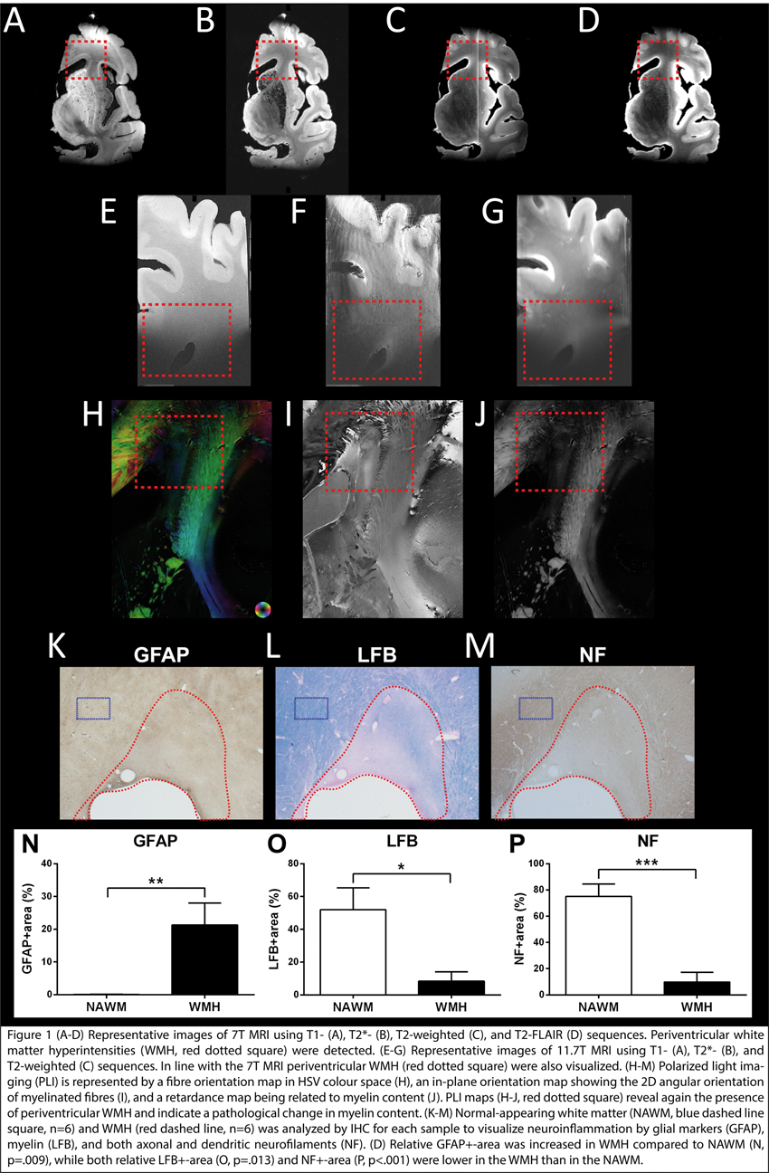
Conclusions
In WMH, neuroinflammation is elevated and accompanied by a loss of myelin (measured using LFB and PLI), and neurofilaments being not visible in NAWM. Ultra-high-field MRI measurements combined with PLI and immunohistochemical stainings will provide a detailed view of the extent of vascular and myelin pathology and the role of inflammatory processes in hypertensive SVD.
PB03-E03
Accumulation of white matter hyperintensities is correlated with decreased structural brain network efficiency in patients with cerebral small vessel disease
1Department of Neurology, Drum Tower Hospital of Nanjing University Medical School, PR China
2Department of Radiology, Drum Tower Hospital of Nanjing University Medical School, PR China
Abstract
Objectives
To evaluate the correlation between the volumetric changes of white matter hyperintensities (WMH) and the topological alterations of structural brain network in patients with cerebral small vessel disease (CSVD).
Methods
From January 2017 to June 2018, fifty-three patients diagnosed with CSVD were recruited in this study. All participants underwent a battery of neuropsychological assessments, including the Mini-mental State Examination (MMSE), Montreal Cognitive Assessment (MoCA), Trail Making Test (TMT) and Stroop Color Word Test (SCWT). MR images were obtained on a 3.0 tesla scanner (Ingenia, Philips Medical Systems), and three-dimensional T1 weighted imaging, three-dimensional fluid attenuated inversion recovery (FLAIR) imaging and diffusion tensor imaging (DTI) were performed. Wisconsin White Matter Hyperintensities Segmentation (W2MHS) toolbox and Pipeline for Analyzing braiN Diffusion imAges (PANDA) were used for WMH segmentation and DTI network construction, respectively. GRaph thEoreTical Network Analysis (GRETNA) toolbox was used to analyze the topological changes of structural brain network. Spearman correlation analysis was performed using the Statistical Product and Service Solutions (SPSS) version 19.0.
Results
Accumulation of WMH volume was correlated with decreased γ(normalized clustering coefficient), increased shortest path (fig. 1) and λ (normalized shortest path), and lower global efficiency (all p value < 0.05). Such correlations were preserved after controlling for age, MMSE and MoCA in partial correlation analysis. The time taken to complete the SCWT, an indicator of a subject’s cognitive processing speed and selective attention, was correlated with increased shortest path (p = 0.010) and λ (p = 0.000). Lower global efficiency of the structural brain network was also found to be correlated with poorer performances on the SCWT (p = 0.010). Correlations remained after controlling for age, MMSE and MoCA (all p value <0.000) in further partial correlation analysis. However, no correlations between the SCWT performances and WMH volumetry were found in this study.

Conclusions
Accumulation of WMH is correlated with dysfunction of topological characteristic of the structural brain network, especially the aspects of global efficiency. Increased shortest path and λ are also correlated with poorer performances on the SCWT, indicating the decline of a subject’s cognitive processing speed. No direct correlations between the WMH volumetric changes and SCWT performances were found in this study, which might suggest the limitations of using the WMH volumetry as an imaging marker for the CSVD.
References
PB03-E04
White matter hyperintensities are associated with impaired oxygenation and worse capillary function than surrounding white matter in aging and late-onset depression
1Department of Radiology (Neuroradiology), Aarhus University Hospital, Aarhus, Denmark
2Center for Functionally Integrative Neuroscience & MINDLab, Aarhus University Hospital, Aarhus, Denmark
3Centre for Psychatric Research, Aarhus University Hospital, Risskov, Denmark
4Mental Health Center Glostrup, Glostrup, Denmark
Abstract
Objectives
White matter hyperintensities (WMHs) of presumed vascular origin are a common finding in magnetic resonance imaging (MRI) of the brain in normal aging. They are believed to reflect cerebral small vessel disease (SVD) and may play an important role in the etiology of complex age- and SVD-related conditions, such as late-onset depression. WMHs may affect the metabolic demands of surrounding normal-appearing white matter (NAWM) by disrupting fiber tracts. Meanwhile, SVD risk factors are thought to reduce tissue oxygenation, not only by reducing regional blood supply, but also by impairing capillary function. To address the balance of white matter oxygen supply and metabolic demand in normal aging and late-onset depression, we used MRI to estimate voxel-wise capillary density as an index of resting white matter metabolism, and combined estimates of blood supply and capillary function to calculate white matter oxygen availability. We hypothesized that capillary dysfunction reduces oxygen availability within WMHs, with respect to the effects of altered perfusion and capillary density.
Methods
We conducted structural, perfusion-, and diffusion-weighted MRI in 21 patients with late-onset depression (aged 57.4 ± 4.7 years) and 21 controls (aged 58.5 ± 6.6 years). We outlined WMHs on fluid-attenuated inversion recovery (FLAIR) images and used tractography to identify the tracts, they intersected. Perfusion data, obtained by gradient echo (GRE) and microvascular weighted spin echo (SE) imaging, comprised cerebral blood flow (CBF), blood volume (CBV), mean transit time (MTT), and relative transit time heterogeneity (RTH), the latter a marker of capillary dysfunction. White matter oxygenation was calculated as the steady state metabolic rate of oxygen (CMRO2) under the assumption of normal tissue oxygen tension (PtO2), and vice versa.
Results
The number, volume, and perfusion characteristics of WMHs did not differ between groups, and we therefore pooled data for the two groups in subsequent analyses. Hemodynamic data (SE/GRE) showed that WMHs have lower CBF and CBV, but higher RTH (16%/11%), than NAWM, resulting in either reduced CMRO2 (-32%/-34%) or PtO2 (-28%; SE data only). Tracts intersected by WMHs showed significantly lower CBF (-10%/-12%), CBV (-8%/-9%), and CMRO2 (-12%/-11%) than did NAWM. Across groups, lower lesion PtO2 was associated withboth higher WMH number and volume.
Conclusion
Our results show that WHMs are associated with impaired oxygenation and worse capillary function (as detected by RTH) than the surrounding white matter in individuals aged 50+ years, independent of parallel late-onset depression. In WMHs, less reduced microvascular blood volume and concomitant capillary dysfunction indicate a severe oxygen supply-demand imbalance, resulting in hypoxic tissue injury. In fiber tracts intersected by WMHs, parallel reductions in oxygenation and microvascular blood volume are consistent with adaptations to reduced metabolic demands. Aging and vascular risk factors may impair perfusion and capillary function to create hypoxic tissue injury within WMHs, which in turn affect the function and metabolic demands of the white matter tracts, they disrupt. We propose that WHMs should be viewed from the perspective of flow-metabolism coupling, where tissue oxygenation is determined by CBF and capillary function in combination.
PB03-E05
Time-domain near-infrared spectroscopy in subjects with cerebral small vessel disease
1San Raffaele Scientific Institute, Neurology Department, Via Olgettina 48, 20132, Milan, Italy
2Politecnico di Milano, Dipartimento di Fisica, Piazza Leonardo da Vinci 32, 20133 Milan, Italy
3Istituto di Fotonica e Nanotecnologie, Consiglio Nazionale delle Ricerche, Piazza Leonardo da Vinci 32, 20133 Milan, Italy
Abstract
Objectives
The microcirculation of the cerebral outer layers and white matter are functionally and anatomically connected [1]. Cerebral small vessel disease (CSVD) predominantly affects the deep and subcortical white-matter. However, MRI and neurophatological studies have reported that diffuse alterations of the cerebral cortex may occur early during the course of CSVD [2]. We hypothesized that the presence of CSVD could affect the oxygenation of cerebral outer layers measured with Time-Domain Near-Infrared spectroscopy (TD-NIRS).
Methods
We selected subjects with age ≥55 years, absence of stenosis ≥50% in cervical extra- and intra-cranial arteries, availability of head MRI-scan imaging. According to Fazekas score [3], we identified 3 groups: subjects with score 0 in both white-matter and periventricular regions (CSVD-0), subjects with score 1–2 in periventricular regions (CSVD-1), and subjects with score 1–2 in both white-matter and periventricular regions (CSVD-2). Resting TD-NIRS measurements from frontal, central, parietal brain regions on each hemisphere provided the average concentration of deoxy-hemoglobin (HbR), oxy-hemoglobin (HbO), total hemoglobin (HbT) and tissue oxygen saturation (StO2).
Results
We enrolled 26 subjects with mean age 68 (±9.5) yrs, 18 were male. Patients with CSVD-0 were significantly younger (58.5 ± 3.0 yrs) compared to CSVD-1 (74.7 ± 9.7 yrs) and CSVD-2 (70.5 ± 7.3 yrs) (p < 0.001), while gender distribution was comparable among CSVD groups (p = 0.304). StO2 was significantly lower in patients with CSVD-2 (55.6 ± 1.0%) compared to CSVD-0 (58.5 ± 1.4%) and CSVD-1 (59.4 ± 1.3%) (p = 0.017). The result persisted significant also after controlling for age (p = 0.042). A cut-off of StO2>58% predicted the absence of CSVD2 with 67% sensitivity and 91% specificity [AUC 81% (CI95% 65–98%)]. The concentration of hemoglobin species was comparable among CSVD groups (HbR p = 0.417; HbO p = 0.428; HbT p = 0.569).
Conclusions
These preliminary data suggest that the presence of extensive CSVD affect the oxygenation of cerebral outer layers as measured by TD-NIRS. In particular, lower StO2 might be a surrogate marker of the presence of extensive white matter-CSVD.
References
PB03-E06
Quantitative regional cerebral flow measured using I123-IMP SPECT and neuropsychological profile in patients with cerebral small vessel disease
1Department of Neurology, Tokyo Women's Medical University, Japan
2Department of Diagnostic Imaging Nuclear Medicine, Tokyo Women’s Medical University
Abstract
Objectives
To investigate the association between regional cerebral blood flow and cognitive function in patients with cerebral small vessel disease (CSVD).
Methods
We enrolled 119 subjects with any vascular risk factors who were suspected of cognitive decline, defined as a score on the Japanese version of the Montreal Cognitive Assessment (MoCA-J) lower than 26 points, and had any CSVD diagnosed based on magnetic resonance imaging (MRI) within the past year in the CSVD prospective registry at Tokyo Women’s Medical University. They underwent neuropsychological testing including the Mini-Mental State Exam (MMSE), and tests of verbal and visual memory, attention-processing speed, executive function, language, visuospatial construction, and depression. Among the 119 subjects, 34 subjects underwent quantitativesingle-photon emission computed tomography (SPECT) using N-isopropyl-p-[123I] iodoamphetamine (I123-IMP)
SPECT analysis using the autoradiography(ARG) method with I123-IMP between September 14, 2017 and June 21, 2018 to assess cerebral blood flow. To investigate the regionality of cerebral blood flow, we projected their cerebral blood flow to surface images using iSSP 5 software and calculated regional quantitative cerebral blood flow using Segmental Extraction Estimation (SEE) software. We identified an association between the calculated cerebral gyrus flow and cognitive function derived from the neuropsychological profile.
Results
Themean age of subjects was 74.0 ± 7.6 years and 22 (65%) were male. The mean duration of education was 14.2 years. The mean MMSE score was 26.8 ± 2.8 and mean MoCA-J score was 21.0 ± 2.7. Regarding neuropsychological domains, decreased blood flow in the bilateral orbital gyri, parahippocampal gyrus, or left precuneus was associated with memory function. Cerebral blood flow in the right frontal lobe or bilateral angular gyri was associated with working memory as determined by the digit span backward. Cerebral blood flow in the bilateral anterior cingulate gyri, bilateral superior frontal gyri, bilateral medial frontal gyri, or left uncus were associated with executive function as evaluated using interference tables of the Stroop test. A low MMSE score and depression as evaluated using the Patient Health Questionnaire-9 were related to decreased flow in multiple areas of the cerebral cortex.
Conclusions
Neuropsychological profiles were associated with quantitative regional cerebral blood flow measured using I123-IMP SPECT in patients with cognitive decline and CSVD. It was suggested that regional cerebral blood flow might affect cognitive functions in patients with cognitive decline and CSVD.
PB03-F01
The dynamics of revascularization after white matter infarction monitored in Flt1-tdsRed and Flk1-GFP mice
1Department of Neurosurgery, Gunma University Graduate School of Medicine, Japan
2Department of Molecular and Cellular Neurobiology, Gunma University Graduate School of Medicine, Japan
3Department of Stem Cells and Human Disease Models, Research Center for Animal Life Science, Shiga University of Medical Science, Japan
Abstract
Subcortical white matter infarction causes ischemic demyelination and loss of brain functions, as the result of disturbances of the blood flow. Although angiogenesis is one of the recovery processes after cerebral infarction, the dynamics of revascularization after white matter infarction still remains unclear.
We induced white matter infarction in the internal capsule of Flk1-GFP::Flt1-tdsRed double transgenic mice by injection of endothelin-1 (ET-1), a vasoconstrictor peptide, together with N(G)-nitro-L-arginine methyl ester (L-NAME), a nitric oxide synthase inhibitor, and followed the changes in Flk1 and Flt1 expression in the vascular system in the infarct area. We used these transgenic mice to monitor the preexisting blood vessels (visualized by tdsRed) and the angiogenic vessel growth (visualized by GFP) after the white matter infarction.
Reduction of Flt1-tdsRed-positive blood vessels 1 day after the injection and increase of Flk1-GFP-strongly-positive blood vessels 3 days after the injection were apparent. The reduction of the area covered by Flt1-tdsRed-positive vessels on the ET-1/L-NAME injected side seemed to continue for a week. In contrast, the area covered by Flk1-GFP-strongly-positive blood vessels on the injected side increased when compared to that on the intact side in a time-dependent manner for a week.
It is well known that the interaction between pericytes and endothelial cells is essential for vessel stability and maturation. Hence we examined the association between pericytes and the blood vessels in the white matter infarction after ET-1/L-NAME injection. PDGFRβ-strongly-positive (PDGFRβ+) cells appeared in the infarct area 3 days after the injection and increased their number thereafter. Three days after the injection, most of these cells were in close contact with Flk1-GFP-positive endothelial cells, indicating these cells are bona fide pericytes. Seven days after the injection, the number of PDGFRβ+ cells increased dramatically, and the vast majority of these cells were not in close contact with Flk1-GFP-positive endothelial cells. The PDGFRβ+ cells also expressed fibronectin, a fibroblast marker, in the demyelinated lesion 7 days after the injection.
Taken together, our results suggest revascularization begins early after the ischemic insult, and the emerging pericytes first ensheath blood vessels and then produce fibroblast-like cells not directly associated with blood vessels.
PB03-F02
Leukocyte RNA expression is associated with cerebral white matter hyperintensity progression over time
1Department of Medicine, Division of Neurology, University of Alberta
2Department of Neurology, University of California Davis
Abstract
Background
Cerebral white matter hyperintensities (WMH) are an important contributor to injury in the aging brain. Progression in WMH volume is associated with cognitive decline and gait impairment. Understanding the factors associated with WMH progression may provide insight to pathogenesis and identify novel treatment targets to improve cognitive health.
Methods
In 60 patients assessed for a cognitive complaint, an MRI brain was obtained at baseline and then repeated at a median of 5.9 years (IQR 3.5–8.2 years) from start of study. WMH was measured by semi-automated segmentation protocol and rate of progression per year determined. A blood sample was acquired at baseline in a PAXgene tube and used to measure whole genome RNA expression by RNA sequencing. The relationship between rate of WMH progression over time and leukocyte RNA expression was analyzed.
Results
The mean age was 76.1 (SD 8.3) years and 61% of participants were female. The median rate of WMH progression over 5.8 years was 1.3 mm3/year (IQR 0.27–3.3mm3/year). The median WMH volume was 5.5 mL (IQR 2.2–14.2). Patients in the quartile with the highest rate of WMH progression had increased leukocyte expression of genes involved in pattern recognition receptors (INFK, NLRP3, OAS1, TGFB1), interferon signaling (IFI6, IFIT1, IFITM3), and leukocyte extravasation (CLDN5, ICAM1, ITGB3, NCF1, TIMP2). A gene model could predict patients likely to experience a high rate of WMH progression over time with >80% accuracy. This remained significant when adjusted for factors associated with WMH progression.
Conclusions
Progression of WMH over time is associated with increased expression of genes involved in leukocyte extravasation, pattern recognition receptor activation and interferon signaling. Patients at risk for WMH progression may be identified by leukocyte RNA expression. Further studies are needed to evaluate the role of peripheral inflammation in relation to rate of WMH progression and contribution to cognitive decline.
PB03-F03
Microglia exacerbate white matter injury via complement C3/C3aR pathway after hypoperfusion
1Dept. of Neurology, Shanghai General Hospital, Shanghai, China
2Neuroscience and Neuroengineering Research Center, Med-X Research Institute and School of Biomedical Engineering, Shanghai Jiao Tong University, Shanghai 200030, China
3Department of Pharmacology and Neuroscience, University of North Texas Health Science Center, Fort Worth, TX 76107, USA
4Department of Physiology, Development and Neuroscience, University of Cambridge, Cambridge, United Kingdom
Abstract
Objective
To determine whether pro-inflammatory microglia aggravate white matter injury via complement C3-C3aR pathway after chronic cerebral hypoperfusion.
Methods
Adult male Sprague-Dawley rats (n = 80) underwent bilateral common carotid artery occlusion for 7, 14, and 28 days. Cerebral vessel density and blood flow were examined by synchrotron radiation angiography [QCE1] and three-dimensional arterial spin labeling. Neurobehavioral assessments, CLARITY imaging, and immunohistochemistry were performed to evaluate microglial activation and the C3-C3aR pathway.
Results
Cerebral vessel density and blood flow were reduced after hypoperfusion (P < 0.05), suggesting that the chronic hypoperfusion model was successful. Myelin basic protein expression levels were decreased, and this effect was associated with an increase in the proportion of M1 microglia and the number of M1 microglia adhering to myelin in the hypoperfusion group (P < 0.05). Moreover, spatial learning and memory deficits were shown; these deficits were accompanied by the up-regulation of complement C3 and its receptors C3aR and ITGAM after hypoperfusion (P < 0.05). Furthermore, the C3aR antagonist SB290157 decreased the number of microglia adhering to myelin (P < 0.05) and attenuated the spatial learning and memory impairments in chronic hypoperfusion rats (P < 0.05).
Conclusions
Our results demonstrated that M1 phenotype microglia aggravate white matter injury via the C3-C3aR pathway during chronic hypoperfusion; these findings indicate that the cross-talk between complement C3 and M1 microglia is a potential therapeutic target for chronic hypoperfusion injury.
PB03-F04
White matter damage and microglial alterations after concussive brain injury
1Neuroprotection Research Lab, Depts of Radiology and Neurology, Massachusetts General Hospital and Harvard Medical School, USA
2Dept. of Neurosurgery, Yokohama City University, Yokohama, Japan
3Dept of Pediatrics, Pediatric Critical Care Medicine, Massachusetts General Hospital, Boston, MA, USA
Abstract
Concussive brain injury is a worldwide public health problem, affecting individuals of all ages and often leading to long-term cognitive deficits and memory loss. A salient feature of concussive brain injury is the absence of brain tissue loss and presumed lack of quantifiable cell death, based on clinical imaging studies in humans and histopathological studies both in the clinical setting and in research using animal models. To understand the pathological basis of these deficits, attention has been given to alterations in brain architecture that consist of pathology in the absence of cell death; such as derangements in brain circuitry. Within this context, recent evidence suggests that white matter damage is an important contributor to the pathophysiology of concussive brain injury.
We have examined white matter changes in a model of mild to severe concussive brain injury (CBI), in which mice under general anesthesia are exposed to calibrated weight drops at different heights. The weight is dropped onto the mouse head between the coronal and lambdoid sutures at midline, resulting in a downward rotation of the head in the dorsal-ventral plane. This injury encompasses elements of human concussion, including impact and rotational acceleration-deceleration forces, loss of consciousness, neuroinflammation, and cognitive deficits.
At 24 h after injury, GST-pi and PDGFR-α, two markers of oligodendrocytes, are decreased in injured mice compared to sham mice. At the same time, Ki67 signal is increased in the SVZ, corpus callosum and cortex, with some signal colocalizing with oligodendrocyte markers.
Microglia morphology and number were also examined. In severe CBI, an increase in microglial number and cell soma size was seen at 6 h after injury in the corpus callosum (CC).
In summary, our data suggest the presence of white matter damage as well as possible regeneration during the acute phase of CBI. Additionally, there is evidence of changes in microglia number and morphology at early time points, raising the question as to whether early microglial activation may be present and may contribute to white matter damage. Further studies will involve clarifying the time course and underlying mechanism, and extent of the white matter damage as well as regeneration.
PB03-F05
The effects of astrocyte and oligodendroscyte lineage cell interaction on white matter injury under chronic cerebral hypoperfusion
1Department of Neurorogy, Juntendo University School of Medicine, Japan
2Department of Neurosurgery, Juntendo University School of Medicine, Tokyo, Japan
3Stroke Center, Jichi Medical University Hospital Division of Neurology, Department of Medicine, Jichi Medical University, Japan
Abstract
Objectives
Gray matter and white matter are the two major components of the CNS. Although most previous studies on the CNS have focused on grey matter, several recent studies have focused on the mechanism of white matter injury under chronic cerebral hypoperfusion such as in vascular dementia or multiple lacunae. Oligodendrocytes (OLGs) differentiate from oligodendrocyte-precursor-cells (OPCs) for myelination in white matter. This differentiation is maintained by cell-cell interactions through trophic factors such as brain-derived-neurotrophic-factor (BDNF). However, differentiation is impaired when white matter injury occurs in a chronic cerebral hypoperfusion model.
Methods
We examined the effects of the interaction between astrocyte and oligodendrocyte lineage cells on myelination regarding the mechanism of impairment. A microcoil was applied to the bilateral common carotid arteries in male C57BL/6 mice as an in vivo cerebral chronic hypoperfusion model (BCAS model). A nonlethal concentration of CoCl2 was added to the primary cell culture from the postnatal rat cortex and incubated in vitro.
Results
White matter injury progressed in the BCAS model as myelin decreased. The numbers of OPCs and astrocytes increased after the operation, whereas that of OLGs decreased at day 28. BDNF continuously decreased until day 28. Differentiation was disrupted under the stressed conditions in the cell culture, but improved after administration of astrocyte-conditioned medium containing BDNF. Astrocytes with BDNF underwent differentiation, but differentiation was impaired under the stressed conditions due to the reduction of BDNF. We examined S100B regarding the mechanism of impairment. S100B is mainly expressed by mature astrocytes, and has neuroprotective and neurotoxic effects inside and outside of cells. GFAP-positive astrocytes increased in the corpus callosum in the BCAS model, whereas the number of mature astrocytes continued to decrease, resulting in reduced BDNF.
Conclusions
The reduction in mature astrocytes due to the discharge of S100B in ischemic conditions caused the reduction in BDNF. Impaired differentiation of the remaining OPCs to OLGs may cause white matter injury, and the reduction of BDNF may play an important role in the impairment under chronic ischemic conditions. Moreover, it is possible that the reduction of mature astrocytes due to S100B excretion causes the decrease in BDNF.
PB03-F06
Epigenetic control in differentiation of oligodendrocyte progenitor cells and modulation of neuroinflammation by sodium valproate treatment in mice after white matter injury
1Department of Neurophysiology and Neuropharmacology, Institute of Special Environmental Medicine, Nantong University, 9 Seyuan Road, Nantong, Jiangsu 226019, China
2Department of Neurology, Affiliated Hospital of Nantong University, 20 XiSi Road, Nantong, Jiangsu 226001, China
Abstract
Objectives
Frequent failure of remyelination contributes to neurodegeneration that correlates with chronic disability in patients. Although many attempts have focused on promoting remyelination and the control of oligodendrocyte progenitor cells (OPCs) differentiation, the underlying mechanisms remain unclear. One promising candidate that may fulfill this role is inhibition of class I/II histone deacetylases (HDACs). In the present study, we investigated the effects and potential mechanisms of HDAC inhibitor, Sodium valproate (VPA) on the proliferation and differentiation of OPCs.
Methods
A demyelination lesion model (DLM) was used to investigate the neuroprotective effects of VPA against white matter demyelination and its underlying mechanisms. DLM was induced by two-point stereotypic injection of 0.25% lysophosphatidylcholine (LPC, 10 μg per point) into the corpus callosum of 8-week-old C57/BL6 mice. VPA was administered at doses of 50 and 100 mg/kg at 2 h after injury until 5 days postlesion. Cognitive function and motor abilities were determined up to 28 days postinjury. Demyelination was evaluated by double immunohistochemistry staining of myelin basic protein (MBP) and neurofilament H nonphosphorylated (SMI-32) monoclonal antibody. White matter inflammation was characterized by the levels of pro-inflammatory cytokines detected by quantitative real-time PCR. In vitro, primary OPCs were harvested from postnatal day 0–2 mouse pup cortex to study the effect of VPA on OPC differentiation.
Results
We found that VPA elicited a dose-dependent attenuation in learning and memory deficits and robust protection of white matter at 10 days after DLM, as demonstrated by reductions in SMI-32 and increases in MBP staining and luxol fast blue-positive myelin. VPA also mitigated the inflammatory responses elicited by LPC at 5 days. Moreover, VPA promoted OPC recruitment, proliferation and the efficiency of subsequent remyelination on day 28 post-DLM. Although the expression of HDAC 1, 2, 3 and 8 was not affected, the levels of acetyl-H3 and acetyl-H4 were remarkably increased after treatment with VPA. In vitro, a fraction of OPCs formed a dramatically complex arborization pattern of MBP+ oligodendrocytes after VPA treatment.
Conclusions
These results indicate that VPA is an effective pharmacological agent that improves neurological outcomes after DLM through potent protection of white matter. HDAC inhibition by VPA may attenuate white matter injury by suppressing inflammation and promoting remyelination. These findings support strategies of regulating protein acetylation targets in demyelination-related diseases.
Keywords
Sodium valproate (VPA), demyelination, protein acetylation, white matter injury, remyelination
PB03-F07
Cardiac rehabilitation is a potential potent neuromodulator of disrupted white matter macrostructure in adults with coronary artery disease
1Lawson Health Research Institute
2Dept. of Medical Biophysics, Western University, London, ON, Canada
3Department of Cardiology, Western University, London, ON, Canada
4Lilibeth Caberto Kidney Clinical Research Unit, London Health Sciences Centre, London, ON, Canada
5School of Kinesiology, Western University, London ON, Canada
Abstract
Objectives
Cognitive decline is linked to the breakdown of the brain’s white matter (WM) macrostructure (WMM), the conduit of functional network domains. WMM integrity can be quantified using diffusion tensor imaging (DTI), a magnetic resonance imaging technique for measuring water molecule diffusion. DTI scalar maps; fractional anisotropy (FA), mean and radial diffusivity (MD/RD) are sensitive to local changes in fiber macrostructure, cellularity/edema, and demyelination, respectively. Changes in these DTI indices are associated with age [1], dementia [2], and recently, increased FA was observed in coronary artery disease (CAD) patients with higher executive function [3]. Because higher cardiorespiratory fitness is positively correlated with intact WMM [4], we examined WMM changes in a cohort of CAD patients in whom we previously observed brain atrophy, hypoperfusion and general cognitive decline [5,6]. We hypothesized that a cardiac rehabilitation (CR), that included moderate aerobic exercise, would improve WMM in these patients.
Methods
Thirty-five CAD patients (age 58 ± 8 years) and 21 controls (age 58 ± 9 years) were scanned on a Siemens 3T Verio system. Nineteen CAD patients (age 60 ± 7 years) were rescanned after six months of CR. DTI images were acquired using a single-shot EPI sequence (64 diffusion encoding directions; b-values = 0 and 1000 s/mm2 and 2 mm3 isotropic voxels). The images were denoised and corrected for subject motion, eddy currents, and field inhomogeneity using FSL (fsl.fmrib.ox.ac.uk) and MRtrix3 (mrtrix.org). DTI scalar maps were calculated from tensor maps generated by non-linear fitting in ExploreDTI [7] and spatially normalized to a standard template using ANTS (http://stnava.github.io/ANTs/). Voxelwise tract-based spatial statistics were performed to compare WMM between CAD and controls and in CAD patients pre- and post-CR. Fitness level was measured by a graded exercise test and breath-by-breath measurements of maximal O2 consumption.
Results
Compared to controls, CAD patients had increased MD in the thalamic radiation, corticospinal tract and longitudinal fasciculus (Fig.1A). Robust changes in WMM were observed after CR, as evidenced by increased FA, decreased MD and RD in several WM tracts linked to cognitive function (Fig.1), including areas of disrupted WMM (increased MD) at baseline.

Conclusions
In general, positive WMM changes were observed in a cohort of CAD patients after CR, in whom we previously observed neuroplastic effects including concomitant increased regional cerebral perfusion, cortical thickening and increased gray matter volume (10–30%) [5,6]. Given the lack of measurable change in fitness level after CR, possibly due to the low-to-moderate levels of exercise training, these robust changes could also be influenced by other positive lifestyle changes associated with CR such as nutrition, psychosocial support, and the general benefits of physical activity [6]. This work lays the foundation for larger population CR studies.
References
PB03-F08
Treadmill exercise suppresses cognitive decline and increases white matter oligodendrocyte precursor cells in a mouse model of prolonged cerebral hypoperfusion
1Neuroprotection Research Laboratory, Departments of Radiology and Neurology, Massachusetts General Hospital and Harvard Medical School, USA
2Department of Neurology, The University of Tokyo Graduate School of Medicine, Japan
Abstract
Objectives
Clinical evidence shows that patients with mild vascular cognitive impairment dementia (VCID) perform better at cognitive tests after exercise[1]. However, the underlying mechanism for this effect remains largely unknown. In the current experiments, to establish a foundation for planning the needed reverse-translational study, we examined how treadmill exercise affects the behavior and the white matter pathology in a mouse model of VCID[2].
Methods
Prolonged cerebral hypoperfusion was induced in 2-month-old (n = 30) male C57BL/6 J mice by bilateral common carotid artery stenosis (day 1). A week later (day 8), the mice were randomly divided into 6-week treadmill exercise (10 m/min, 60 min, 5days/week) group (n = 15) and sedentary group (n = 15) for observation. Temporal change of working memory was evaluated by Y-maze (day 13 and 48). After the mice were sacrificed at day 48, right gastrocnemius muscle was isolated to observe physical changes due to treadmill exercise (n = 5/group). At the same time, the corpus callosum was carefully dissected from each mouse to semi-quantify the amount of myelin basic protein (MBP) and platelet derived growth factor receptor α (PDGFRα) by western blot analysis (n = 5/group). Spatial distribution of the PDGFRα-positive cells (Oligodendrocyte precursor cells: OPCs) was also analyzed by immunohistochemistry (n = 5/group).
Results
Mice in the treadmill group tended to have lower number of small muscle fibers and higher number of large fibers, validating the effect of exercise. Notably, compared to sedentary mice, treadmill mice retained working memory in Y-maze test (P = 0.04). While there was no significant difference in myelin integrity between the two groups, mice with treadmill exercise showed increased amount of PDGFRα in the corpus callosum (P = 0.02). Immunohistological findings revealed that the treadmill group exhibited larger population of PDGFRα-positive cells in the subventricular zone (SVZ; P = 0.01), i.e. the "hot spot" for the proliferation of OPCs.
Conclusions
We have demonstrated that treadmill exercise suppresses cognitive decline in mice with prolonged cerebral hypoperfusion that mimics the pathophysiology of VCID. This supportive effect of exercise could partly be explained by an increase of OPCs in the white matter, presumably migrating from the SVZ.
References
PB03-F09
Biphasic roles of pentraxin 3 in cerebrovascular responses after white matter stroke
1Neuroprotection Research Laboratory, Departments of Radiology and Neurology, Massachusetts General Hospital and Harvard Medical School
2Department of Neurology, Mie University Graduate School of Medicine
Abstract
Objectives
Stroke is one of the devastating diseases in the central nervous system. After stroke onset, ischemic injury induces several inflammatory responses to accelerate cerebrovascular dysfunction. However, these phenomena may be necessary for tissue repair and remodeling at the recovery phase (Ref 1). Within the neurovascular unit (NVU), several mediators are involved in this biphasic mechanism after stroke. Recently, we proposed that pentraxin 3 (PTX3) may be another NVU mediator that regulates stroke pathology, i.e. astrocytes produce PTX3 to support blood-brain barrier (BBB) integrity in the acute phase of stroke (Ref 2). PTX3 belongs to a pentraxin super family and is known to be upregulated under the conditions of cardiovascular and cerebrovascular dysfunction in human as well as animal models. However, the roles of PTX3 in the chronic phase of stroke are still mostly unknown. Since some NVU mediators that are harmful for brain in the acute phase of stroke would be in turn beneficial during the chronic phase of stroke (Ref 3), we hypothesized that PTX3 may suppress compensatory responses in the repairing phase of stroke.
Methods
We used a mouse model of white matter stroke (endothelin-1 injection into mouse corpus callosum) to examine the roles of PTX3 because white matter area is vulnerable for ischemic stress but the pathological mechanisms of white matter stroke remain to be elucidated. For in vivo experiments, endogenous PTX3 expression was suppressed by a siRNA approach. For in vitro experiments, we treated cerebral endothelial cultures with recombinant PTX3 to examine whether PTX3 supports in vitro endothelial tightness and suppresses in vitro angiogenesis.
Results
In a mouse model of white matter stroke, PTX3 expression was upregulated in GFAP-positive astrocytes around the affected area at least up to 21 days. PTX3 upregulation at the acute phase of stroke may be a compensatory response because when PTX3 expression was suppressed by injecting PTX3 siRNA at day 0, BBB damage at day 3 after white matter stroke was exacerbated. On the contrary, when PTX3 siRNA was treated at day 7, the compensatory angiogenesis at day 21 was promoted, suggesting that endogenous PTX3 may in turn suppress the repairing system after stroke. These in vivo findings were supported by in vitro cell culture experiments. In an endothelial permeability assay using transwell system, PTX3 supported in vitro endothelial tightness. However, in a matigel tube formation assay, PTX3 suppressed in vitro angiogenesis.
Conclusions
This study demonstrates that PTX3 is an important mediator for stroke pathology in white matter. PTX3 has biphasic roles after white matter stroke, i.e. beneficial in acute phase but detrimental in chronic phase. Understanding of PTX3 roles may help us to elucidate the complex mechanisms of stroke pathology, and also, PTX3 itself would be an attractive therapeutic target to stroke.
References
PB03-F10
Functional efficiency may mediate the association between white matter hyperintensities and cognitive impairment
1Department of Neurology, Drum Tower Hospital of Nanjing University Medical School, PR China
Abstract
Objective
White matter hyperintensity (WMH) could induce cognitive decline in the elderly. We aimed to characterize the functional efficiency alterations in subjects with WMH and its relationship with WMH load and cognitive impairment.
Methods
38 healthy controls (HC), 36 WMH with normal cognition (WMH-NC) and 38 WMH with mild cognitive impairment (WMH-MCI) were recruited. The functional network was constructed from resting-state functional magnetic resonance imaging. The six subnetworks (default mode network; cingulo-opercular network; fronto-parietal network; occipital network; sensorimotor network; and cerebellum network) were obtained based on Dosenbach atlas. The measures of efficiency were calculated based on graph theory. The relationship between WMH load, network measures and cognitive performance were assessed by linear regression analyses and mediation analyses. The functional network measures were subsequently applied to discriminant analysis using a support vector machine (SVM) classification method.
Results
We found that the Patients with WMH-MCI not only had lower global efficiency of whole brain, but also lower subnetwork functional efficiency, especially the fronto-parietal network and cingulo-opercular network. Furthermore, reduced nodal efficiency appeared in prefrontal, parietal and anterior cingulate cortex regions. Mediation analysis reveled that functional efficiency mediated the associations between periventricular WMH and information processing speed. Using the SVM algorithm, WMH-MCI was differentiated from subjects with WMH by the selected functional efficiency measures and the accuracy reached up to approximately 81.08% with leave-one-out cross-validation. CONCLUSION: WMH mainly disrupted functional network efficiency in fronto-parietal network, cingulo-opercular network. Functional efficiency might mediate the association between WMH and cognitive performance. The classification results indicate that functional network efficiency measures might provide an useful tool as disease diagnosis.
PB03-F11
Effect of hypertension on white matter microstructure and its correlation with cognitive function
1Department of Neurology, Drum Tower Hospital of Nanjing University Medical School, PR China
Abstract
Objective
The aim of this study was to explore the effect of hypertension on the white matter microstructure and the association between white matter microstructure and cognitive function based on automatic fiber quantification.
Methods
Subjects enrolled from Department of Neurology of Nanjing Drum Tower Hospital between January 2017 to July 2018 were recruited in this study sequentially, finally we got 44 subjects in hypertension with cognitive impairment (HPN-CI) group,50 subjects in hypertension without cognitive impairment(HPN-nonCI) group and 29 subjects in normal control(NC) group. All subjects underwent MRI examination and neuropsychological tests. Automatic fiber quantification was used to identify 20 major white matter trajectories,then we compared the differences of these fibers between NC group and HPN-nonCI group,as well as HPN-nonCI group and HPN-CI group. We explore the association between white matter microstructure and cognitive function via linear regression analysis.
Results
(1)The FA values of 20 white matter trajectories in these 3 groups showed an decreasing trend, while MD value showed a increasing trend. (2)The FA values of left thalamic radiation tract, callosum forceps major, callosum forceps minor were significantly different between NC group and HPN-nonCI group. The MD values of left thalamic radiation tract, right thalamic radiation tract, left corticospinal tract, right cingulum hippocampus, callosum forceps major,callosum forceps minor,left uncinate were significantly different between NC group and HPN-nonCI group.(3)The FA values of left cingulum cingulate and left inferior fronto-occipital fasciculus were significantly different between HPN-nonCI group and HPN-CI group. There was a significant correlation between left cingulate cingulate and MoCA score(Standardized β-values = 0.268 P = 0.029). The MD values of right thalamic radiation, callosum forceps minor, right inferior longitudinal fasciculus were significant different between HPN-nonCI group and HPN-CI group, in which the right inferior longitudinal fasciculus and memory ability(Standardized β-values = -0.243 P = 0.047),as well as executive ability(Standardized β-values = -0.284 P = 0.021) were significantly correlated.
Conclusion
In people with hypertension, the destruction of white matter microstructure integrity is universal, but some segments are more susceptible to hypertension. The integrity of cingulate cingulateis correlated with the overall cognitive level, and the integrity ofinferior longitudinal fasciculus is correlated with memory ability and executive ability.
PB03-F12
The effect of physical fitness (VO2) on white matter hyperintensity volume and cognition in older adults from the generation100 study
1Department of Neuromedicine and Movement Science, NTNU. Norway
2Department of Radiology and Nuclear Medicine St Olavs Hospital, Trondheim University Hospital, Norway
3Department of Circulation and Medical Imaging, NTNU, Trondheim, Norway
Abstract
Objectives
Investigate if cardiovascular fitness assessed by VO2peak is associated with white matter hyperintensities (WMH) and/or cognitive functioning in older adults from the general population.
Methods
A random selection of older adults (n = 104), born between 1936–42 taking part in the randomized clinical trial Generation100 (https://clinicaltrials.gov/ct2/show/NCT01666340), underwent structural MRI at 3T Skyra scanner with a 32 channel head coil (Siemens) at baseline. 3D FLAIR images (TR 500 ms, TE 389 ms, TI 1650 ms, voxel size 1x1x1mm3, no interslice gap, FOV 256 mm) were used for manual delineation of WMH in Mango. Intra-observer reliability evaluation was performed in a blinded manner on 10 scans equally distributed through the cohort. A 3D T2-weighted space sequence was available for reference for WMH, and used in combination with the 3D MPRAGE scans to calculate total intracranial volume (ICV). WMH volumes were normalized to ICV. Cognitive testing of processing speed, verbal working memory, verbal and spatial memory, and pattern separation was performed. Aerobic physical fitness was assessed using graded maximal exercise testing on a motor-driven treadmill or exercise bike. Since 34% of the participants did not achieve VO2max, VO2peak was used. VO2peak reflects the highest value of oxygen uptake attained in a maximal exercise test where no plateau in oxygen consumption is observed. The registered VO2peak values were normalized to weight measured with an eight-polar bioelectrical impedance analyser (Inbody 720). Statistical analyses were performed in SPSS. To obtain a normal distribution, WMH volumes, were ln-transformed. VO2peak data were also stratified into a high and a low VO2peak group using sex specific SD values for the high (mean ≥+1SD) and low (mean ≤-1SD) groups. Associations and group differences between VO2peak and low versus high VO2peak group versus WMH volume and cognitive test scores were assessed. Threshold for statistical significance was set at p < 0.05.
Results
Table 1 shows participant characteristics. The intra-rater reliability of 0.99 (95%CI = 0.98–0.99) indicated excellent agreement for manual WMH delineation. There was no significant association between WMH volume and VO2peak. In the direct comparison between the VO2peak high versus the VO2peak low group, those in the high VO2peak group had a 45% reduction in mean WMH volume, but this difference was not statistically significant. Of the cognitive tasks, VO2peak was significantly associated only with processing speed. This finding was replicated in the direct comparison of processing speed in the dichotomized high VO2peak versus the low VO2peak group.

Conclusions
In an older general population sample, aerobic fitness measured as VO2peak was not associated with WMH volume. This might be due to participants being rather fit (Table 1). However, a particular positive effect of higher VO2peak on processing speed was demonstrated. Given the importance of processing speed for several everyday tasks such as driving, aerobic fitness appears to provide some albeit highly circumscribed, benefits in health aging.
PB03-F13
Does working memory training impact white matter in aging? a longitudinal diffusion tensor imaging study
1Department of Neuroradiology, Klinikum rechts der Isar, Technische Universität München, Germany
2Graduate School of Systemic Neurosciences, Ludwig-Maximilians-Universität, Germany
3Department of Nuclear Medicine, Klinikum rechts der Isar, Technische Universität München, Germany
Abstract
Objectives
The benefits of working memory training in older adults have been observed before, but the white matter (WM) changes underlying the effects of these interventions remain unclear. Using diffusion tensor imaging (DTI) we are able to investigate the structural brain changes occurring after a specific training to gain a better understanding of the training-induced neuronal plasticity during this life period. There are lines of evidence showing that working memory training can even prevent aging- and dementia-related cognitive impairment [1]. Against this background, the aim of this study is to investigate the effects of an online 8-weeks-working-memory-training on WM microstructure using the tract-based spatial statistics (TBSS) approach as implemented in the FSL software [2].
Methods
A total of 48 participants matched for age and sex were randomly assigned to the experimental or the active control group. The former performed verbal and visual adaptive n-back training whereas the latter participated in low-level n-back training. All these participants underwent simultaneous acquisition of 18-F-flourodeoxyglucose (FDG) PET and MRI on a hybrid PET/MR system and neuropsychological testing shortly before and after the end of the training to study behavioral practice and transfer effects. We corrected DTI images for motion, eddy currents and venetian blind artefacts [3]. We optimized the TBSS protocol for longitudinal analysis by performing halfway registration to minimize registration-related bias [4]. We investigated changes in FA, mean diffusivity (MD) and axial diffusivity (AD), which is assumed to indicate axonal morphology, and radial diffusivity (RD), which is hypothesized to mainly reflect myelin characteristics.
Results
We did not find any significant changes in FA, MD, AD or RD when correcting for multiple comparisons. Regarding neuropsychological data, there were significant training effects as well as practice effects (p < 0.001) but we did not find any near- or far-transfer effects in these healthy participants.
Conclusions
The present study led us to conclude that this specific online training program may not go along with significant WM changes or that the spatial resolution of DTI might not be sufficient to detect potential training-related changes in WM. Our results suggest that this kind of training significantly improved working memory performance (i.e., practice effects) but did not cause any improvements in other related cognitive fields (i.e., transfer effects). Subgroup analysis will follow to investigate whether there are WM changes only in the participants that took advantage of the working memory training.
References
PB03-G01
Plaque characteristics of patients with symptomatic mild carotid artery stenosis
1Department of Neurosurgery, Kawasaki Medical School, Japan
2Department of Stroke Medicine, Kawasaki Medical School, Japan
3Department of Pathology, Kawasaki Medical School, Japan
Abstract
Background,Object
Severe carotid artery stenosis with vulnerable plaque or ulcer may be considered to perform carotid endarterectomy(CEA),but symptomatic low-grade stenosis(<50%) with vulnerable plaque or ulcer remains uncertain. We discussed the cases that we performed revascularization to patients with low-grade carotid artery stenosis (<50% NASCET) by angiography
Subjects
The subjects of this study were 18 patients with symptomatic mild stenosis (<50%) on angiography from among 175 patients who underwent revascularization in our department. The plaques were evaluated by black-blood MRI (BB-MRI) and ultrasonography (US) and classified into 2 types: Type 1 (n = 15), a lesion with an ulcer or mobile plaque or thrombosis on angiography or US; and Type 2 (n = 3), a lesion without any of the above. Fourteen patients underwent carotid endarterectomy (CEA), and 4 patients underwent carotid artery stenting (CAS).
Result
The average NASCET stenosis by angiography was 27.2%(from5 to 41%) and the average carotid artery stenosis in area by carotid echo was 69.8%(from 44.5 to 97%). Stenosis rate of this two method was not at all correlated. In Type I that CEA was performed, 10 patients out of 11 patients were observed vulnerable plaque in pathological findings, and 1 patient was observed thrombus. In Type 2 plaque that underwent CEA, all patients had vulnerable plaque by histopathology. One patient had 1/4 visual field deficit (5.3%) after CEA. During the follow up period, none of the patients had restenosis and stroke.
Conclusion
The findings of US and BB-MRI in patients with symptomatic mild stenosis (<50%) on angiography are important for determining treatment. If BB-MRI and/or US show the findings of vulnerable plaque in mild stenosis, surgical treatment may be considered for these patients.
PB03-G02
Novel examination method using carotid ultrasonography for intracranial vertebrobasilar artery stenosis validation
1Division of Neurology, Department of Internal Medicine, Jichi Medical University School of Medicine, Tochigi, Japan
Abstract
There are several examinations for intracranial artery stenosis validation such as cranial MRI, 3D-CTA, and cerebral angiography. However, we are not able to perform these examinations near bedside in the emergency room. Using the transcranial color-coded sonography-Examination method enables us to validate the intracranial artery stenosis, however, it is difficult or impossible in many cases especially in Asian. Thus, we aimed to develop the novel examination method for intracranial artery stenosis validation which is simple, speedy, and possible near bedside.
We developed the novel examination method named “Fujimoto Method” using carotid ultrasonography. In “Fujimoto Method”, the endo-diastolic velocity (EDV) was calculated in 0° and 30° head elevation position in the clinical study. The EDV ratio, the EDV in 30°/ the EDV in 30°, was calculated, and analyzed the correlation between the EDV ratio and the intracranial vertebrobasilar artery stenosis presence. Whether there was the intracranial vertebrobasilar artery stenosis or not was validated by the cranial MRI retrospectively. We examined the 99 patients in our hospital from 2017 July to 2018 October. In 99 patients, 32 patients have the intracranial vertebrobasilar artery stenosis and 67 patients have no intracranial vertebrobasilar artery stenosis. The EDV in 30° significantly more decreased than in 0° (p < 0.05). The sensitivity was 75.0% and the specificity was 83.5% with EDV ratio cutoff 0.78.
Both of the carotid ultrasonography and head up tilt examinations are easily, speedy, and less invasive. “Fujimoto Method” is the novel examination method which is combination of the carotid ultrasonography and the modified head up tilt examination. The blood flow volume defines the blood pressure and the vascular resistance. The gravity change induces the blood velocity and the EDV decreases when the position elevates. However, the EDV decrease is compensated by the autoregulation of the vascular resistance increase. We speculate the compensation is not enough in case there is intracranial artery stenosis because the vascular resistance is already increased than there is no stenosis.
In conclusion, the novel examination method named “Fujimoto Method”, which is combination of the carotid ultrasonography and the modified head up tilt examination, enable to validate the intracranial vertebrobasilar artery stenosis.
PB03-G03
Bursting the bubble: cerebral air emboli during cardiac surgery are not significantly associated with new MRI brain lesions, cerebral microbleeds or cognitive decline
1Department of Cardiovascular Sciences, University of Leicester
2University Hospitals of Leicester NHS Trust, UK
3Open University Department of Physical Sciences, Milton Keynes, MK7 6AA, UK
4Xinapse Systems, UK
Abstract
Objectives
Bubbles released into the cerebral circulation during cardiac surgery have long been recognised as a potential source of brain injury. This study utilises recent advances in the detection and analysis of cerebral air emboli using transcranial Doppler (TCD) ultrasound [1] to directly compare the size, number, and total volume of bubbles entering the brain during cardiac surgery with post-operative MRI and neuropsychological outcomes. The aim of this study was to determine whether bubbles experienced during surgery are significantly associated with the presence of new ischaemic lesions, cerebral microbleeds, or post-operative cognitive decline.
Methods
Adults undergoing cardiac surgery (CABG and/or valve surgery) received intra-operative TCD embolus detection monitoring for the duration of surgery. TCD recordings were analysed using a novel bubble sizing algorithm [1] to investigate the number and estimated sizes of bubbles entering the middle cerebral artery territories during surgery [2]. MRI Fluid Attenuated Inversion Recovery (FLAIR) [3], Susceptibility Weighted Imaging (SWI) [4] and neuropsychological outcomes [3,4] were evaluated before and after surgery to identify new ischaemic lesions, cerebral microbleeds (CMBs), and post-operative cognitive decline (POCD). MR and cognitive outcomes were then modelled as a function of demographic and peri-operative factors, with bubble properties as the parameter of interest, to assess whether the number, size, volume, or duration of bubble showers are linked to new brain injury.
Results
Forty-four cardiac surgery patients (41 male) with complete bilateral TCD recordings and pre- and post-operative outcome measures were included for analysis. Of these 44 patients, 36% developed new ischaemic lesions, 72% developed CMBs, and 48% exhibited symptoms of cognitive decline 6 weeks post-surgery. There were no significant differences in the proportion of patients receiving new ischaemic lesions (p = 0.7) or incidence of cognitive decline (p = 0.8) between procedure types. Patients who underwent valve surgery received 2.3 times the number of bubbles compared to CABG patients, and 7 times as many macro-bubbles (>0.1 mm). We found no evidence of any association between post-operative outcomes and bubble properties. Patient age was significantly associated with the likelihood of developing POCD. Duration of aortic cross-clamping was significantly linked to the risk of developing new cerebral microbleeds with a 10% increase in the odds of acquiring new CMBs for every minute of clamping time (OR: 1.103, 95% CI: 1–1.22, p = 0.049).
Conclusions
We found no evidence for any association between the number, size, or duration of bubble showers and post-operative outcome (ischaemic lesions, CMBs, or POCD). The main risk factors for brain injury were advanced age for POCD, and aortic cross-clamping time for acquiring new cerebral microbleeds.
References
PB03-G04
Influence of cerebral aneurysms on the redistribution of cerebral blood flow: clinical measurements
1Lavrentyev Institute of Hydrodynamics SB RAS
2Novosibirsk State University
3Meshalkin clinic for circulation pathology (Novosibirsk, Russia)
Abstract
Cerebral aneurysms are a widespread pathology of cerebral vessels. In the literature there are numerous studies devoted to the causes of the formation of cerebral aneurysms, but so far the cause of their formation, as well as the complex effect exerted by such a pathology has not been fully investigated.
In the present work, an assessment of the changes in cerebral blood flow of five patients of the Meshalkin Clinic with cerebral aneurysms was compared with the literature data on the distribution of cerebral blood flow (Zarinkoob et al. JCBFM 2015). The measurements were carried out with the unique Volcano Combowire intravascular measurement sensor at the Meshalkin Clinic (Khe et al. J Appl Mech Tech Phy (2017) 58: 763). It turned out that individual locations of cerebral aneurysms have little effect on the distribution of blood flow, while others have a statistically significant effect on the redistribution of cerebral blood flow.
This work was supported by a grant from the Russian Science Foundation 17–11-01156

PB03-G05
Regional temperature, cerebral blood flow and metabolism responses to cortical spreading depolarization in human brain
1Dept. of Neurosurgery, Yamaguchi University, Japan
2Organization for Innovation and Excellence, Kumamoto University, Japan
3Dept. of Electrical and Electronics Engineering, Shizuoka University, Japan
Abstract
Background and Purpose
Cortical spreading depolarization (CSD) has been detected in patients with traumatic brain injury and ischemic or hemorrhagic strokes and now recognized as one of the pathologies of secondary brain injury in those patients. As supply- demand mismatch is the main mechanisms of the deleterious effect of CSD in the injured brain, monitoring not only electrocorticographic depolarization but also blood flow and metabolism may help us to diagnose the situation of the patients correctly. On this purpose, we have generated a new multimodal brain monitoring sensor which contains electrode, thermistor and near infra-red spectroscopy (NIRS). Using this sensor we monitored regional temperature, oxy- and deoxy- hemoglobin (Hb) response to cortical spreading depolarization in patients with severe brain injury including subarachnoid hemorrhage (SAH) and trauma.
Methods
The strip sensor used here consisted of 6 units. One unit included electrocorticography electrode, thermistor and NIRS and total 6 units are arranged in a line on the polymide strip sheet. The length, width and thickness of the sensor were 65 mm, 8 mm and 20um respectively. We placed this sensor on the brain surface after the decompressive craniotomy. Following surgery, monitoring was continued for 7 to 14 days. The data were collected and analyzed using Powerlab.
Results
We have used this novel sensor for 19 patients so far and there were no apparent adverse effects caused by the sensor. In 7 patients we could detect cortical spreading depolarization (CSD). Three were traumatic brain injury and 4 were SAH. Using the data from those patients, we analyze the regional cerebral temperature, blood flow and metabolism response to CSD. During CSD, monophasic increase in brain temperature was detected with a high reproducibility. Brain surface temperature rose to a peak increase of 0.03 ± 0.01°C on average. Duration of this change (10% maximum amplitude) was 250 ± 74 seconds and it was significantly longer than that of DC shift (=182 ± 47 seconds, P < 0.01). Looking at perfusion and metabolism, deoxy-Hb always showed monophasic increase pattern along with CSD which indicates an increase in metabolism. On the other hand, oxy-Hb showed two different patterns. One was a monophasic increase and the other was a monophasic decrease. In the former, it may indicate the normal hyperemic response to a metabolic increase. In the later, it may indicate the so-called “spreading ischemia” which is inversed hemodynamic response to CSD and assumed to be related to brain injury.
Conclusions
We showed the initial results of our new multimodal sensor. This novel sensor can simultaneously monitor electrocorticography, brain temperature, blood flow and metabolism. Using this sensor, we might be able to detect CSD easier and can distinguish harmful CSD from others.
PB03-G06
VCAM-1 targeted alpha-particle therapy for early brain metastases
1Normandie Univ, UNICAEN, CEA, CNRS, ISTCT/CERVOxy group, GIP CYCERON, 14000 Caen, France
2Grand Accélérateur National d'Ions Lourds (GANIL), CEA/DRF-CNRS/IN2P3, 14000 Caen, France
3Cancer Research UK and Medical Research Council, Oxford Institute for Radiation Oncology, Department of Oncology, University of Oxford, OX3 7LJ, Oxford, UK
Abstract
Objectives
Brain metastases (BM) frequently develop from primary breast cancer. Despite external beam radiotherapy (EBRT), overall survival is low (mean, 6 months from diagnosis). The therapeutic challenge is to treat in a targeted manner at an early stage when relatively few metastatic tumor cells have invaded the brain. Vascular cell adhesion molecule-1 (VCAM-1), overexpressed by endothelial cells during the early stages of BM development, is a potential target. The aim of this study was to investigate the therapeutic value of targeted alpha-particle radiotherapy combining 212Pb with anti-VCAM-1 antibodies (212Pb-αVCAM-1).
Methods
Human breast carcinoma cells that metastasize to the brain, MDA-231-Br-GFP, were injected into the left cardiac ventricle in nude mice. 21 days after injection, 212Pb-αVCAM-1 specificity towards BM was determined in a biodistribution study and systemic/brain toxicity evaluated. Therapeutic efficacy, assessed using anatomical MRI and histology, and overall survival after 212Pb-αVCAM-1 treatment were compared to that observed after EBRT.
Results
With a tumor/healthy brain dose deposition ratio of 6 (5.52e108 and 0.92e108 disintegrations per g of BM and healthy tissue, respectively), 212Pb-αVCAM-1 showed good specificity. MRI analyses showed a significant reduction in metastatic burden after 212Pb-αVCAM-1 treatment compared to EBRT (p < 0.001), translating to an increase in overall survival of 28.99 ± 7.25% (p < 0.01). No major toxicity was observed.

Conclusions
The present investigation demonstrates combining anti-VCAM-1 antibodies with an alpha-emitting radionuclide, 212Pb (212Pb-αVCAM-1) provides a very specific and efficient treatment for tumor cells, whilst minimizing healthy brain tissue damage due to the short range of the alpha-particles.
Corresponding author
Dr Aurélien Corroyer-Dulmont, Unité ISTCT, GIP Cyceron, Bd H Becquerel, BP 5229, 14074 Caen Cedex, phone: 33 2 31 47 02 09, fax: 33 2 31 47 02 22. aurelien.corroyer-dulmont@cnrs.fr
As first author of this abstract I confirm I am eligible for the « Early Career Investigator Travel Bursary » and the « Niels Lassen Award » and that I am applying for.
Acknowledgements
We thank Drs Patricia Steeg and Brunilde Gril (National Cancer Institute, USA) for providing the MDA-MD-231Br-GFP cell line. CNRS, CEA, Université de Caen-Normandie, the French Ministère de l'Enseignement Supérieur et de la Recherche, the Conseil Régional-Normandie and the European Union-Fonds Européen de Développement Régional (FEDER), the Fédération pour la Recherche sur le Cerveau par l’opération Rotary “Espoir en tête » (FRC), ANR-11-LABEX-0018-01; ANR-10-EQPX1401. MSS and NRS were supported by Cancer Research UK (grant number C5255/A15935).
PB03-G07
Involvement of phosphorylated cPLA2 in thenovel action mechanism of temozolomide
1Mol. Pharmacol., Dept. of Biofunct. Eval., Gifu Pharmaceutical. University, Japan
2Dept. of Dent Pharmacol.,Grad. Sch. Dent., Asahi University, Japan
3Dept. of Neurosurg., Grad. Sch. Med., Gifu University, Japan
4Histopathol and Cytopathol., Dept. of Lab. Sci, Gunma University, Japan
Abstract
Objectives
Glioblastoma multiforme (GBM) is one of the most lethal brain tumors. Although surgical operations are performed as the standard treatment of GBM, it is quite difficult to remove tumor tissues completely. After surgery, temozolomide is used as the only chemotherapeutic treatment that can be expected to prolong survival for GBM patients. Temozolomide shows the anti-tumor activity via alkylation of DNA. However, its detailed mechanism is still unknown. The alkylating agent, sulfur mustard suppresses cell growth in the murine neuroblastoma-rat glioma hybrid NG108 cell line through both the activation of cytosolic phospholipase A2 (cPLA2) and release of arachidonic acid from cell membranes (1). Moreover, arachidonic acid suppresses the cell growth and promotes apoptosis in breast cancer (2). Therefore, the anti-tumor effects of temozolomide may be also involved with phosphorylated-cPLA2 (p-cPLA2). This study aimed to investigate the anti-tumor effect and its mechanism of temozolomide by in vitro and in vivo experimental models with some glioblastoma cell lines.
Methods
We performed several analyses including cell viability, cell migration and apoptosis to study the effects of temozolomide at 0 to 750 µM on anti-tumor effects using three glioblastoma multiforme cell lines (GL261, U251MG and T98G). GL261 is a murine glioblastoma cell line which expresses a low level of O6-metylguanine DNA methyltransferase (MGMT). Both U251MG and T98G are human glioblastoma cell lines, and express low and high level of MGMT. To make the murine orthotropic glioblastoma model, 8 week-old mice were received intracranial injection of 1 ´ 105 GL261 cells in 2 µl of PBS using at the following co-ordinates: 1 mm anterior, 2 mm lateral (left of midline) to bregma, and a depth of 3 mm from the dural surface. Brains were fixed in 4% paraformaldehyde imbedded in paraffin 14 days after implantation, cut into 5-mm thick sections, and processed for hematoxylin-eosin staining. In this study, we evaluated the effects of temozolomide on tumor size with 5 days oral administration of temozolomide at 50 and 150 mg/kg at 7 days after implantation. Finally, we investigated the p-cPLA2 expression in GL261 cells treated with 300 µM temozolomide with immunoblotting and clarified whether the phosphorylation of cPLA2 affects cell growth by cPLA2 inhibitor (AACOCF3).
Results
Temozolomide at 0 to 750 µM suppressed cell proliferation in GL261 and U251MG and migration in GL261 and increased apoptosis in U251MG in vitro. In addition, temozolomide showed anti-tumor effect, 47% (50 mg/kg) and 43% (150 mg/kg) reduction of the maximum cross-sectional tumor area and 43% (50 mg/kg) and 25% (150 mg/kg) reduction of tumor volume in the murine orthotropic glioblastoma model in vivo. Furthermore, 300 µM temozolomide increased about 9 times as the expression of p-cPLA2. In addition, AACOCF3 inhibited the suppression of GL261 cell growth induced by temozolomide. However, in T98G, temozolomide did not suppress cell proliferation or cause phosphorylation of cPLA2.
Conclusions
These findings indicate that temozolomide suppresses GBM growth by phosphorylation of cPLA2.
References
PB03-G08
Riluzole acts synergistically with temozolomide against MGMT-positive glioblastoma
1Molecular Pharmacology, Department of Biofunctional Evaluation, Gifu Pharmaceutical University, Gifu, Japan
2Department of Neurosurgery, Gifu University Graduate School of Medicine, Gifu, Japan
Abstract
Objective
Glioblastoma multiform (GBM) is one of the most common and the most aggressive tumor in the central nervous system. Temozolomide (TMZ) is almost the only chemotherapeutic agent against GBM with ability to cross the blood-brain barrier. In spite of the current combined-modality therapy including chemotherapy with TMZ, the prognosis of GBM remains poor. It has been reported that the O6-methylguanine DNA methyltransferase (MGMT) is a DNA repair enzyme which causes chemoresistance to TMZ. Riluzole (RIL), a metabotropic glutamate receptor 1 inhibitor, approved for the treatment of amyotrophic lateral sclerosis (ALS), has been reported to inhibit the growth of GBM via suppression of PI3K/AKT/mTOR pathway. However, the efficacy of the addition of RIL to TMZ against GBM has been unclear. We evaluated the effectiveness of the combination therapy of TMZ and RIL on GBM.
Methods
To evaluate the effects of the addition of RIL on the growth of GBM cell lines, we carried out isobologram analysis of the combination of TMZ and RIL. To elucidate the mechanism of RIL on MGMT transcription, quantitative real-time RT-PCR analysis was conducted. We evaluated the efficacy of the combination therapy of TMZ and RIL, using orthotopic mouse allograft model of MGMT-positive GBM.
Result
RIL enhanced the anti-tumor effect of TMZ synergistically in MGMT-positive GBM cells. RIL suppressed MGMT transcription directly, and the addition of RIL to TMZ also suppressed TMZ-induced up-regulated MGMT transcription significantly (p < 0.01). Furthermore, the combination therapy of TMZ and RIL significantly suppressed the growth of the MGMT-positive tumor compared with the control group (p < 0.05). The mean size of the tumor was lower in the combination therapy group compared with TMZ monotherapy group.
Conclusion
These findings indicate that RIL suppresses TMZ-induced up-regulated MGMT transcription and enhances the anti-tumor effect of TMZ on MGMT-positive GBM. Therefore, the addition of RIL to TMZ could be a treatment option for MGMT-positive GBM patients.
PB03-G09
Endovascular stenting of the superior sagittal sinus stenosis by meningioma invasion presenting intracranial hypertension; a case report
1Yuri-Kumiai General Hospital, Department of Neurosurgery, Akita, Japan
2Akita University Graduate School of Medicine, Department of Neurosurgery, Akita, Japan
Abstract
Background
Surgical removal of meningioma partially invading the superior sagittal sinus (SSS) is difficult, because it may require reconstruction of the SSS and occlusion of the SSS may cause venous infarction.
We have experienced a case with intracranial hypertension due to a meningioma partially occluding the SSS which was successfully treated by stenting and stereotactic radiosurgery.
Case presentation
A 60-year-old female was admitted to our hospital presenting burred vision and bilateral papilledema. A lumbar puncture showed markedly increased intracranial pressure (34 cm H2O). MRI revealed a meningioma, 2 cm in size, invading and narrowing the SSS (90% stenosis on digital subtraction angiography (DSA). The angiography revealed tumor stain from the right middle meningeal artery (MMA). Increasing intracranial pressure was considered due to the stenosis of the SSS by the meningioma. We planned a combination of an endovascular stenting of the SSS to improve intracranial hypertension and a stereotactic radiosurgery (SRS) for the tumor control. The SRS was first performed to avoid metal artifacts of the stent
Prior to stenting, intraoperative measurement of blood pressures of distal and proximal to the stenosis were 35 and 6 mmHg[h1] , respectively. After placement of a self expanding stent, those measured 14 and 8 mmHg, respectively. Two months after stenting, the stenosis of the SSS was improved, MRI showed[h2] a shrinkage of the meningioma, and the blurred vision and the papilledema improved.
Conclusion
A meningioma invading and partially occluding the SSS to cause an intracranial hypertention was successfully treated by SRS and stenting of the SSS.
PB03-G10
Modified whole-brain network organization in focal epilepsy: A systematic review, meta-analysis and meta-regression
1Biomedical MR Imaging and Spectroscopy Group, Center for Image Sciences, University Medical Center Utrecht, The Netherlands
2Department of Pediatric Neurology, Brain Center Rudolf Magnus, University Medical Center Utrecht, The Netherlands
Abstract
Objectives
Psychiatric and neurological disorders are associated with pathological brain network disorganization, both structurally and functionally. In epilepsy, global modifications of network integration and segregation are commonly found, even in focal epilepsies were pathology is locally confined. A better understanding of global network modifications may shed light on the frequent comorbidities, such as cognitive deficits, associated with brain-wide network damage in these patients. However, small sample sizes from individual studies prevent i) robust estimation of the extent of network deviations and ii) hamper the association of these network deviations with clinical characteristics. We therefore aimed to more accurately estimate whole-brain functional and structural brain network deviations in people with focal epilepsy and map associations with clinical characteristics. The study extends a much smaller meta-analysis conducted in 2014.1
Methods
We collected brain network data from literature from ∼1600 whole-brain networks (806 people with epilepsy and 800 controls). We systematically aggregated network integration and segregation deviation metrics and analyzed these data with random-effects meta-analysis and meta-regression, separately for structural and functional brain networks. We studied normalized network deviations in terms of the average shortest path length (measure of integration) and average clustering coefficient (measure of segregation) (Figure panel A). Associations between network deviations were assessed for disease duration, presence of structural brain lesion and antiepileptic drug usage.
Results
The average shortest path length in structural networks significantly deviated in people with epilepsy compared to controls with an absolute standardized mean difference of -0.3542 (p-value0.047). The functional shortest path length was overall not significantly altered (Figure panel B). This also applies for both the structural and functional clustering coefficient. Considerable variations were found between individual studies. The extent of network alterations in the direction as presented in the included studies was not associated with disease duration, structural brain lesions, and antiepileptic drug use (Figure panel C). However, by absolutizing the network alterations, we found a significant association between the extent of change in the functional path length and antiepileptic drug use and a trend between the extent of functional path length change and disease duration (Figure Panel D). For the functional clustering coefficient, absolute alterations were significantly linked with disease duration and antiepileptic drug use.

Conclusions
We show a consistent but relatively small shift in functional and structural whole-brain network organization in focal epilepsy. The extent of deviation corresponds with the presence of a structural lesion and drug usage. The variation between individual studies may be attributed to network reconstruction differences or underpowered study design and emphasizes the need for sufficiently powered network acquisitions in mapping brain modifications in neurological disorders. Also, substandard statistical analyses often used in studies on brain network organization might obfuscate clinical relevant network disruptions in epilepsy. We therefore advocate to use more advanced statistical approaches such as machine and deep learning algorithms in characterizing brain network modifications in people with epilepsy.
Reference
PB03-G11
Functional brain network topology of deaf and hearing in relation to sign language experience
1Biomedical MR Imaging and Spectroscopy Group, Center for Image Sciences, University Medical Center Utrecht and Utrecht University, Utrecht, The Netherlands
2Department of Pediatric Neurology, Brain Center Rudolf Magnus, University Medical Center Utrecht, Utrecht, The Netherlands
3Reabilitação Baseadana Comunidade (RBC) Effata, Bissorã, Oio, Guinea-Bissau
4CBR Effata, Omorodu Iseke Ebonyi LGA, Ebonyi State, Nigeria
Abstract
Objectives
Prolonged auditory sensory deprivation leads to extensive reorganization in the brain, which is characterized by widespread functional enhancement and altered connectivity in remaining sensory systems. This is believed to reflect compensatory and cross-modal plasticity[1,2]. To better understand such network changes, we evaluated differences in functional electroencephalography (EEG) brain network topology between deaf and hearing individuals. We expected differences in network activities between deaf and hearing individuals in an eyes-open versus eyes-closed paradigm[3]. Lastly, we anticipated a relationship between functional network backbone characteristics and sign language experience.
Methods
The study pipeline is shown in
Results
We found increased functional connectivity strength between the auditory, visual and somatosensory cortices, in deaf individuals (
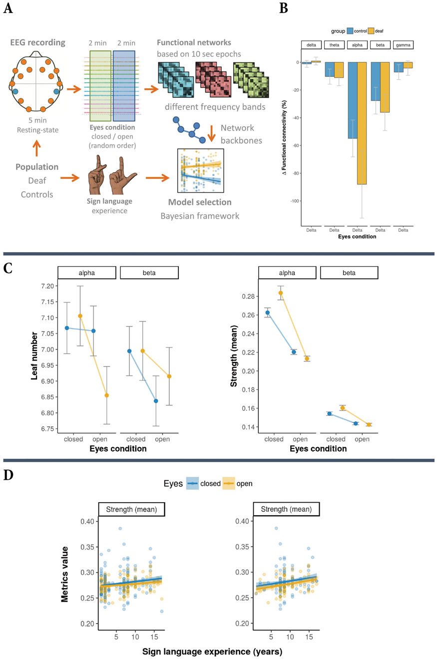
Conclusions
Our study provides original insights into the reorganization of brain networks derived from resting-state EEG measurements in deaf people and hearing controls. We showed functional network backbone differences between eyes-closed and eyes-open conditions, which were larger for deaf individuals than for hearing controls. These effects are arguably caused by auditory deprivation and cross-modal plasticity mechanisms. Moreover, our study demonstrated a relationship between functional network backbone characteristics and sign language experience, which reflects the ability of ongoing neuronal brain adaptions in people with hearing disabilities.
Keywords
Cross-modal plasticity; Deafness; Sign language; EEG; Functional networks; Minimum spanning tree.
Funding
Funded by the Netherlands Organization for Scientific Research (NWO-VENI 016.168.038), and the Dutch Brain Foundation [F2014(1)-06].
References
PB03-G12
Cerebrovascular reactivity decrease sharper in young patients with obstructive sleep apnea
1Dept. of Vascular Ultrasonography, Xuanwu Hospital, Capital Medical University,China
Abstract
Objectives
Obstructive sleep apnea syndrome (OSAS) is a breathing disorder characterized by intermittent and repetitive collapse of the upper airway with resulting transient hypoxemia, arousals, and apneas. The impaired cerebrovascular reactivity (CVR) of patients with OSAS has been determined contributed to the increased risk of ischemic stroke. Although CVR decrease with advancing age in normal people, whether it decreased faster in older patients with OSAS is still not clear. The aim of this study was to compare the change of CVR in elder and young patients with OSAS via transcranial Doppler (TCD).
Methods
According to the recommendation of American Academy of Sleep Medicine Task Force, the severity criteria of OSAS was divided into mild, moderate and severe degrees via apnea-hypopnea index (AHI). A total of 20 elder patients (>60 y) and 40 young patients (<60 y) matched with severe degree (1:2) and gender were included in this study. After 5minutes rest, participants were fitted with a head frame, and the TCD ultrasound 2-MHz probe (EMS-9PB, Delica) was fixed in place. The averaged mean velocity of bilateral middle cerebral artery (MCAmv) was recorded with normal breath as baseline. Then the breath holding (BH) was started and maintained for 40–50 seconds. The maximum MCAmv apnea and the duration of breath-holding were recorded. Breath holding index (BHI) was calculated according to the equation of BHI = (MCAmv apnea-MCAmv baseline) × 100/ (MCAmv baseline × time of BH). The whole procedure was repeated once after 10minutes rest, the averaged BHI was used for analyzing.
Results
There were no significant differences of BMI and vascular risk factors such as hypertension, coronary artery disease, diabetes and hyperlipidemia between young and elderly group. Although the elder people had lower BHI in mild degree of OSAS (0.92 vs. 1.46, P<0.01), there were no differences in moderate (0.81 vs. 0.93, P = 0.57), or severe degree (0.76 vs. 0.79, P = 0.90). The drop of BHI was found in both groups with severity increased. Surprisingly, we found the young patients had faster declining compared with elder patients (P<0.01) (
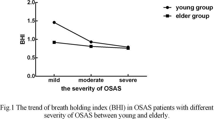
Conclusions
CVR is impaired in all patients with OSAS. Although CVR was declined as the severity increases, young patients have sharper decreasing trend than the elderly.
References
PB03-G13
Changes in cerebral and muscular hemodynamics induced by central sleep apnea events under normal sleep and under continuous positive airway pressure titration
1Center of Sleep Medicine and Sleep Research, Clinic Barmelweid AG, Switzerland
2Department of Neurology, Inselspital, Bern University Hospital and University of Bern, Switzerland
Abstract
Objectives
Sleep apnea syndromes (SAS) are high risk factors of stroke. Obstructive sleep apnea (OSA) is the most common type of SAS and previous studies have reported changes in cerebral hemodynamics induced by OSA. Central sleep apnea (CSA) is less common than OSA, but its pathophysiological mechanisms are more complicated. The cerebral hemodynamic changes associated with CSA events are still unknown. While OSA is effectively treated with continuous positive airway pressure (CPAP), it is well-known that some initial CPAP titrations result in emergence of CSA, i.e., treatment-emergent CSA. We aim to characterize the cerebral and muscular hemodynamics induced by ‘normal’ CSA and treatment-emergent CSA by CPAP.
Methods
33 patients (age: 54.9 ± 13.5 years, Male: 29, Female: 4, BMI: 36.1 ± 7.4) with sleep apnea did CPAP titration. They first did 1-hour baseline sleep without CPAP and then slept with CPAP in the rest of night. A standard all-night Video-Polysomnography (Embla RemLogic, Embla Systems LLC, Tonawanda, NY, USA) measurement was recorded from each patient. Frequency-domain multi-distance near-infrared spectroscopy (FDMD-NIRS) (Imagent, ISS, Champaign IL, USA) measurements were conducted over the middle of left forehead and the left bicep brachii muscle. The sleep stages, respiratory and limb movement events were scored in 30-s epochs based on the Video-Polysomnography measurements. The changes of blood volume (BV) and tissue oxygen saturation (StO2) measured by FDMD-NIRS were compared between CSA baseline (i.e., 2 seconds before CSA onset) and during CSA events with paired t-test (P < 0.05) in the 1-hour baseline sleep and under CPAP, respectively. The maximal decrements of StO2 during CSA events were also compared between baseline sleep and sleep under CPAP (unpaired t-test, P < 0.05).
Results
22/33 patients showed increased CSA-index during CPAP compared to 1-h baseline, including 13 patients showed no CSA events at baseline. In baseline sleep we recorded validated FDMD-NIRS signals in 145 and 191 CSA events in the brain and muscle, respectively. Cerebral StO2 decreased approximately 2.22% (P < 0.001, 95% conference interval (CI): [-2.75, -1.68]) and cerebral BV decreased 1.25 arbitrary unit (a.u.) (P < 0.001, 95% CI: [-1.56, -0.94]) during CSA events compared to CSA baselines; Muscular StO2 and BV decreased 0.65% (P < 0.001, 95% CI: [-0.75,-0.55]) and 0.55 a.u. (P < 0.001, 95% CI [-0.67,-0.44]), respectively. We recorded validated FDMD-NIRS recordings in 1236 and 1209 CSA events under CPAP in the brain and muscle, respectively. Cerebral StO2 and BV decreased 1.47% (P < 0.001, 95% CI: [-1.57, -1.37]) and 0.81 a.u. (P < 0.001, 95% CI: [-0.89, -0.72]) in CSA events, respectively. These decrements were 1.2% (P < 0.001, 95% CI: [-1.36,-1.04]) and 0.93 a.u. (P < 0.001, 95% CI: [-1.08,-0.79]) in the muscle, respectively. The StO2 decreased more in the brain compared to muscle in both baseline sleep and under CPAP titration (P < 0.001). Compared to baseline sleep, the decrements of StO2 under CPAP were smaller in the brain (P = 0.008), but larger in the muscle (P < 0.001).
Conclusions
CSA events are associated with tissue desaturation in both brain and muscle, but the degree of desaturation is larger in the brain. Although CPAP can induce treatment-emergent CSA, the degree of desaturation caused by CSA is reduced by CPAP compared to baseline sleep.
PB03-G14
Cerebral blood flow in adolescents with post concussive symptoms is related to recovery time
1Department of Pharmacology, Physiology and Neuroscience, Rutgers, The State University of New Jersey, Newark, NJ, USA
2The MARCS Institute for Brain, Behaviour and Development, Western Sydney University, Sydney, Australia
3Sydney Children's Hospital Network, Children’s Hospital Institute of Sports Medicine, Children’s Hospital Westmead, Sydney, Australia
4Discipline of Child and Adolescent Health, Sydney Medical School, The University of Sydney, Sydney, Australia
5War Related Illness and Injury Study Center, New Jersey Veterans Affairs Healthcare System, East Orange, NJ, USA
Abstract
Objectives
Concussion affects up to 1.9 million children and adolescents in the USA every year1. Despite the high incidence of brain injury, there is a lack of objective measures of diagnosis and markers of recovery. Monitoring cerebral blood flow is a promising physiological marker to monitor recovery from injury2. Body posture is an important consideration when monitoring cerebral blood flow as supine cerebral blood flow is typically greater than seated cerebral blood flow in healthy conditions.
Methods
We conducted a pilot study investigating the percent change in internal carotid cerebral blood flow between seated and supine positions in 18 concussed children (12 male, 6 female; mean age = 13 ± 2 years) who were admitted to an outpatient sports clinic presenting with persistent concussive symptoms lasting >8 days. Eight non-concussed children (5 male, 3 female; mean age = 13 ± 2 years) who were admitted to the clinic for musculoskeletal injuries served as control participants. Internal carotid artery (ICA) blood flow was obtained in participants using duplex ultrasonography during resting conditions in the seated and supine positions. Beat-by-beat blood pressure (Finapres), ECG and end-tidal CO2 were also obtained.
Results
There was no difference in ICA blood flow between concussed and control children (seated ICA: concussed = 597.7 ± 165.1 vs control = 533.5 ± 116.0 ml/min; p = 0.331; supine ICA: concussed = 610.9 ± 137.1 vs control = 601.9 ± 140.4 ml/min; p = 0.877). There was a greater ratio of supine to seated ICA blood flow in control compared to concussed children, but this was not statistically significant (control = 113 ± 13.5% vs concussed = 105.2 ± 19.7%; p = 0.312). Interestingly, the percent increase in CBF when supine compared to seated in concussed children was significantly correlated with the number of days of recovery (mean recovery = 40 ± 33 days; r = 0.601, p = 0.008). Additionally, this relationship was present in the males (r = 0.694; p = 0.012), but not in the females (r = 0.526; p = 0.283).
Conclusions
This data suggests supine cerebral blood flow increases with recovery time in concussed male children. It is possible females do not recover supine cerebral blood flow as quickly as the male concussed children, which would be consistent with slower recovery times in females3. However, further work is needed to confirm these findings in a larger sample of male and female concussed children before conclusions can be made. This work was supported by Western Sydney University (P. Breen).
References
PB03-G15
Potential serum biomarkers for the prediction of escitalopram efficacy on depression
1The Affiliated Hospital of Jiangsu University
Abstract
Objective
Although several pharmacological selections for depression are currently available, a large proportion of patients still cannot achieve a complete remission or respond adequately in first antidepressant therapy, whose characteristics remain vaguely known. This study aimed to explore the application of serum biomarkers in the prediction of escitalopram efficacy on depression, so as to guide the clinical drug selection.
Method
In this study, 306 depression patients were treated with escitalopram (10 mg) for 6 weeks. The patients were divided into the escitalopram-sensitive group (ES, n = 172) and escitalopram-insensitive group (EIS, n = 134) according to the result of HAMD-24 scale after 6 weeks. All participants’ serum samples were collected on the first day, and 10 types of serum biomarkers were analyzed. Next, data of 100 patients in the ES group and 100 patients in the EIS group were used to build the logistic regression model, and the receiver operating characteristic (ROC) curve was drawn. Then, to validate the accuracy of our model, another 72 patients in the ES group and 34 patients in the EIS group were studied.
Results
Of the 10 selected serum biomarkers, 4 were screened to build the regression model, BDNF, FGF-2, TNF-α and 5-HT. The regression equation was Z = 1/[1+e – (-5.065 +0.145 (BDNF)+0.029 (FGF-2) – 0.368 (TNF-α) + 0.813 (5-HT))], and the 4 biomarkers-combined detection achieved an AUC (area under the ROC curve) of 0.929 and a predictive accuracy of 88.70%.

Conclusion
The logistic regression model and ROC curves based on serum biomarkers in this study provide a more reliable means to predict the curative effect on escitalopram in patients with depression at an early stage, and to provide clinical evidence for drug selection.
References
PB03-G16
Clinical evaluation of a prototype ‘brain tv’ (tissue velocimetry) ultrasound system for emergency assessment of suspected acquired brain injury
1University of Leicester, UK
2University Hospitals of Leicester NHS Trust, UK
3Nihon Kohden, Japan
Abstract
Objectives
Acquired brain injury (ABI) is known to be a leading cause of death worldwide. As brain tissue pulsations connect tissue biomechanical properties, such as stiffness, to blood flow and cerebrospinal fluid dynamics, measurement of brain tissue pulsatility using Transcranial Tissue Doppler (TCTD) may prove valuable for screening patients for vascular pathology, or triage and monitoring of patients with acquired brain injury,(figure 1). Here we describe the design of an ongoing clinical trial evaluating Brain TV (tissue velocimetry) TCTD measurements for diagnosis and monitoring of ABI.
Methods
A protocol for prospective observational clinical evaluation of Brain TV, including diagnostic accuracy assessment according to ‘STARD’ criteria [1], was developed. The objectives of the study are to provide ‘confidence in concept’ data confirming alterations in tissue pulsatility in the presence of brain injury and to facilitate an initial diagnostic assessment of the sensitivity of Brain TV for detection of brain injuries as a function of injury sub-type, location and severity. Prior to commencing the study, the safety of Brain TV was independently assessed by Clinical Engineers from the University Hospitals of Leicester NHS Trust and sponsorship was provided by the University of Leicester. Our nationally approved protocol includes permission to obtain consultee assent for patients with severe injuries who are unable to provide written informed consent [2].
Results
Preliminary observations from this ongoing Brain TV clinical evaluation study will be presented. Our study design includes prospective triage using Brain TV of up to 250 patients arriving at the Leicester Royal Infirmary Emergency Department with suspected acute ABI. Brain tissue pulsation properties will be compared to the severity of brain injury assessed using standardised clinical assessment scores (e.g. Glasgow Coma Scale and National Institute for Health Stroke Score). A sub-set of patients will be asked to wear markers during their CT scan, to aid positioning of the ultrasound probe with respect to the location of injury. A cardiac-gated aMRI scan will also be performed to reveal any disruption of brain tissue motion in the presence of injury. Patients admitted to hospital will receive daily Brain TV (TCTD, ECG), physiological monitoring (BP, CO2) and follow-up of clinical outcome to explore the prognostic potential of Brain TV as a physiological monitoring tool.

Conclusion
This study aims to generate a database of BTP estimates and imaging data from patients with brain injury, enabling us to better understand brain biomechanics and explore the clinical potential of Brain TV for diagnosis and monitoring. In future work, Brain TV measurements will be compared to archived recordings from healthy subjects to determine the sensitivity and specificity of Brain TV for detection of abnormal brain biomechanics and to relate the extent of disruption of Brain TV waveforms to the severity of injury.
References
PB03-G17
Novel c-Jun N-terminal kinase inhibitor IQ-1S modulates human platelet aggregation
1Cardiology Research Institute, Tomsk NRMC, Tomsk, Russia
2Kizhner Research Center, Tomsk Polytechnic University, Tomsk, Russia
3Research Institute of Biological Medicine, Altai State University, Barnaul, Russia
4Department of Microbiology and Immunology, Montana State University, Bozeman, USA
5Cardiovascular Research Center, Cardiology Division, Massachusetts General Hospital, Harvard Medical School, Charlestown, Massachusetts, USA
Abstract
Objectives
Abnormal platelet aggregation is essentially involved in ischemic stroke. A novel c-Jun N-terminal kinase (JNK) inhibitor IQ-1S exerts neuroprotective effects and improves survival in cerebral ischemia-reperfusion injury in mice [1]. IQ-1S suppresses pro-inflammatory cytokine production and donate nitric oxide (NO) in experimental models [1]. Considering that JNK- and NO-mediated signaling mechanisms are closely associated during ischemia-reperfusion whereas platelet aggregation is sensitive to NO, compounds with dual action donating NO and inhibiting JNK are promising as agents targeting thrombophilia and exerting neuroprotection in ischemic stroke. Objective of this study was to elucidate the effects of IQ-1S and its analogues on human platelet aggregation.
Methods
Effects of IQ-1S analogues on platelet aggregation were assessed in human platelet-rich plasma and isolated platelets. Peripheral venous blood was sampled from the median cubital vein. Platelet-rich plasma was obtained by blood centrifugation for 7 min at 1,500 rpm and supernatant sampling. For platelet isolation, prostaglandin E1 (1 μM) and apyrase (0.1 U/mL) were added to platelet-rich plasma, which was then incubated for 15 min at 37°C, centrifuged for 15 min at 3,000 rpm, and washed twice. The final concentration of platelets was standardized. Platelet aggregation was triggered by 2-mg/mL collagen. Platelet-rich plasma and isolated platelets were incubated with IQ-1S and its inactive analogues AK-29 and AK-30 in the cuvette at concentrations of 2, 5, 10, 25, 50, and 100 μM for 10 min at 37°C. Platelet aggregation degree and rate were assessed with two-channel laser analyzer 220 LA NPF Biola (Russia). Wilcoxon signed-rank test was used to assess statistical differences. Differences of p < 0.05 were considered significant.
Results
IQ-1S (2 and 5 μM) significantly attenuated platelet aggregation degree and rate in platelet-rich plasma, but not in isolated platelets. Application of 2 and 5 μM of IQ-1S decreased platelet aggregation from 72.28 (n = 6, p = 0.02) to 69.36 and 71.05 U (n = 6, p = 0.04) according to light-transmission curves; platelet aggregation rate decreased from 40.62 to 25.45 (n = 6, p = 0.02) and 28.33 U/min (n = 6, p = 0.04) according to mean aggregate size curves and from 81.03 to 65.55 and 65.93 U/min (n = 6, p = 0.02) according to light-transmission curves, respectively. Inactive IQ-1S analogues, AK-29 and AK-30, did not dose-dependently affect human platelet aggregation though, at high concentration (100 μM), AK-29 and AK-30 slightly attenuated platelet aggregation rate whereas AK-30 also attenuated platelet aggregation degree in platelet-rich plasma.
Conclusions
The ability of IQ-1 to exert slight to moderate attenuation of human platelet aggregation may be beneficial in stroke injury. The absence of IQ-1S effects on aggregation of collagen-stimulated isolated platelets suggests that plasma-associated co-factor is required for the response. Obtained results provide fundamental basis for further development of new agents aimed at prevention and treatment of ischemia-reperfusion injury.
Acknowledgements
The study was supported by the Ministry of Education and Science of the Russian Federation (No. 4.8192.2017/8.9 and AAAA-A15-115123110026-3); histoquantitative study was supported by the Russian Science Foundation (No. 17–15-01111).
Reference
PB03-H01
Generation and characterization of the human iPSC line from blood cells of a CADASIL patient carrying a NOTCH3 mutation
1Clinical Neurosciences Research Laboratory, Department of Neurology, University Clinical Hospital, Universidade de Santiago de Compostela, Health Research Institute of Santiago de Compostela (IDIS), Santiago de Compostela, Spain
2Cellular Fate Reprogramming Unit, Department of Molecular and Translational Medicine, University of Brescia, Brescia, Italy
3Stroke Unit, Department of Neurology, Hospital Clínico Universitario, Santiago de Compostela, Spain
Abstract
Objective
CADASIL is the most common form of hereditary stroke disorder. It is caused by mutations of the Notch3 that led to a progressive degeneration of the smooth muscle cells in blood vessels. Nowadays there is no treatment for this disorder. Our aim was to prove if it is possible to reprogram peripheral blood cells from a patient with CADASIL to induced pluripotent stem cells, setting the basis for future differentiation to the affected cell type and drug screening.
Methods
A feeder-free, non-integrative reprogramming with Sendai virus was performed using a sample of peripheral blood cells from a patient with CADASIL. Immunocitochemistry and qPCR techniques were carried out for characterization, and RT-PCR and PCR techniques were performed to check the absence of the virus and the exogenous Yamanaka´s factors. Formation of embryoid bodies was also induced.
Results
We achieved several stable induced pluripotent stem cells colonies (9.6% of efficiency). The characterization was positive for the pluripotent markers NANOG, SOX2, OCT4, SSEA4 and TRA-1-60 and the expression levels of endogenous OCT4, SOX2 and NANOG was also positive. Viruses were completely absent in cells at passage 5. We were also able to induce the formation of embryoid bodies.
Conclusion
Reprogramming of peripheral blood cells from CADASIL patients into induced pluripotent stem cells suggests that Notch3 mutation does not limit the cell reprogramming and offers an unprecedented opportunity for studying and modeling CADASIL pathology.
PB03-H02
Hepcidin decreases rotenone induced a-synuclein accumulation via autophagy in SH-SY5Y cells
1Department of Physiology and Hypoxic Biomedicine, Academy of Special Environmental Medicine, Nantong University, 9 Seyuan Road, Chongchuan District, Nantong, Jiangsu 226019, China
2Department of Neurophysiology and Neuropharmacology, Institute of Special Environmental Medicine, Nantong University, 9 Seyuan Road, Nantong, Jiangsu 226019, China
Abstract
Objectives
Parkinson’s disease (PD) is the most common motor disorder affecting the elderly. Strong evidences showed that both iron deposition and a-synuclein (α-syn) aggregation were hallmarks of PD. Hepcidin is a newly identified iron metabolism regulating hormone, which could reduce iron both in central nervous system and peripheral system. These knowledges prompted us to hypothesize that hepcidin might be able to reduce iron and further decrease a-syn accumulation. This study was designed to investigate the effects of hepcidin on the expression and aggregation of a-syn and the mechanisms involved in vitro.
Methods
Human neuroblastoma SH-SY5Y cells were used as a model which introduced by rotenone and α-syn into the cell model to mimic pathological progress of PD. MTT was used to identify the cell viability after SH-SY5Y cells treated with rotenone. Western blot and immunohistochemistry were used to measure the a-syn aggregation induced by rotenone or clearance by hepcidin. Graphite furnace atomic absorption spectrophotometer analysis was used to determine the iron content in cells. To clarify the mechanisms involved in the process, the ubiquitin-proteasome system and autophagy system was detected by using inhibitor of the two systems in physiological and transfection conditions, respectively.
Results
Compared with the control groups, the cell viability was impaired in dose-dependent manner after rotenone treatment. Long-term (3 days) and low concentration rotenone (25 nM) could increase a-syn accumulation as well as iron contents in SH-SY5Y cells. a-syn expression and iron contents were decreased after treating with hepcidin peptide (100 nM) in rotenone-induced SH-SY5Y cells. Besides, hepcidin peptide could also repress rotenone effects on a-syn in a-syn overexpression conditions.
Decreased a-syn expression after treating with hepcidin could be prevented by autophage inhibitors (both 3MA and CQ) but not by proteasome inhibitor (MG132).
Conclusions
Hepcidin could eliminate α-syn expression induced by rotenone in SH-SY5Y cells. The clearance effect of hepcidin on a-syn aggregation was confirmed to be mediated through autophagy pathway by pre-treatment with autophagy inhibitors. This study illustrated that hepcidin may offer the potential therapeutic perspective in a-syn aggregation diseases, especially in PD.
Keywords
Parkinson’s disease (PD); rotenone; hepcidin; a-synuclein, iron; autophage
Acknowledgements
This research was supported by Chinese Natural Science Foundation grants 81873924 (to QQ Luo).
PB03-H03
Pathological study and genetic analysis of myotonic dystrophy Type I
1Department of Pathology, Nanjing Drum Tower Hospital, China
2Department of Neurology, Nanjing Drum Tower Hospital, China
Abstract
Objective
To investigate the clinical, pathological and genetic features of myotonic dystrophy type I (DM1) and improve the diagnosis and cognition of the disease.
Methods
Clinical and pathologic data of 15 patients with DM1 were studied retrospectively. Muscle biopsy was performed to study pathological features and triplet primed-polymerase chain reaction (TP-PCR) was used to detect DMPK gene amplification.
Results
Five of 15 patients had a positive family history. Muscle weakness was the most common initial syndrome. EMG shows myogenic damage (seven cases)myotonia potentials (six cases) in DM1. Seven of 15 paitents had an increased number of repeat sequence copies of trinucleotides [cytosine-thymine-guanine, (CTG) n] in the 3′-untranslated region of the DMPK gene[1] (>50 repeat sequences and far beyond the normal level) (figure 1). Our results showed: 1) There was a significant statistical difference (p<0.05) in type I skeletal muscle in DM1 compared with normal control group (61.99 ± 5.76 versus 41.43 ± 2.85). 2) The diameter of type I fibers in DM1 group was significantly lower than that in normal control group (p < 0.05). Although the diameter of type II muscle fibers in the DM1 group was slightly higher than that in the type I muscle fibers, it did not reach statistical significance (P > 0.05). The results indicated that DM1 patients mainly suffered from type I muscle fiber atrophy, and type II muscle fibers could be moderate compensatory hypertrophy. 3) The ratio of central nuclear muscle fibers in the normal control group was 2.67 ± 1.41, and the proportion of central nuclear muscle fibers in the DM1 patients was 36.39 ± 5.45, which was statistically significant (p < 0.05). It indicated that the proportion of intramuscular nucleus migration in DM1 patients was significantly higher than that in the control group. The marked increase of internalized nuclei, a preferential atrophy of type I fibers, and sarcoplasmic masses in DM1 has pathological diagnosis significance.

Conclusions
The main clinical characteristics of DM1 are delayed relaxation after skeletal muscle contraction. The nuclear ingression of muscle fibers, nuclear chain fibers and abnormal distribution of muscle fibers are the pathological characteristics of DM1[2]. Skeletal muscular biopsy is of most importance for diagnosis. Furthermore, the detection of CTG-repeats of DMPK gene may provide an additional effective way for genetic diagnosis.
References
PB03-H04
Olfactory type g-protein alfa subunit related changes in the striatum underlie the genesis of L-DOPA-induced dyskinesia
1Dept. of Neurosurgery, Tokushima University, Japan
2Parkinson’s Disease and Dystonia Research Center, Tokushima University Hospital, Japan
3Dept. of Neurosurgery, Takamatsu Municipal Hospital, Japan
4Dept. of Neurodegenerative Disorders Research, Tokushima University, Japan
Abstract
Objectives
Prolonged and pulsatile exposure to L-3,4-dihydroxyphenylalanine (L-DOPA) causes L-DOPA-induced dyskinesia (LID) in patients with Parkinson’s disease (PD)1. Although dopamine D1 receptors (D1Rs) hyperactivity is supposed to underlie the genesis of LID, recent studies revealed that an adenosine 2A receptors (A2ARs) antagonist, which is used for treatment in patients with PD, also causes LID as a major adverse effect1. Olfactory type G-protein alfa subunit (Gαolf) couples with D1Rs in the striatonigral “direct” medium spiny neurons (MSNs) and A2ARs in the striatopallidal “indirect” MSNs1. Gαolf represents the rate-limiting factor for the D1Rs- and A2ARs-dependent cAMP production and could serve as a major contributor to the circuit level dysfunction observed in LID1. In this study, we aimed to reveal the Gαolf -related changes in LID.
Methods
LID mice were developed by daily administration of L-DOPA to 6-hydroxydopamine lesioned hemi-parkinsonian mice. Immunostainings against D1Rs, A2ARs, Gαolf or c-Fos in the dorsolateral striatum were performed using the tyramide signal amplification method. Dual-antigen recognition to detect couplings of two molecules were conducted in the dorsolateral striatum using in situ proximal ligation assay (PLA) with an antibody against Gαolf in combination with antibodies against D1Rs or A2ARs. The optical densities of immunoreactive products and the number of c-Fos positive nuclei after dopamine administrations were assessed. We performed statistic analyses using Stat View 5.0 software. P-value of less than 0.05 were considered statistically significant.
Results
Gαolf-immunoreactivity (IR) showed significant increases only in PD on the lesioned side compared to non-lesioned side (n = 10). D1Rs- and A2ARs-IR demonstrated no difference among naïve, PD and LID striatum. D1R-Gαolf PLA-IR significantly increased in PD and LID and A2AR-Gαolf PLA-IR significantly decreased only in LID on the lesioned side when compared to naïve control. Density of c-fos positive nuclei after acute dopamine administration significantly increased in both PD and LID on the lesioned side striatum and globus pallidus when compared to naïve control.
Discussions
The Gαolf protein levels in the striatum are regulated by posttranslational usage-dependent mechanism through the activation of D1Rs and A2ARs1. Indeed, we found Gαolf-IR up-regulation in PD due to the disuse and its normalization in LID attributable to the daily stimulation of D1Rs. Increase in D1R-Gαolf PLA-IR of PD and LID striatum indicated the increased coupling of D1Rs and Gαolf, i.e. the increased responsiveness of D1R signaling which was verified by the increased c-fos density in the striatum. Decrease in A2AR-Gαolf PLA-IR of LID suggested the decreased responsiveness of A2AR signaling which was confirmed by increased c-fos density in the globus pallidus. Our findings suggested that A2ARs antagonists might be effective in reducing the priming of LID, but they could exacerbate dyskinesia after the establishment of LID.
Reference
PB03-H05
FABP3 ligands inhibiting α-synuclein oligomerization in Neuro2A cells
1Dept. of Pharmacol, Grad. Sch. Pharm. Sci., Tohoku Univ. Japan
Abstract
Introduction
In Parkinson's disease (PD), α-synuclein (αSyn) accumulation and inclusion triggers dopamine neuronal death and synapse dysfunction in vivo. We previously reported that fatty acid-binding protein 3 (FABP3) is highly expresses in dopaminergic neurons and aggravates αSyn oligomerization when exposure to 1-methyl-1,2,3,6-tetrahydropiridine (MPTP) in vivo and in vitro (J Biol Cem 2014;289:18957). We here discovered FABP3 ligands inhibiting α-synuclein oligomerization induced by arachidonic acid (AA).
Method
FABP ligands were modified from FABP4 inhibitor BMS309403 and assessed their inhibitory action on AA-induced α-synuclein oligomerization using FABP3 and αSyn co-overexpressed Neuron2A cells. The αSyn oligomerization levels were measured using western blotting assay and immunohistochemical analyses.
Result
The AA treatment triggered αSyn oligomerization in Neuro2A cells in FABP3-dependent manner. A potent FABP3 ligand 1 totally blocked αSyn oligomerization induced by FAPB3 and AA. In addition, ligands 7 and 8 also elicited inhibition of αSyn oligomerization in Neuro2A cells. Aggregation of αSyn in FABP3 and AA treated cells is also blocked by FABP3 ligand 1 in culture Neuro2A cells.
Discussion
The long-chain unsaturated fatty acid such as AA induces αSyn oligomerization with FABP3, which highly expresses in dopaminergic neurons. In this work, we found that AA stimulates αSyn oligomerization in FABP3-dependent manner. We furthermore developed the low molecular compound, which reduce αSyn oligomerization. Taken together, the new FABP3 ligand is attractive therapeutic candidate for Parkinson and Lewy body diseases.
PB03-H06
Beneficial and protective role of Ashwagandha (roots of Withania somnifera) against 3-nitropropionic acid-induced oxidative stress in a rat model of huntington's disease
1Department of Biotechnology, Vivekananda Global University, India
Abstract
Objectives
Huntington's disease (HD) is a neurodegenerative disorder that results from the destruction of neurons in the basal ganglia, and oxidative stress has been implicated in its pathogenesis. Ashwagandha (roots of Withania somnifera) is used in traditional Indian medicine (Ayurveda) for general debility, consumption, nervous exhaustion, insomnia, and loss of memory. Withania Someniferais (WS) restrain brain aging and help in regeneration of neural tissues besides producing antistress, adaptogenic and memory enhancing effect.
The present study examined the potential therapeutic effects of WS against 3-nitropropionic acid (3-NP)-induced oxidative stress in a rat model of HD and explored the mechanisms of action.
Methods
80 Male SD rats were pretreated with methanolic extract of WS (200 and 400 mg/kg b.w.) orally prior to the intraperitoneally (i.p.) administration of 3-NP (12 mg/kg b.w.) for 15 days. Nimodipine (12 mg/kg, po) was used as positive control drugs. The body weight, grip strength and behavior were monitored within 5th, 10th and 15th day after 3-NP treatment. Then the animals were sacrificed, neuronal damage in striatum was estimated using Nissl staining. The learning and memory function were assessed by Morris water maze test. The oxidative stress indicators [glutathione reductase (GRx), superoxide dismutase (SOD), neurolipofusicin and malondialdehyde (MDA)] and inflammatory cytokines (TNF-a, IL-1β, and IL-6) were measured in hippocampus using corresponding commercial kits. The mRNA and protein levels of PPARγ were evaluated by real time (RT)-PCR and Western blot analysis were analyzed in rat model of HD.
Results
Present results shown that administration of 3-NP resulted in a marked reduction in the body weight, memory, grip strength locomotion activity and significantly increased lipid peroxidation, PPARγ, inflammatory cytokines and depleted antioxidant enzyme accompanied by progressive striatal dysfunction. WS (200 and 400 mg/kg) treated animals exhibited a significant improvement in behavioural, biochemical, histological alterations and oxidative stress parameters in comparison to only 3-NP treated animals. Present results shown dose-dependently improved 3-NP-induced biochemical, behavioral and enzymatic changes. Similar effects were obtained with the positive control drugs nimodipine.
Conclusion
WS exerts a protective action against 3-NP-induced oxidative stress in the rat model of HD, which is associated with its neuroinflammatory with anti-oxidant activity, and consequently improves behavioral deficits.
PB03-H07
Possible remyelination by inflammatory-induced endogenous neural stem cells of spinal cord in a mouse model of experimental autoimmune encephalomyelitis
1Dept. of Ophthalmology, Hyogo College of Medicine, Japan
2Dept. of Surgical Pathology, Hyogo College of Medicine, Japan
3Institute for Advanced Medical Sciences, Hyogo College of Medicine, Japan
4Dept. of therapeutic progress in brain diseases, Hyogo College of Medicine, Japan
5Division of Neurology, Dept. of Internal Medicine, Hyogo College of Medicine, Japan
Abstract
Objectives
The processes of myelination and remyelination play a crucial role in the clinical course of multiple sclerosis and are apparent in its animal model, experimental autoimmune encephalomyelitis (EAE) (1, 2). There is considerable evidence to show that a number of stimuli can activate endogenous stem/progenitor cells in the central nervous system (CNS), including the spinal cord, but their possible contributions to remyelination under inflammatory conditions, such as EAE remains unclear. Many studies have demonstrated that neural stem/progenitor cells (NSPCs) can proliferate, migrate, and differentiate after CNS injuries, including brain and spinal cord injuries (3–7). In this study, we used a mouse model of myelin oligodendrocyte glycoprotein (MOG)-induced EAE, to investigate whether adult spinal cord-derived NSPCs have the potential of remyelination following CNS inflammatory injuries.
Methods
EAE mice were established as previously described (8). Lumbar cords were harvested from control or EAE mice on days 7, 28, and 56 after treatment. Spinal cord sections were subjected to immunohistochemistry using antibodies against markers for NSPCs (nestin) and oligodendrocyte lineages, such as oligodendrocyte specific protein, myelin basic protein, and myelin associated glycoprotein. Additionally, cells isolated from the spinal cords of EAE or control mice were incubated to promote the formation of neurospheres, followed by further incubation in medium to promote neuronal differentiation.
Results
Immunohistochemistry showed that cells expressing nestin emerged in the spinal cords of mice after MOG treatment, mainly at the site of demyelination, where many inflammatory cells were present, whereas nestin+ cells were rarely observed in PBS-treated control mice. In vitro, nestin+ cells isolated from the spinal cords of MOG-treated mice could differentiate into multi-neural lineages, including oligodendrocytes.
Conclusions
These results show that nestin+ stem/progenitor cells activated by EAE can differentiate into oligodendrocyte lineages, indicating that spinal cord-derived stem/progenitor cells have the potential of remyelination under inflammatory conditions.
References
PB03-H08
Progranulin is involved in α-synuclein pathology through autolysosome formation
1Mol. Pharmacol., Dept. of Biofunct. Eval., Gifu Pharmaceutical Univercity, Japan
Abstract
Objectives
Parkinson’s disease (PD) is a major progressive neurological disease, which is characterized by the display of tremor, bradykinesia, muscle rigidity and impaired balance. Lewy bodies are accumulated in the brain of PD patients and largely consisted of α-synuclein. Namely, α-synuclein is the pathological hallmark of PD. Recently, it has been reported that some PD patients with α-synuclein lesions have a progranulin gene mutation (1, 2). Progranulin is a multipotent protein that contributes to various pathology such as inflammation and tumorigenesis. Absence of progranulin gene causes the onset of frontotemporal lobar degeneration and neural ceroid lipofuscinosis. However, the role of progranulin for α-synuclein accumulation is unclear. We examined the effects of progranulin against α-synuclein accumulation.
Methods
We examined brain α-synuclein expression level in progranulin deficient mice. We evaluated the effects of progranulin against MPP+ damage in human derived neuroblastoma cells (SHSY-5Y cells). Alpha-synuclein, autophagosome formation factors (AMPK, mTOR, LC-3) and autophagosome degradation factor (p62) were evaluated by Western blotting. To clarify the mechanism of progranulin against autophagy degradation, we evaluated autolysosome formation using DAL-green, a specific autolysosome detector.
Results
Insoluble and aggregated α-synuclein was detected in progranulin deficient mice. While SHSY-5Y cells, progranulin treatment suppressed the increase of α-synuclein expression, did not change AMPK or mTOR activation. Furthermore, progranulin reduced LC-3II/LC3-I ratio and p62 expression. Interestingly, progranulin rescued the decrease of autolysosome formation by MPP+.
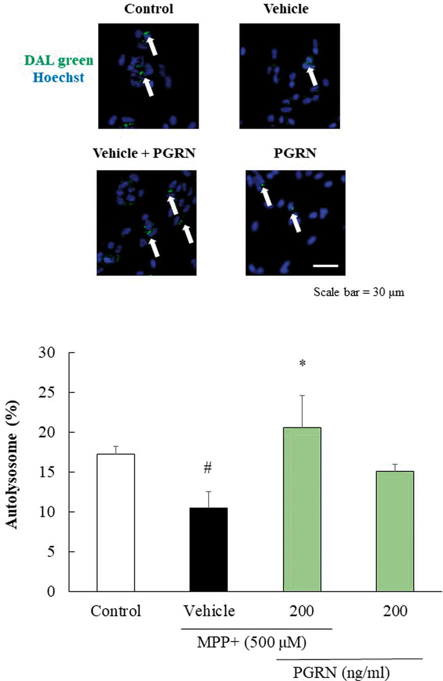
Conclusions
These findings indicate that progranulin promotes autophagy degradation by inhibiting autolysosome formation and reduces α-synuclein accumulation. Therefore, progranulin may be one of new therapeutic targets of PD.
References
PB03-H09
Therapeutic benefits of edaravone for enhanced oxidative stress in mice model of amyotrophic lateral sclerosis
1Dept. of Neurology, Okayama University, Japan
Abstract
Oxidative stress is associated with the degeneration of both motor neurons and skeletal muscles in amyotrophic lateral sclerosis (ALS). A free radical scavenger edaravone has been proven as a therapeutic drug for ALS patients, but the neuroprotective mechanism for the oxidative stress of ALS has not been fully investigated. In this study, we investigated oxidative stress in ALS model mice bearing both oxidative stress sensor nuclear erythroid 2-related factor 2 (Nrf2) and G93A-human Cu/Zn superoxide dismutase (Nrf2/G93A) treated by edaravone. In vivo Nrf2 imaging analysis showed the accelerated oxidative stress both in spinal motor neurons and lower limb muscles of Nrf2/G93A mice according to disease progression. These were significantly alleviated by edaravone treatment accompanied by clinical improvements. The present study suggests that in vivo optical imaging of Nrf2 is useful for detecting oxidative stress in ALS, and edaravone alleviates the degeneration of both motor neurons and muscles related to oxidative stress in ALS patients.
PB03-H10
Arterial spin labeling MR imaging for the detection of cerebellar hypoperfusion in patients with spinocerebellar degeneration
1Second Department of Internal Medicine (Neurology), University of Fukui, Japan
2Biomedical Imaging Research Center, University of Fukui, Japan
3Department of Radiology, University of Fukui, Japan
4Faculty of Nursing and Social Welfare Science, Fukui Prefectural University, Japan
Abstract
Objectives
Spinocerebellar degeneration (SCD) is characterized by progressive cerebellar ataxia and atrophy. Perfusion SPECT usually shows cerebellar hypoperfusion in patients with SCD and aids the diagnosis; however, the availability of SPECT has many restrictions. The arterial spin labeling (ASL) MR imaging allows noninvasive measurement of cerebral blood flow (CBF). The aim of this study was to evaluate the clinical utility of ASL imaging for the detection of cerebellar hypoperfusion in patients with SCD.
Methods
We retrospectively investigated the cerebellar blood flow measured by ASL MRI and 123I-IMP SPECT in patients with SCD and healthy controls in this study. ASL and SPECT images were spatially normalized. Regional CBF were obtained from ASL and SPECT images by volume-of-interest analysis in patients (n = 16). Regional CBF were also measured by ASL in age-matched controls (n = 19) and by SPECT in separate controls (n = 17). The cerebellar CBF values were normalized to the CBF values for the whole gray matter (nCBF) in ASL and SPECT.
Results
The mean cerebellar nCBF measured by ASL was lower in patients with SCD (0.70 ± 0.09) than in the controls (0.91 ± 0.05) (p < 0.001), which was consistent with the comparison using SPECT (0.82 ± 0.05 vs. 0.98 ± 0.05, p < 0.001). The cerebellar nCBF measured by ASL significantly correlated with that determined by SPECT in patients (r = 0.56, p < 0.001). The Bland-Altman plot of the cerebellar nCBF measured by ASL and SPECT showed a good agreement between the two modalities.
Conclusions
ASL imaging showed decreased cerebellar blood flow, which correlated with that measured by SPECT, in patients with SCD. These findings suggest that the clinical utility of noninvasive MRI with ASL for detecting cerebellar hypoperfusion. ASL imaging can be an alternative to SPECT imaging in the diagnosis of SCD, with great advantages that include the simultaneous evaluation of cerebellar atrophy.
Reference
PB03-J01
Effect of yokukansan on nitric oxide production and hydroxyl radical metabolism during cerebral ischemia and reperfusion in mice
1Department of Neurology, Saitama Medical University, Japan
2Department of Pathology, Saitama Medical University, Japan
3Department of Neurology, Tottori Medical Center, Japan
Abstract
Objectives
The purpose of this study was to investigate the effects of yokukansan on forebrain ischemia. Because we can measure nitric oxide production and hydroxyl radical metabolism continuously, we investigated the effect of yokukansan on nitric oxide production and hydroxyl radical metabolism in cerebral ischemia and reperfusion.
Methods
Yokukansan (300 milligrams per kilogram per day) was mixed into feed and given to eight mice for 10 days. Eight additional mice received normal feed (control). Nitric oxide production and hydroxyl radical metabolism were continuously monitored using the salicylate trapping method. Forebrain ischemia was produced in all mice by occluding the common carotid artery bilaterally for 10 minutes. Levels of the nitric oxide metabolites nitrite and nitrate were determined using the Griess reaction. Survival rates of hippocampal CA1 neurons were calculated and 8-hydroxydeoxyguanosine-immunopositive cells were counted to evaluate the oxidative stress in hippocampal CA1 neurons 72 hours after the start of reperfusion.
Results
Arterial blood pressure and regional cerebral blood flow were not significantly different between the two groups. The level of nitrate was significantly higher in the yokukansan group than in the control group during ischemia and reperfusion. Levels of 2,3- and 2,5- dihydroxybenzoic acid were significantly lower in the yokukansan group than in the control group during ischemia and reperfusion. Although survival rates in the CA1 did not differ significantly, there were fewer 8-hydroxydeoxyguanosine-immunopositive cells in animals that had received yokukansan than in control animals.
Conclusion
These data suggest that yokukansan exerts reducing hydroxyl radicals in cerebral ischemic injury.
PB03-J02
Beneficial effect of chlorpromazine and promethazine (C+P) on hyperglycolysis and activation of NADPH- Nicotinamide adenine dinucleotide phosphate (NADPH) oxidase (NOX) in ischemic stroke
1China-America Institute of Neuroscience, Beijing Luhe Hospital, Capital Medical University, China
2Department of Neurosurgery, Wayne State University School of Medicine, USA
Abstract
Introduction
Activation of NOX, a superoxide-producing enzyme, exacerbated ischemic injury through the overproduction of reactive oxygen species (ROS). NOX is dependent on glucose metabolism. The increased catabolism of glucose (hyperglycolysis), in turn, increased ROS production via NOX. Previous studies have demonstrated depressive or hibernation-like roles of combined C+P in brain activity, leading to neuroprotection. We further assessed C+P’s effect on hyperglycolysis and thus NOX activation.
Methods
Sprague-Dawley rats were subjected to 2 h middle cerebral artery occlusion (MCAO) followed by 6 or 24 h reperfusion. At the onset of reperfusion, rats received an IP injection of saline or C+P (8 mg/kg), or inhibitors of NOX (apocynin, 2.5 mg/kg), glucose transporter (GLUT)-1(phloretin,100 mg/kg), or GLUT3 (cyclin B, 0.5 mg/kg). Infarct volume and neurological deficits were determined at 24 h post reperfusion. Hyperglycolysis was detected by brain levels of ATP and glucose, protein expression of GLUT1
Results
C+P reduced infarct volume and neurological deficits. C+P reduced elevations in glycolytic (GLUT1, GLUT3 and PFK) and LDH protein levels and also reduced glucose and lactate while raising ATP levels in the brain. In ischemic rats with GLUT1 and GLUT 3 inhibitor, glucose and lactate levels decreased as ATP levels increased, while GLUT1, GLUT3, and PFK protein levels decreased. These results together suggest reduced cellular glucose uptake and utilization, in association with reduced brain acidosis. C+P decreased stroke-induced NOX activation and brain damage by reducing enzymatic activity, as observed with inhibitors of NOX and GLUT1/3. Expression of NOX subunit gp91phox, P22phox and P47phox was reduced with C+P, similar to those with NOX and GLUT inhibitors.
Conclusion
C+P exerts neuroprotection, by attenuating hyperglycolysis and associated NOX activation through inhibition of GLUT and NOX.
PB03-J03
The importance and limitation of reperfusion in crafting a neuroprotection strategy after ischemic injury
1China-America Institute of Neuroscience, Beijing Luhe Hospital, Capital Medical University, China
2Department of Neurosurgery, Wayne State University School of Medicine, USA
Abstract
Introduction
Although the achievements of the most recent endovascular thrombectomy protocol, including longer treatment windows (up to 16–24 h) and a high rate of revascularization (∼71%), have been encouraging, morbidity and mortality data still show substantial room for improvement: at 90 days, 40% of patients who underwent this procedure incurred severe disability (mRS: 3–5), and 15.3% had died. It is therefore apparent that additional avenues to improve these outcomes must be explored. In this study, we attempt to do so by investigating both the contribution of reperfusion (reoxygenation) to cellular damage following oxygen-glucose deprivation (OGD), and how the application of an adjuvant neuroprotective strategy can mitigate this damage.
Methods
Human neural SH-SY5Y cell lines were subjected to OGD for 1, 2, 4, 6, 12, or 24 h, followed by re-oxygenation for 23, 22, 20, 18, 12 h, or no reoxygenation. Neuroprotectants, either TAT-NR2B9c (8 µM) or uric acid (300 µM), were added to all groups; a dose-response curve was calculated in advance to determine a maximally effective dose. After 24 h, cell viability was detected by WST-1, and cell damage was assayed by LDH release into the supernatant.
Results
Cell survival rates were significantly reduced by OGD for 1, 2, 4, 6, 12 and 24 h to 80%, 73%, 64%, 55%, 25% and 8%, respectively. All reoxygenated groups displayed a time-dependent increase in cell viability: 95%, 88%, 79%, 72% and 39%, respectively, as compared to OGD without reoxygenation; these data suggest the importance of early reperfusion. In addition, LDH release did not increase in 2 and 4 h OGD without reoxygenation, but did increase in the reoxygenation groups, and in the 24 h OGD without reoxygenation groups (p < 0.05): these results suggest an oxidative mechanism of injury. Moreover, neuroprotection with TAT-NR2B9c and uric acid further increased (p < 0.05) cell survival and decreased (p < 0.05) LDH release in 2 and 4 h OGD with reoxygenation, while no effect was found on 24 h OGD without reoxygenation.
Conclusion
Reperfusion is an essential but highly time-sensitive strategy for cell salvage after ischemia. Adjuvant neuroprotection is also beneficial, but must be administered upon reperfusion.
PB03-J04
The impact of collaterals on reperfusion after stroke
1Department of Neurology, University Hospital Zurich and University of Zurich, Switzerland
2Institute of Pharmacology and Toxicology, Experimental Imaging and Neuroenergetics, University of Zurich, Switzerland
3Neuroscience Center Zurich, University and ETH Zurich, Zurich, Switzerland
Abstract
Objectives
Despite improvements in acute recanalization treatments, stroke remains one of the leading causes of death and disability worldwide. To achieve the best possible outcome for the individual patient, therapies have to be administered rapidly. Here, we propose that 1) treatment success depends on collateral flow to the ischemic area, and that 2) dynamic changes in cerebral blood flow (CBF) go beyond the ischemic zone, affecting also remote areas. Furthermore, we hypothesize that CBF changes within and outside the affected territory may predict stroke outcome.
Methods
For induction of experimental ischemia in two mouse strains with differences in the naive collateral network, thrombin was injected into the left middle cerebral artery (MCA) of C57BL/6 and Balb-C mice. 30 min later, thrombolysis was initiated through intravenous injection of recombinant tissue plasminogen activator (rt-PA) or saline (for controls). CBF was monitored using laser speckle imaging during stroke and treatment. Furthermore, multi-wavelength spectroscopy was performed before and after stroke to differentiate arteries from veins. Functional deficits were assessed by the sticky tape test and a composite neurological score. On day 7, animals were euthanized. Triphenyl-tetrazolium chloride (TTC) was used to stain brain slices for infarct size quantification.
Results
In vehicle-treated mice with poor naïve collaterals (Balb-C), stroke led to a persisting CBF drop; however, rt-PA administration led to significant reperfusion of the MCA territory. In mice with better collaterals (C57BL/6), irrespective of rt-PA administration, CBF recovered to only about 50%. Although reperfusion was not much enhanced during the 1.5 h after rt-PA administration, C57BL/6 mice had less functional deficits and significantly smaller infarct areas after rt-PA compared to control animals. Interestingly, blood flow was highly dynamic during ischemia and reperfusion, involving not only the ipsilateral hemisphere and leptomeningeal collaterals, but also remote areas. In both strains, the administration of rt-PA reduced infarct volumes. Under the same treatment, mice equipped with rich collaterals had better outcomes compared to mice with poor collaterals.
Conclusion
In our stroke model including clot formation and i.v. thrombolysis, closely mimicking the clinical situation we found that, mice equipped with better collaterals had less functional deficits and smaller infarct volumes after stroke. However, the immediate CBF response to rtPA and the magnitude of reperfusion was better in Balb-C mice with poor collaterals. Our data suggest that early reperfusion of the ischemic territory does not necessarily predict functional outcome. Further analyses are under way.
References
PB03-J05
Effect of reperfusion on the associations between selected outcome measures following middle cerebral artery occlusion in mice
1Vascular Biology and Immunopharmacology Group, Department of Physiology, Anatomy and Microbiology, La Trobe University, Melbourne, Australia
2Cardiovascular Disease Program, Biomedicine Discovery Institute and Department of Pharmacology, Monash University, Melbourne, Australia
3Department of Mathematics and Statistics, La Trobe University, Melbourne, Australia
4Stroke & Ageing Research, Department of Medicine, School of Clinical Sciences at Monash Health, Monash University, Melbourne, Australia
Abstract
Pre-clinical stroke studies model the pathophysiology of clinical stroke where a range of parameters are measured to assess the severity of outcome. However, post-stroke pathology is complex and variable, and associations between specific outcome parameters are difficult to identify. Better understanding of relationships between post-stroke parameters could thus provide valuable knowledge to assist translation of pre-clinical findings into the clinic. To date, a large-scale analysis of data from multiple experimental protocols controlled for covariates has not taken place. Here, we performed retrospective analyses on data from 726 C57Bl/6 mice (6–26 week-old) subjected to intraluminal filament-induced middle cerebral artery occlusion (MCAO) to explore evidence for associations between parameters. Analyses were performed with R. When analysed as two separate experiments, infarct and edema volumes were significantly correlated in mice that received 1 h MCAO + 23 h reperfusion (ρ = 0.6, p < 0.0001, n = 214) but not in mice subjected to MCAO without reperfusion (ρ = -0.2, p > 0.05, n = 51). This relationship remains when rank of the ratio of edema and infarct between the two groups were compared (p < 0.01). Multiple regression showed that edema was significantly associated with the timing of infarct assessment (β = 22.8, p < 0.01), age (β = 0.8, p = 0.05), female mice (β = -32.1, p = 0.02) and infarct (β = 0.4, p < 0.01). The type of experiment (early reperfusion at 1 h vs ischemia for
PB03-J06
Transdural revascularization by cranial burr hole and systemic erythropoietin pretreatment in mild and severe ischemic rat model
1Dept. of Biomedical Sciences, Ajou University Graduate School of Medicine, Republic of Korea
2Dept. of Neurology, Ajou University School of Meddicine, Republic of Korea
Abstract
Objectives
Cranial burr hole with an angiogenic boosting agent (erythropoietin, EPO) can be an effective revascularization method for acute stroke patients with cerebral perfusion impairment. We compared the efficacy of the combination therapy in mild and severe ischemic rat models. In addition, we are to elucidate the roles of reactive oxygen species (ROS) on revascularization in the setting of the combination therapy.
Methods
In male Sprague-Dawley rats (250 to 270 g), mild and severe ischemic rat models with cerebral perfusion impairment were induced by permanent bilateral internal carotid artery ligation (bICAL) in combination with or without transient middle cerebral artery occlusion (tMCAO) for 30 minutes. Rats received intraperitoneal injection of recombinant human erythropoietin (EPO, 5,000 U/kg) or saline for 3 consecutive days after ischemic injury. The cranial burr hole and small dura mater crack was performed on the right hemisphere. To inhibit ROS, a radical scavenger (N-acetyl-L-cysteine (NAC)) or a nicotinamide adenine dinucleotide phosphate (NADPH) oxidases (NOX) inhibitor (apocynin) were additionally injected. Modified neurological severe score (mNSS), infarct volume, revascularization take rate, hemodynamics, and histological and molecular analysis were evaluated up to 28 days.
Results
Mild model showed a successful transdural revascularization under the combination therapy, but severe model did not at 28 days. Severe model also had a significant decrease in associating factors on hypoxia and angiogenesis and a significant increase of ROSs, although there was cerebral blood flow (CBF) reduction up to 14 days in both models. Amount of ROS expression was inversely correlated with hypoxic and angiogenic factors in severe model and it was directly correlated with cell death signals. Moreover, severe model had the upregulation of NOX2 and NOX4 and their suppression after administration of apocynin as NOX inhibitor. Interestingly, pretreatment of apocynin led to a successful trasdural revascularization even in severe model. They also attenuated the infarct volume and improved neurological outcome as well as cerebral hemodynamics.
Conclusions
Our finding suggest that targeting of early generated-ROS might play a role for transdural revascularization in the setting of the combination therapy.
PB03-J07
Postoperative luxury perfusion on arterial spin labeling MRI indicates reperfusion injury and hemorrhagic complication after endovascular mechanical thrombectomy
1Dept.of Neurosurgery, Nagasaki harbor medical center, Japan
2Dept.of Neurosurgery, Nagasaki University, Japan
3Dept.of Radiology, Nagasaki University, Japan
4Dept.of Neurology, Nagasaki University, Japan
Abstract
Background and Purpose
Endovascular thrombectomy is of benefit to most patients with acute ischemic stroke caused by major intracranial vessel occlusion. Reperfusion injury is, however, one of the critical complications after successful recanalization, which has not been fully evaluated. The aim of this prospective study was to assess the impact of hyperintensity signal on postoperative arterial spin labeling (ASL) MR imaging on hemorrhagic complications and clinical outcome after endovascular treatment.
Methods
Consecutive patients showing acute stroke with major intracranial vessel occlusion were prospectively analyzed. All the patients underwent endovascular thrombectomy, and MR imaging including ASL was performed at pre-,postoperative day 1 and day 7. Clinical and radiological outcomes were evaluated especially focusing on arterial spin labeling findings.
Results
Of 97 patients, 39/97 (40.2%) showed hyper-intensity signal on ASL (ASL+) at postoperative day 1. There were no significant differences in baseline characteristics between the groups with and without ASL+, including preoperative NIH stroke scale, door to puncture time, occlusion vessel and TICI score. Interestingly, 29/39 (72.5%) patients developed minor bleeding or hemorrhagic changes in ASL+ group, which was detected only in 19/58 (33.9%) in ASL- group. Modified Rankin Scale was not different between the groups at postoperative day 90 under strict control of blood pressure. Multivariate regression analysis showed that ASL+ is significantly related to postoperative hemorrhagic complications (Adjusted OR:21.68, P = 0.004).
Conclusions
Postoperative hyper-intensity signal on ASL MRI indicates vasoparalysis and luxury perfusion even successful recanalization after endovascular thrombectomy, which has a risk for hemorrhagic complications.
PB03-J08
Intrinsic cerebral hemodynamic changes in the acute stage after combined revascularization surgery for adult moyamoya disease: N-isopropyl-p-[123I] iodoamphetamine single-photon emission computed tomography study
1Department of Neurosurgery, Kohnan Hospital, Sendai, Japan
2Department of Neurosurgery, Tohoku University, Sendai, Japan
Abstract
Objective
Surgical revascularization such as superficial temporal artery-middle cerebral artery anastomosis for adult moyamoya disease prevents cerebral ischemic attack by improving cerebral blood flow, and could also reduce the risk of re-bleeding in hemorrhagic-onset patients. The present study sought to clarify the cerebral hemodynamic changes in the acute stage after direct/indirect combined revascularization surgery for adult moyamoya disease.
Materials and methods
The present study includes 54 consecutive adult patients with moyamoya disease (21 to 76 years old, 43.1 in average), undergoing superficial temporal artery-middle cerebral artery anastomosis with indirect pial synangiosis on 65 affected hemispheres. We prospectively performed single-photon emission computed tomography (SPECT) at postoperative day (POD) 1 and 7 after 65 surgeries. Perioperative management management protocol included strict blood pressure control (100–130 mmHg) and intravenous minocycline hydrochloride administration.
Results
The outcome of 65 surgeries was favorable in all cases, except for one (1.5%) manifesting as delayed intracerebral hemorrhage due to local hyperperfusion. The postoperative SPECT revealed the characteristic cerebral blood flow improvement pattern with transient local hyperperfusion (POD1) and subsequent distribution of cerebral blood flow in wider vascular territory (POD7) on 37 hemispheres (56.9%, 37/65).
Conclusion
The revascularization surgery is a safe and effective treatment for adult moyamoya disease, while transient local hyperperfusion should be strictly managed by intensive perioperative care.
References
PB03-J09
Prediction of postoperative hyperperfusion after surgical revascularization for moyamoya disease and carotid stenosis – A SPECT study
1Dept. of Neurosurgery, Toyama University, Japan
2Dept. of Radiology, Toyama University Japan
Abstract
Purpose
Surgical revascularization is quite useful to improve the outcome in patients with moyamoya disease (MMD) and internal carotid artery stenosis (ICS), but postoperative hyperperfusion is known as one of major causes leading to neurological morbidity and mortality. This study was aimed to extract the strongest predictor for postoperative hyperperfusion for patients with MMD and ICS.
Methods
This study included 59 hemispheres of 41 patients who underwent STA-MCA anastomosis and indirect bypass for MMD, and 84 patients who underwent carotid endarterectomy (CEA) or carotid artery stent (CAS) for ICS. Preoperatively, cerebral blood flow (CBF) and its cerebrovascular reactivity (CVR) to acetazolamide were quantified in all 125 patients, using 123I-IMP SPECT. To detect postoperative hyperperfusion, CBF was monitored just after surgery and serially measured 2 and 7 days post-surgery. Receiver operating characteristic (ROC) analysis was performed to determine the accuracy to predict the occurrence of postoperative hyperperfusion by calculating the area under the ROC curve (AUC).
Results
Postoperative hyperperfusion was detected in 14/59 hemispheres in MMD and 11/84 patients with ICS. The sensitivity, specificity, and AUC to predict it in MMD were 72.8%, 81.1%, and 0.71 for CBF, 88.3%, 85.6%, and 0.81 for CVR, and 92.8%, 89.2% and 0.88 for both CBF and CVR, respectively. Likewise, these values in ICS were 78.8%, 71.4%, and 0.67 for CBF, 84.1%, 80.5%, and 0.79 for CVR, and 89.4%, 88.0%, and 0.86 for both CBF and CVR, respectively.
Conclusion
This study strongly suggests that the patients with decreased CBF and CVR are at highest risk for postoperative hyperperfusion after surgical revascularization for both MMD and ICS.
PB03-J10
Characteristics of cerebral hemodynamics assessed by CT perfusion in moyamoya disease
1Dept. of Neurosurgery, Sapporo Medical University, Japan
Abstract
Objectives
Patients with ischemic disease suffer from various degrees of hemodynamic stress, and the degree depends on the difference in collateral circulation. This circulation can be evaluated with PET or SPECT at the gold standard. Due to the recent development of multidetector row CT, hemodynamic parameters can now be conveniently obtained with CT perfusion. The purpose of this study is to characterize the hemodynamic parameters of CT perfusion in moyamoya disease, and to discuss the differences in collateral circulation between moyamoya disease and atherosclerotic disease.
Methods
A total of 16 hemispheric sides of 15 patients with moyamoya disease and 10 hemispheric sides of 9 patients with atherosclerotic disease who underwent bypass surgery were included. The median patient age was 30.0 (7.0–39.0) years. Of the 15 patients, 14 showed repeated TIA and the remaining patient experienced ischemic stroke. The MRA score was 4.0 (3.3–6.0). CT perfusion was performed in that same period that performed 123I-IMP SPECT. Cerebral blood flow (CBF), cerebral blood volume (CBV), and mean transit time (MTT) values obtained by CT perfusion using standard singular value decomposition as the deconvolution algorithm in moyamoya disease were calculated. Preoperative values of these parameters were compared with those of atherosclerotic disease. Then, the postoperative changes of these parameters were analyzed.
Results
In the impaired side, CBF as measured by CT perfusion was correlated with that measured by 123I-IMP SPECT. In moyamoya disease, CBV as measured by CT perfusion was significantly increased compared to in atherosclerotic disease, yet CBF was significantly decreased in atherosclerotic disease. Postoperatively, the asymmetry ratios of MTT were significantly improved, especially in atherosclerotic disease compared with moyamoya disease.
Conclusion
On CT perfusion, the parameters included transit time and arrival time. CBV increase in moyamoya disease, were unique characteristics. This represents a pathological difference and depends on the difference in collateral circulation and the compensation mechanisms. In moyamoya disease, ischemia was compensated for by arterial complexity and divergence and meandering as observed in moyamoya vessels, meandering vessels, and microvascularization of the pial arteries. As a result, the time integral value of the cerebral blood volume calculated from an arteriole to the brain tissue was increased, and CBV was subsequently increased significantly. Moreover, postoperative improvement of MTT was markedly especially in atherosclerotic disease. From the viewpoint of the range covered by bypass surgery, blood flow through the bypass could stream to M1-2 bifurcation retrogradely, and covers a relatively wide range in atherosclerotic disease. In this pathology, the tissue transit time was not affected. On the other hand, vascular lesions often extend to cortical vessels or more distal arterioles in moyamoya disease.
References
PB03-J11
Intra- and extra-hospital improvement in patients withacute ischemic stroke: influence of reperfusion therapy and molecularmechanisms associated
1Clinical Neurosciences Research Laboratory, Health Research Institute of Santiago de Compostela
Abstract
Background
Neuroprotective treatments in acute ischemic stroke (IS) are focused to reduce the pernicious effect of excitotoxicity, oxidative stress and inflammation, although some of those mechanisms are known to have beneficial effects during the late stages of IS. In this study we aim to investigate the early and late improvement of IS patients in relation with reperfusion, glutamate and IL-6 levels.
Methods
We included 4295 IS patients. Intra and extra-hospital improvement were defined as the differences between the NIHSS on admission and at discharge, and between the NIHSS at discharge and at 3 months respectively. Glutamate and IL6 levels were analyzed on admission and 24 hours afterwards.
Results
Intra-hospital improvement was related with effective reperfusion (OR: 38.13, 95%CI:5.26–276.64) and with good outcome at 3 months (p < 0.0001), while extra-hospital improvement was negatively related with effective reperfusion (OR:0.02, 95%CI:0.01–0.03). High glutamate levels on admission were associated with a worst intra-hospital improvement (OR:0.99, 95%CI: 0.99–0.99). High glutamate and IL-6 levels at 24 hours were associated with better extra-hospital improvement (OR:1.10, 95%CI, 1.07–1.12 and OR:1.10, 95%CI, 1.08–1.11).
Discussion
Effective reperfusion after recanalization showed the best clinical outcome. However, the long term recovery is less marked in patients with effective reperfusion. The variations of glutamate and IL6 levels in the first 24 hours clearly showed a relationship between the molecular components of the ischemic cascade and the clinical outcome of patients.
Conclusion
Our findings suggest that the rapid reperfusion after recanalization treatment blocks the molecular response to ischemia that is associated with repairing processes.
PB03-K01
Temporally and spatially-distinct blood flow impairment and their impact on brain structure and function: from single-vessel occlusion to hypertension
1Neurobiology, Biochemistry and Biophysics School, Tel Aviv University, Israel
2Sagol School for Neuroscience, Tel Aviv University, Israel
Abstract
An increasing amount of clinical and pre-clinical body of work points at different types of impaired brain blood flow dynamics as emerging factors behind neurodegenration and cognitive decline. Although it is clear that under extreme ischemic conditions, such the occlusion of a main artery, brain function is directly impacted by neuronal death, it is less clear how (and if) more subtle hemodynamic changes affect neuronal structure and function, eventually leading to prominent behavioural changes. Further, while it is straightforward to classify vascular impairments by the nature of their spatial and temporal changes induced to blood flow, the outcome of such changes on neuronal structure and function is sometimes unpredictable and can have a larger impact, both in the affected area and over time.
We address this issue by systematically testing different combinations of temporal and spatial vascular perturbations. First, we demonstrate the overlooked long-range effect of single cortical micro infarct formed by the occlusion of a penetrating vessel. Here we discovered that, weeks after the vascular insult, non-contiguous tissue (even in the contra-lesion hemisphere) displays microstructural changes as observed by DTI. Further, this highly localized vascular insult triggers a major migration of myeloid cells over the white matter, extending several millimetres beyond its epicentre. On the flip side, we find that following weeklong systemic changes to blood flow the brain vasculature is able to compensates these changes, returning to baseline conditions. Despite these transient changes, neuronal activity (as observed in awake mice using multiphoton microscopy) remained affected after the return to blood flow baseline suggesting long-term homeostatic changes. We hypothesize that this homeostatic changes is rooted in the susceptibility of different inhibitory cell types to blood flow supply, even at very short time scales of several seconds. To test this, we will show a novel surgical procedure that allows us to reduce flow to large brain areas by reversely occluding flow through the common carotid artery (CCA) while simultaneously measuring inhibitory and excitatory neuronal activity.
PB03-K02
Increased stroke vulnerability of N-lineage C57BL/6 mouse substrains. refined timeline for fixation of responsible mutation(s) and evidence for sex-specific parent-of-origin effects
1Department of Neurology, University of Tennessee Health Science Center, Memphis, USA
2Department of Genetics, Genomics and Informatics, University of Tennessee Health Science Center, Memphis, USA
3Department of Anatomy and Neurobiology, University of Tennessee Health Science Center, Memphis, USA
Abstract
Objectives
Previous studies demonstrated greater stroke vulnerability of several N-lineage C57BL/6 substrains relative to C57BL/6 J, and indicated that the responsible mutation(s) occurred in the NIH colony between the divergence of ByJ and NCrl substrains in 1961 and 1974, respectively (1,2). In addition, sex differences seen in the J lineage were absent from N-lineage mice. Such variation presents a significant source of confound in experimental stroke, since genetically modified mice are often on unknown or mixed substrain backgrounds, and substrain mismatches have occurred in some studies. The present experiments extended this analysis to include the NCrSlc substrain (available in Japan) that originated at a time point intermediate to ByJ and NCrl (3). Preliminary studies also compared stroke outcomes in the offspring of reciprocal crosses of J and NJ parents to begin examining the impact of mixed substrain background in such models.
Methods
C57BL/6NCrSlc male mice were from Japan SLC (Hamamatsu, Japan). Mice from in-house J and NJ colonies were bred to produce JxNJ and NJxJ (female x male) F1 crosses. All mice (∼3 months old) were subjected to focal cerebral ischemia by permanent tandem occlusion of the right middle cerebral and ipsilateral common carotid arteries. Infarct volumes (24 h) were evaluated as the region of pallor following triphenyltetrazolium chloride staining. Comparison data for J, ByJ, NCrl, and NJ substrains are from Zhao et al. (1).
Results
Male NCrSlc mice exhibited the larger infarct phenotype characteristic of subsequently derived N-lineage substrains (Figure panel A), being significantly larger than J and ByJ but not different from NCrl and NJ (Kruskal-Wallis and Dunn’s multiple comparison tests). Female mice were not available for export. Preliminary results are consistent with a larger infarct phenotype for all F1 progeny except the daughters of J dams (J x NJ females), which tend to exhibit the smaller infarcts characteristic of the maternal parent (Figure panel B).
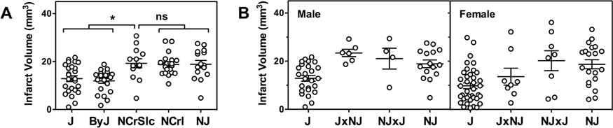
Conclusions
These results establish that the mutation(s) increasing stroke vulnerability occurred prior to the introduction of the N lineage to Japan. Stroke studies involving J- and N-lineage mice commonly available in Japan are therefore subject to the same potential confounds due to mismatched substrain backgrounds previously identified for mice of these lineages from other sources (1). This also identifies ByJ and NCrSlc as the most closely related of existing C57BL/6 substrains to show a difference in stroke outcome, further limiting the pool of candidate underlying genetic variants. Results for the F1 crosses provide evidence for a complex inheritance pattern, with a differential effect of parental genotype on the vulnerability of male and female offspring. How this extends to subsequent generations, and its implications for intrinsic stroke phenotypes of genetically modified mice derived through complex breeding strategies involving mixed substrain backgrounds, remain to be established.
References
PB03-K03
Balanced single-vector co-delivery of VEGF / PDGF-BB improves functional collateralization in chronic cerebral ischemia
1Department of Neurosurgery and Center for Stroke research Berlin, Charité – Universitätsmedizin Berlin, Berlin, Germany
2Department of Biomedicine, University Hospital Basel and University of Basel, Switzerland
3Global Medical Affairs, Bayer Pharma AG, Wuppertal, Germany
4Department of Neurosurgery, Faculty of Medicine, University of Tsukuba, Tsukuba, Ibaraki, Japan
Abstract
Objectives
Patients with chronic cerebral hemodynamic impairment due to progressive stenosis or occlusion of major cerebral arteries carry a risk of recurrent transient ischemic attacks and cerebral infarction. The myoblast-mediated delivery of angiogenic genes represents a cell-based approach for targeted induction of therapeutic collateralization. Here, we tested the superiority of myoblast-mediated co-delivery of vascular endothelial growth factor-A (VEGF) together with platelet-derived growth factor-BB (PDGF-BB) on transpial collateralization of an indirect encephalomyosynangiosis (EMS) in a model of chronic cerebral ischemia.
Methods
Mouse myoblasts expressing a reporter gene alone (empty vector; EV), VEGF164 (VEGF), hPDGF-BB (PDGF) or VEGF and PDGF-BB through a single bi-cistronic vector (VIP) were implanted into the temporal muscle of an EMS following permanent ipsilateral internal carotid artery occlusion in adult, male C57BL/6N mice. Over 84 days, myoblast engraftment and gene product expression, hemodynamic impairment, transpial collateralization, angiogenesis, pericyte recruitment and post-ischemic neuroprotection were assessed.
Results
On day 21, fusion of myoblasts with temporal muscle was observed in all experimental groups. In the temporal muscle, the exogenous gene product transcription and protein of VEGF164 and/or hPDGF-BB in VEGF-, PDGF- and VIP-myoblasts were confirmed on day 42, respectively. By day 42, animals received VIP showed significantly earliest and most pronounced cerebrovascular reserve capacity (CVRC) recovery in cerebral cortex after acetazolamide challenge. VIP-treated animals had a significantly higher number of positive spontaneous transpial collateralization in the brain-muscle interface, reduced cortical infarct volume and rescued neuronal cells in the ischemic cerebral cortex after middle cerebral artery occlusion. Vessel density with pericyte coverage in the cerebral cortex was significantly increased in the VIP-treated animals. X-gal staining, rtPCR and Western blot analysis confirmed positive reporter gene expression, gene product transcription and increased VEGF and/or PDGF-BB levels at the muscle / brain interface of the EMS on day 42. Also, supplementation of PDGF-BB resulted in a striking astrocytic activation with intrinsic VEGF mobilization in the cortex below the EMS. Next to increased astrocytic activation, a marked elevation of phospho-PDGF-Rb signal intensity was seen in the GFAP positive-reactive astrocytes in the cortical region below the EMS.
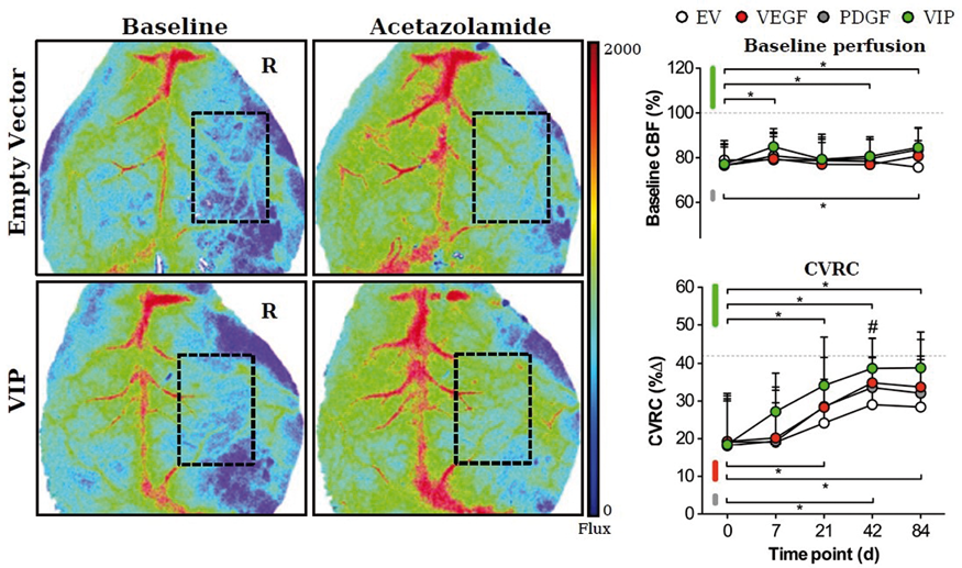
Conclusions
Treatment with VEGF164 and hPDGF-BB co-expressing VIP myoblasts not only resulted in improved hemodynamic rescue, transpial collateralization and ischemic protection, but also in a higher pro-angiogenic activity with significantly improved pericyte recruitment as a sign of vascular maturation. EMS surgery together with myoblast-mediated co-delivery of VEGF/PDGF-BB may have the potential to serve as a novel treatment strategy for augmentation of collateral flow in the chronically hypo-perfused brain.
Reference
PB03-K04
Potential role of HMGB1 in astrocytic TLR4 signaling following transient focal cerebral ischemia
1Dept of Neurology, University of Wisconsin School of Medicine and Public Health
2Dept. of Neurology, University of Maryland School of Medicine, Baltimore, MD
3University of Maryland School of Medicine, Dept. of Neurosurgery, Neurosurgery Research Laboratories, 10 S. Pine St, MSTF, Room 634B, Baltimore, MD, 21201–1595, US
4University of Maryland School of Medicine, Department of Neurosurgery, University of Maryland School of Medicine, Dept. of Neurosurgery, 22 S. Greene St., Suite S12D, Baltimore, MD 21201–1595, US
Abstract
Objective
Limited options are available for treatment of acute stroke and new approaches to treatment are needed. One such approach involves a better understanding of innate immune response to brain injury such as acute focal cerebral ischemia. This includes understanding the temporal profile, and specificity, of Toll-like receptor (TLR) 4 signaling in brain cell types, such as astrocytes, following focal cerebral ischemia and the endogenous ligands that may mediate such signaling.
Methods
We used the transient Middle Cerebral Artery occlusion (MCAO) model and Wistar rats to determine the presence of TLR4, and downstream mediator expression, in penumbral astrocytes, following acute and chronic focal cerebral ischemia. We also used the injection model, in which we stereotactically injected either High Mobility Group Box 1 (HMGB1); an endogenous TLR4 ligand or Phosphate Buffer Saline (PBS) into normal rat cortex, to determine the role of HMGB1 in cortical astrocyte TLR4 expression and signaling. Double immunolabeling analysis of brain sections, from all models, using specific antibodies, was used to determine astrocyte TLR4 expression and signaling. To corroborate these findings, cultured astrocytes, from the DI TNC 1 astrocyte cell line were stimulated with recombinant HMGB1 and RNA extracted, and reverse transcribed, for quantitative PCR to determine TLR4 and inducible nitric oxide synthase (iNOS) message levels. The one-way ANOVA or Student t test, was used to determine statistical differences between groups; p < 0.05.
Results
Expression of TLR4 and downstream mediators such as iNOS occurs in penumbral astrocytes in acute and chronic focal cerebral ischemia, but not in the cortical astrocytes in the control contralateral hemisphere. In the injection model, injection of recombinant HMGB1, and not PBS, leads to TLR4 and iNOS expression in perilesional astrocytes. Consistent with these results, In the in vitro model, stimulation of the DITNC 1 astrocyte cell line, with recombinant HMGB1, led to increased TLR4 and iNOS message levels compared to cells PBS-stimulated cells.
Conclusions
Both acute and chronic focal cerebral ischemia increase TLR4 expression in penumbral astrocytes. These findings suggest that HMGB1, an endogenous TLR4 ligand, is an important physiological ligand for TLR4 signaling, in penumbral astrocytes, following acute and chronic ischemia and HMGB1 amplifies TLR4 signaling in astrocytes.
PB03-K05
Loss of Wip1 aggravates brain injury after ischemia/reperfusion by overactivating microglia
1XuanWu Hospital, Capital Medical University, China
2Institute of Military Cognition and Brain Sciences, Academy of Military Medical Sciences, China
Abstract
Background and Purpose
Wild-type p53-induced protein phosphatase 1 (Wip1), belongs to PP2C family, is a serine/threonine phosphatases. Wip1 expressed in several tissues, especially in multiple cancers, and observed overexpression and played as an oncogene. A growing body of researches suggested that Wip1 played critical role in inflammation and regulated the inflammation of peripheral tissue negatively. It might contribute to brain injury by regulating central nervous system inflammation through microglia/macrophage functions after brain ischemia/reperfusion.
Methods
In the present study, we used focal brain ischemia/reperfusion model in Wip1 knockout(KO) mouse and wild-type mouse (WT), primary microglia cells was stimulated by LPS to identify its role in the neuroinflammation. Rescue with lentivirus-Ppm1d (genes that express Wip1) through Lateral cerebral ventricle injection and add to medium were controlled by vehicle virus. Behavioral outcomes were assessed by TreadScan system analysis gaits. We used western blot to detect the level of protein, immunofluorescence to mark the microglia which were classically activated (M1) and alternatively activated (M2), ELISA to detect the level of inflammation factors.
Results
Wip1 KO mouse displayed more severe brain injury and impaired motor outcome with increased brain infarct volum and higher expression of inflammatory cytokine in brain. Studies in microglia/macrophage activation after brain ischemia/reperfusion, we found that Wip1KO increased microglia/macrophage activation after brain ischemia/reperfusion and primary microglia cells were stimulated with LPS. Rescue with lentivirus-Ppm1d lateral cerebral ventricle injection and add to medium could reverse the aggravation of damage by Wip1 knockout.
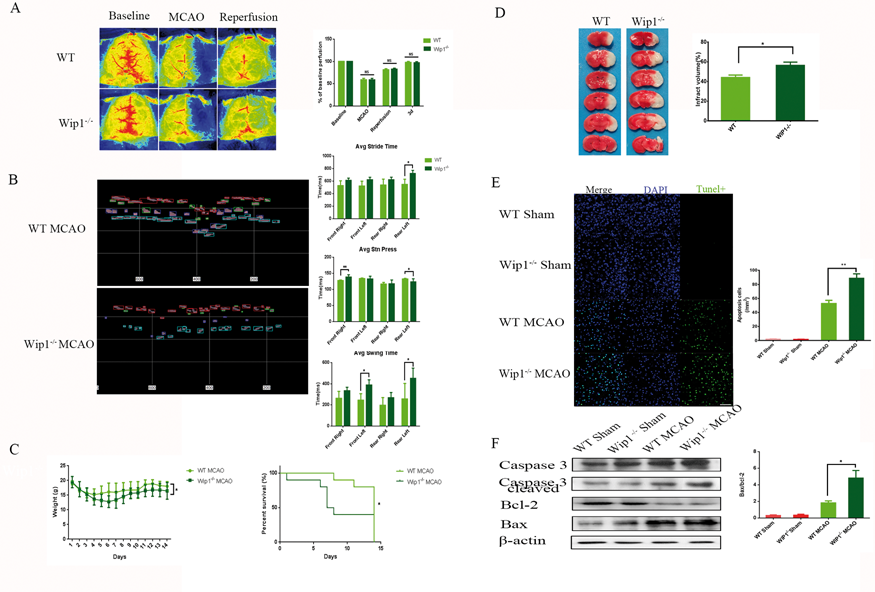
Conclusions
Thus, our results suggested that Wip1 as a negative regulator of neuroinflammation perhaps through inhibiting microglia/macrophage activation after brain ischemia/reperfusion injury process.
PB03-K06
TNF alpha is not the mediator of microglia-induced delayed neuronal death after cardiac arrest
1Anesthesiology and Perioperative Medicine, Oregon Health and Science University
Abstract
Survivors of cardiac arrest suffer from memory loss and cognitive dysfunction that greatly reduce quality of life. Much of this deficit is caused by neuronal loss in ischemia-vulnerable brain regions such as the hippocampus. We previously found that microglia mediate both neuronal death and memory deficit after cardiac rest (1). Others have shown that TNF alpha is a major contributor to microglia-mediated neuronal death in vitro (2). As microglial expression of TNF alpha increases after cardiac arrest (3), we hypothesized that TNF alpha released by activated microglia is a main driver of neuronal death and memory dysfunction.
Methods
We crossed TACEfloxmice and CX3CR1CreER mice expressing tamoxifen-inducible Cre recombinase. Offspring lack the TNF alpha converting enzyme TACE (aka ADAM17) in microglia and peripheral macrophages (CX3CR1CreER+/- – TACEflox+/+). These mice produce TNF alpha, but are unable to release it from their CX3CR1 positive microglia and macrophages (4). We induced Cre-recombination with a tamoxifen pulse 30 days before exposing mice to cardiac arrest and resuscitation. This delay allows for the short-lived peripheral macrophages to be completely turned over and replaced by TACE intact cells, while the low-turnover brain microglia continue to lack functional TACE. We used CX3CR1CreER-/- – TACEflox+/+ mice that do not undergo Cre recombination and have intact TACE as controls. Control mice received tamoxifen to exclude nonspecific drug effects. Mice were subjected to potassium chloride induced cardiac arrest. Cardiopulmonary resuscitation (CPR) was initiated after 10 minutes of cardiac arrest by injection of epinephrine and chest compressions at a rate of 300/minute. Neuronal death in the ischemia sensitive CA1 region was quantified after H&E staining 3 and 11 days after CPR. Memory function was tested using trace fear conditioning testing 10 days after CPR and neurogenesis in the dentate gyrus was assessed by counting doublecortin-positive newborn neurons.
Results
Neuronal death in the hippocampus was not different between groups. Mice lacking microglial TACE performed worse in fear conditioning testing and had fewer newborn neurons after cardiac arrest than control mice.

Conclusion
Mice who cannot release TNF alpha from their microglia are not protected from delayed neuronal death after cardiac arrest, suggesting that microglial TNF alpha is not a major driver of neuronal death. Surprisingly, memory deficit is exacerbated in mice with TACE deficient microglia, suggesting that microglial TNF alpha may be beneficial for functional recovery. This may be related to the significant suppression of post-ischemic neurogenesis we observed in TACE deficient mice. Future investigations are needed to define the role of microglia TNF alpha in neurogenesis and synaptic plasticity after cardiac arrest.
References
PB03-K07
Positive correlation between PDGFRβ-mediated tissue repair and neurorestoration in a permanent middle cerebral artery occlusion stroke model in mice
1Department of Medicine and Clinical Science, Graduate School of Medical Sciences, Kyushu University
Abstract
Background and Purpose
Platelet-derived growth factor receptor β (PDGFRβ) is expressed highly in pericytes and smooth muscle cells, and participates crucially in fibrotic response within infarct areas after ischemic stroke. However, how PDGFRβ-expressing cells promote tissue repair and whether the repair leads beneficially or detrimentally to neurorestoration remain largely elusive. We aimed to elucidate the mechanisms underlying the PDGFRβ-mediated tissue repair and neurorestoration after acute ischemic stroke.
Methods
Brain infarcts were induced by permanent occlusion of the right middle cerebral artery (pMCAO) using a laser-induced photochemical reaction in wild-type and PDGFRβ heterozygous knockout (PDGFRβ+/−) mice. We examined infarct volume, post-stroke arteriogenesis, blood flow recovery in ischemic areas, tissue repair, peri-infarct reorganization, and neurologic restoration.
Results
Infarct volume was comparable between wild-type and PDGFRβ+/-mice on day 1 after pMCAO; however, it was significantly larger in PDGFRβ+/-mice from day 7 to 28. Although cerebral blood flow, assessed by a 2-dimensional laser blood flow meter, was reduced equally on day 1 in both mice groups, its recovery was dependent on the extent of leptomeningeal collateral development, which was significantly suppressed in PDGFRβ+/-mice on day 7 compared to wild-type mice. PDGFRβ+/-mice showed less recruitment of α smooth muscle actin (αSMA)-positive cells around the collateral vessels. Furthermore, PDGFRβ+/-mice had a lesser extent of CD31-positive endothelial cells in ischemic areas followed by greater amounts of blood-brain barrier leakage than in wild-type mice. Quantitative polymerase chain reaction using brain homogenates of ischemic areas demonstrated that expressions of various genes related to angiogenesis, tissue repair, and neurorestoration were lower in PDGFRβ+/-mice than in wild-type mice. In peri-infarct areas, the number of mature oligodendrocytes assessed by adenomatous polyposis coli (APC) and glutation S transferase (GST)-π was significantly smaller in PDGFRβ+/-mice than in wild-type mice on day 28, while the number of olig2-expressing oligodendrocyte-precursor cells (OPC) was not significantly different between the groups, indicating that PDGFRβ appears to regulate oligodendrogenesis. Finally, neurologic functions assessed by Rota-rod, cylinder test, and modified neurological severity score (mNSS) on later than day 14 were restored worse in PDGFRβ+/-mice compared to wild-type mice.
Conclusions
PDGFRβ enhances post-stroke pial collateral development, resulting in the effective recovery of blood flow in ischemic areas after pMCAO. The PDGFRβ-mediated enhanced recovery of blood flow may contribute to better tissue repair and subsequent neurorestoration in per-infarct areas partly through the induction of oligodendrogenesis from OPC.
PB03-K08
Investigation of inflammasome expression in chronic cerebral hypoperfusion
1Department of Neurology, Mie University Graduate School of Medicine, Mie, Japan
2Department of Internal Medicine, Nanakuri Memorial Hospital, School of Medicine, Fujita Health University, Tsu, Mie, Japan
Abstract
Objectives
Inflammation plays an important role in cerebral ischemia. Recently, some reports indicate that inflammatory response triggered by tissue damage is mediated through a multiple-protein complex called inflammasome (Martinon et al., 2002). The AIM2 (absent in melanoma 2) and the NLRP3 (NOD-like receptor family, pyrin domain containing 3) inflammasome complex triggers caspase-1-mediated maturation of IL-1β and IL-18. This study was undertaken to test our hypothesis that inflammasomes may contribute to brain damage using bilateral common carotid artery stenosis (BCAS) model that is frequently used as an animal model of vascular cognitive impairment (VCI) worldwide (Shibata et al., 2004; Bink et al., 2013).
Methods
C57BL/6J mice were subjected to sham or BCAS operation as a chronic cerebral hypoperfusion using microcoils with an internal diameter of 0.18 mm. At 2, 4 weeks after BCAS, these mice were sacrificed and examined (n = 5 for each group). The coronal sections were stained with anti-NLRP3 and anti-AIM2 antibodies, and the number of NLRP3- or AIM2-positive cells were compared with IL-18 or IL-1β levels, which were determined by ELISA. In human autopsy brains, we examined patients with cerebral infarction (CI group, n = 4) and neurodegenerative diseases without cerebral infarction (Cont. group, n = 4). Brain sections were immunostained with anti-NLRP3 and anti-AIM2 antibodies in the white matter lesions.
Results
There was a numerical increase of NLRP3/GFAP- or AIM2/CD68-double positive cells in the corpus callosum from 2 and 4 weeks after BCAS (for NLRP3; P ≦ 0.05 at 2 weeks and P≦0.05 at 4weeks, for AIM2; P≦0.05 at 2 weeks). Similarly, IL-18 and IL-1β levels significantly increased at 2 and 4 weeks after the BCAS (IL-1β; P≦0.001 at 2 weeks and P≦0.001 at 4 weeks, IL-18; P≦0.01 at 2 weeks and P≦0.001 at 4 weeks). In the peri-infarct territory of the human brain, the immunoreactive area was larger in the CI group than in the Control group for NLRP3 (P≦0.05, mean ± SD; 0.96 ± 0.49 and 0.22 ± 0.09%, respectively) and AIM2 (P≦0.05, mean ± SD; 0.28 ± 0.24 and 0.05 ± 0.04%, respectively).
Conclusions
These results indicate that chronic cerebral hypoperfusion induces upregulation of NLRP3 and AIM2-inflammasomes, likely leading to a cytokine increase including IL-18 and IL-1β. Inflammasomes may play a pivotal role in neuroinflammation after chronic cerebral hypoperfusion.
References
PB03-K09
Molecular switching from ubiquitin-proteasome to autophagy pathways in mice stroke model
1Dept. of Neurology, Okayama University, Japan
Abstract
Objectives
The ubiquitin-proteasome system (UPS) and autophagy are two major pathways to degrade misfolded proteins that accumulate under pathological conditions. When UPS is overloaded, the degeneration pathway may switch to autophagy to remove excessive misfolded proteins. In the present study, we thus investigated the alterations of UPS and autophagy activities in relation to the key proteins after a transient middle cerebral artery occlusion (tMCAO) in mice, and examined whether there was crosstalk between these two pathways after cerebral ischemia.
Methods
Mice were subjected to tMCAO and the changes of UPS and autophagy were evaluated by immunohistochemical analysis, immunofluorescent histochemistry and western blot.
Results
In the present study, tMCAO resulted in accelerated ubiquitinpositive protein aggregation from 0.5 h of reperfusion in mice brain after 10, 30 or 60 min of tMCAO. In contrast, significant reduction of p62 and induction of LC3-II were observed, peaking at 24 h of reperfusion after 30 and 60 min tMCAO. Western blot analyses showed an increase of BAG3 and HDAC6 at 1 or 24 h of reperfusion that was dependent on the ischemic period. In contract, BAG1 decreased at 24 h of reperfusion after 10, 30 or 60 min of tMCAO after double immunofluorescent colocalization of ubiquitin, HSP70, p62 and BAG3.
Conclusions
These data suggest that a switch from UPS to autophagy occurred between 10 and 30 min of cerebral ischemia depending on the BAG1/BAG3 ratio and level of HDAC6.
PB03-L01
Role of brain-type fatty acid binding protein in ischemic neoronal injury and ischemia-induced neurogenesis after transient forebrain ischemia
1Dept. of Neurosurgery, University of Yamanashi, Japan
2Dept. of Organ Anatomy, Tohoku University
Abstract
Objectives
Fatty acid-binding proteins (FABPs) are intracellular low-molecular-weight (14–15 kDa) polypeptides and are key molecules in the uptake, transportation and storage of long-chain polyunsaturated fatty acids. Among various FABPs, brain-type FABP (FABP7) has been shown to be expressed in the adult rodent brain, especially in reactive astrocytes and neural stem/progenitor cells, and has a pivotal role against traumatic injury and regulates neurogenesis during the brain development; however, its roles on neuronal injury and neurogenesis after cerebral ischemia have not been elucidated. In this study, the expression of FABP7 after ischemia was studied, and the effects of genetic FABP7 inhibition on neuronal injury and neurogenesis after ischemia were investigated.
Methods
Male FABP7 knockout (KO) mice with a C57BL/6 J background and their wild-type (WT) littermates were subjected to transient forebrain ischemia for 20 minutes. Histological injury in the hippocampi 10 days after ischemia was evaluated by cresyl violet staining. The expression of FABP7 was investigated by immunohistochemistry and western blot. Bromodeoxyuridine immunohistochemistry was used for the study of proliferation and differentiation of progenitor cells.
Results
There was no difference in the level of neuronal injury between the WT and KO mice. FABP7 expression was observed in neural stem/progenitor cells and significantly increased 7 to 10 days after ischemia (n = 4, P < 0.05), which was consistent with the peak of hippocampal neurogenesis. In the KO mice, neurogenesis was significantly decreased compared with the WT mice under both physiological and ischemic conditions (n = 7, P < 0.05). The differentiation from newborn cells to immature neurons was more activated in the KO mice (n = 7, P < 0.05), but there was no difference in the number of cells differentiated to mature neurons between both mice.
Conclusion
The findings of this study imply that FABP7 expressed in neuronal stem/progenitor cells regulates the proliferation and maintenance of newborn cells after cerebral ischemia.
References
PB03-L02
The role of oligodendrocyte precursor cells in angiogenesis after brain ischemia
1Dept of Neurosurgery, Kyoto University, Japan
2Dept of neurology,Kyoto University
3Department of Neurosurgery, Graduate School of Medicine, Tokushima University
4Department of Developmental Neurobiology, KAN Research Institute, Inc
Abstract
Objectives
Oligodendrocyte precursor cells (OPCs) have been shown to play more diverse and crucial roles than previously recognized. OPCs regulate neuronal, glial and vascular systems in a direct and reciprocal fashion. However, the roles of reactive OPCs under ischemic conditions are largely unknown. Thus, our aim is to evaluate how OPCs contribute to neurovascular damage and repair after ischemic stroke.
Methods
The behavior of OPCs was evaluated in a mouse model of ischemic stroke produced by transient middle cerebral artery occlusion (MCAO) in vivo. For in vitro experiments, the phenotypic change of OPCs after oxygen glucose derivation (OGD) was examined using primary rat OPC culture. The function of hypoxic OPCs on vascular system was assessed by media transfer experiments from OPCs to endothelial cells in vitro. Furthermore, the therapeutic potential of hypoxic OPCs was evaluated in a mouse model of MCAO in vivo.
Results
We classified OPCs into 3 types according to their morphology and position in relation to cerebral endothelium; parenchymal OPCs, perivascular OPCs and intermediate OPCs. Perivascular OPCs in cerebral cortex were increased alongside post-stroke angiogenesis in a mouse model of MCAO. In vitro RNA-seq analysis revealed that primary cultured OPCs increased the gene expression of numerous pro-angiogenic factors after OGD. Hypoxic OPCs secreted a greater amount of pro-angiogenic factors, such as VEGF and angiopoietin-1, compared with normoxic OPCs. Hypoxic OPC-derived conditioned media (CM) increased the viability and tube formation of endothelial cells. In vivo studies also demonstrated that five consecutive daily treatment with hypoxic OPC-CM beginning on day 2 after MCAO facilitated post-stroke angiogenesis, alleviated infarct volume, and improved functional disabilities.
Conclusions
Following cerebral ischemia, the phenotype of OPCs in cerebral cortex shifts from parenchymal to perivascular subtype, which can promote angiogenesis. The optimal usage of the “reactive” OPCs after cerebral ischemia would provide a novel therapeutic option for stroke.
PB03-L03
Temporal profile of endogenous multilineage-differentiating stress enduring (Muse) cells in mouse middle cerebral artery (MCA) occlusion model
1Dept. of Neurosurgery, Toyama University, Japan
2Dept. of Neurosurgery, saisei kai toyama hospital
Abstract
Objective
Recent studies have indicated that SSEA-3+Muse cells are mobilized from the bone marrow to the peripheral blood and play a critical role to repair the damaged tissue. However, their temporal profile in acute stage of ischemic stroke is still obscure. This study was aimed to reveal their temporal profile in the peripheral blood, brain, spleen, and lung, using murine permanent middle cerebral artery occlusion (pMCAO) model.
Materials and Methods
Nine mice were subjected to pMCAO. Using FACS, the number of SSEA-3+cells in theperipheral blood mononuclear cells (PBMCs) fraction were quantified before, 7 days and 28 days after MCA occlusion. Histological analysis was performed to assess their distribution. As the control, the distribution of mesenchymal stromal cells (MSC, Sca-1+) in these organs were also evaluated.
Results
The fraction of SSEA-3+cells were 0.8 ± 0.3% before pMCAO, and significantly decreased to 0.6 ± 0.3% and 0.5 ± 0.1% at 7 and 28 days after pMCAO, respectively (P = 0.0146). Immunostaning revealed a significant correlation between the accumulation of SSEA-3+cells in the spleen and their decrease in peripheral blood (R2 = 0.74). SSEA-3+cells were not detected in the lung, but they were distributed around cerebral infarct and some of them expressed Tuj-1. On the other hand, the number of Sca-1+cells in the lung and spleen were significantly larger in the mice subjected to pMCAO than the controls (P < 0.01), but they were not detected in the brain. Average cell size of SSEA-3+and Sca-1+cells in the bone marrow were 5.3 ± 0.4 and 7.8 ± 0.3 μm, respectively (P < 0.01).
Conclusions
These findings strongly suggest that the consumption of Muse cells may exceed their mobilization from the bone marrow in acute stage of ischemic stroke. Muse cells, but not MSCs, can migrate toward the infarct brain, because the MSCs are trapped in the lung due to their bigger size.
PB03-L04
Effects of spreading depolarization on development of cerebral infarction after middle cerebral artery occlusion in Na+, K+-ATPase α2 subunit-deficient mice
1Dept. of Neurology, Keio University School of Medicine, Japan
2Div. of Biology, Center for Molecular Medicine, Jichi Medical School, Japan
3Brain Science Inspired Life Support Research Center, University of Electro-Communications, Japan
4Department of Functional Brain Imaging Research, National Institute of Radiological Sciences, Japan
5Dept. of Physiology, International University of Health and Welfare School of Medicine, Japan
6Dept. of Neurology, Shonan Keiiku Hospiral, Japan
Abstract
Background
The α2 subunit of Na+, K+-ATPase, mainly expressed in astrocytes in the adult brain, is encoded by the gene ATP1A2, and point mutation of ATP1A2 is a cause of familial hemiplegic migraine type 2. We previously reported enhanced susceptibility to cortical spreading depression (CSD) and prolonged recovery from CSD in Atp1a2 knockout mice (1). On the other hand, spreading depolarization (SD), a similar phenomenon to CSD, often occurs spontaneously and may exacerbate ischemic infarction. At the previous meeting, we presented the results of that the occurrence of SD during ischemia might influence the development of infarction.
Objectives
The aim of the present work was to evaluate the occurrence of SD and its effect on development of cerebral infarction after middle cerebral artery occlusion (MCAO) in Na+, K+-ATPase α2 subunit-deficient mice.
Methods
Heterozygotes of two types of Atp1a2-defective heterozygous mice, Atp1a2tm1Kwk (C-KO)(2) and Atp1a2tm2Kwk (N-KO)(3) were used and compared with their wild-type littermates (N = 51). The middle cerebral artery was exposed through a burr hole in the temporal bone under isoflurane anesthesia, and transiently occluded for 45 min by a modification of Tamura’s method(4). Cerebral blood flow (CBF) was continuously recorded over the ipsilateral parietal bone by laser speckle flowgraphy and occurrence of SD was evaluated in terms of spatiotemporal changes of CBF. Brains were excised 24 hr after the experiment and infarct volume was evaluated in the coronal plane with Nissl staining.
Results
CBF decreased by 35.5 ± 7.1% at the ischemic core region and increased by 104.3 ± 66.2% after reperfusion, and there was no significant difference between the two genotypes of deficient mice. SD spontaneously occurred zero to three times, but the occurrence and change of CBF during passage of SD were not significantly different between mice with the two genotypes. Cerebral infarction was not detected in mice without SD and showed a tendency to be larger in mice with a higher incidence of SD. The same trend was found in both genotypes of deficient mice, and no significant difference was found.

Conclusion
Two genotypes of Atp1a2-deficient heterozygous mice, which show high susceptibility to CSD, demonstrated comparable occurrence of SD and ischemic infarction. Na+, K+-ATPase activity might contribute little to the spontaneous occurrence of SD during ischemia and development of ischemic infarction.
References
PB03-L05
Non-spreading anoxic depolarization: A novel phenomenon?
1Department of Medical Physics and Informatics, Faculty of Medicine, Faculty of Science and Informatics, University of Szeged
Abstract
Objectives
Spreading depolarization (SD) has been recognized as the underlying mechanism of lesion development in acute brain injury, and a correlate of the aura phase of migraine. SD events and the associated CBF response display great variations in duration and magnitude, and have been accepted to define a spectrum or continuum.1,2 Here we set out to explore specific propagation properties of SD and the associated CBF response in the rodent cortex exposed to hypoxia/ischemia of various severity.
Methods
Spontaneous spreading depolarization occurred in response to forebrain ischemia (iSD), which was initiated by the bilateral occlusion of the common carotid arteries in isoflurane-anesthetized, old (18 months) male Sprague-Dawley rats (n = 16). Thirty minutes later, the complete withdrawal of O2 from the anesthetic gas mixture, maintained for 4–5 min (arterial blood pO2 = 35 ± 10 mmHg) gave rise to reversible anoxic depolarization (AD). The depolarization events and the associated cerebral blood flow (CBF) response were recorded either with two glass capillary microelectrodes and adjacent laser-Doppler probes positioned over the parietal cortex 3–4 mm apart (n = 15), or visualized with multi modal imaging involving a voltage-sensitive dye (RH-1838), and laser speckle contrast analysis (LASCA) (n = 1). Mean arterial blood pressure (MABP) was continuously monitored through a femoral artery catheter, which also served blood sampling for blood gas analysis. MABP and the CBF trace were used offline for the calculation of the cerebrovascular autoregulatory index (rCBFx).
Results
As expected, iSDs evolved as a propagating wave, characterized by a transient negative DC shift that occurred with a delay at the recording site more distant to the origin of the event, and as a wave of increased RH-1838 fluorescence intensity travelling across the field of view at a rate of 2.31 mm/min (Fig. 1). In contrast, AD, which was reversible on the restoration of oxygen supply, was detected at both electrodes synchronously, and emerged as a rapid increase of RH-1838 fluorescence intensity involving the entire field of view simultaneously. The CBF response to both iSD and AD was typically a transient drop of CBF (to 10.5 ± 3.0 and 8.4 ± 4.0%, iSD and AD, respectively). However, the CBF response was different for iSD and AD in that iSD was coupled by spreading ischemia independent of any variations in MABP (rCBFx remained below 0.3), whereas AD caused a non-spreading reduction of CBF tightly coupled to the drop of MABP at the same time (rCBFx = 0.56 ± 0.31).

Conclusions
The data suggest the existence of a yet un-identified type of depolarization event in hypoxic/ischemic brain injury, which can be classified as non-spreading anoxic depolarization. In addition, the CBF response to non-spreading AD is a passive drop of CBF following the concomitant reduction of MABP, rather than spreading ischemia thought to be brought about by high concentration potassium. The place of non-spreading AD on the continuum of spreading mass depolarization1,2 needs to be carefully considered.
References
PB03-L06
Therapeutic potential of valproic acid against cerebral ischemia: Beyond epileptic disorders
1Department of Neurology, Graduate School of Medicine, Nippon Medical School
2Department of Emergency and Critical Care Medicine, Graduate School of Medicine, Nippon Medical School
3Department of Biochemistry and Molecular Biology, Graduate School of Medicine, Nippon Medical School
Abstract
Objective
Valproic acid (VPA), widely used in clinical contexts for the treatment of epilepsy, migraine, and bipolar disorder, has neuroprotective properties in cellular and animal models. However, the precise mechanisms underlying its neuroprotection against cerebral ischemia remain unknown.
Methods
First, we used a rat transient focal ischemia model to examine whether post-insult and single injection treatment with VPA (300 mg/kg) has neuroprotective effects, if so, to explore the therapeutic time window. To simulate a typical human stroke, the Stroke Therapy Academia Industry Roundtable (STAIR) Committee endorses the testing of therapeutics in models of permanent ischemia and in animals with comorbidities such as diabetes and hypertension. Next, we used a permanent ischemia model in streptozotocin-induced hyperglycemic rats to VPA ameliorates neurological symptoms and to examine the potential mechanisms of any protective effects seen.
Results
In the first experiment, VPA injected immediately or 90 min but not 270 min after ischemia induction significantly reduced infarct volume and improved neurological deficit compared with vehicle (P < 0.05). VPA significantly reduced TUNEL-positive cells, myeloperoxidase (MPO)-positive cells, ionized calcium binding adapter molecule 1(Iba-1)-positive cells, 4-hydroxy-2-nonenal-positive cells, and 8-hydroxy-deoxyguanosine-positive cells compared with vehicle in the ischemic boundary zone (P < 0.05). In the second experiment, VPA significantly alleviated the aggravation of neurological deficits and rotarod test scores accompanied by suppressing MPO positive cells, Iba-1-positive cells, von Willebrand factor-positive cells, and Fluoro-Jade C-positive cells compared with vehicle in the ischemic boundary zone (P < 0.05).
Conclusions
VPA ameliorated neurological deficits by modulating the inflammatory response, suppressing oxidative stress, and protecting against endothelial damage, leading to reduced neuronal cell degeneration following cerebral ischemia. Although, further studies determining the appropriate VPA doses and/or administration frequencies will be needed for clinical application, it might be promising as a candidate therapy for human stroke.
PB03-L07
Effects of edaravone on NO production, OH- metabolism and nNOS activity during cerebral ischemia and reperfusion
1Dept. of Neurology, Saitama medical University, Japan
Abstract
Objective
Edaravone, which acts as a free radical scavenger, has been used clinically in Japan for the treatment of cerebral infarction since 2001. Various studies have reported on the neuroprotective effect of Edaravone. However, the effects of Edaravone on nitric oxide (NO) production and hydroxyl radical metabolism during cerebral ischemia and reperfusion in vivo have not been thoroughly investigated. The purpose of our study is to investigate the effectts of edaravone on the nitric oxide (NO) production, hydroxyl radical (OH-) metabolism and nNOS activity during cerebral ischemia and reperfusion.
Methods
C57BL/6 mice [n = 13] were used. Animals were initially anaesthetized with 2% halothane in air supplemented with O2 and maintained with 0.5%–1% halothane. Rectal temperature was maintained at 37.0–37.5°C with a disposable heat pack and small fan. A polyethylene catheter was inserted into the right femoral artery to measure blood pressure. Edaravone 3 mg/ kg was given by intravenous injection in 8 mice just before reperfusion, and others [n = 5] were uesd for contol group. NO production and hydroxyl radical metabolism were monitored continuously using in vivo microdialysis. A microdialysis probe was inserted into the striatum in each hemisphere and perfused with Ringer’s solution at a constant rate of 2 µl/min. The in vivo salicylate trapping method was used to monitor hydroxyl radical formation via 2,3- and 2,5-dihydroxybenzoic acid (DHBA). A laser Doppler probe was placed on the surface of the skull of the right hemisphere. After 2 hours of equilibration, fractions were collected every 10 minutes. Forebrain cerebral ischemia was produced by occlusion of both common carotid arteries for 10 minutes. Levels of NO metabolites, nitrite (NO2-) and nitrate (NO3-), in the dialysate were determined using the Griess reaction. Brain sections were immunostained with an anti-nNOS antibody. To determine the fractional area density of nNOS-immunoreactive pixels to total pixels of the whole field, the captured images were analyzed. Mann-Whitney U test was used for group comparisons.
Results
Blood pressure and cerebral blood flow: There were no significant differences between the groups. NO2−: Edaravone group (96.1 ± 37.3%; mean ± SD) showed significantly lower values than that of the control group (127.1 ± 17.2) after repurfusion (p < 0.05). NO3-: Edaravone group (173.8 ± 29.2%) showed significantly higher levels than that of the control group (130.4 ± 16.0) after repurfusion (p < 0.05) (Figure). 2,3-DHBA: Edaravone group (95.0 ± 4.8%) showed significantly lower values than that of the control group (108.7 ± 7.2) after repurfusion (p < 0.01). nNOS activity: Edaravone group (1.2 ± 0.2%) was significantly lower than that of the control group (1.7 ± 0.3) during ischemia (p < 0.01).

Conclusion
Our data suggest that edaravone effects not only on NO and OH- metabolites but also nNOS activity during cerebral ischemia and reperfusion.
PB03-L08
Posterior cerebellar strokes in mice cause memory and hippocampal synaptic plasticity impairments
1Department of Anesthesiology, University of Colorado, Anschutz Medical Campus
2Pediatrics, Division of Neonatology, Children's Hospital Colorado
Abstract
Objective
Each year in the U.S. there are an estimated 20,000 strokes resulting in cerebellar infarction. The neurological impairments observed in these patients include motor coordination and motor learning impairments. Surprisingly, cerebellar stroke patients also exhibit cognitive and affective symptoms. Importantly, infarcts to anterior cerebellum are more likely to result in motor deficits, while infarcts in posterior cerebellum are associated with cognitive and affective symptoms. Establishing a model of cerebellar stroke that recapitulates aspects of human cerebellar infarction will allow us to perform mechanistic studies of network perturbations that contribute to motor and cognitive deficits. The goal of this study was to develop and characterize a photo-thrombotic mouse model of focal cerebellar stroke.
Methods
Adult male and female mice were head-fixed in a stereotaxic frame, administered Rose Bengal (150 ug/g) and the superior cerebellar artery (SCA) was illuminated for 15 minutes with a cold white LED light source to produce an anterior or posterior cerebellar infarct. Histological analysis of ischemic infarct, blood-brain barrier permeability and gliosis were performed at 1 and 7 days after stroke induction. Behavioral testing of motor and memory function was performed in mice subjected to sham procedure or photo-thrombosis at 7 days following surgery. We also performed extracellular field recordings at 7 days post-injury in the CA1 region of the hippocampus to assess long-term potentiation (LTP).
Results
Cerebellar stroke volumes were larger at 1 day than 7 days, likely do to edema at the earlier time point. Lesion volumes were similar between males and females at both time points. We observed blood-brain barrier barrier leakage of Evan’s blue and ovalbumin-Alexa647 within the infarcted area at 1 day after stroke onset. We also observed increases glial fibrillary acid protein in the infarct and peri-infarct region at 7 days after stroke onset. Mice with anterior cerebellar strokes displayed motor incoordination on a balance beam task while we failed to detect deficits in mice with posterior cerebellar strokes. Conversely, memory deficits in the contextual fear-conditioning task were observed only in mice with posterior strokes. Memory deficits correlate with contralateral hippocampal long-term plasticity impairments at 7 days after stroke in the posterior but not anterior cerebellar stroke mice.
Conclusions
Photothrombosis of the SCA results in histological hallmarks of ischemic stroke observed in other brain regions. Importantly, we now have a reproducible model of location-specific deficits seen in the cerebellar stroke population, with anterior strokes causing motor deficits and posterior strokes causing cognitive deficits. Our observation that posterior cerebellar strokes cause synaptic plasticity impairments in the hippocampus provide a molecular mechanism that we can target to improve memory function after stroke that is independent of acute neuroprotection. This novel model will be a valuable tool for understanding how cerebellar infarction alters functional connectivity with other brain regions to produce motor and non-motor deficits.
PB03-L09
Assessing functional deficits in the mild mouse dMCAO model
1School of Life Sciences, University of Nottingham
Abstract
Objectives
Despite the advantages of low mortality and reproducible cortical lesions, functional deficits are generally mild and difficult to detect in the distal middle cerebral artery occlusion (dMCAO) model. This study evaluated a battery of sensorimotor behavioural tests for use with dMCAO in combination with ipsilateral carotid artery occlusion (CAO) to try and characterize subtle deficits. We also employed novel tests, including lickometry1 and high speed whisker tracking2.
Methods
Permanent dMCAO + CAO was performed on 33 male C57BL/6 J mice (sham, n = 15, dMCAO, n = 18) via electrocoagulation of the MCA and ligation of the carotid artery (CA). Between 24 hrs-8wks post-surgery mice underwent a series of commonly employed behavioural tests: the mouse grimace score, a neurological score, adhesive removal, corner test, cylinder test, grip strength, disengage test, and staircase reaching. Two additional, but less-well characterized approaches were also employed: lickometry and whisker tracking. Infarct volume was quantified using immunohistochemistry (NeuN).
Results
Lesion size at 24 hrs was 15.41 +/- 2.99 (% hemisphere) and 5.38 +/- 1.035 at 8wks. Mild, but not significant deficits were observed in removal time (sec) in adhesive removal, and % success in the staircase task. A significant reduction in consumption per 1000 licks (g) was seen at one month using lickometry, though no differences were observed from comprehensive assessment of whisker abilities (Figure 1).

Conclusions
Consistent with other reports, despite the improved welfare benefits associated with this model, lesions and functional deficits were mild. We recommend the use of more sensitive assessments (adhesive removal and staircase paw-reaching) in comparison to neurological score. Lickometry was sensitive enough to detect long-term stable deficits at 4 weeks in this model and future investigations can expand upon whisker tracking.
Figure legend. (A) A timeline of the experimental design. (B) Time to remove stimuli in the adhesive removal test. (C) overall consumption of water across 1000 licks via lickometry chambers for both sham (black squares and dMCAO + CAO groups (red circles).
PB03-L10
Crossed cerebellar diaschisis following transient middle cerebral artery occlusion in rats
1Dept. Neurological Surgery, Okayama University Graduate School of Medicine, Dentistry and Pharmaceutical Sciences, Japan
Abstract
Objectives
Hypoperfusion and hypometabolism in the contralateral cerebellar hemisphere are caused by supratentorial lesion, such as stroke. This phenomenon is defined as crossed cerebellar diaschisis (CCD) and mainly detected by nuclear medicine study in human, but its pathology is not fully understood. In this study we evaluated cerebral blood flow (CBF) and histology of cerebellum using a rat model of focal ischemic stroke.
Methods
Male Wister rats underwent 90 minute right middle cerebral artery occlusion (MCAO) followed by reperfusion. CBF was evaluated by SPECT/CT using 123I-IMP 3 and 7 days after MCAO. Volumes of interest were set in each side of cerebral and cerebellar hemispheres respectively, and the ratio between right and left hemispheres was calculated. Histological evaluation has been performed using Nissl staining and TUNEL method.
Results
123I-IMP SPECT evaluation showed marked hypoperfusion in the right MCA territory on day3 (R/L = 0.36). This laterality slightly recovered on day7 (R/L = 0.62). There was also significant hypointensity in the left cerebellar hemisphere, contralateral to the supratentorial infarct (day3: L/R = 0.87, day7: L/R = 0.93). A significant reduction in cell density of left lateral cerebellar nucleus was noted in cerebellar sections. TUNEL assay revealed that the apoptosis in the left cerebellar cortex were induced after MCAO.
Conclusions
We detected CCD in rat stroke models using SPECT. Histological change was observed in the contralateral cerebellar hemisphere after supratentorial injury.
PB03-L11
Local cooling inhibits the expression of necroptosis-related protein after transient spinal cord ischemia in rabbits
1Department of Cardiovascular Surgery, Kyushu University Graduate School of Medicine
2Department of Clinical Research, National Hospital Organization Sendai Medical Center
3Department of Neurology, Okayama University Graduate School of Medicine
Abstract
Objective
As a potential mechanism of delayed paraplegia, we investigated the role of necroptosis, which is a type of regulated necrosis, in normothermic and hypothermic spinal cord ischemia, by immunohistochemical analysis of the necroptosis-related proteins receptor interacting protein kinase (RIP) 1, RIP3, and cellular inhibitor of apoptosis protein (cIAP) 1/2.
Methods
We used rabbit normothermic and hypothermic transient spinal cord ischemia models. Neurological function was assessed according to a modified Tarlov score at 8 hours, 1, 2, and 7 days after reperfusion. Morphological changes in the spinal cord were examined using hematoxylin and eosin staining. Western blot and histochemical analyses of RIP1, RIP3, and cIAP1/2, and double-label fluorescent immunocytochemical studies were performed.
Results
There were significant differences in physiological function between the normothermic and hypothermic groups (P < .001). In the normothermic group, most of the motor neurons were selectively lost at 7 days after reperfusion (P < .001 compared with the sham group), and they were preserved in the hypothermic group. Western blot analysis revealed the up-regulation of RIP1, RIP3 and cIAP1/2 at 8 hours in the normothermic group (RIP1,P = .032; RIP3, P < .001; cIAP1/2, P = .041 compared with sham), and the expression of RIP3 was prolonged until 2 days. In the hypothermic group, the expression of these proteins was not apparent. The double-label fluorescent immunocytochemical study revealed the induction of RIP3 and cIAP1/2 in the same motor neurons.
Conclusions
These data suggest that transient normothermic ischemia induced necroptosis, a potential factor in delayed motor neuron death, and hypothermia may inhibit necroptosis.
PB03-M01
Single cell transcriptome profiling of brain mononuclear cells after ischemic stroke in type 2 diabetic mice
1Dept. of Neurosurgery, UCSF, San Francisco, USA
2Dept. of Neurosurgery, SFVAMC, San Francisco, USA
3Dept. of Neurosurgery, Tohoku University Graduate School of Medicine, Sendai, Japan
4Bakar Computational Health Sciences Institute, UCSF, San Francisco, USA
Abstract
Objectives
Type 2 diabetes mellitus (T2DM) is a major risk factor of ischemic stroke and is associated with poor outcome after stroke, yet the mechanisms underlying the detrimental effects of diabetes in stroke remain largely unclear. Our previous work suggests that T2DM expanded subsets of myeloid cells in the blood, which may potentiate a more severe inflammatory response in the brain upon ischemic injury. We thus determined the transcriptome profiles in each single cell and associated molecular networks of brain mononuclear cells (BMNs) under diabetic and ischemic conditions using a mouse T2DM model, db/db.
Methods
We performed single-cell RNA sequencing (scRNAseq) in Percoll gradient-isolated BMNs from middle-aged db/db and control mice db/+ with or without stroke (N = 3 each) three days after distal middle cerebral artery occlusion (dMCAO) or sham-operation. We first determined the cell types of BMNsaccording to gene expression patterns in reference databases such as Immgen and mouseRNAseq, followed by comparing the transcriptome among the 4 groups of mice. We also assessed the biological roleof differentially expressed (DE) genes between groups by pathway enrichment analysis.
Results
Unsupervised Seurat-based clustering revealed a total of 20 clusters of cells among ∼22,000 cells from 4 experimental groups, primarily consisting of myeloid, endothelial and ependymal cells. Minor populations of fibroblasts, oligodendrocytes and neuroprogenitor cells were also captured in the analysis. Among the myeloid cells, macrophages of the MFIO5.II-480INT phenotype were greatly enriched in db/db sham BMNsand in stroke samples of either genotype. db/+ sham BMNshad the least number of myeloid cells and greatest number of endothelial and ependymal cells. Unlike the myeloid cells in the blood, fewer brain myeloid cells expressed Ly6C2. Stroke and T2DM shared a large number of up- or down-regulated genes among the myeloid cells including most highly induced genes Spp1, Rbm3, Ctsb and CD63, and most highly down regulated genes Meg3, Enpp2 and Snhg11. Wfdc17 was upregulated in db/db sham myeloid cells compared to db/+ sham myeloid cells; however, it was downregulated after stroke in the db/+ myeloid cells. Interestingly, some of the myeloid DE genes were also detected in the endothelial cells in a similar fashion. Pathway enrichment analysis revealed that stroke or T2DM activated pathways involved in lysosome activity, apoptosis, chemotaxis and inflammatory responses, while they tended to suppress pathways in antigen presentation or adaptive immune responses.
Conclusions
The present studydemonstrated altered gene expression profile and molecular networks across various cell types in BMNsin response to diabeticand ischemic conditions in single cell, providing a clue to the underlying mechanism of the exacerbating effect of T2DM onnative immunity and response to stroke.
PB03-M02
Altered clock expression with aging and after ischemic stroke in female mice
1Dept Pharmacology, Medical Coll., Ewha Womans Univ., Seoul, Korea
2Thomas Jefferson High School for Science and Technology, USA
Abstract
Objectives
Circadian rhythm with a 24-h period is important for biological homeostasis, and therefore disruption of circadian rhythm is associated with numerous diseases such as metabolic disorder, cancers and neurodegenerative diseases. At the molecular level, circadian rhythm is regulated by a set of clock genes including CLOCK. CLOCK is involved in ovulation and regulation of estrogen receptor expression, but the role of CLOCK in the brain of female is largely unknown. This study was aim to define whether CLOCK expression is altered by aging and ischemic stroke, an age-related diseases, in female mice.
Methods
C57BL6 female young (2-3 months) and aged (20 months) mice were used. The tissues of the brain, liver and visceral fat were collected day and night at 12 h interval, and CLOCK protein levels were examined. Ischemic stroke was induced by a transient middle cerebral artery occlusion for 30 min, and CLOCK protein levels were determined at 12 h interval for 3 days after reperfusion.
Results
In young mice, CLOCK expression in the brain was increased at night time compared with that at day time. However, the rhythmic expression of CLOCK was not observed in the ipsilateral and contralateral brain after ischemic stroke. In aged mice, CLOCK level was significantly increased in the normal brain at both day and night time compared with those in young mice. After ischemic insult, CLOCK level was further enhanced in the ipsilateral brain and visceral fat tissues at night time until the 2 nd day after reperfusion.
Conclusions
The data suggest that CLOCK expression changes with aging, and ischemic insult alters the pattern of CLOCK expression in the brain and peripheral tissues. It should be further determined whether disruption of diurnal expression of CLOCK is related with the susceptibility to ischemic brain damage in estrogen-deficient elderly women.
References
PB03-M03
Focal ischemia alters the abundance of the epitranscriptomic mark m6A on mRNAs in mouse brain
1Dept. of Neurological Surgery, Univ. of Wisconsin
2Cell and Molecular Pathology Graduate Program
3William S. Middleton VA Hospital
Abstract
DNA and histones undergo many epigenetic modifications that are known to modulate post-stroke brain damage. Recent studies showed that RNAs can also undergo >100 types of chemical modifications that are known as epitranscriptomic changes which are thought finely control protein expression together with noncoding RNAs. Methylation of the adenosine at N6 position (m6A) at the stop codon, 3’UTR and long exon regions is the most abundant modification that mRNAs undergo in the adult brain. This activity-dependent modification is known to play a significant role in synaptic plasticity and cognition. Dysregulated m6A of specific RNAs is also known to precipitate many CNS-related pathologies including brain tumor growth, stress response and addiction. Using a mouse transient middle cerebral artery occlusion (MCAO) model of focal ischemia, we analyzed the m6A alterations as a function of reperfusion time in the peri-infarct cortex that can be salvaged with a therapy. In adult male mice, global m6A levels increased significantly at 12 h and 24 h reperfusion following transient MCAO compared to sham control (by 3.3 to 4 fold; p < 0.05; n = 5/group). The m6A RNA methylation is controlled by a highly-specific set of readers that methylate (METTL3, METTL14 and WTAP) and demethylate (ALKHB5 and FTO). Of these, only FTO mRNA and protein expression as well as its demethylase activity down-regulated significantly at 12 h and 24 h reperfusion (by 50 to 60%; p < 0.05; n = 5/group) following transient MCAO compared with sham. FTO is known to be abundantly expressed in neurons. FTO immunostaining in the peri-infarct neurons decreased significantly at 12 h reperfusion following transient MCAO compared to sham. These changes were not sex-specific as m6A levels were also increased at 12 h reperfusion following transient MCAO compared to sham in in adult female mice (by 3-fold; p < 0.05). We conducted microarray analysis of m6A modified transcriptome following immunoprecipitation with a specific m6A antibody in a cohort of adult, male mice subjected to transient MCAO and 12 h reperfusion or sham surgery. Microarray profiling showed increased m6A methylation in 126 mRNAs (>5-fold; p < 0.05; n = 5/group) in the peri-infarct cortex following stroke compared to sham. Overall, our studies show that decreased demethylation activity due to down-regulation of FTO leads to increased m6A levels in a subset of mRNAs in mouse brain following focal ischemia. This is a first study that shows that stroke influences epitranscriptomic changes in RNAs that might has a role in secondary brain damage together with epigenetic factors and noncoding RNAs.
PB03-M04
Caveolin-1 role in neovascularization and astrogliosis after stroke and effects of cavtratin as a neuroprotectant
1Dept. of Clinical Neuroscience, CHUV, Lausanne, Switzerland
2Brain Molecular Imaging lab, CNRS UMR 5287, INCIA, University of Bordeaux, France
3Basic Science Department, Loma Linda University School of Medicine, Loma Linda, CA, USA
Abstract
Background
Complex cellular and molecular events occur in the neurovascular unit after stroke and contribute to neuronal death, neurological deterioration and mortality. Caveolin-1 (Cav-1) is present in brain endothelial cells and astrocyte cultures. Cav-1 has a dual mode of action. On one hand, Cav-1 is associated with caveolae formation involved in endocytosis and transcytosis. On the other hand, Cav-1 can act through a scaffolding domain to modulate signaling pathways1. Therefore, Cav-1 is likely to be an important player in the context of NVU dysfunction. However, its role after stroke is still controversial and the effect of Cavtratin, a cell-permeable peptide containing the scaffolding domain of Cav-1 has never been investigated2.
Objectives
The goal of the study was to investigate the role of Cav-1 during the first week after stroke and evaluate the potential neuroprotective effect of the Cavtratin peptide.
Methods
We first compared wild type (WT) and genetically modified Cav-1 knock-out (KO) mice in an ischemia-reperfusion model using transient Middle Cerebral Artery Occlusion (tMCAO). Outcome measures including lesion volume, behavioral tests, and immunofluorescence staining were collected at various time-points and up to 7 days after injury. We then performed blinded and randomized IP injections of Cavtratin or control scrambled peptide (dose 2.5 mg/kg) 3 hours after the reperfusion and evaluated the same outcome measures as above.
Results
After tMCAO, Cav-1 expression was increased in new blood vessels within the lesion and we showed for the first time its presence in reactive astrocytes in the peri-lesion. Cav-1 KO mice exhibited a more severe post-stroke outcome with larger lesions and worse behavioural scores than WT mice in all tests. Cav-1 KO mice exhibited reduced neovascularization and modified astrogliosis compared to WT mice 3 days post injury associated with the aggravated functional deficits. Preliminary results of the outcomes after Cavtratin or control peptide injection show that mice injected with Cavtratin perform better in behavioral tests and display some features of neuroprotection.
Conclusions
Altogether, these results point towards a potential protective role of endogenous Cav-1 in the first days after ischemia by promoting both neovascularization and astrogliosis3, and single injection of Cavtratin may facilitate the recovery post-stroke.
References
PB03-M05
Do death and subsequent contraction of brain pericytes contribute to “no-reflow”? A study on rat brain pericytes in primary culture
1Dept of Physiology, Faculty of Medicine, Kuwait University
Abstract
When the middle cerebral artery (MCA) in mouse was reopened after 2 h occlusion, brain pericytes remain contracted[1]. Another in vivo study shown that brain pericytes died quickly and remained contracted (rigor mortis). These findings led to a hypothesis that pericytes' prolonged contraction after stroke constricts capillaries, causing the “no-reflow”[2]. These findings were later challenged by an in vivo study, which found that brain pericytes did not possess a complete contractile apparatus[3]. To clarify these conflicting data, we explored effects of oxygen glucose deprivation (OGD) on viability of cultured pericytes, in the presence or absence of various cytokines, and on morphological parameters that indicate contraction of their processes.
Methods
Cell viability and apoptosis were assessed in purified cultures of brain pericytes from Sprague-Dawley rats [4], which were subjected either to OGD protocols or to control conditions (5%CO2 in air, 5 mM glucose). In some cases, cytokines that are released from the neurovascular unit (NVU) during hypoxia were added to the medium before OGD protocols. To assess whether OGD protocols caused contraction of pericytes, cell images were acquired every 5 min for a period of 15 h before and after OGD protocols. Stacks of images of randomly selected 11–22 cells for every experimental condition were analyzed using a software that estimated single cell membrane mobility (SCMM), area/perimeter ratio (APR) and fractal dimension (FD). Two-way ANOVA was used to analyze effects of OGD on viability of the cells. All data were normally distributed, as confirmed by Shapiro-Wilk's test. Paired and unpaired t-tests were used to determine significance of differences in SCMM, APR and FD.
Results
Cell viability was marginally and significantly reduced (p < 0.05, Figure 1) and the number of apoptotic cells has increased (p < 0.05) after 2 h and 6 h OGD protocols. Large and significant (p < 0.001) reduction in the cell viability, which was accopanied by a large increase in the number of apoptotic cells was observed after 24 h OGD protocol. Addition of NVU-derived cytokines significantly affected cell viabilities during OGD protocols. Rat recombinant erythropoietin (rrEPO) alone or together with vascular endothelial growth factor (VEGF) significantly worsened detrimental effects of 24 h OGD on cell viability. However, when three main cytokines (EPO, VEGF and Angiopoietin 1) were added together to the medium, they exerted a significant protective effects during 24 h OGD (p < 0.001 vs. 24 h OGD without cytokine addition, Figure 1); cell viability under these conditions exceeded 70%.

We could not find evidences that pericytes remained contracted for a long period of time after OGD protocols. Twenty minutes and 1 h OGD protocols caused a significant reduction in SCMM (p < 0.05 and p < 0.01 vs. pre-OGD values, respectively). There were a significant increases of APR after 20 min and FD after 3 h OGD protocols and a significant decrease in APR after 3 h OGD protocol.
Conclusions
We could not find evidences that pericytes die rapidly during OGD protocols, especially when NVU-derived cytokines were added to ther medium. Also, pericytes contracted after 20 min and 1 h OGD protocols, but they did not remain contracted for extended time periods.
References
PB03-M06
Roles of lncRNA-1810034E14Rik in modulating functions of microglial cells after ischemic stroke and the underlying mechanisms
1Department of Neurology, Drum Tower Hospital of Nanjing University Medical School, PR China
Abstract
Objective
Neuro-inflammation induced by ischemic stroke directly affects prognosis of patients[1]. Activation status and function of microglia regulate neuro-inflammation and affect the outcome of ischemic stroke [2]. It has been reported that a variety of long non-coding RNA (lncRNA) differentially expressed in microglia cells after ischemic stroke. These lncRNAs may be involved in regulating the function of microglia, but the specific mechanism has not been clarified [3]. LncRNA-1810034E14Rik is widely distributed in microglia, but the function of LncRNA-1810034E14Rik is still unknown. In this study, we discussed the change of lncRNA-1810034E14Rik after ischemic stroke, and whether lncRNA-1810034E14Rik can regulate microglia function after ischemic stroke. In addition, we further explored the molecular mechanism of lncRNA-1810034E14Rik regulating microglia function.
Methods
In vitro, primary microglia were extracted from the cortex of C57BL/6 suckling mice, and RNA was extracted after OGD for microarray analysis of lncRNAs. Microglia were transfected with a lentivirus that overexpress lncRNA-1810034E14Rik 24 hours before OGD. The secretion of inflammatory factors was detected by ELISA, the activation of microglia was observed by immunofluorescence, the migration ability of microglia was detected by scratch test, and the phagocytic function was detected by fluorescent microspheres. At the same time, microglia and neurons were co-cultured in vitro. CCK-8 and LDH release analysis were used to detect the survival status of neurons. In vivo, we established a model of middle cerebral artery occlusion (MCAO) in C57BL/6 mice. A lentivirus that overexpress lncRNA-1810034E14Rik was injected into the cortex of C57BL/6 mice. MCAO was performed two weeks later. Immunofluorescence was taken to detect the activation of microglia in ischemic penumbra and ELISA was used to detect the infiltration of inflammatory factors in infarcted cortex. Western blotting was used to detect the NF-kappa B signaling pathway in vivo and in vitro.
Results
1) The expression of lncRNA-1810034E14Rik in microglia after OGD was significantly down-regulated (>5 fold change, p < 0.05).
2) Overexpression of lncRNA-1810034E14Rik significantly reduced the activation of microglia and down-regulated the level of inflammatory cytokine.
3) Overexpression of lncRNA-1810034E14Rik had no significant effect on migration and phagocytosis of microglia.
4) Overexpression of lncRNA-1810034E14Rik could reduce the neurotoxicity of microglia after OGD.
5) Overexpression of lncRNA-1810034E14Rik in cortex could improve motor function damage, reduce brain edema and reduce infarcted volume in MCAO mice.
6) The NF-kappa B pathway was activated in the infracted cortex and OGD-induced microglia. Overexpression of lncRNA-1810034E14Rik significantly inhibited the phosphorylation of p65.
Conclusions
1) LncRNA-1810034E14Rik could reduce infarcted volume and improve neurological function after ischemic stroke; LncRNA-1810034E14Rik could significantly reduce neuro-inflammation after ischemic stroke.
2) LncRNA-1810034E14Rik could reduce the activation of microglia by inhibiting the NF-kappa B signaling pathway.
References
PB03-M07
Differential susceptibility of human neural progenitors and mature neurons to ischaemic injury
1School of Medicine, University of Tasmania, Hobart, Tasmania, Australia
2Florey Institute of Neuroscience and Mental Health, Victoria, Australia
3Department of Neuroscience, Central Clinical School, Monash University, The Alfred Centre, Victoria, Australia
4Illawarra Health and Medical Research Institute Centre for Molecular and Medical Bioscience, University of Wollongong, New South Wales, Australia
5Stem Cell Core Facility, Stem Cells Australia, The University of Melbourne, Victoria, Australia
Abstract
Background
Translational neuroscience presents many challenges because of the complexity of the brain and its interdependence with all the other organs of the body. Nevertheless, moving from successful animal experiments to successful human clinical trials has proven unexpectedly difficult. The breadth of this failure strongly suggests we lack important knowledge specific to human brain responses to injury and disease.
Methods
Here we report the use of human embryonic stem cell (hESCs) derived neural progenitors (5 days) and morphologically complex neurons (49 days) to study human neuronal responses to ischaemic injury. Cells of H9 hESC lineage were induced to differentiate into neurons using a noggin-based protocol. After different durations in culture to allow development of increasing arbour complexity, the neural progenitors and mature neurons were subject to oxygen glucose deprivation (OGD) and OGD with reperfusion (OGD-R) to mimic the major events of untreated and treated ischaemic stroke. Glucose deprivation was achieved by replacing NBM with glucose-free Neurobasal Medium (Thermo Fisher, A2477501). The plates were then placed in a hypoxic chamber (STEMCELL, #27310), flushed with nitrogen for 10 minutes and placed in a 37°C incubator for 1, 2, 4 or 6 h to induce different degrees of OGD injury. OGD-R groups were given fresh NBM after the OGD injury to replace the deoxygenated medium and returned to the normoxic 37°C incubator until the total incubation time reached 24 h. Energy dysfunction was detected by MTT assay. A nuclear cell death and viability staining kit (ENZO-53004) was used to assess cell death and MAP2 staining (Millipore, MAB5622, 1:200 dilution with a donkey anti-rabbit secondary (Life Tech, A10042, diluted 1:500) was used to assess morphological change. Power analysis was used to determine cohort sizes and randomisation and blinding were used to minimise risk of bias.
Results
Neural progenitors did not die after OGD but did experience progressive loss of metabolic activity as detected by the MTT assay. Mature neurons showed minimal cell death initially but 44% and 78% died after 4 and 6 hrs OGD, respectively. Metabolic dysfunction was more severe in mature neurons (∼70%) than in neural progenitors and was evident after only 1 hr of OGD, long before detection of neuronal death at 4 hrs. OGD-R salvaged metabolic activity but not cell death in mature neurons. In neural progenitors there was little metabolic salvage by reperfusion and cell death was now induced (50% and 65% at 4 and 6 hrs OGD-R, respectively).

Conclusions
Our results demonstrate differences in the responses of human neural progenitors and their structurally mature neuronal derivatives to two clinically relevant ischaemic injury conditions. These results imply we may be missing opportunities to maximise protection of the stroked brain during reperfusion but also that reperfusion therapies may pose additional risks to immature brains. Our data also suggest that hESC-derived neural cultures may provide an important test-bed for exploring human ischaemic biology and screening of candidate therapies.
PB03-M08
Monoacylglycerol lipase inhibitors reduce neuroinflammatory response in experimental ischemic stroke
1National Institute of Neurological Disorders and Stroke, National Institutes of Health, USA
2Dept. of Neurobiology, University of Pittsburgh, USA
Abstract
Recent studies have uncovered that monoacylglycerol lipase (MAGL) functions as the principal regulator of simultaneous endocannabinoid receptor activation and eicosanoid synthesis in the central nervous system. Therefore, it is possible that inhibition of MAGL may attenuate neuroinflammation by enhancing endocannabinoid receptor-dependent signaling and decreasing prostaglandin synthesis. In this study, we have studied the mechanisms of MAGL inhibitor in the improvement of stroke outcome in rat models of ischemic stroke. Spontaneously hypertensive rats were subjected to an intracortical microinjection of the potent vasoconstrictor endothelin-1, permanent middle cerebral artery occlusion via craniectomy, or transient middle cerebral artery occlusion by the intraluminal suture method. MAGL inhibitor JZL184, MJN110, or NF1819 was administered 60 minutes after focal cerebral ischemia. JZL184 and/or the cannabinoid receptor 1 (CB1) antagonist, AM251, or the cannabinoid receptor 2 (CB2) antagonist, AM630 were administered to rats 30 min before MAGL inhibitor to determine whether its effects were mediated through CB1 and/or CB2 receptors. Infarct volume and functional outcome, as well as brain levels of endocannabinoids, cytokines, and chemokines were assessed by magnetic resonance imaging, behavioral tests, histology, liquid chromatography-mass spectrometry, real-time PCR, and enzyme-linked immunosorbent assay. Pharmacological inhibition of MAGL significantly attenuated infarct volume and ameliorated sensorimotor deficits. MAGL inhibition also induced a significant decrease in expression levels of brain pro-inflammatory molecules and microglial activation. These protective effects of MAGL inhibition were not fully abrogated by selective antagonists of cannabinoid receptor, suggesting that the beneficial effects of MAGL inhibitor might be mediated by reduction in neuroinflammatory response. Our results suggest that MAGL may contribute to the pathophysiology of focal cerebral ischemia and is thus a promising therapeutic target for the treatment of ischemic stroke.
PB03-M09
Combined genetic deletion of Th2 cytokines does not affect ischemic injury in mice
1Istituto di Ricerche Farmacologiche Mario Negri IRCCS, aDepartment of Neuroscience, via La Masa 19, 20156 Milan, Italy
2Adaptive Immunity Laboratory, Humanitas Clinical and Research Center, Via Manzoni 56, Rozzano, 20089 Milano, Italy
3Department of Biomedical Sciences, Humanitas University, Via Rita Levi Montalcini 4, Pieve Emanuele, 20089 Milano, Italy
Abstract
Objective
After ischemic injury microglia and macrophages (M/M) may promote either neurotoxicity (M1-like activation) or tissue repair and neuroprotecion (M2-like activation). This suggests M/M as legitimate targets for therapeutic intervention. There is evidence that immunomodulatory manipulation, including via Th2 cytokines, exerts neuroprotection after ischemia. We investigated the consequences of genetic deletion of Th2 cytokines, (IL-4, IL-5, IL-9, IL-13) on the neuroanatomical outcome, on the M/M markers, and on the ischemic microenvironment after ischemic stroke.
Methods
Wild type (WT) and IL-4, IL-5, IL-9, IL-13 deficient mice (4KO) were subjected to permanent occlusion of the middle cerebral artery (pMCAo) and followed up to 5wks after pMCAo. Outcome was assessed at 24 h 7days by examination of the ischemic lesion, neuronal count and M/M activation by CD11b, CD45high, CD68. M1-like and M2-like activation was assessed by CD16/32, Ym1, CD206 and Arginase1 immunohistochemistry. Gene expression was investigated at7 days. At 5wks histopathological outcome was assessed by neuronal count, brain atrophy, collagen deposition and GFAP immuno-histochemistry.
Results
4KO mice showed no difference in lesion and neuronal count 7days after pMCAo compared with WT. Ischemic 4KO mice had lower CD16/32 expression at 24 h, lower CD11b and CD16/32 expression at 7days than WT. 4KO mice had higher CD206 expression at 24 h than WT and higher CD206 and Arginase1 expression at 7days and increased mRNA for CXCL9. Additional histopathological analysis, including brain atrophy, gliotic scar and collagenous scar confirmed no difference between genotypes at 5wks.
Conclusions
This study casts light on the proposed neuroprotective function of Th2 cytokines, showing that combined IL-4, IL-5, IL-9, IL-13 deletion does not affect the outcome of ischemic stroke. Our findings indicate that Th2 cytokines are not an essential neuroimmunological cue able to drive the brain’s ischemic outcome.
References
PB03-M10
Possible association of PCSK9 expression and inflammation in acidic condition by ischemic stroke
1Dept. Neurology, Tokai University School of Medicine
Abstract
Purpose
Proprotein Convertase Subtilisin/Kexin Type 9 (PCSK9) is a novel therapeutic target to dyslipidemia and atherosclerosis. On the other hand, the correlation of PCSK9 with inflammation after ischemic stroke is unclear. Recently, sterol regulatory element-binding protein2 (SREBP2) has been reported to be regulated under acidic condition such as malignancy and sepsis. Further it contributes to increase PCSK9. We made a hypothesis that acidosis after ischemia may induce to increase SREBP2 and PCSK9, and it can be related with brain injury. The purpose of this study is to clarify it and assess the role of PCSK9 and SREBP2 in ischemic stroke.
Subjects and Methods
SH-SY5Y cell, which is human neuroblastoma cell, was used. To make acidic condition, we adjusted medium pH to 6.8 using hydrochloric acid, and cells were grown under acidic condition for 24hours. We also grow the other cells to add lipopolysaccharide (LPS) into medium (pH7.4) as inflammation model. These two groups were compared with control group, which were grown conventional medium (pH 7.4). We assessed the expression of both PCSK9 and SREBP2 in nucleus and cytoplasm by immunohistochemistry. We also assessed the expression using western blot (WB).
Results
Both PCSK9 and SREBP2 were significantly increased in acidic condition and LPS-induced inflammation, especially in nucleus, compared to control (p < 0.001). Overexpression of these proteins were also confirmed in acidic condition using WB.
Conclusion
Upregulation of PCSK9 is induced by SREBP2 in ischemic stroke and it can be a novel therapeutic target.
PB03-N01
Anoxic depolarization induces glutamate excitotoxicity and causes cell death in rat brain slices
1Department of Medical Physics and Informatics, Faculty of Medicine and Faculty of Science and Informatics, University of Szeged, Hungary
2Lyon Neuroscience Research Center, University Claude Bernard Lyon I, Lyon, France
Abstract
Objectives
Glutamate excitotoxicity has been recognized as a central mechanism of neuronal loss after ischemic stroke. Extreme elevation of extracellular glutamate concentration (>30 uM) over–activates neuronal ionotropic glutamate receptors such as the N-methyl-D-aspartate receptor (NMDAR) and causes cellular calcium overload. In turn high intracellular calcium concentration activates enzymatic pathways and lead to apoptotic or necrotic cell death. Anoxic depolarization (AD) induces extensive extracellular glutamate elevation and serves as the hallmark of ischemic cell death (Hinzman et al., 2015; Satoh et al., 1999; Ueda et al., 1992). Our aim was to examine the role of NMDA receptors in AD evolution and the AD-related excitotoxic cell damage.
Methods
AD was induced by oxygen-glucose deprivation (OGD) in rat coronal brain slices (350 µm, n = 13). AD occurrence was confirmed by simultaneous monitoring of local field potential (LFP) and extracellular glutamate concentration using enzymatic biosensors. NMDA receptors were pharmacologically blocked by a non-competitive NMDA antagonist; MK-801 (100 µM). Infarct volume related to AD was determined by using triphenyl tetrazolium chloride (TTC) staining.
Results
MK-801 treatment decreased AD amplitude (9.30 ± 0.54 vs. 15.62 ± 5.08 mV; MK-801 vs. control). MK-801 did not significantly modify the glutamate peak in response to AD (95.12 ± 78.8 vs. 71.35 ± 40.7 µM; MK-801 vs. control), but delayed AD onset (807.26 ± 294.1 vs. 497.75 ± 151.67 s; MK-801 vs. control). Furthermore, NMDA antagonism enhanced OGD-related infarct volume (99.98 ± 0.01 vs. 58.67 ± 36.78% MK-801 vs. control).

Conclusions
Taken together, NMDA receptor blockade failed to prevent AD occurrence or to reduce the profound glutamate release during AD. Based on these findings, we propose that alternative NMDA receptor independent glutamate release mechanisms operate in brain ischemia. The complexity of the pharmacological profile of glutamate excitotoxicity thus warrants further research.
Funding
EFOP-3.6.3-VEKOP-16-2017-00009; GINOP-2.3.2-15-2016-00048; EFOP-3.6.1-16-2016-00008, NRDIO (NKFIH, K-120358; PD-128821)
References
PB03-N02
MicroRNA-210-3 p protects endothelial progenitor cells against oxygen-glucose deprivation injury by targeting RGMA
1Dept. of Neurology, School of medecine, Shanghai Jiaotong University, China
Abstract
Objective
This study aims to explore the effect of microRNA-210-3 p (miR-210-3 p) on endothelial progenitor cells (EPCs) under normal and oxygen-glucose deprivation conditions
Methods
EPCs were extracted from umbilical cord blood of the healthy parturient women. We transfected miR-210-3 p mimic and inhibitor into EPCs, and after 24 h, we exposed them under oxygen-glucose deprivation (OGD) conditions for 4 h to simulate ischemia. We detected miR-210 by real-time polymerase chain reaction (RT-PCR) and tested the ability of proliferation, migration and tubeformation of normal EPCs and OGD-treated EPCs by CCK-8, transwell chamber and matrigel, respectively. The direct target of miR-210-3p was predicted using miRWalk targets library and verified by dual-luciferase reporter system.
Results
Compared with normal EPC, higher miR-210-3p was found in OGD-treated EPCs (p < 0.05). Up-regulation of miR-210-3p promotes proliferation, migration and tubeformation in EPCs under normal and OGD conditions (p < 0.05). Down-regulation of miR-210-3p inihibits their abilities in OGD-treated EPCs (p < 0.05). Repulsive guidance molecule A (RGMA), a negative regulator of angiogenesis, was predicted as a target of miR-210-3p. Up-regulation of miR-210-3p could inhibit its expression on protein level in OGD-treated EPCs, whereas facilitate (p < 0.05). Dual-luciferase reporter system confirmed that RGMA was the direct target of miR-210-3p.
Conclusions
MiR-210-3p protects EPCs against oxygen-glucose deprivation injury. MiR-210-3p overexpression enhances angiogenesis via inhibiting RGMA.
PB03-N03
RNF213 gene regulation in endothelial cells under hypoxia
1Department of Biomedical Sciences, Ajou University Graduate School of Medicine, Suwon, Republic of Korea
2Department. of Neurology, Ajou University School of Medicine, Suwon, Republic of Korea
Abstract
Objectives
Moyamoya disease (MMD) is a rare and progressive occlusive disorder of cerebral vasculature around the circle of Willis with abnormal compensatory formation of fragile collateral vessels. The Ring-finger protein 213 (RNF213, a specific susceptibility gene of MMD) has been identified, which can contribute to a familial type as well as progressive intracranial artery stenosis with the specific genetic mutation on chromosome 17q25 (moyamoya disease-2; MYMY2). Until now, such a genetic expression has still been unclear under chemical or environmental hypoxic conditions. We are to investigate phosphodiesterase type-3 (PDE-3) inhibitor (ie. Cilostazol) whether it restores the endothelial dysfunction under hypoxia with specific mechanisms by RNF213 gene expression because it effectively regulates the intracellular cyclic adenosine monophosphate (cAMP).
Methods
Human Umbilical Vein Endothelial Cells (HUVECs) were incubated simultaneously with cobalt chloride [CoCl2 (400 µM/ml), Sigma aldrich] and cilostazol (3, 10, 30 µM/ml, Sigma aldrich) for 12 hours. In addition, for the physiological hypoxic insult, HUVECs were transferred to an anaerobic chamber in a time-dependent manner. The culture medium was replaced with the saturated with N2 gas before incubation in an anaerobic chamber.
Results
We confirmed that the tube formation ability of HUVECs on Matrigel were significantly damaged by hypoxia(20%). Cilostazol restores the suppressed ability of tube formation of HUVECs(95%). Furthermore, cilostazol downregulated both mRNA and protein expression of RNF213, VEGFR2, and HIF-1α under hypoxia in HUVECs(>50%). The treatment of cilostazol has a recovery effect for impaired endothelial function accompanied by reduced cell death signaling.
Conclusions
Our results suggest that endothelial function is closely associated with a hypoxic stress by suppressing RNF213 gene. HUVECs under hypoxia were dramatically improved the endothelial function by cilostazol. Cilostazol might be beneficial because RNF213 was downregulated by cilostazol through the suppressed hypoxia and cell death. Furthermore, cilostazol attenuated the increased expression of RNF213 with a suppression of cell death signaling in endothelial cells under the hypoxia. This concept might be generalized into a therapeutic application as a sort of modulator for Moyamoya patients with impaired RNF213 expression.
PB03-N04
Protein arginine methyltransferases in cerebral ischemia
1Dept. of Cellular Biology and Anatomy, Louisiana State University Health Sciences Center Shreveport
2Dept. of Neurology, Louisiana State University Health Sciences Center Shreveport
Abstract
Cardiopulmonary arrest (CA) is one of the leading causes of mortality world-wide. Disruption of cerebral blood flow (CBF) via CA results in acute hyperemia (increase in CBF) and chronic hypoperfusion (decrease in CBF). This long-lasting phase of chronic hypoperfusion can contribute to neuronal cell death and cognitive impairment. Our long-term goal is to identify novel pharmacological therapies that can combat CBF depression, afford neuroprotection, and enhance the patient’s quality of life. We previously discovered that palmitic acid methyl ester (PAME) is a novel vasodilator/neuroprotection agent derived from the superior cervical ganglion (SCG, sympathetic nervous system). Administration of PAME, but not palmitic acid (PA) alone, caused endothelium-independent vasodilation and enhanced cortical CBF in vivo. PAME is released in the presence of arginine derivatives, such as L-arginine and Nω-Nitro-L-Arginine (NLA) [nitric oxide synthase (NOS) inhibitor]. Interestingly, arginine derivatives are substrates for protein arginine methyltransferases (PRMTs) that can methylate various biological targets causing pre or post-transcriptional/translational modifications. Since PAME’s release is enhanced in the presence of arginine derivatives, our hypothesis is that methylation of palmitic acid (PA) to form PAME via PRMTs is essential to enable PAME’s therapeutic actions against ischemia, by providing enhancements in CBF, neuroprotection, and functional recovery. To investigate if the methylation of PA is needed for CBF enhancement after ischemia, we utilized a model of global cerebral ischemia [asphyxial CA (ACA, 6 min)]. We measured CBF via laser speckle flowmetry before and 24 hours after ACA. Post-treatment with PAME significantly enhanced CBF (21.07% ± 1.02%) to suggest that PAME can alleviate ACA-induced hypoperfusion. We also assessed cell morphology and neurodegeneration in the CA1 region of the hippocampus with hematoxylin and eosin, and Fluoro-Jade C (FJC) staining. Our results suggest that the number of normal neurons in ACA-treated rats (492.30 ± 95.65) was decreased as compared to control (909.60 ± 20.16) and ACA + PAME-treated animals (804.70 ± 27.26). Additionally, ACA resulted in an increase in FJC-positive (neurodegenerative) neurons (564.10 ± 75.36), while ACA + PAME had alleviated degenerative neurons (170.70 ± 10.24). Functional cognitive outcomes were tested post-ACA via spontaneous alternation test (T-maze). ACA animals displayed poor functional outcomes with reduced alternation ratio (0.261 ± 0.049) and enhanced side-bias preference (0.821 ± 0.046). Post-treatment of PAME after ACA their enhanced alternation ratio (0.487 ± 0.039) and reduced their side-bias preference (0.641 ± 0.025). To investigate the role of PRMT enzymes in the methylation of PA to form PAME, we assessed mRNA and protein expression. Our results suggest that mRNA/protein expression of PRMT8 was enhanced in the presence of arginine derivatives as compared to control. Currently, PRMT8 has been identified in the central nervous system with limited function. Knock out of PRMT8 reduced endogenous regional CBF laser speckle measurements (282.30 ± 5.19) as compared to their wild-type background (321.50 ± 8.17), suggesting a possible role in the methylation of PA to form PAME. Overall, our results suggest that methylation of PA to form PAME via PRMTs, can enhance CBF, neuroprotection, and functional outcomes after CA.
PB03-N05
Cleaved b-actin may contribute to DNA fragmentation following very brief focal cerebral ischemia
1Department of Neurology, MIND Institute, University of California at Davis
Abstract
Objective
Our previous study demonstrated caspase independent DNA fragmentation after very brief cerebral ischemia1, the mechanism of which was unclear. Other studies2–4 imply that structural changes of actin might modulate DNase I activity and promote apoptosis. In addition, we have shown many changes of actin gene expression in peripheral blood including assembly of actin filaments and actin stress fibers, interaction of actin filaments, formation of an actin comet, and biogenesis of the action cytoskeleton following brief cerebral ischemia5. In this study, we explore whether actin is cleaved following focal cerebral ischemia, and whether these structural changes of actin might modulate DNA fragmentation observed following focal ischemia.
Methods
Multiple brain samples from 12 adult male SD rats were studied. Focal cerebral ischemia was produced by occluding the middle cerebral artery (MCA) using the intraluminal suture technique. Occlusion lasted 5 minutes, 10 minutes, or 2 hours before removal of the suture and reperfusion (n = 3 in each group). Sham-operated control rats (n = 3) were used for control. Western blot analysis was used for identification of cleavage of actin. In vitro Granzyme B and Caspase-3 activities was analyzed to identify the cleavage of actin. In vitro DNA degradation by ischemic cerebral extract was performed using calf DNA as substrate. Data are given as mean ± SE. One-way ANOVA was performed with Fisher LSD Post Hoc test; p value of <0.05 was considered statistically significant.
Results
β-actin was cleaved at 24 hours following 10-min and 2-hour focal cerebral ischemia in rats. Though granzyme B and caspase-3 cleaved β-actin in vitro, the fragment size of β-actin cleaved by granzyme B was the same as those found after 10-min and 2-hour focal ischemia. This was consistent with increases of Granzyme B activity after 10-min and 2-hour ischemia compared to controls. Cerebral extracts from 10-min and 2-hour ischemic brains degraded DNA in vitro. Adding intact β-actin to these samples completely abolished DNA degradation from the 10-min ischemia group but not from the 2-hour ischemia group.

Conclusions
β-actin is likely cleaved by Granzyme B by 24 hours following 10-min and 2-hour focal cerebral ischemia. Intact β-actin inhibits DNase, and cleavage of β-actin activates DNase which leads to caspase-independent DNA fragmentation.
References
PB03-N06
Ischemic postconditioning silences NMDA receptor currents through the mitochondrial permeability transition pore opening in the mice hippocampal neurons
1Department of Neurosurgery, Nara Medical University, Japan
2Department of Phisiology I, Nara Medical University, Japan
Abstract
Background
Cerebral ischemic postconditioning (IPoC) has been shown to reduce infarct volume in cerebral ischemia/reperfusion (I/R) injury and has association with a mitochondrial ATP-dependent potassium (mK+ ATP) channel. Recently, some reports have demonstrated that N-methyl-D-aspartate receptor (NMDAR) silencing induced by mild opening of the mitochondrial permeability transition pore (mPTP) was a crucial determinant of neuroprotection. In the present study, we examine the precise mechanisms of IPoC using electrophysiological approach.
Methods
C57BL/6J mice hippocampal slices were used in all experiments. We simulated severe neuronal ischemia by exposing slices to nitrogen-contained solution. We measured the NMDAR currents and intracellular Ca2+ concentration, mitochondrial membrane potential with patch-clump technique and the fluorescent probes in mice hippocampal neurons.
Results
NMDARs and anoxia mediated increase in Ca2+ were silenced during IPoC (P < 0.05) and mK+ ATP channel opener diazoxide prevented the anoxia-mediated increase in Ca2+ and reduction in NMDAR currents (P < 0.05). The mPTP blocker cyclosporine A prevented the IPoC effect that NMDAR currents reduced (P < 0.05). Furthermore, Φm depolarization was induced by the activation of mK+ ATP channels during IPoC.
Conclusion
The present study indicates that mitochondria plays a pivotal role for neuroprotection of IPoC induced by opening of mK+ ATP channels through NMDAR silencing by mild or transient mPTP opening.
PB03-N07
Ischemic postconditioning prevents surge of presynaptic glutamate release by activating mitochondorial ATP-dependent potassium channels in the mouse hipppocampus
1Dept. of Neurosurgery, Nara Medical University, Japan
2Dept. of Neurophysiology, Nara Medical University, Japan
Abstract
A mild ischemic load applied after a lethal insult reduces the subsequent ischemic-repefusion injury, and is called ischemic postconditioning(PostC). We studied the effect of ischemic PostC on synaptic glutamate release using a whole-cell patch-clamp technique. We recorded spontaneus excitatorypost-synaptic currents(sEPSC) from CA1 pyramidal cell in mouse hippocampal slice. The perfusion of an artificial cerebrospinal fluid(ACSF) equilibrated with a mixed gas (95% N2 and 5% CO2) was used as the ishemic load. The ischemic load was applied for 7.5 min, followed by ischemic PostC 30 s later, consisting of 3 cycles of 15 s reperfusion and 15 s of re-ischemia. We found that a surging increase in eEPSC in sEPSCs frequwncy occurred during the immediate-early reperfusion period after the ischemic insult. Ischemic PostC significantly suppressed this surge of sEPSCs. The mitochondrial KATP (mito-KATP) channel opener, diazoxide, also suppressed the surge of sEPSCs when applied for 15 min immediately after the ischemic load. The mito-KATP channel blocker, 5-hydroxydecanoate (5HD), significantly attenuated the suppressive effect of both ischemic PostC and diazoxide application on the surge of sEPSCs. Furthermore, there was a significant negative correlation between cumulative sEPSCs and the number of live CA1 neurons (r = −0.68;p = 0.03). These results suggest that the opening of mito-KATP channels is involved in suppressive effect of ischemic PostC on synaptic glutamate release and protection against neuronal death. We hypothesize that activation of mito-KATP channels reduces Ca2+ uptake into the mitochondrial matrix during immediate-early reperfusion period after ischemia, and prevents opening of mitochondrial permeability transition pore.
PB03-N08
Remote limb preconditioning ameliorates focal cerebral ischemic injury by regulating the expression and function of adenosine kinase
1Dept. of Neurology, Ruijin Hospital, Medical School of Shanghai Jiaotong University
2Dept. of Neurology, Affiliated Hospital of Nantong University
Abstract
Remote limb preconditioning (RPC) can promote neurological function and also can reduce infarct size and after cerebral ischemia; however, the molecular mechanism of RPC remains unclear. Adenosine kinase (ADK) maintains neuronal function by regulating adenosine level, which regulates the pathophysiological process of cerebral ischemia. For exploring the role of ADK in RPC, we generated distal middle cerebral artery occlusion model of rat. Western blot analysis and immunofluorescent staining suggested that RPC alleviated ADK up-regulation after cerebral ischemia, which was associated with ischemia-induced astrogliosis. Terminal deoxynucleotidyl transferase-mediated biotinylated-dUTP nick-end labeling (TUNEL) showed that neuronal apoptosis was associated to ADK expression and astrogliosis after brain ischemia, which were also reduced by RPC. For further investigate the role of ADK in astrogliosis-induced neuronal death during ischemia and RPC, we employed primary cultured astrocytes and neurons, and oxygen-glucose deprivation (OGD) model was built. The results showed OGD mediated ADK up-regulation, astrogliosis, pro-inflammatory cytokines secretion and neuronal death, which was prevented by RPC. Then, ADK knock-down astrocytes were built, which also protected against neuronal death in OGD model. And ADK inhibition reduced iNOS, TNF-α and IL-1β expression; but astrocytic ADK inhibition didn’t ameliorate astrocyte proliferation. Taken these together, ADK was associated with astrogliosis after ischemia, its inhibition reduced astrogliosis-induced neuronal death. Our findings extended the current understanding on the role of ADK in astrogliosis and also provided new evidence for the mechanism of PRC induced brain protection.
PB03-N09
Detrimental role of serum/glucocorticoid-regulated kinases 1 in cardiac arrest-induced brain injury
1Department of Neurology, Louisiana State University Health Sciences Center-Shreveport
Abstract
Cardiac arrest (CA) is a major cause of death and disability across the globe affecting up to 17.5 million people each year with less than 10% survival rate. One of the major hallmarks of CA is the inherent cerebral blood flow (CBF) derangements, in particular the hypoperfusion (a decrease in CBF) that lasts from hours to days after ischemia plays a vital role in ischemia-mediated neuroinflammation, neuronal cell death, and neurological deficits. Therefore, the major challenge of post-resuscitative care is to alleviate hypoperfusion. Serum/glucocorticoid-regulated kinases 1 (SGK1), a serine/threonine kinase, is crucial for regulating homeostasis, inflammation, and cell fates in various organs (heart, liver, kidney), but under investigated in the brain. We previously discovered that mRNA and protein expression of SGK1 in brain regions that are vulnerable to ischemia (i.e. CA1 region of the hippocampus) were significantly enhanced with the occurrence of hypoperfusion 24 hrs after CA. We sought to inhibit SGK1 expression following CA via GSK 650394 (specific SGK1 inhibitor) to explore the pathophysiological role of SGK1 in CA-induced hypoperfusion and brain injury. A rat model of global cerebral ischemia (6 mins asphyxial cardiac arrest, ACA) was utilized. Results from intra-vital two-photon laser scanning microscopy and laser speckle contrast imaging indicate pre-treatment with GSK 650394 (1.2 μg/kg, intracerebroventricular injection) (32.40 ± 11.94%) immediately before ACA attenuated cortical hypoperfusion (-0.66 ± 5.43%) 24 hrs after ACA. Intriguingly, pre-treatment with GSK 650394 reduced CA-induced neuroinflammation (via capillary immunoassay), while enhanced neuronal survival (via hematoxylin/eosin and fluoro-Jade C stain) in the CA1 region of the hippocampus. Finally, we evaluated cognitive/behavioral function (via Y-maze and novel object recognition test) to assess the rats’ functional learning/memory after ACA. Rats pre-treated with GSK 650394 (0.61 ± 0.04) presented with better neurological outcomes than untreated rats (0.45 ± 0.05) after ACA. In conclusion, SGK1 is detrimental during ischemia. Attenuation of SGK1 expression via GSK 650394 affords neuroprotection against CA-induced hypoperfusion, neuroinflamation, neuronal cell death, and neurological deficits. The use of GSK 650394 to inhibit SGK1 can lead to novel therapies/targets preventing ischemic brain injury.
PB03-N10
Combined treatment of rehabilitation and novel glycosominoglycan treatment abolishes forelimb impiarments following motor cortex stroke in mice
1Department of Anatomy, Brain Health Research Center and Brain Research New Zealand, University of Otago, Dunedin, New Zealand
2Department of Neurology, University of California Los Angeles, USA
3Ferrier Institute, Victoria University, Wellington, New Zealand
Abstract
Questions
Stroke is a leading cause of death and disability worldwide, with millions of individuals suffering long-term impairments each year. Due to new advances in biosynthesis, glycosominoglycans (GAGs) can now be synthesized in a cost-effective manor allowing them to be investigated as potential therapeutics for various diseases. Post-stroke recovery is always accompanied with rehabilitation however, most preclinical research doesn’t replicate that. The lack of physical rehabilitation accompanied with promising treatments may explain why effects don’t translate into the clinic. Therefore, we combined remedial therapy using a pasta matrix reaching task with administration of a novel GAG to improve motor functional recovery in a mouse model of stroke.
Methods
Mice (2–3 month old) were trained on a pasta matrix paradigm to make 100 reaches in 15 minutes. Focal stroke was induced by photothrombosis (n = 30) to the left motor cortex (MC). Treatment with the GAG compounds (90ug/ml) was via subcutaneously implanted ALZET minipumps from 3-days post-stroke. Mice were trained on a 3 days on / 1 day off schedule on the pasta matrix task beginning 5-days post-stroke. At 1 and 4-weeks post-stroke mice were tested on the grid walking and cylinder tasks to quantify forelimb impairments. Statistical significance was calculated using a two-way ANOVA.
Results
Quantification of forelimb impairment showed no difference in performance at 1 and 4-weeks in sham animals (p > 0.9999). Stroke mice with only rehabilitation were impaired at 1-week post-stroke on both the grid walking (33.3%, SEM 7.27, p < 0.0005) and cylinder (39.6%, SEM 4.7, p = 0.0381) tasks. Stroke mice receiving combined rehabilitation and GAG treatment preformed significantly better than the stroke rehabilitation only group (p = 0.0255) and showed no differences compared to sham animals at 1 and 4-weeks.
Conclusions
Mice receiving a combined physical rehabilitation along with chronic GAG intervention show reduced forelimb impairment as early as 1-week following MC stroke.
PB03-O01
Differential roles of epigenetic regulators in the survival and differentiation of oligodendrocyte precursor cells
1Dept. of Radiology and Neurology, Massachusetts General Hospital and Harvard Medical School, USA
2Dept. of Neurology, Kyoto University, Japan
3Dept. of Pediatrics,Massachusetts General Hospital and Harvard Medical School, USA
Abstract
During development or after brain injury, oligodendrocyte precursor cells (OPCs) differentiate into oligodendrocytes to supplement the number of oligodendrocytes. Although mechanisms of OPC differentiation have been extensively examined, the role of epigenetic regulators, such as histone deacetylases (HDACs) and DNA methyltransferase enzymes (DNMTs), in this process is still mostly unknown. Here, we report the differential roles of epigenetic regulators in OPC differentiation. We prepared primary OPC cultures from neonatal rat cortex. Our cultured OPCs expressed substantial amounts of mRNA for HDAC1, HDAC2, DNMT1, and DNMT3a. mRNA levels of HDAC1 and HDAC2 were both decreased by the time OPCs differentiated into myelin-basic-protein expressing oligodendrocytes. However, DNMT1 or DNMT3a mRNA level gradually decreased or increased during the differentiation step, respectively. We then knocked down those regulators in cultured OPCs with siRNA technique before starting OPC differentiation. While HDAC1 knockdown suppressed OPC differentiation, HDAC2 knockdown promoted OPC differentiation. DNMT1 knockdown also suppressed OPC differentiation, but unlike HDAC1/2, DNMT1-deficient cells showed cell damage during the later phase of OPC differentiation. On the other hand, when OPCs were transfected with siRNA for DNMT3a, the numberof OPCs was decreased, indicating that DNMT3a may participate in OPC survival/proliferation. Taken together, these data demonstrate that each epigenetic regulator has different phasespecific roles in OPC survival and differentiation.
PB03-O02
Effect of fingolimod on oligodendrocyte maturation under prolonged cerebral hypoperfusion
1Department of Neurology, Kyoto University, Graduate School of Medicine, Japan
2Department of Neurology, National Cerebral and Cardiovascular Center, Japan
3Department of Regenerative Medicine and Tissue Engineering, National Cerebral and Cardiovascular Center, Japan
4Department of Neurosurgery, Kyoto University, Graduate School of Medicine, Japan
Abstract
Objectives
Oligodendrocytes (OLGs) support neuronal system and have crucial roles for brain homeostasis. As the renewal and regeneration of OLGs derived from oligodendrocyte precursor cells (OPCs) are inhibited by various pathological conditions, the restoration of impaired oligodendrogenesis is a therapeutic strategy for OLG-related diseases such as subcortical ischemic vascular dementia (SIVD). Fingolimod, a drug for multiple sclerosis, is reported to elicit a cytoprotective effect on OPCs in vitro. However, the effects of fingolimod against ischemia-induced suppression of OPC differentiation remain unknown. The purpose of present study was to investigate the effect of fingolimod against ischemia-induced suppression of oligodendrogenesis.
Methods
For in vitro experiments, we prepared primary culture of OPCs and OLGs obtained from neonatal rats. To induce prolonged hypoxic conditions, OPCs were incubated with non-lethal concentration of CoCl2 and were treated with or without fingolimod. Western blot analysis and immunocytochemistry were performed to examine the effect of fingolimod on OPC differentiation under ischemic condition. For in vivo experiments, we used a mouse model of prolonged cerebral hypoperfusion and white matter ischemic lesions generated by bilateral common carotid arteries stenosis (BCAS). Fingolimod was administered intraperitoneally to the BCAS-operated mice. On day 28 after surgery, mice were euthanized and western blot analysis and immunohistochemistry were performed.
Results
In vitro studies demonstrated that low concentration of fingolimod directly rescued ischemia-induced suppression of OPC differentiation via PI3K-Akt pathway, as shown by western blot and immunocytochemistry. Western blot analysis of in vivo studies revealed that fingolimod treatment significantly ameliorated the decreased levels of myelin basic protein expression in the BCAS-operated mice. Furthermore, BCAS induced microglial activation, which was attenuated by fingolimod treatment. Immunohistochemistry showed that the number of newly generated mature OLGs in the corpus callosum was increased in fingolimod-treated mice on day 28 after BCAS.
Conclusions
The present study demonstrates that fingolimod can promote oligodendrogenesis under ischemic conditions and may be a therapeutic candidate for SIVD.
PB03-O03
MicroRNA (miR)20a-3p preserves astrocyte mitochondrial function under ischemic conditions
1Dept. of Neuroscience and Experimental Therapeutics, Texas A&M HSC College of Medicine
Abstract
Objectives
Astrocytes play a critical role in ischemic injury including maintenance of the blood brain barrier, production of trophic substances and detoxification of the intercellular environment. Our previous work has shown that astrocytes from middle-aged reproductively senescent (acyclic) rats, who typically sustain larger infarct volumes after stroke, have a reduced functional capacity for glutamate clearance and production of trophic factors compared to astrocytes from adult (normally cycling) rats, who typically have smaller infarct volumes. Epigenetic analysis of adult and middle-aged astrocytes revealed greater H3K4 trimethylation of the promoter region of the mir15–92 cluster in adult astrocytes (Chisholm et al., 2015). Subsequent qPCR analysis confirmed significant elevation of all members of this cluster in adult astrocytes, with a dramatic 240-fold elevation of mir20a-3p. Bioinformatic analysis indicates that mir20a-3p modulates mitochondrial genes and inflammatory mediators. The following pilot study tested the effect of mir20a-3p on mitochondrial function under normoxic and ischemic conditions in vitro.
Methods
Cultures of immortalized human astrocytes were stained with Mitotracker deep red and imaged using confocal microscopy. Cells were cultured until confluent and assigned either normoxic (21% oxygen, 25 mM glucose) or ischemic (1% oxygen, 0 mM glucose) conditions for 6 hours. Cells were treated with 50 nM mir20a-3 p, 50 nM scrambled control miR or vehicle. Mitochondrial function was assessed by Fluorescent Recovery After Photobleaching (FRAP), through photobleaching a small area of a cell via a 405 nM laser for 3 minutes and quantifying the subsequent fluorescent recovery.
Results
The rate of fluorescent recovery is a measure of the continuity of the mitochondrial membranes. Photobleaching rapidly reduced fluorescent Mitotracker signal, and recovery was monitored for 5 minutes. Human astrocytes subject to OGD or normoxia achieved maximal recovery by ∼60 seconds and displayed stable fluorescent signal thereafter. However, cells exposed to OGD had approximately a 10% decline in fluorescent recovery compared to normoxic cells, which was maintained throughout the recording period. The effect of mir20a-3p was tested under normoxic and ischemic conditions. Pretreatment with mir20a-3p or scrambled for 6 hours increased recovery after photobleaching to similar levels under normoxic conditions. However, under ischemic conditions, recovery occurred earlier and to a greater extent in mir20-3p conditions compared to scrambled mir treated cultures.
Conclusions
These data suggest that mir20a-3p either promotes mitochondrial fusion or inhibits mitochondrial fission, resulting in greater recovery. Mir20a-3p is thought to repress several mitochondrial genes, including ROCK1. ROCK1 is shown to recruit Drp1, which is instrumental in mitochondrial fission (Wang et al., 2012). Additionally, ROS have been shown to be upstream promoters of Drp1. Our hypothesis is that (a) ROS synthesized in response to OGD led to increased mitochondrial fission, resulting in a decrease in fluorescent recovery and that (b) ROCK1 expression is repressed by mir20a-3p treatment, resulting in greater fluorescent recovery.
References
PB03-O04
MiR-424 prevents astrogliosis after cerebral ischemia in middle-aged mice by enhancing repressive H3K27me3 via targeting NFIA/DNMT1 pathway
1Institute of Cerebrovascular Disease Research and Department of Neurology, Xuanwu Hospital, the First Clinical Medical College of Capital Medical University, Beijing, China
2Beijing Geriatric Medical Research Center and Beijing Key Laboratory of Translational Medicine for Cerebrovascular Diseases, Beijing, China
3Beijing Institute for Brain Disorders, Beijing, China
Abstract
Objectives
Previous studies have shown that global DNA and histone methylation patterns in astrocytes are influenced by age following ischemia. However, whether aberrant site-specific DNA and histone methylation of reactive astrogliosis in elderly rodents need further study. We have proved that microRNA-424 (miR-424) protect against cerebral ischemic injury in acute stage by suppressing activation of microglia, this study investigates the effects and its potential mechanism of miR-424 on astrogliosis in middle aged mice following cerebral ischemic stroke.
Methods
Cerebral ischemia was induced by transient middle cerebral artery occlusion (MCAO) in 9-month old male C57BL/6 J mice with intracerebroventricular injection of lenti-miR-424. Promoter methylation of GFAP was validated by pyrosequencing assay. Luciferase assays were performed to validate interactions between miRNAs and potential targets. U87 and U251 cells were treated with miR-424 agomir with or without oxygen and glucose deprivation (OGD). Protein levels, cell cycle, and apoptosis were evaluated by western blot, immunofluorescence and flow cytometry respectively.
Results
The p-STAT3 levels increased consistently with GFAP at 1 day, 3 days and 14 days post-reperfusion in mice subjected to transient MCAO. However, the methylation of STAT3 binding site in the promoter of GFAP gene increased in MCAO mice at 3 days and 14 days post-reperfusion. By contrast, the repressive H3K27me3 decreased and permissive H3K4me3 increased, and colocalized with GFAP in ipsilateral cortex. These results indicated that the decreased levels of repressive H3K27me3 levels and increased levels of permissive H3K4me3 might result in GFAP upregulation in reactive astrocytes in the brain tissues of middle-aged mice. DNMT1 expression in astrocytes was increased in the ischemic brain of middle-aged mice, which indicated that DNMT1 might be involved in reactive astrogliosis of middle-aged MCAO mice. In vitro, miR-424 agomir targeting NFIA and increased DNMT1 and H3K27me3 levels in U87 cells after OGD, and induced cell cycle arrest in astrocytes. In vivo, the GFAP labeling-positive cells were enhanced markedly at 14 days after ischemia, while intracerebroventricular injection of miR-424 significantly decreased the expression of GFAP induced by I/R injury. Furthermore, western blot and immunofluorescence examination revealed that the MAP-2 labeling-positive cells were enhanced markedly in the ischemic cortex after treated with miR-424. Therefore, miR-424 suppressed reactive astrogenesis and promoted axonal regeneration in recovery stage of middle-aged MCAO mice.
Conclusions
Our findings reveal that miR-424 prevents astrogliosis in cerebral ischemia in middle-aged mice by enhancing repressive H3K27 trimethylation via targeting NFIA/DNMT1 pathway.
References
PB03-O05
Astrocyte heterogeneity after stroke: functional changes and new targets for neural repair
1Dept of Neurology, University of California – Los Angeles, Los Angeles, California, USA
2Semel Institute for Neuroscience and Human Behavior, University of California – Los Angeles, Los Angeles, California, USA
3Dept of Neurobiology, University of California – Los Angeles, Los Angeles, California, USA
Abstract
Objectives
Stroke is one of the most common forms of death and disability worldwide, yet few treatment options exist. Tissue repair and recovery after injury is a complex process, involving both scar formation around the site of the injury that likely isolates the damage and prevents spread of immune-related damage molecules as well as remodeling of the neighboring and, in some cases, contralateral tissue. Astrocytes are central to these processes and respond differently in these distinct compartments. While it has long been clear that astrocytes respond to damage in a gradated fashion, the details of these responses are unknown. Here, we use morphologic and phenotypic tools to identify distinct zones of reactive astrocytes after stroke, which we used to inform a transcriptomic analysis to comprehensively map zone-specific astrocytic responses to both white matter and cortical stroke. We are using these transcriptomic maps to identify potential intervention points to promote repair and recovery, with a particular focus on astrocytic control of vascular development after white matter stroke.
Methods
In order to map the morphologic changes astrocytes undergo after stroke, we developed astrocyte-specific spaghetti monster reporter lentiviruses, injected them sparsely into the cortex or white matter at the time of stroke, and assessed individual astrocyte morphology at 7 days post-stroke. We also assessed phenotypic changes in post-stroke astrocytes, using markers for proliferation, reactivity, and proteins previously identified as differentially regulated by stroke. To map the transcriptomic changes zone-specific astrocytes undergo after stroke, we used GFAP-Cre/Ribotag mice (Sanz et al.), in which astrocytic ribosomes are tagged, to selectively isolate astrocyte-enriched mRNA in laser captured reactive astrocyte zones. These samples were analyzed via RNAseq to create an unbiased, comprehensive analysis of the gene changes occurring in astrocytes in distinct zones 7 days after white matter or cortical stroke.
Results
Astrocytes undergo stereotyped morphologic and phenotypic changes in both stroke models based on the distance from the infarct border. These differences were used to define zones of reactivity that were then analyzed transcriptomically. While some transcriptomic changes are preserved between white matter and cortical stroke, others are not, with potential functional consequences for the recovering tissue. In particular, cortical astrocytes appear to trigger greater angiogenesis and vessel maturation after stroke than do white matter astrocytes. Angiogenesis has been associated with increased recovery after gray matter stroke; therefore, we are exploring the function of astrocyte-induced angiogenesis in white matter stroke.
Conclusions
Astrocytes change in stereotyped ways after an ischemic injury; these changes vary both by distance from the infarct and by the region of infarcted tissue and can be quantified morphologically, phenotypically, and transcriptomically. Both the similarities and the differences in these responses provide targets for novel post-stroke interventions to promote repair, including astrocyte-controlled region-specific angiogenesis.
Reference
PB03-O06
Keap1/Nrf2 system-dependent astroglial neuroprotection against dopamine
1Department of Neurology, Keio University School of Medicine
2Department of Neurology and Stroke, Saitama Medical University International Medical Center
3Department of Neurology, Graduate School of Medicine, Osaka City University
4Clinical and Translational Research Center, Keio University School of Medicine
5Department of Biochemistry, Keio University School of Medicine
6Department of Biology, Keio University School of Medicine
7Department of Neurology, Shonan Keiiku Hospital
Abstract
Purpose
The present study examined the dopamine-induced astroglial protective function through the activation of the pentose-phosphate pathway (PPP) to reduce reactive oxygen species (ROS).
Methods
In vitro experiments were performed using striatal neurons and cortical or striatal astroglia prepared from Sprague-Dawley rats or C57BL/6 mice. The rates of glucose phosphorylation in astroglia were evaluated using the [14 C]deoxyglucose method. PPP activity was measured using [1-14 C]glucose and [6-14C]glucose after acute (60 min) or chronic (15 hours) exposure to dopamine. ROS production was measured using H2DCFDA. The involvement of the Keap1/Nrf2 system was evaluated using Nrf2 gene knockout mice, immunohistochemistry, and quantitative RT-PCR analysis for HO-1.
Results
Acute exposure to dopamine elicited increases in astroglial glucose consumption with lactate release. PPP activity in astroglia was robustly enhanced independently of Na+ dependent monoamine transporters. In contrast, chronic exposure to dopamine induced moderate increases in PPP activity via the Keap1/Nrf2 system. ROS production from dopamine increased gradually over 12 hours, whereas it decreased in the presence of astroglia. Dopamine induced neuronal cell damage that was prevented by coculturing with astroglia, but not with Nrf2-deficient astroglia.
Conclusions
Dopamine-enhanced astroglial PPP activity in both acute and chronic manners may possibly reduce neuronal oxidative stress.
PB03-O07
Effect of sevoflurane preconditioning on neural glial dynamics and neural network formation after cerebral ischemia and reperfusion in rats
1Dept. of Anesthesiology, Huashan Hospital, Fudan University, China
Abstract
Objectives
The effects of sevoflurane preconditioning on the neural glial cell, promoting or suppressing their activation, remains a controversy. We aimed to determine whether sevoflurane preconditioning can protect brain via changing astrocytes and microglia/macrophage dynamics after cerebral ischemia and reperfusion (I/R).
Methods
Adult rats were randomly divided into sham, ischemia or sevoflurane-preconditioning (Sevo) group. Transient middle cerebral artery occlusion (tMCAO) model was used to mimic I/R injury. Rats were given 1.2% sevoflurane 1 hour for 4 consecutive days before MCAO in Sevo group. TTC staining and Garcia’s behavioral scores were utilized to evaluate cerebral infarction and neurological outcome from day 1 to day 3 after surgery. Astrocytes and microglia/macrophage dynamics were identified by immunofluorescence staining, as well as neural network information till 4 weeks after injury.
Results
Sevoflurane preconditioning was identified to accelerate microglia/macrophage migrating to and invasion in the ischemic core from day 1 to day 5 after damage. Significant accumulation of amoeboid and phagocytic microglia/macrophages was observed in sevoflurane group from day 3 to day 5 after ischemic injury. In addition, sevoflurane pretreatment also promoted the proliferation of microglia/macrophage (Iba1+/Ki67+) dramatically in the ischemic core on day 3 post-insult. Subsequently, sevoflurane preconditioning substantially promoted the astrocytic activation and migration from the penumbra to the infarct with microglial activation from day 3 after I/R injury. The formation of astrocytic scaffolds facilitated neuroblasts migrating from the subventricular zone to the lesion sites on day 14 after injury. Neural networks increased in the infarct of sevoflurane preconditioned rats, consistent with decreased infarct volume and improved neurological scores after I/R injury.

Conclusions
These findings demonstrate that sevoflurane preconditioning confers neuroprotection, partly by accelerating astrocytes and microglia/macrophage spatial and temporal dynamics, which facilitates neural reconstruction after brain ischemia. The current study might help to approach more relevant glial cell-based strategy for human stroke therapy.
PB03-O08
Microglial responses after phagocytosis: e. coli bioparticles, but not cell debris or amyloid beta, induce matrix metalloproteinase-9 secretion in vitro
1Neuroprotection Research Laboratory, Dept. of Radiology and Neurology, Massachusetts General Hospital, Harvard Medical School
2Dept. of Pediatrics, Massachusetts General Hospital
Abstract
Objectives
Microglia behaves as a sentinel in the central nervous system. Upon infection or brain damage, microglia is activated to play several roles in immune responses, including releasing soluble factors and phagocytosis. Even under physiological conditions, microglia contributes to maintaining brain homeostasis by scavenging unnecessary damaged cells or infectious particles. However, little is known about what microglia do after phagocytosis. In this study, we tested if microglia secrete a well-known neurovascular mediator matrix metalloproteinase-9 (MMP-9) after phagocytosis.
Methods
Primary microglial cultures were prepared from neonatal rat brains. As bait for microglial phagocytosis, we used fluorescent-tagged E. coli, fluorescent-tagged cell debris that was prepared from glial cultures, or fluorescent-tagged amyloid beta. Microglial cultures were incubated with these baits for 2 hours, and then, the cells were washed out and maintained in standard microglial culture media (DMEM/F12) for up to 24 hours. MMP-9 secretion was detected with a standard gelatin zymography using conditioned media from microglia.
Results
Our microglial culture system was pure and functional because more than 98% of cells were positive with a microglial marker Iba1 and they phagocytosed all of the fluorescent-tagged E. coli bioparticles, fluorescent-tagged cell debris, and fluorescent-tagged amyloid beta. Gelatin zymography showed that microglia secreted MMP-9 starting at 12 hours after phagocytosing the fluorescent-tagged E. coli bioparticles and constantly produced MMP-9 at least by 24 hours after phagocytosis. On the other hand, microglia that incoorporated cell debris or amyloid beta did not produce MMP-9. A p38 MAP kinase signaling pathway was involved in the MMP-9 secretion by E. coli particle, because a p38 MAP kinase inhibitor SB203580 (20 µM) suppressed it. However, 20 μM SB203580 did not suppress microglial phagocytosis. Lipopolysaccharides (LPS), the major component of the outer membrane of E. coli, also induced MMP-9 secretion in a dose-response manner in our microglial culture system, and the response was inhibited by either a toll-like receptor (TLR) 4 inhibitor TAK242 (50 µM) or SB203580 (20 µM) treatment, which suggests that LPS-TRL4 pathway, but not phagocytotic activity itself, plays a role in releasing MMP-9 from microglia after E. coli stimulation.
PB03-O09
P2Y12 receptors are essential for physiological tissue distribution, 3D morphology and motility of microglia
1Laboratory of Neuroimmunology, Institute of Experimental Medicine, Hungarian Academy of Sciences, Budapest, Hungary
2Szentágothai János Doctoral School of Neuroscience, Semmelweis University, Budapest, Hungary
Abstract
Objectives
There is growing evidence indicating the importance of microglia in maintaining proper neuronal activity and function. During their surveillance activity, motile microglial processes constantly monitor their environment1, while purinergic signalling via the P2Y12 receptor (P2Y12R) is required for microglial process recruitment to sites of injury2,3 and for the elimination of terminally injured cells by microglia4 in several pathological states. Although this receptor is microglia-specific in the brain, the role of P2Y12R signalling in microglial responses and surveillance activity under physiological conditions remained largely unexplored. Thus, the aim of our study was to examine the role of P2Y12R signalling in microglial morphology and function under physiological conditions in mice.
Methods
The role of microglial P2Y12R was studied in CX3CR1GFP/+ x P2Y12+/+ and CX3CR1GFP/+ x P2Y12-/- mice as well as by the injection of PSB-0739 (a P2Y12R antagonist) into the cisterna magna. Microglial process motility was studied by in vivo 2-photon microscopy. For morphological analysis, we used confocal laser-scanning microscopy and performed 3-dimensional morphology analysis of microglia as described before5.
Results
The lack of microglial P2Y12R resulted in significant disturbances of microglial localization in the neocortex of mice. P2Y12R-/- animals had elevated density of microglial cells, while the ratio of satellite microglia was also significantly increased. The absence of P2Y12R altered microglial morphology, leading to the disappearance of the majority of fine processes. In addition, acute and selective blockade of P2Y12R drastically decreased the motility of surveilling microglial processes in vivo.

Conclusions
We show that P2Y12R function is essential for proper microglial actions even under physiological conditions. Disturbance of P2Y12R signalling has fundamental effects on microglial distribution, morphology and function. Since P2Y12R is only expressed by microglia in the brain potentially allowing cell-specific targeting, further studies are necessary to fully understand the consequences of P2Y12R-dependent interventions.
References
PB03-O10
Priming of microglia with interferon-γ slows neuronal gamma-band oscillations in situ
1Institute of Physiology and Pathophysiology, University of Heidelberg, Germany
Abstract
Objectives
Interferon-γ (IFN-γ, type II interferon) is a proinflammatory T lymphocyte cytokine that serves in priming of microglia – resident CNS macrophages (innate immunity) – during the complex microglial activation process under pathological conditions. Priming generally permits an exaggerated microglial response to a secondary inflammatory stimulus. The impact of primed microglia on physiological neuronal function in intact cortical tissue (in situ) is widely unknown, however.
Methods
We explored the effects of chronic IFN-γ exposure (72 hours) on microglia in organotypic hippocampal slice cultures of the rat, i.e., postnatal cortical parenchyma lacking leukocyte infiltration (adaptive immunity). We focused on fast neuronal network waves in the gamma-band (30–70 Hz) that were evoked by application of cholinergic receptor agonist, acetylcholine. Such gamma oscillations are fundamental to higher brain functions, such as perception, attention and memory, and exquisitely sensitive to metabolic and oxidative stress.
Results
IFN-γ induced substantial morphological changes and cell population expansion in microglia as well as moderate upregulation of activation markers, MHC-II, CD86, IL-6 and inducible nitric oxide synthase (iNOS), but not TNF-α. Cytoarchitecture and morphology of pyramidal neurons and parvalbumin-positive inhibitory interneurons were well-preserved. Notably, gamma oscillations showed a specific decline in frequency of up to 8 Hz. This rhythm disturbance was caused by moderate microglial nitric oxide (NO) release demonstrated by pharmacological microglia depletion and iNOS inhibition.
Conclusion
IFN-γ priming induces substantial proliferation and moderate activation of microglia that is capable of slowing neural information processing. This mechanism might contribute to cognitive impairment in chronic brain disease featuring elevated IFN-γ levels, blood-brain barrier leakage and/or T cell infiltration, well before neurodegeneration occurs. Prominent examples are (post) stroke, multiple sclerosis and Alzheimer's disease.
PB03-P01
Electrophysiologyand regional cerebral blood flow during potassium-induced cortical spreadingdepression in a mouse model of familial hemiplegic migraine 2
1Dept. of Neurology, Keio University School of Medicine, Japan
2Div. of Biology, Center for Molecular Medicine, Jichi Medical School, Japan
3Div. of physiology, International University of Health and Welfare, Japan
4Dept. of Neurology, Shonan Keiiku Hospital, Japan
Abstract
Background
Familial hemiplegic migraine (FHM) is an autosomal dominant subtype of migraine, and at least one first- or second-degree relative has migraine aura including hemiparesis (one-sided motor weakness). FHM2 is caused by mutations in the ATP1A2 gene, which encodes the α2 subunit of the Na+, K+-ATPase and is mainly expressed in astrocytes in adult brain. The E700K mutation in ATP1A2 exon 15, which replaces glutamic acid with lysine, was identified in three migraine patients from one family. Affected individuals showed a stereotypic pattern of migrainous headache associated with hemisensory and hemiparetic attacks as auras. Cortical spreading depression (CSD) has been hypothesized to be the underlying mechanism of the migraine aura, and we demonstrated enhanced susceptibility to CSD in Atp1a2-deficient mice[1].
Objective
To clarify the pathogenesis of FHM2 by comparing the characteristics of CSD in adult wild-type mice and mice harboring the E700K mutation in Atp1a2 gene.
Methods
CSD was elicited by application of a gradient KCl over the occipital cortex surface in transgenic mice, C57BL/6 J-Tg(Atp1a2*E700K)9151Kwk (Fig.1) (Tg, N = 30, both males and females) and their wild-type littermates (WT, N = 38) under urethane anesthesia and artificially ventilated, and the responsiveness and sensitivity to CSD were examined.
Results
In total, Tg mice exhibited faster propagation velocity (Tg, 4.76 ± 0.18 mm/min; WT, 3.89 ± 0.18 mm/min; P = 0.001)and a longer full-width-at-half-maximum (FWHM), reflecting slower recovery from DC deflection, compared to WT mice (Tg, 87.1 ± 6.5 s; WT, 61.5 ± 5.1s; P = 0.003). Both male and female groups showed the same trend. The threshold for initiating CSD tended to be lower in the Tg group, especially in female mice. The electroencephalogram (EEG) was suppressed immediately after the first CSD wave passed and then gradually recovered, and no difference of maximum EEG suppression was observed between Tg and WT. The initial decrease of regional cerebral blood flow (rCBF) elicited by the first CSD seemed to be similar in each group, but the subsequent transient increase of rCBF in the Tg group was slightly but significantly larger than that in WT in the case of males. Post-CSD oligemia and the temporal changes of rCBF in response to second and subsequent CSD were quite similar between Tg and WT. Physiological parameters such as mean arterial pressure, heart rate, respiratory rate and expiratory CO2 concentration were well maintained during experiments, and were not significantly different among groups.

Conclusion
We previously showed that susceptibility and responsiveness to CSD may differ depending upon the knockout strategy for the gene disruption in two types of Atp1a2-deficient mice[1]. Although E700K mutant mice showed a similar threshold to WT for KCl-induced CSD, the effect of CSD might be greater, as indicated by rapid propagation and slow recovery.
Reference
PB03-P02
A novel ischemia-reperfusion rat model with asymmetric brain damage for post-cardiac arrest syndrome
1Dept. of Biomedical Sciences, Ajou University Graduate School of Medicine, Republic of Korea
2Dept. of neurology, Ajou University School of Medicine, Republic of Korea
Abstract
Objectives
Although brain damage after post-cardiac arrest syndrome is a culprit for poor impending outcome, its effective treatment has not still been developed. Four-vessel occlusion (4-VO) model has been existed as the specific model mimicking the brain damage after post-cardiac arrest patients although it had a relatively less damage. To mimic the brain damage after post-cardiac arrest syndrome, we need a novel animal model that has the specific behaviors such as coma and brain stem signs in such brain damage. Therefore, we are to investigate whether this model can be explainable as a practical application of therapeutic hypothermia, as compared to previously affirmed models for post-cardiac arrest syndrome.
Methods
We made asymmetric ischemia-reperfusion model "chimera model" with different ischemic mode in each hemisphere for mimicking global ischemia and for obtaining asymmetric ischemia-reperfusion contrast between two hemispheres. This model was established in male Sprague-Dawley rats through combination with transient middle cerebral artery occlusion (tMCAO) for 30 minutes in the right hemisphere and transient four-vessel occlusion (4-VO) for 8 minutes. We confirmed cardiac arrest behavior (Coma, Stretch, White-eye balls) during 4-VO. We sequentially measured cerebral blood flow (CBF), modified neurological severe score (mNSS), brain stem signs (NDS) at 1, 2, 3, 5, and 7 days after injury. We measured cell death through cresyl violet staining, TTC, and MRI. Temperature management (hypothermia (33°C), normothermia (37°C)) was maintained in a temperature controller for 120 minutes after whole surgical procedure.
Results
Chimera model had the general features of tMCAO model (CBF reduction during MCAO (50%) and mNSS (>10 score)) and 4-VO model (CBF reduction during 4-VO (12%) and cardiac arrest behaviors). They also showed loss of brain stem signs without any differences during tMCAO and 4-VO models. At 7 days after injury, Chimera and tMCAO models (vs. 4-VO) had broad infarction with cortical area and increased NSE (neuron specific enolase) level in plasma. Chimera and 4-VO models (vs. tMCAO) induced neuronal damage and glial activation in hippocampal CA1 region. In addition, chimera model caused appropriate brain damage (cortical and hippocampal CA1 region). The mortality rate had 30% for chimera model, 20% for tMCAO model, and 10% for 4-VO model. Therapeutic hypothermia (vs. normothermia) in chimera and 4-VO models led to the decreases in infarct volume, hippocampal damage, inflammation, and brain stem signs at 7 days after injury.
Conclusions
Chimera model is a suitable model with severe infarction and cardiac arrest behavior, indicating to mimic the post-cardiac patients. Our model can be used for the development of novel therapeutic modality.
PB03-P03
A mouse model of focal cerebellar infarct and the effect of ROCK inhibitor fasudil
1Neurovascular Research Laboratory, Department of Radiology, Harvard Medical School, Massachusetts General Hospital, Charlestown, MA, United States of America
2Department of Physical Medicine and Rehabilitation, Health University School of Medicine, Istanbul, Turkey
3Department of Physiology, Hacettepe University School of Medicine, Ankara, Turkey
4Stroke Service and Neuroscience Intensive Care Unit, Department of Neurology, Harvard Medical School, Massachusetts General Hospital, Boston, MA United States of America
Abstract
Objective
Cerebellar infarcts account for ∼3% of all strokes and are often found incidentally on neuroimaging. Experimental models representing this unique infarct location are scarce. We aimed to develop a small cerebellar stroke model, characterize the lesion and neurological deficits, and test the effects of Rho-kinase inhibitor fasudil in mice.
Methods
The potent vasoconstrictor L-NIO (100 mM) with 2.4uL (n = 17) or vehicle (n = 21) was stereotaxically injected into the cerebellar lobule IV-V in male C57bl/6 J mice. Fasudil (10 mg/kg, IP) or vehicle (saline 0.1 ml, IP) treatment started 24 hours after injection and continued twice a day for 2 weeks. We assessed behavior a battery of 7 sensory, motor and cognitive tests at multiple time points over 4 weeks. We confirmed the size of the brain lesions with H&E and luxol-fast blue staining. In addition, we used laser doppler flowmetry (LDF) to test the effect of L-NIO on cerebral blood flow on intact cerebral cortex.
Results
L-NIO reproducibly induced small and highly focal infarcts (0.089 ± 0.013 mm3) mainly involving white matter of cerebellar lobules IV-V, when examined on day 28. Compared with sham, cerebellar infarct group (n = 17) showed increase in right (p = 0.035) and left stride length (p = 0.004), decrease in the distance between the center of the plantar of the front paw and the corresponding hind foot (CR) (p = 0.024), and increase in variation of CR (p = 0.004). In static rod test, 70% of cerebellar infarct mice fell from the 6 mm rod, compared with 20% in the sham group (p = 0.001). Cerebellar infarct also increased forepaw foot faults compared to sham (p = 0.020). We did not find any difference in cognitive tests such as Morris water maze, Barnes maze and novel object recognition. Fasudil (n = 10) reduced infarct volumes by a third and improved the performance in the foot fault test compared with vehicle (n = 9). LDF study showed that L-NIO causes 73% ± 5% reduction in cerebral blood flow and persisted at least for 2 hours; therefore, ischemia was a likely mechanism of lesion formation after L-NIO.
Conclusion
This is the first description of a focal cerebellar infarct model in mice that induces reproducible lesions and neurological deficits (altered balance and motor coordination). Fasudil reduced lesion volumes and some deficits.
PB03-P04
Moyamoya-like vasculopathies observed in a novel mouse surgical model
1Department of Neuroscience, University of Kentucky
2Department of Neurology, University of Kentucky
3Department of Neurosurgery, University of Kentucky
4Sanders-Brown Center on Aging, University of Kentucky
5Center for Advanced Translational Stroke Science, University of Kentucky
Abstract
Objectives
Moyamoya is an unusual chronic cerebrovascular condition that is characterized by the progressive stenosis of the internal carotid arteries (ICA) and their major branches. The vascular stenosis is accompanied by the formation of an abnormal vascular network of collaterals at the base of the brain, all of which can result in ischemic and hemorrhagic strokes. Current treatments include antiplatelet therapy and surgical bypass, which provides additional collateral flow to the ischemic tissue. However, to determine the etiology of moyamoya and possibly develop novel therapies, experimental animal models are needed. Recently, investigations have linked a susceptibility gene (RNF213) and developed a transgenic model for idiopathic moyamoya disease, which occurs primarily in East Asian children. However, there is no animal model which mimics the acquired vasculopathy of moyamoya syndrome, which occurs in adults in their 20’s -40’s and is associated with underlying conditions such as auto-immune disease or coagulopathy. We report here, the characterization of a novel surgical model of moyamoya, using the placement of microcoils on the ICA of mice.
Methods
Male C57Bl/6 J mice (4 months old) underwent surgery for the unilateral placement of a microcoil (0.16 mm) onto the proximal ICA or sham control. After 14, 28 or 60 days (N = 6–8/time point), the blood vessels were examined for changes in diameter, number of anastomoses, and development of new collaterals using an injection of DiI. Brain tissue was examined for ischemia and hemorrhage using cresyl violet and Prussian blue stains and cross-sections of blood vessels were examined for intimal thickening using Verhoeff-van Gieson stain to differentiate elastic fibers and collagen. Expression of previously identified genes associated with human moyamoya syndrome (VEGF, SDF-1 a, MMP-9, bFGF, VCAM-1, and MCP-1) was quantified by qPCR.
Results
After 28 days, the distal ICA and anterior cerebral artery (ACA) had significantly decreased diameters at the Circle of Willis, with an initial decrease in the number of cortical anastomoses. By 60 days, we observed the formation of new cortical collaterals in patterns typical of moyamoya. Histology demonstrated changes in the various layers of the blood vessels, and indicated possible intimal thickening of affected blood vessels. A significant upregulation in both VEGF and SDF-1α occurred, indicative of angiogenesis and hypoxia-induced vasculogenesis, consistent with moyamoya.
Conclusions
We report the development of an animal model with vasculopathies which mimic those observed in patients with moyamoya syndrome. While further characterization of this model is needed, it provides a critical novel tool to test new therapies for this cerebrovascular disease.
PB03-P05
Adeno-associated virus-mediated overexpression of survivin prior to transient middle cerebral artery occlusion reduced infarction volume
1Div. of Genetic Therapeutics, Center for Molecular Medicine, Jichi Medical University, Japan
2Dept. of Neurology, Juntendo University Urayasu Hospital, Japan
3Dept. of Neurosurgery, Jichi Medical University
Abstract
Objective
We hypothesized that adeno-associated virus (AAV)-mediated survivin overexpression in the ischemic boundary zone of the rats ameliorates ischemic damage after transient middle cerebral artery occlusion (MCAO).
Methods
Four-week-old male Sprague-Dawley rats were divided into three groups, including sham-operated group, AAV-survivin group, and AAV-green fluorescent protein (GFP) group. In the AAV groups, rats were injected with 4 × 10⁹ vg of AAV carrying His-tagged GFP or survivin into the right striatum under stereotaxic apparatus. Three weeks later, the right MCAOs of the rats were transiently occluded for 90 mins. Rats were evaluated with neurological score and their brains were taken for histological analysis 24 hours after transient MCAO (n = 4, each group).
Results
The neurological scores did not show significant difference between AAV-GFP and AAV-survivin groups (sham vs. AAV-GFP, p = 0.0198; sham vs. AAV-survivin, p = 0.0647; AAV-GFP vs. AAV-survivin, p > 0.9999). Nissl staining showed the significant reduction of infarction area of the AAV-survivin group compared to the AAV-GFP group (AAV-GFP: 67 ± 3% vs. AAV-survivin: 51 ± 2%, p = 0.0017). Immunohistochemistry of myeloperoxidase, a marker for neutrophil, did not show the significant difference in the proportion of immuno-positive cells between AAV-GFP and AAV-survivin groups in the ischemic boundary zone (AAV-GFP: 35 ± 4% vs. AAV-survivin: 30 ± 6% per visual field, p = 0.7017). On the other hand, cells positive for the active caspase-3 and terminal deoxynucleotidyl transferase-mediated deoxyuridine triphosphate nick end labeling (TUNEL) were significantly reduced in the AAV-survivin group compared to the AAV-GFP group in the ischemic boundary zone (sham 6.8 ± 2.6%, AAV-GFP: 32.0 ± 1.8% vs. AAV-survivin: 22.2 ± 1.0% per visual field, p = 0.0147, for the active caspase-3; sham: 0%, AAV-GFP: 66.0 ± 2.4% vs. AAV-survivin: 49.5 ± 3.5% per visual field, p = 0.0029, for the TUNEL).
Conclusion
Overexpression of survivin in the ischemic boundary zone rescued the ischemic damage after transient MCAO in rats through the anti-apoptotic effect, not the anti-inflammatory effect.
PB03-P06
Efficient gene transducution in gerbil hippocampus using adeno-associated virus
1Div. of Genetic Therapeutics, Center for Molecular Medicine, Jichi Medical University, Japan
2Dept. of Neurosurgery, Jichi Medical University
Abstract
Objective
We compared gene transduction efficiency of two different viral capsids of adeno-associated virus (AAV) including AAV5 and AAVrh10 under the control of three different promoters to elucidate the relationship between viral capsids and gene promoters.
Methods
We stereotaxically injected 1 × 10¹⁰ vg of AAV5 or AAVrh10 into the right side of hippocampus of 4-week-old male gerbils, which expressed green fluorescent protein (GFP) driven by three different promoters including cytomegalovirus (CMV), chicken ß-actin promoter with CMV immediate-early enhancer (CAG), and synapsin I (SynI). Three weeks later, the gerbils were decapitated and the brains were cryosected to measure the area of transgene expression in the hippocampus (n = 3, each).
Results
The widest GFP expression was observed in the rh10-CMV and rh10-CAG groups (rh10-CMV: 7.1 ± 1.1 mm2, rh10-CAG: 6.0 ± 0.5 mm2, p < 0.001, for both, compared with the control). The second widest expression was observed in the rh10-SynI and 5-CMV groups (rh10-SynI: 4.2 ± 0.2 mm2, 5-CMV 4.2 ± 0.2 mm2, p < 0.05, for both, compared with the control). The smallest expression was observed in the 5-SynI group (0.2 ± 0.2 mm2, not significant, compared with the control).
Conclusion
Although the most of the combinations of viral capsids and gene promoters are thought to be appropriate for the AAV-mediated gene transduction in the central nervous system, we showed the possibility of the risk of failure in some combinations.
PB03-P07
A novel model of cerebral hyperperfusion with blood brain barrier breakdown, white matter injury, and cognitive dysfunction
1Dept. of Neurosurgical Engineering and Translational Neuroscience, Graduate School of Biomedical Engineering, Tohoku University, Japan
2Dept. of Neurosurgical Engineering and Translational Neuroscience, Tohoku University Graduate School of Medicine, Japan
3Dept. of Neurosurgery, Tohoku University Graduate School of Medicine, Japan
4Dept. of Neurosurgery, Menoufia University Graduate School of Medicine, Egypt
5Dept. of Neurosurgery, Kohnan Hospital, Japan
Abstract
Cerebral Hyperperfusion (CHP) is associated with considerable morbidity; its pathophysiology involves disruption of the blood brain barrier (BBB) with subsequent events such as vasogenic brain edema, and ischemic and/or hemorrhagic complications. Researchers are trying to mimic the condition of CHP; however, a proper animal model is still lacking. In this work, we report a novel CHP model induced surgically that mimics the reported pathophysiology of clinical CHP including BBB breakdown, white matter (WM) injury, inflammation, and cognitive impairment.
Cerebral blood flow was significantly raised in the cerebral cortex after surgical CHP induction in rats. CHP induced BBB breakdown evident by Evans blue dye extravasation and MMP-9 was identified as a possible culprit. WM degeneration was evident in the corpus callosum and corpus striatum. Immunohistochemistry revealed macrophage activation and glial cell upregulation as an inflammatory response to CHP in the striatum and cerebral cortex. CHP also caused significant impairments in spatial learning and memory compared to the sham operated animals.
Our surgically produced model can be used for studying the pathophysiology of CHP as well as developing targeted therapies to manage CHP.

PB03-P08
An experimental model of hyperperfusion following chronic cerebral ischemia in rats: effect of estrogen deficiency and hypertension
1Dept. of Neurosurgery, Akita University Graduate school of medicine, Japan
2Dept. of Neurosurgery, Tohoku University Graduate school of medicine, Japan
3Department of Neurosurgical Engineering and Translational Neuroscience, Tohoku University Graduate School of Biomedical Engineering, Japan
Abstract
Clinically the hyperperfusion syndrome is a serious consequence that often occurs in first few days after carotid endarterectomy when long-lasting preoperative hemodynamic ischemia is present. Although oxidative stress and matrix metallopeptidase activity after postoperative cerebral blood flow (CBF) restoration may relate to this phenomenon, precise mechanisms are still under investigation. The authors hypothesized that vascular endothelial dysfunction may contribute to the mechanism and investigated a rat model of postoperative hyperperfusion under a condition of estrogen deficiency and hypertension.
Methods
SD rats were divided into one of two groups (n = 4, each). Group 1 received a surgery to make left common carotid artery (CC) occlusion and right CC severe stenosis by using sutures. Group 2 had the same CC surgery 2 weeks after bilateral oophorectomy and ligation of posterior renal arteries followed by 1% saline for drinking water. Seventy two hours after the carotid surgery, all rats were subject to CBF restoration by removal of the suture used to make the stenosis. Laser speckle flowmetry (LSF) was used to monitor changes in CBF of bilateral cerebral hemispheres before and after the CBF restoration. Immunohistochemical staining of vascular endogenous nitric oxygen (eNOS) was performed in rats of both groups to investigate degree of endothelial function of various sites of cerebral arteries.
Results
Systolic arterial blood pressures of the group 2 were as high as around 200 mmHg, while it was around 100 mmHg in the group 1. After the CC surgery, bilateral CBF decreased about a half. Upon CBF restoration by removing the suture of the right CC, bilateral mild hyperperfusion developed within 30 minutes which lasted until at least 3 days later in group 1. In group 2, immediate CBF recovery and clearer hyperperfusion occurred bilaterally, but the left side showed some delay compared to the right side.
Conclusion
The difference between the groups 1 and 2 is bilateral oophorectomy and ligation of posterior renal arteries followed by 1% saline intake, which is known to introduce experimental cerebral aneurysms in rats as a result of arterial endothelial injury produced by estrogen deficiency and hypertension. In the present model, similar arterial endothelial injury may have occurred and the immunohistochemical results will be presented in the meeting. The authors will then discuss about mechanism of hyperperfusion after blood flow restoration following chronic cerebral ischemia.
PB03-P09
A rat model of venous sinus occlusion; impact on cerebral blood flow
1Dept. of Neurosurgery, Akita University Japan
2Dept. of Neurosurgery, Tohoku University Japan
3Dept. of Neurosurgical Engineering and Translational Neuroscience, Tohoku University Japan
Abstract
Objectives
The purpose of this study was to introduce rat models of acute superior sagittal sinus occlusion (SO) and to examine temporal cerebral blood flow (CBF) changes upon the SO.
Methods
Sprague-Dawley rats were used to introduce SO under anesthesia. In model 1, through craniotomy over the calvarium, 40% FeCl3 solution was topically applied just on the sinus for 10 minutes1). In model 2, the sinus was ligated at two portions 5 mm apart. In both models, laser speckle flowmetry (LSF) was performed continuously from pre-occlusion to 100 minutes after the FeCl3 application or ligation. After the experiment (n = 4, each), rats were sacrificed by perfusion fixation and brains were histologically examined after HE staining.
Results
In model 1, LSF showed gradual decrease of the sinus flow (approximately 20% of the pre-occlusion level) over 20–40 minutes after application of FeCl3 demonstrating sinus thrombosis. Bilateral cortical veins running into the sinus also showed occlusion. CBF in the bilateral parasagittal cortex adjacent to the occluded sinus also showed gradual decease by 30–40% compared to the pre-occlusion level over 40–50 minutes, and then slowly recovered but did not reach pre-occlusion levels. In models 2, sinus occlusion was immediate upon ligation, but some residual flow remained (30% of the pre-occlusion level) suggesting incomplete SO. Cortical veins showed flow reduction but not complete occlusion as well. CBF in the bilateral parasagittal cortex adjacent to the occluded sinus acutely deceased by 20–30% compared to pre-occlusion in 20 minutes, but then gradually recovered to pre-occlusion levels over the next 30–40 minutes. In both models, histological brain damages were not evident.
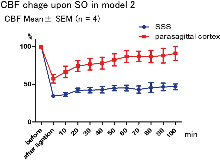
Conclusions
One of the major differences in the two SO models is that SO is almost complete including adjacent cortical veins in the model 1 and that they were not complete in the model 2. These suggest that clinically, even partial recanalization of the sinus may have beneficial effects.
Reference
PB03-P10
Blood brain barrier breakdown, neuroinflammation and widespread lipid disturbances evident with mass spectrometric imaging after cerebral venous thrombosis in rats
1Dept. of Neurosurgery, Tohoku University, Japan
2Dept. of Neurosurgical engineering and Translational Neuroscience, Tohoku University, Japan
Abstract
Background
Cerebral venous thrombosis (CVT) is a rare form of cerebral stroke that causes a wide range of presentations ranging from mild headache to severe morbidity or death in the severer forms. Treatment of CVT is classically in the form of anti-coagulation or thrombolysis, but there is a little focus on developing new therapies for its treatment. In this work we aimed to analyze the pathophysiology of CVT, in order to reveal pathways that share in its pathology and that can be potential drug targets.
Methods
CVT was induced in rats by exposing the SSS and applying ferric chloride soaked filter paper to induce thrombosis. Rats were analyzed by Histology, Immunohistochemistry, western blotting, gel zymography, qPCR and mass spectrometric imaging (MSI) to evaluate cellular changes, MMP2 and MMP9 activation, inflammation, cell stress and lipid changes in the brain at different time points after CVT.
Results
Our results showed early MMP9 activation and BBB disruption after 6 hours of CVT, followed by over inflammation, inflammasome activation and pyroptotic cell death that peaked at three days. These changes were focused in the parasagittal cortex and around the SSS. MSI, on the other hand, showed lipid perturbations to extend beyond the area of venous infarction, with several phosphatidylcholine (PC) and ceramide (Cer) species showing changes in the temporal cortex, hippocampus or Thalamus. Lipid changes correlated with cellular and inflammatory changes in the venous infarct area and qPCR analysis of enzymes regulating these lipid species was well correlated with their changes.
Conclusion
SSS thrombosis causes BBB breakdown, inflammation and inflammasome activation in the parasagittal cortex. However, molecular perturbations were observed well beyond the area of infarction at the metabolic level, indicating that CVT induces global pathologic changes in the brain, which can explain some of the symptoms that accompany it in clinical practice.
PB03-P11
Intracellular S1P levels dictate fate of different regions of hippicampus following transient global cerebral ischemia
1Dept. of Neurosurgery, Tohoku University, Japan
2Dept. of Neurosurgical engineering and Translational Neuroscience, Tohoku University, Japan
3Dept. of Medical Science, Tohoku University Graduate School of Biomedical Engineering, Japan
Abstract
Background
Sphingosine-1-phosphate (S1P) is a sphingolipid molecule that possess anti-inflammatory, pro-proliferative and anti-apoptotic properties, as well as a diverse set of intracellular actions, and is produced by the action of sphingosine kinase 1 (SphK1) and sphingosine kinase 2 (SphK2) on Sphingosine (SPH). S1P metabolizing enzymes have important roles in apoptosis, development and other processes either via controlling intra and extracellular S1P levels or via direct interactions with other proteins. However; the role of S1P metabolizing enzymes in apoptosis is still under debate. Therefore; we attempted to shed a light on sphingolipid metabolism changes during intrinsic apoptosis that follows TGCI, to understand the role of S1P and S1P metabolizing enzymes in neuronal apoptosis and survival.
Methods
TGCI was induced in male Sprague-Dawley rats weighing 280–320 gm, by bilateral common carotid arteries occlusion combined with hypotension for 5 minutes. Rats were sacrificed at 6, 24, 48 and 72 hours. Cell death and cellular changes was evaluated with immunohistochemistry. S1P levels were evaluated with L-MS. mRNA levels of SphK1, SphK2, Sgpl1, Spns2, HSP70 and BCL2 were assessed with qPCR. We also analyzed the gene expression levels of S1P metabolizing enzymes in PC12 neuronal cell line after oxygen-glucose deprivation stress. Finally, we evaluated the impact of FTY720 in-vivo and in-vitro on cell death as well as THI (S1P lyase inhibitor) and ABC294640 (SphK2 inhibitor) in-vitro.
Results
Neuronal apoptosis was observed after 72 hours in CA1 sub-region only, followed by activated microglia infiltration. S1P was upregulated early in CA3 and not in CA1. SphK1 was upregulated in both CA1 and CA3, however SphK2 was downregulated in CA3 following an initial upregulation. S1P lyase was also downregulated in CA3 and not in CA1. Spinster 2 was initially intensely upregulated in both regions; however it was severely downregulated in CA3 by 24 hours and remained downregulated by 48 hours to return to normal levels by 72 hours, contrary to CA1 where it gradually declined to be below baseline only after 72 hours. FTY720 was protective in-vitro, however it failed to improve the outcome in-vivo. THI reduced cell viability at higher concentrations and ABC294640 improved cell viability at lower doses while it reduced cell viability at higher doses.
Conclusion
Early S1P upregulation is linked to CA3 sub-region resistance to TGCI. However; this should by studied in the light of S1P metabolizing enzymes, as this protective effect is linked to not only SphK1 upregulation, but to the downregulation of the S1P lyase and the S1P exporter; Spinster 2. SphK2 which has pro-apoptotic motif was also downregulated in CA3 and might contribute to CA3 resistance to TGCI.
PB03-P12
LabCIRS – a critical incident reporting system for academic research departments
1Charité – Universitätsmedizin Berlin, Department of Experimental Neurology, Berlin, Germany
2Charité – Universitätsmedizin Berlin, Center for Stroke Research Berlin, Berlin, Germany
3QUEST Center for Transforming Biomedical Research, Berlin Institute of Health (BIH), Berlin, Germany
Abstract
Errors and mistakes occur more or less frequently in complex working environment like biomedical research departments. Although they have negative impact on the research results, errors are seldom reported for multiple reasons, e.g. fear of negative consequences. This was recognized in areas where error management is crucial for safety reasons and led to development of anonymous critical incident reporting systems (CIRS) to lower the reporting threshold and improve the error culture.
Two years ago we presented LabCIRS, a CIR system tailored specifically for research labs. This open-source web application is written in Python and was developed and implemented in the department of Experimental Neurology at Charité Berlin.
With increasing publicity other local research departments intended to use LabCIRS, e.g. the central animal facility. However the original system was designed for use in a single department only. Therefore every new division requires a separate instance of LabCIRS including an own server infrastructure. This would have led to high administrative effort and waste of resources. Therefore we decided to introduce some improvements and developed a new version which allows providing separate CIRS for each research department by single installation.
The highlights of the new version are:
• Multi-tenancy – one installation can serve multiple departments in one organization. The reported incidents are visible only to the members of the respective department.
• Support for multiple languages for the user interface and for the content – Besides translatable user interface the published incidents can be presented in different languages.
• Comments on the reported incident – the reporter can add comments or explanations to the incident if necessary still preserving anonymity.
For all interested research institutions LabCIRS is freely available under GPL.
PB03-Q01
Whole-brain visualization and quantification of region-specific neuroplasticity and axonal connectivity using serial two-photon tomography
1Dept. of Neurology & Neurotherapeutics, UT Southwestern Medical Center, USA
2Peter O’Donnell Jr. Brain Institute, UT Southwestern Medical Center, USA
3Dept. of Neuroscience, UT Southwestern Medical Center, USA
Abstract
Objectives
Stroke is the leading cause of disability in the United States, leading to significant impairments in motor function.[1] Ischemic injury induces profound alterations in the architecture of the brain.[2] Understanding the complex role of neuroplasticity in stroke recovery has been limited by standard slide-based imaging methods, which are not well suited for visualization and analysis of the whole rodent brain.[3] These limitations have led to the recent development of new whole-brain mesoscale imaging techniques useful for examining neuroplasticity and axonal connectivity after stroke, such as serial two-photon tomography.[4] Machine learning algorithms and automated registration workflows can be applied to the resultant volumetric images to quantify alterations in descending motor tracts.[5]
Methods
Young male C57/B6 mice received a photothrombotic stroke in the primary motor cortex or sham surgery. 15 days later, mice were injected with psuedotyped rabies virus (PRV-152), a trans-synaptic retrograde virus carrying green fluorescent protein (GFP) into the affected forelimb. Mice were sacrificed 6 days post-injection. Neuronal cell bodies and axons in descending tracts to the forelimb musculature were fluorescently labeled. Serial two-photon tomography (STP; Tissuecyte 1000) generated high-resolution images of coronal sections of the entire mouse brain. We have developed a pipeline for whole brain image analysis that includes supervised machine learning (pixel-wise random forest models via the “ilastik” software package) followed by registration to a standardized 3D atlas of the adult mouse brain (Common Coordinate Framework v3.0; Allen Institute for Brain Science). These procedures allow the visualization and quantification of cellular fluorescent signals throughout the brain in an unbiased manner. The fluorescent signals of neuronal cell bodies and axons in specific areas of the brain in injured and uninjured mice were compared. Finally, we generated 3D reconstructions of the brains for a global view of the post-stroke brain 3 weeks after injury.
Results
Innovative techniques such as STP allow us to visualize post-stroke neuroplasticity and changes in axonal connectivity in the corticospinal tract and other supraspinal motor tracts that descend to the distal musculature of the affected forelimb. These alterations in brain structure and connectivity have been quantified using our automated analysis workflows. By comparing the normalized probabilities of fluorescent signals derived from labeled cell bodies and axons of brains from both injured and naïve mice, we can identify brain regions that undergo dynamic changes in connectivity at late post-stroke time points.
Conclusions
Post-stroke neuroplasticity and changes in axonal connectivity can be visualized with unprecedented resolution using viral tracing techniques and novel imaging methods such as STP. With the assistance of machine learning algorithms to isolate relevant fluorescent signals from STP-generated images and the availability of a standardized 3D mouse brain atlas for image registration, it is possible to not only visualize neuroplasticity, but also to quantify it in an unbiased region-specific manner. Future studies can utilize these novel volumetric imaging and analysis techniques to identify new therapeutic targets to improve motor recovery after stroke.
References
PB03-Q02
Fluorescence resonance energy transfer (FRET) based quantitative analysis of c-jun n-terminal kinase (JNK) in oxidative stress-induced cell death
1Department of Biological Sciences, The University of Tokyo, Japan
2The University of Tokyo Hospital, Japan
3JCHO Tokyo Shinjuku Medical Center, Tokyo, Japan
Abstract
Objectives
Oxidative stress plays a critical role in neuronal cell death including cerebral ischemic injury. JNK functional activity are a suitable indicator of stress-induced conditions in the neuronal cell, particularly after excitotoxic injury. Phospho-JNK has been observed in the bordering zone around an ischemic core and mitogen-activated protein kinase phosphatase-1 expression was induced in the brain of rat MCA-O reperfusion model.1)
The immortalized mouse hippocampal HT22 cell is widely used for evaluating the mechanisms of oxidative stress-associated neuronal cell death2). The HT22 cells do not possess glutamate receptors, and treatment with high levels of glutamate can inhibit the synthesis of intracellular glutathione. Typically, cystine is rapidly reduced to cysteine, which is necessary for the formation of glutathione and then, reactive oxygen species (ROS) levels become increased.
It has been clearly demonstrated that the activation JNK is detected during the death of HT22 cells by Western blotting assay, MAPK activity apparently decides the fate of cell: Survival or Death under stress. Western blotting reflects the average status of a specific cell population and we cannot understand the spatiotemporal dynamics of signaling molecules within a single cell. To overcome these limitations, the Fluorescence Resonance Energy Transfer (FRET) biosensors, which quantifies molecular interactions in biology and chemistry, can be used. We applied the JNK-FRET probes and performed real-time visualization of phosphorylation in a single cell nucleus.
Methods
FRET biosensors for JNK (JNKAR1) were gift from Dr. Aoki.3) To generate cell lines that express stably FRET biosensors, we utilized a PiggyBac transposon system. We induced oxidative stress with glutamate of varied concentration (0–10 mM). We confirmed cell death by Propidium Iodide staining. Cells were seeded onto 35 mm glass-based dishes. Time-lapse FRET images were obtained by epifluorescence microscope(IX83, Olympus) and were analyzed using CellProfiler and Python. The ratio of yellow to cyan emissions were calculated at each time-points and normalized by the emission ratio before stimulation. This study was supported by the Japan Society for the Promotion of Science (JSPS) KAKENHI Grant Number (18K16578).
Results
In the cells expressing JNK-FRET probe, FRET efficiency were elevated in response to glutamate addition, indicating the increase of phosphorylation of JNK. All cells which exhibited apparent JNK activation died, whereas, those without JNK activation survived.(fig)

Conclusions
We developed a new model for real-time visualization of JNK activities under oxidative stress in HT22 cells and confirmed that phosphorylation of JNK can be seen in cell death. This promising model can be used not only for research of the mechanisms of neuronal cell death related to ROS, but also can have practical application, such as evaluation of treatment methods. Cerebral infarction requires quickly treatment, and understanding the mechanisms of neuronal damage in early phase of cell death is necessary to develop new treatments. In future, we expect that the FRET probe could be used in in-vivo models.
References
PB03-Q03
Separability of calcium slow waves and functional connectivity during wake, sleep, and anesthesia
1Dept. of Radiology, Washington University in St. Louis, USA
2Dept. of Neurology, Washington University in St. Louis, USA
3Dept. of Biomedical Engineering, Washington University in St. Louis, USA
Abstract
Objectives
During non-rapid eye movement (NREM) sleep as well as under certain types of anesthesia, there is an approximately 1 Hz slow oscillation that represents a globally continuous hyper/de-polarization of neurons across cortex1. This slow oscillation has previously been found to significantly alter functional connectivity (FC) architecture during induction by ketamine/xylazine anesthesia2, however, it remains unclear if this alteration is consistent across types of anesthesia and natural sleep. Further, it has yet to be reconciled whether there are discrepancies between neural and hemoglobin brain mapping and whether these potential discrepancies are brain state or frequency dependent. Lastly, whether the 1 Hz slow oscillation replaces FC typical of the wake state or is rather superimposed remains unclear.
Methods
Wide field imaging was conducted using sequential LED firing (454nm (GCaMP6 excitation); 523 nm, 595 nm, 640 nm (hemoglobin)) and a cooled, frame-transfer EMCCD camera at 16 Hz. We report imaging of excitatory neurons under wakefulness (W), anesthesia (ketamine/xylazine (K/X), and dexmedetomidine “dex” (D) + atimapezole (DR)) and NREM (N), staged by EEG/EMG in mice (N = 5) expressing GCaMP6 driven by a Thy1 promoter.
Results
Infraslow (0.009–0.08 Hz) FC analysis yielded topographically similar maps regardless of brain state and dynamic analyzed (hemoglobin/calcium) (Figure 1A). However, anesthesia delta (0.7–3.0 Hz) FC showed a significant increase in large, correlated contiguous cortical regions with a strong anti-correlation for distant cortical areas. NREM delta FC showed a similar topography, but to a lesser extent compared to anesthesia. Given that the slow oscillation involves the entire cortex, we hypothesized that it was responsible for these drastic FC changes and that it could be separated from other activity. We used principal component analysis (PCA) on the time-series imaging data, and found that in the anesthesia and sleep conditions, the slow wave phenomenon could be recreated by summing the first three PCs. Subtracting these components and reapplying FC analysis revealed contralateral homotopic correlations and anticorrelations between functionally distinct regions; patterns that were markedly similar to unmodified delta wake FC (magenta outline). A fraction of delta map coverage was computed by summing pixels unique to each condition compared to wake after thresholding by random effects analysis (Figure 1B) which captured the brain state dependency of FC topographies before PC removal, and the loss of dependency after.

Conclusions
We found significant changes across GCaMP6 delta FC architecture, while only modest changes in hemoglobin FC. Further, PCA revealed a separable 1 Hz slow oscillation that upon removal, left correlation structures strikingly similar to the wake condition. These results indicate that the slow oscillation superimposes onto canonical wake FC architecture. This work delineates two types of spontaneous activity for future study, in particular for understanding how sleep and Alzheimer’s disease interact, or how anesthesia influences post-operative delirium.
References
PB03-Q04
Simultaneous measurement of infraslow cerebral evoked potentials and cerebral blood flow during cortical spreading depression enabled by transparent graphene electrode array and laser speckle flowmetry
1ICFO-Institut de Ciències Fotòniques, The Barcelona Institute of Science and Technology, Spain
2Instituto de Microelectronica de Barcelona, IMB-CNM (CSIC), Esfera UAB, Bellaterra, Spain
3Centro de Investigación Biomédica en Red en Bioingeniería, Biomateriales y Nanomedicina (CIBER-BBN), Madrid, Spain
4Institut d’Investigacions Biomèdiques August Pi i Sunyer (IDIBAPS), Barcelona, Spain
5Catalan Institute of Nanoscience and Nanotechnology (ICN2), CSIC and The Barcelona Institute of Science and Technology (BIST), Campus UAB, Bellaterra, Barcelona, Spain
Abstract
Introduction
Cortical spreading depression (CSD) is a slow propagating wave with an excessive transient depolarization in the neurons thus is accompanied by the localized change in evoked electrical activity and cerebral blood flow (CBF). This depolarization can last up to a minute [1]. In this abstract we present simultaneous measurement of hemodynamic signal with laser speckle flowmentry (LSF) and electrical brain signals with a novel graphene transistor [2]. Where, a graphene solution-gated field effect transistors (gSGFET) array was used to record the change in evoked potential at multiple locations, thus being capable to recover spatially resolved maps of infraslow brain activity [2].
Methods
An adult Wistar rat (400 g) was anesthetized isoflurane, and two craniotomies and durotomies were performed on the left hemisphere, in a frontal cortex and somatosensory cortex (Fig. 1(a)). The CSD was induced topically in the frontal cortex with 6 mM concentration of potassium chloride (KCl). To retrieve infraslow cerebral evoked potential, a graphene, 4x4, 400 mm grid spaced gSGFET was used, Fig. 1(b). The array was placed in direct contact with the somatosensory cortex (Fig. 1(c)), [2]. For the recovery of hemodynamic changes, a continuous wave laser diode (785 nm) was set for a full-field homogeneous illumination and a commercial CCD camera with an exposure time of 5 ms were placed on the top to capture the diffused light scattered from the imaging area, [3]. The speckle contrast was calculated for the predefined region, Fig. 1(c), at each pixel over 100 frames in temporal domain, [2]. In the moment of KCl injection, simultaneous 10 min recording of electrophysiological and hemodynamic signal started.
Results
Relative cerebral blood flow (ΔrCBF) and evoked potentials (|U|) are shown in Fig. 1(d). Increase (hyperemia) due to cortical spreading depression in the ΔrCBF signal can be observed. This is also seen in the evoked potential (blue (darker) line). Relative cerebral blood flow was compared to the electrical signal obtained from the graphene sensor, Fig. 1(d).

Fig. 1. (a) Illustration of rat brain with a position of the graphene (gSGFET) sensor and KCl injection. (b) 4x4 gSGFET transistor was used for recovery the electrical brain signal (c) laser speckle image showing position of the graphene sensor, ROI (yellow squares) and vessels in the brain of the rat. comparison of the electrical signal (blue) and cerebral blood flow (red) for each graphene transistor position are shown in the panel (d).
Conclusion
Previously, hemodynamic changes in the blood flow have been shown with laser speckle flowmetry, however, they were not directly compared with the electrical activity due to single point measurement. Here, by taking advantage of the gSGFET technology and LSF, a simultaneous measurement of evoked potential and cerebral blood flow, respectively, are shown.
References
PB03-Q05
Functional high-density speckle contrast optical tomography (SCOT) in small animals and in human brain
1ICFO-Institut de Ciencies Fotniques, The Barcelona Institute of Science and Technology, Spain
2POLIMI – Politecnico di Milano – Dip. Elettronica Informazione e Bioingegneria, Milano, Italy
3Department of Brain Ischemia and Neurodegeneration, Institute for Biomedical Research (IIBB), Spanish Research Council (CSIC), Institut d'Investigacions Biomèdiques August Pi iSunyer (IDIBAPS), Barcelona, Spain
4Institucio Catalana de Recerca i Estudis Avancats (ICREA), 08015 Barcelona, Spain
Abstract
Introduction
Novel high-density speckle contrast optical tomography (HDSCOT) [1,2], is introduced for non-invasive, high-density three-dimensional cerebral blood flow (CBF) maps during functional activation in small animals and humans.
Methods
Five adult rats (male, Wistar) were anesthetized with isoflurane and placed on the stereotaxic frame, Fig.1 (a). Scalp was removed and the baseline measurement was acquired with multi-exposure speckle contrast imaging (MESI). Anaesthesia was switched to intravenous α-chlorarose before the start of the electrical forepaw stimulation. Eight blocks consisting of baseline (18 s), stimulation (30 s, 6 Hz) and 90 s recovery, were used first to stimulate right forepaw, followed by eight blocks stimulation of the left forepaw. In the end five blocks of sham measurements were performed.
However, this was a minimally invasive approach, where the scalp was removed but the skull was kept intact. To show HDSCOT in non-invasive manner a volumetric change during of the CBF was recovered in humans during sensorimotor task. A male bold subject was asked to sit on a chair, and a sensorimotor region was located. The HDSCOT system was focused on the region-of interest and eight source positions were scanned around it, Fig.1 (b). For the activation of the sensorimotor task (finger tapping (3 Hz)) was used, where the subject was asked to look at the screen for 1 min, followed by finger tapping of 1 min and 1 min recovery. Stimulation block was repeated 10 times. Contralateral and ipsilateral finger tapping were performed.
Results
In the Fig. 1(c) the reconstructed blood flow change averaged over five animals is shown for the forepaw stimulation and a sham measurement. Where an increase in the flow can be observed during the functional activation (shaded region). In the Fig. 1(d) a CBF change can be observed for different depth in the adult human brain. Increase in blood flow can be observed during contralateral activation, while during the ipsilateral a much smaller increase in the CBF can be observed.
 Fig. 1: Small animal and adult brain experimental setups are shown in panels (a) and (b) respectively. In the panel (c) a change in cerebral blood flow can be observed during forepaw stimulation in animal models. In the panel (d) a cerebral blood flow change during finger tapping with contralateral and ipsilateral hand are presented.
Conclusion
Here we have demonstrated functional high-density SCOT for resolving blood flow changes at different depths, from small animals to humans. This novel method paves the way for new novel applications of diffuse optics.
Acknowledgments
The project was funded by Fundació CELLEX Barcelona, Ministerio de Economía y Competitividad FEDER (PHOTODEMENTIA, DPI2015-64358-C2-1-R), Instituto de Salud Carlos III / FEDER (MEDPHOTAGE, DTS16/00087), the “Severo Ochoa” Programme for Centres of Excellence in R&D (SEV-2015-0522), the Obra social “la Caixa” Foundation (LlumMedBcn)
References
PB03-Q06
Ultrasound speckle decorrelation-based velocimetry of entire rodent brain for stroke study
1Department of Biomedical Engineering, Boston University, USA
2Athinoula A. Martinos Center for Biomedical Imaging, Massachusetts General Hospital, Harvard Medical School
Abstract
In vivo imaging of cerebral blood flow (CBF) not just in the cortical but also in the subcortical regions is of critical importance to study vascular dysfunction during the brain disease progression. Current available imaging methods are limited either by shallow penetration which only allows imaging superficial cortical layers (eg. optical imaging) or by low spatiotemporal resolution (eg. MRI and CT). The ability to bring together the functional and structural information from a large field-of-view, at a high spatiotemporal resolution would allow many new hypotheses to be explored. Thanks to the progress of ultrafast ultrasound plane wave imaging, it is now possible to have sufficient spatiotemporal resolution (∼100 μm and up to 30 KHz frame rate) to resolve acoustic speckle decorrelation in the imaging plane. For the first time, the normalized field autocorrelation function-based velocimetry analysis, which is primarily used in optical imaging is applied for ultrasound-based blood flow velocimetry measurement of the entire rodent brain. The developed imaging technology will expand the capabilities for studying pre-clinical models of cerebrovascular pathophysiology such as aging, Alzheimer’s disease, stroke, intracerebral hemorrhage, migraine, epilepsy, and traumatic brain injury. In this study, we show the preliminary results of stroke study in a mouse model using the developed technology.

PB03-Q07
Brain tissue pulsation in healthy subjects using transcranial tissue doppler (TCTD) ultrasound
1University of Leicester, UK
2University Hospitals of Leicester NHS Trust, UK
3University of Washington, USA
4Nihon Kohden, Japan
5Broadview Laboratories, USA
Abstract
Objectives
This study investigates the feasibility of using a novel application of Transcranial Doppler ultrasound to characterise brain tissue pulsations (BTP) over the cardiac cycle in healthy volunteers. It is well known that the brain ‘pulses’ with each cardiac cycle [1], but the magnitude of tissue displacements, measured using Transcranial Tissue Doppler (TCTD), in healthy subjects has not yet been characterised. This cross-sectional study aims to quantify healthy brain motion over the cardiac cycle. We investigate whether the magnitude of BTP varies with sample depth, heart rate (HR), mean arterial pressure (MAP), pulse pressure (PP), and demographic factors such as age and sex.
Methods
TCTD ultrasound measurements of BTP were obtained from 107 healthy volunteers (56 male, 51 female) with ages ranging from 20–81 years using a commercially available TCTD system (Spencer Technologies, USA). Each 8 second TCTD measurement provides real-time displacement estimates from 30 depths ranging from ∼2 to 8 cm beneath the skin. ECG measurements were acquired in synchrony with the BTP measurements to investigate the amplitude and timing of brain motion over the cardiac cycle. An exploratory statistical analysis was performed to extract summary statistics and investigate relationships between parameters using linear regression modelling.
Results
Pulsations up to a maximum of 250 μm were observed with a median value of ∼10 μm. Pulsation waveform shape and timing was highly consistent between subjects, however, considerable variations in pulsation amplitude were observed. The median pulsation amplitude estimated from all 107 subjects, was 4.43 μm (IQR: 2.69, 7.12) at the shallowest depth (∼2 cm) to 14.05 μm (IQR: 7.78, 23.05) nearer to the ventricles (∼8 cm). Linear regression analysis suggests that average BTP amplitude increases by ∼1 μm [95% CI: 1.01, 1.02] for each 1 mmHg increase in PP (p < 0.001), and decreases by ∼1 μm [95% CI: -1.02, -1.01] with each 1 mmHg increase in MAP (p < 0.001). Univariate analysis suggested a significant decrease in pulsation amplitude with age in the over 50’s (p = 0.035), but this did not prove to be robust after adjustment for MAP. Average pulsation amplitude was observed to be slightly (∼2 μm) higher in men (p = 0.015). Pulsation amplitude did not appear to be related to heart rate.
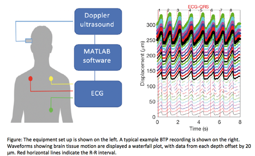
Conclusions
This study is the first to report BTP estimates from a cohort of healthy subjects measured using TCTD. BTP measured using TCTD was consistent with previous measurements using an alternative phased-array Doppler ultrasound Tissue Pulsatility Imaging (TPI) technique (Kucewicz,et al.) [2] and MRI (Weaver, et al.) [3]. Our normative data confirms consistency in waveform shape across the cardiac cycle, and quantifies variations in pulsation amplitude with depth, and an effect of sex, MAP and PP. This normative dataset provides a useful model of healthy BTP for comparison with clinical studies.
References
PB03-Q08
Dynamic laser speckle imaging of cerebral blood flow
1Neurophotonics Center, Boston University, USA
2Faculty of Health and Medical Sciences, Copenhagen University, Denmark
Abstract
Interpretation of the data measured by the laser speckle intensity imaging techniques, such as laser speckle contrast imaging (LSCI), multi-exposure laser speckle imaging (MESI) and laser Doppler flowmetry (LDF), relies on the choice of the appropriate mathematical model which describes the relationship between measured intensity dynamics and the correlation time. The relationship is defined by the electrical field (g1) and intensity (g2) temporal auto-correlation functions [1]. The relation of g2 to g1 is affected by static scattering and speckle averaging effects and was thoroughly studied in the past years both theoretically and experimentally [2]. The field correlation function, which describes the relation of g2 to the correlation time and, thus, to the blood flow speed, is generally represented by one of three forms, which are defined by the light scattering (i.e. single or multiple scattering) and particle motion (i.e. ordered or unordered) regimes [3]. Most commonly, the single form that corresponds to both single scattering unordered motion or multiple scattering ordered motion regimes is applied [1]. The applicability of this model to all imaged tissues or regions of the brain, however, was never verified.
We introduce dynamic laser speckle imaging (DLSI) – the first technique to allow quantification of the laser speckle intensity temporal auto-correlation function in wide-field for every pixel individually. We use it to show that the generally accepted form of the field correlation function is applicable only to the vessels of the specific size and may lead to a significant misinterpretation otherwise. Particularly we explain that the high relative cerebral blood flow (rCBFcore∼30–40%) observed in the ischemic core of the stroke with both LSCI and LDF [4] is in a significant degree caused by the use of the wrong g1 model, and show that the rCBFcore is ∼8–16% when the intensity correlation is measured and analyzed directly with DLSI. We then demonstrate how LSCI interpretation can be significantly improved by applying the correct field correlation model, but still neglecting static scattering and speckle averaging effects, resulting in rCBFcore∼12–20%. We also show that, by using the static scattering and coherence degree parameters estimated with DLSI, one can further improve the LSCI interpretation resulting in the relative blood flow very similar to the one measured with DLSI (rCBFcore∼8–18%).
References
PB03-Q09
Enhanced spatiotemporal reconstruction of neural activities using simultaneous electroencephalography and near-infrared spectroscopy
1Department of Biomedical Engineering, Carnegie Mellon University, United States
2Department of Electrical and Computer Engineering, Carnegie Mellon University, United States
3Department of Radiology, University of Pittsburgh, United States
4Department of Bioengineering, University of Pittsburgh, United States
5Center for Neural Basis of Cognition, United States
Abstract
Objectives
Electroencephalography (EEG) and near-infrared spectroscopy (NIRS) are commonly adopted methods for monitoring brain activities non-invasively and without the confinement of an MRI scanner. While source reconstruction provides insight into the location of brain activities, both modalities suffer from limitations: EEG has a high temporal resolution, but poor spatial resolution; on the other hand, NIRS, which measures slow hemodynamics, has limited temporal resolution, but can be spatially more precise than EEG. We are therefore developing an algorithm which integrates simultaneously recorded EEG and NIRS signals, and performs an enhanced reconstruction of neural activities with high spatiotemporal resolution.
Methods
We demonstrate our algorithm using simulated data based on a realistic head model. A finite element method (FEM) model is constructed based on a pre-segmented brain atlas, and then loaded into FieldTrip [2] and NIRFAST [3] toolboxes to calculate the forward matrices for EEG and NIRS respectively. A 32-channel EEG is used in the standard 10–20 configuration. 9 sources (750 and 850 nm) and 8 detectors are used in NIRS, forming 24 channels, covering approximately the right sensory-motor cortex, in which regions (A and B) of activations with a separation of approximately 2 cm are simulated (Fig. 1a). They are assumed to be activated with a 50 ms inter-stimulus interval. In total we simulate 100 trials, spaced by 200 ms for each region (Fig. 1a,b). Electrical and hemodynamic signals are assumed to be colocalized. White noise is added to the simulated scalp recordings. Scalp EEG data is then averaged across the trials, and NIRS changes are averaged across the whole experiment.
To demonstrate the improvement using the proposed algorithm, we reconstruct EEG and NIRS separately, and compare these with joint reconstruction using the reconstructed oxygenated hemoglobin (HbO, obtained from NIRS) activities as the spatial prior for EEG. The joint reconstruction algorithm is inspired from similar algorithms in joint localization using fMRI and EEG.
Reconstruction is performed using a hierarchical Bayesian framework, in which structural assumptions are made on the covariance matrices of sensors and source voxels (detailed in [4]). When performing EEG reconstruction with spatial prior, the source voxels are assumed to be independent, but the variances are assigned such that they are larger if HbO has larger activation and smaller otherwise. The same algorithm is applied to each of the time steps.
Results
The improvement of using HbO as the spatial prior for EEG reconstruction is demonstrated at representative timesteps in Fig. 1c,d. The algorithm resolves both activation regions spatially and temporally, especially that it recovers region B, which is hardly visible when using only EEG.

Conclusions
Multimodality localization using EEG and NIRS has received little attention from the community. Our simulation results strongly suggest that our algorithm, which combines the high spatial resolution of NIRS and high temporal resolution of EEG, can achieve a high-resolution spatiotemporal reconstruction of neural activities.
References
PB03-Q10
The use of photoacustics to measure cerebral blood volume and cerebral oxygenation
1Dept. of Neurosurgery, Heidelberg University, Germany
2Division of Computer Assisted Medical Interventions, German Cancer Research Center, Heidelberg, Germany
3Faculty of Physics and Astronomy, Heidelberg University, Heidelberg, Germany
4Medical Faculty, Heidelberg University, Heidelberg, Germany
Abstract
Introduction
There is a need to monitor whole brain oxygenation and brain hemodynamics in neurocritical care patients. Here, we show that the use of nanosecond laser pulses delivered into the brain produces a thermoelastic expansion which in turn gives rise to acoustic waves that can be detected to produce high resolution images.
Methods
We used five 30–35 Kg german landrace swine to induce changes in cerebral blood flow using 1 Mol KCl stimulations on the brain cortex. Brain electrical activity was monitored using electrocorticography. The surface of the brain was covered with a gel pad and a photoacoustic probe together with a ultrasound probe. A hybrid photoacoustic ultrasonic imaging system is based on (1) an ultrasound (US) research system with a featuring a linear US transducer with a operating on a center frequency of 7.5 MHz and broad acoustic response combined with (2) a near infrared fast tuning optical parametric oscillator laser. The system acquires multispectral sequences with corresponding US images for each photoacoustic image.
Results
Photoacustic imaging enables the study of brain hemodynamics deep in the gyrencephalic brain with high spatiotemporal resolution. As rapid neuronal depolarization causes tissue hypoxia, we could measure the decreases on oxygenation and changes in blood volume. Global brain hypoxia and spreading depolarizations were detected. Due to its high resolution, promising imaging depth and high contrast, this novel approach to cerebral hemodynamics imaging could lead to groundbreaking advances in stroke and brain injury research.
Conclusions
We present the first photoacoustic imaging of brain hemodynamics gyrencephalic brain.
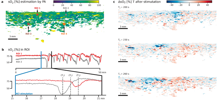
PB03-Q11
19F-magnetic resonance spectroscopic imaging visualizes the distribution of matrix metalloproteinases in ischemic brain using in vivo
1Center for Integrated Human Brain Science, Brain Research Institute, University of Niigata
Abstract
Matrix metalloproteinases (MMPs) damage the neurovascular unit, promote blood–brain barrier (BBB) disruption following ischemic stroke and play essential roles in hemorrhagic transformation (HT), which is a part of the most severe side effects of thrombolytic therapy. However, no biomarkers have presently been identified that can serve to track track changes in the distribution of MMPs in brain. Here, we developed a new 19F molecular ligand, TGF-019, for visualizing the distribution of MMPs in vivo using 19F magnetic resonance spectroscopic imaging (19F-MRSI).
Synthesize of TGF-019
We first synthesized TGF-019 that is fluorinated ligand of pan-MMP inhibitor GM6001. The ability of TGF-019 to interact with MMP2, MMP3 and MMP9 was assessed using an in vitro inhibition assay. TGF-019 was found to completely inhibit MMP2 and MMP9, with a somewhat weaker, but significant inhibition of MMP3.
In vivo sensitivity of TGF-019
To demonstrate TGF-019 has sufficient sensitivity for the specific MMPs suspected in evoking HT during ischemic stroke, 3 μl of 0.1 μg/μl pro-MMP2, MMP3, pro-MMP9 or activated-MMP9 was injected into MMP2 knockout mice brain using a 30 gauge Hamilton syringe. The injection was performed over 15 min and was followed by TGF-019 intravenous administration and MR measurement. All four MMPs were visualized clearly on MRSA images.
Visualizing ischemic lesion by TGF-019
We then utilized it to assess those MMPs at 22 to 24 hours after experimental focal cerebral ischemia on MMP2-null mice, as well as wild-type mice with and without the systemic administration of recombinant tissue plasminogen activator (rt-PA). Transient focal cerebral ischemia was induced using suture method occluding MCA artery. The signal intensity of TGF-019 within the ischemic lesion delineated by T2-weighted imaging was significantly stronger than the non-ischemic contra-lateral hemisphere in sufficient signal-to-noise ratio (23.7 in ischemic cortex). Of those groups, TGF-019 signal intensities were strongest in the t-PA group followed by the WT-saline group, while those of the MMP2 KO group were found to be the weakest (p < 0.05, ANOVA with Bonferonni correction). Optical zymographic densities also showed significantly higher densities in the ischemic hemisphere than the contra-lateral hemisphere. Conversely, MMP3 did not show a significant increase among all areas in this experimental procedure. Signal Intensities of TGN-019 and zymographic optical densities of MMP2/9 were found to be highly correlated in the range shown by the experiments.

Conclusion
The 19F-MRSI of TGN-019 administered mice showed high signal intensity within ischemic lesions that correlated with total MMP2 and MMP9 activity, which were confirmed by zymographic analysis of ischemic tissues. Based on the results of this study, 19F-MRSI following TGN-019 administration can be used to assess potential therapeutic strategies for ischemic stroke.
PB03-Q12
Multi-channel MR-compatible flexible microelectrode for deep brain stimulation and electrophysiological recording
1Dept. of Neurology, University of North Carolina at Chapel Hill, Chapel Hill, NC, United States
2Interdisciplinary Institute of Neuroscience and Technology, Zhejiang University, Hangzhou, China
3Neuroscience Curriculum, University of North Carolina Chapel Hill, Chapel Hill, NC, United States
4Blackrock Microsystems, Salt Lake City, UT, United States
5Biomedical Engineering, National Yang-Ming University, Taipei, Taiwan
Abstract
Objectives
Functional MRI (fMRI) is one of the leading tools to map neural circuit function and connectivity. Despite ongoing advances in MR data acquisition and analysis, we still face tremendous challenges to unequivocally interpret fMRI data. Specifically, the use of a hemodynamic signal as a surrogate marker of neuronal activity presents a major challenge to any straightforward interpretation of fMRI data. In recognition of this caveat, it is prudent to examine fMRI signals against more “ground truth” measures of neuronal activity using electrophysiological recordings. Here, we present a polyimide-based MR-compatible microelectrode array with an MR-compatible head-stage preamplifier and demonstrate the feasibility of recording BOLD, LFP, and single-unit activity simultaneously in vivo. We also demonstrate the dual-functionality of this microelectrode, which allows site-selective microstimulation at targeted brain nucleus with unprecedented spatial resolution.
Methods
Polyimide-based 16-channel microelectrodes were stereotactically implanted into ventroposterior thalamus (VPTh) for deep brain stimulation (DBS)-fMRI or primary somatosensory cortex (S1) for the electrophysiology-fMRI experiments in rats (n = 10). fMRI data were acquired by gradient-echo-EPI sequence on a Bruker 9.4T scanner. For DBS-fMRI experiment, bipolar stimulation was performed between two adjacent channels with the stimulation paradigm of the 60 s(Off)-[30s(On)-120(Off)]x2. Stimulation parameters are tuned to evoke robust and repeatable BOLD responses (0.05 ms, 20 Hz, 1mA). For the electrophysiology-fMRI experiment, an MR-compatible preamplifier custom-designed for this project (Blackrock Microsystem, Ohio, USA) was used. A pair of needle electrodes were inserted to the left forepaw to induce sensory stimulation with the paradigm of the 60 s(Off)-[30s(On)-60(Off)]x2 during fMRI scanning (0.5 ms, 9 Hz, 3mA). All data preprocessing pipeline and analysis followed standard protocols stated in our previous publications.
Results
The electrode in an agarose gel phantom shows negligible artifact on both spin-echo and gradient-echo images at 75 um isotropic resolution (
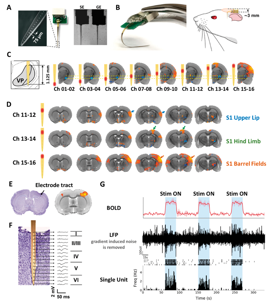
Conclusions
We demonstrated a novel tool that allows concurrent DBS-fMRI and electrophysiology-fMRI. We expect this technique to bring a profound impact to the brain imaging community as it adds concurrent mesoscopic brain modulation and recording capacity to the macroscopic fMRI acquisition.
PB03-Q13
Blood vessel segmentation using MP2RAGE sequence at 7t MRI
1Center for Information and Neural Networks, NICT, Osaka, Japan
2Graduate School of Frontier Biosciences, Osaka University, Osaka, Japan
3Siemens Healthcare K.K., Osaka, Japan
4Advanced Clinical Imaging Technology, Siemens Healthcare AG, Lausanne, Switzerland
Abstract
Objectives
Multiple contrasts can be acquired by using magnetization prepared two rapid acquisition gradient echoes (MP2RAGE) simultaneously. This feature has an advantage to segment different brain structures, such as the brain tissues [1]. Among the brain structures, a blood vessel is also an important brain structure that addresses specific anatomical territories [2]. But it is hard to segment the blood vessels in T1w image due to the low contrast or inhomogeneous intensity. In this study, we proposed a new approach to segment not only the brain tissues but also the vessels simultaneously with submillimeter spatial resolution using MP2RAGE sequence at 7T magnetic resonance imaging (MRI).
Methods
Four healthy normal subjects without a history of neurological disease or any other medical conditions participated in this study after providing their written informed consent. The experiments were performed on a 7 T MRI scanner (MAGNETOM 7T, Siemens Healthcare, Germany). MP2RAGE sequences [3] were used with the following parameters: voxel resolution = 0.7 mm3, repetition time (TR) = 5000 ms, echo time (TE) = 3.43 ms, inversion time (800 ms/2600 ms), and flip angle = 4°/5°. We also acquired 3D Time-of-Flight (TOF) image with the following parameters: voxel resolution = 0.3 x 0.3 x 0.4 mm3, repetition time (TR) = 20 ms, echo time (TE) = 4.47 ms, and flip angle = 18°. We applied our calculations and Frangi filtering (http://www.vmtk.org, 2008) on MP2RAGE images. In addition, we applied the same filter to TOF images and registered individual native T1w images. Then, we masked both the images with the same mask image for a direct comparison.
Results
We segmented the three different brain tissues as well as the blood vessels using MP2RAGE images. The mean absolute voxel difference and dice coefficient scores between the vessel masks of MP2RAGE segmentation and TOF segmentation were 23.3% and 66.2%, respectively. The masks of the large arteries, such as the anterior and middle cerebral arteries, were well-matched between MP2RAGE and TOF segmentations but the smaller blood vessels were not (Fig. 1) due to the relatively lower contrasts resulting from the B1 bias field. However, this limitation could be overcome by using B1 bias field correction technique [4] or by changing the acquisition parameters.

Conclusions
We successfully segmented the blood vessels as well as the brain tissues using MP2RAGE multi-contrast images and Frangi filtering. The vessel segmentation using MP2RAGE sequence at 7T has the potential 1) to be acquired alongside other brain tissue segmentation from the same MR sequence, 2) to be used for the correction of white matter segmentation, and 3) to provide precise anatomical territory information with submillimeter spatial resolution.
References
PB03-Q14
FLAIR-based estimation of the stroke onset time: a magnetic field dependent study
1Department of Radiology, University of Pittsburgh
Abstract
Objectives
The knowledge of stroke onset time is a prerequisite for thrombolytic treatment, but this information is unknown in ∼25% of acute ischemic stroke patients. Recently, a fluid-attenuated inversion recovery (FLAIR)-based MRI index (e.g., DWI-FLAIR mismatch) has been suggested as a surrogate to determine whether the stroke duration is within the treatment window [1], but it is still unclear whether the efficacy of this index is dependent on the magnetic field strength. In this work, we aim to evaluate the field-dependence of the FLAIR signal in stroke with computer simulations and with experiments at a high field of 9.4 T.
Methods
Results and discussions
With typical imaging parameters (TR, TI, and TE) used for the T2-FLAIR, the simulated MRI signals initially drop below the baseline due to the rapid change of blood flow and oxygenation effects, and then slowly increase. The rate of the increase is highly dependent on B0. At 1.5 T, the zero-crossing time (when FLAIR signal intensity becomes higher than baseline) is less than 2–3 hours. At 3 and 7 T, the zero-crossing times are longer and range between 3–6 h and 6–12 h, respectively. At 9.4 T, the signal intensity is less than the baseline for all the stroke durations. This result is confirmed by our 9.4 T experimental data of MCAO rats. The FLAIR signal of the ischemic tissue (as identified by DWI) is less than that of the contralateral normal tissue at the acute phase (1–5 h) and at 24 h post occlusion, i.e., no positive FLAIR contrast can be detected.
Conclusions
The stroke duration when a positive FLAIR contrast appears between ischemic and normal tissue increases with B0 and is dependent on the imaging parameters. This may explain previous reports that the efficacy of using the DWI-FLAIR mismatch as a biomarker for stroke onset estimation reduced at 3 T than 1.5 T [4,5]. Thus, a FLAIR-based estimation of stroke onset should be designed or interpreted with care.
References
PB03-Q15
Inter-individual variation in arterial blood T1: an error source of pCASL CBF
1Akita Research Institute of Brain and Blood Vessels
Abstract
Objectives
Cerebral blood flow (CBF) quantification in ASL requires arterial blood T1 (T1a) for correcting the T1 decay of labeled blood. In the standard pCASL procedure T1a is fixed to a normal value (1,650 ms at 3T); however, individual T1a depends strongly on blood hematocrit (Ht) [1,2]. Therefore, the standard calculation with fixed T1a results in erroneous CBF estimates for patients having Ht values outside the normal range, such as severely anemic patients [3]. The aim of this study was to demonstrate that the fixed T1a assumption is a potential error source in pCASL CBF quantification even for non-anemic subjects and the use of Ht-based individual T1a improves the quality of a cross-sectional analysis.
Methods
Data from patients with occlusion or severe stenosis of the unilateral ICA or MCA were retrospectively analyzed (n = 32). The single-PLD pCASL study (PLD = 2,000 msec) was performed on a 3T scanner (MAGNETOM Verio Dot, Siemens) with 3D GRASE readout (prototype sequences) [4]. CBFs were calculated with 1) fixed T1a (1,650 ms) and 2) individual T1a estimated from Ht (blood sampling), using the Ht-T1a formula [1]. Regions-of-interest (ROIs) covering MCA territory were defined on the contralateral (normal) hemisphere with respect to steno-occlusive lesions, and hemispheric CBF values were measured.
Results
Individual Ht values varied between 33% and 48%, corresponding to ±100 ms differences in estimated T1a values and ±10% errors in CBF estimates with fixed T1a. Average and standard deviation (SD) of CBF (mL/100 g/min) are 41.1 ± 7.3 (fixed T1a) and 41.0 ± 6.3 (estimated T1a); the use of individually-estimated T1a reduced inter-individual variations in pCASL CBF estimates. Correlation analyses showed that CBF with fixed T1a was strongly, negatively correlated with Ht (r = −0.568) but the correlation was markedly reduced when using individually-estimated T1a (r = −0.293) (Figure).

Conclusion
CBF estimates with the standard pCASL procedure rely on the assumption of T1a. Care should be taken when performing a correlation analysis of pCASL CBF with Ht-related factors such as sex, age, and pathology.
Acknowledgements
We thank Dr. Josef Pfeuffer for providing the 3D ASL prototype and Dr. Hideto Kuribayashi of Siemens Healthineers for the collaborative research project.
References
PB03-Q16
Validation of an optical method for measuring cerebral blood flow in adults
1Department of Medical Biophysics, Western University, London, Ontario, Canada
2Imaging Division, Lawson Health Research Institute, London, Ontario, Canada
3Department of Physics and Astronomy, University of Pennsylvania, Philadelphia, USA
4Department of Anesthesiology and Critical Care, University of Pennsylvania, Philadelphia, USA
5Department of Neurology, University of Pennsylvania, Philadelphia, USA
6Department of Neurosurgery, University of Pennsylvania, Philadelphia USA
Abstract
Objectives
Monitoring cerebral blood flow (CBF) is central to the management of critical care patients with neurological emergencies considering delayed cerebral ischemia is a major cause of secondary brain injury. Since transporting critical-care patients to imaging facilities is risky, there is a need for bedside methods. One promising approach is dynamic contrast-enhanced (DCE) near-infrared spectroscopy (NIRS), which requires measuring the time-varying concentration of an optical contrast agent, Indocyanine Green (ICG), in the brain [1]. However, like all NIRS methods, signal contamination from the extracerebral tissue can result in substantial errors in the CBF estimates [2]. This study presents a novel DCE-NIRS method using a combination of time-resolved (TR) detection to enhance depth sensitivity and a multi-layer modelling approach to determine the cerebral ICG curve. The accuracy of the method was assessed by comparison to CBF measurements from the MRI-based method, arterial spin labelling (ASL).
Methods
The study involved two sites: Western University (ten healthy subjects, 3 females, age = 30 ± 11 y) and the University of Pennsylvania (seven healthy participants, 4 females, age = 26 ± 4 y). Under the Western protocol, NIRS/MRI data were acquired simultaneously at normocapnia and hypercapnia to determine if DCE-NIRS was sensitive to changes in CBF. For the UPenn protocol, DCE NIRS and ASL data were collected sequentially at normocapnia. Moment analysis was applied to the TR data to isolate the brain signal, which was analyzed to determine the time-varying change in the absorption coefficient, Δµa(t). This relationship is typically assumed to be linear; however, this is not valid with ICG due to the strong light absorption. Instead, a non-linear model based on a two-layer forward solver was developed. The resulting Δµa(t) was converted to cerebral ICG concentration, from which CBF was determined.
Results
ICG data sets from a representative subject at normocapnia and hypercapnia are shown in Figure 1(A), along with the corresponding ASL CBF images. Both techniques showed the expected increase in CBF during hypercapnia. Comparison of CBF estimates from DCE-NIRS and ASL for all subjects from both sites are presented in Figure 1(B). Good agreement was confirmed with a regression slope of 0.99, an intercept of −1.7 mL/100 g/min, and R2 = 0.88.
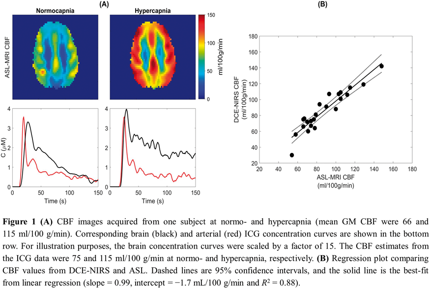
Conclusions
The data presented in this study show that DCE-NIRS combined with depth-sensitive detection can quantify CBF in the adult brain. In addition to showing good agreement with ASL at baseline, the experiments also showed DCE-NIRS is sensitive to changes in flow. These results highlight the potential of this technology for measuring CBF at the bedside of critical-care patients. In addition, continuous CBF monitoring is possible by combining this technique with diffuse correlation spectroscopy.
References
PB03-Q17
Construction of the fractal analysis system for DaT-SPECT
1Dept. of Neurology, Japanese Red Cross Okayama Hospital, Japan
2Dept. of Medical Radiation, Tokyo Medical College, Japan
3Dept. of Neurology, Okayama University Graduate School of Medicine, Dentistry and Pharmaceutical Sciences, Japan
Abstract
Background
Parkinsonian syndrome, including Parkinson's Disease (PD), and dementia with Lewy bodies (DLB) are movement impairing diseases in which striatal dopamine neurons degenerate, and is characterized by a decrease in dopamine transporter (DAT) density present at the nerve endings. Nuclear medicine images such as DAT-SPECT (Single Photon Emission Computed Tomography) images and DAT-PET (Positron Emission Tomography) images are used to diagnose degenerative diseases of striatal dopamine neurons, and 123I-FP-CIT (DAT Scan: registered trademark), 123I-β- CIT, 99mTc-TRODAT-1, 11C-PE21, 11C-CFT and the like are known as DAT imaging agents. Also, fractal analysis using the concept called fractal proposed by Madelbrot in 1973 makes it possible to quantify complex morphology and apply it to radiology.
Purpose
The purpose of this is to develop an information system for the differential diagnosis of several degenerative diseases in striatal dopamine neurons based on stored fractal data for each disease by setting the region of interest (ROI) which includes the striatum of the SPECT image and calculating the fractal dimension based on the image data in the ROI.
Materials and methods
Forty PD patients (16 males, 24 females (average age of 77.4 years ± 6.2)) diagnosed based on clinical diagnostic criteria of PD, DLB and progressive supranuclear palsy (PSP) in outpatient and inpatient clinic in our hospital. Three DLB patients and nine patients with PSP were subjected to DaT-SPECT, and fractal analysis was performed to examine whether they can be distinguished from each other.
Result
Through this analysis, a significant difference between PD and PSP and between DLB and PSP were observed. From the results, it is possible to distinguish between PD and PSP and to distinguish between DLB and PSP by using fractal value by pixel counting method.
PB03-R01
Effects of basal condtions on evoked funtional MRI and CBV in mouse
1Center for Neuroscience Imaging Research (CNIR), Institute for Basic Science (IBS), Suwon, Republic of Korea
2Department of Health science and technology (SAIHST), Sungkyunkwan University, Seoul, Republic of Korea
3Department of Biomedical Engineering, Sungkyunkwan University, Suwon, Republic of Korea
Abstract
Objectives
Blood-oxygenation-level-dependent (BOLD) functional MRI is useful to investigate functions of diseased animal models and mapping of neural networks in a whole brain. BOLD fMRI of anesthetized mouse has been of a great interest, but its findings are inconsistent among literature. One potential reason is the varied baseline physiological conditions induced by breathing condition and anesthetics, which can greatly influence the magnitude and dynamics of BOLD responses (1-3). High carbon dioxide levels in blood (pCO2) elevate the basal CBF with vasodilation, resulting in the reduced BOLD magnitude and increased time-to-peak. However, until now, due to the small size of the mouse, mouse fMRI experiments have been carried out without careful control of vascular physiology. Thus, we investigated whether different pCO2 levels can change hemodynamic responses evoked by forepaw stimulation in anesthetized mice.
Methods
Experiments were conducted to investigate the degree of pCO2 level and vessel dilation depending on breathing conditions and anesthetic conditions in mice. Three other groups were tested in which the different anesthesia and breathing conditions were matched: Ketaminexylazine-mechanical, ketaminexylazine-self-breathing, and isoflurane-mechanical (n = 35). Arterial blood gas analysis were conducted to compare the physiological parameters. Using optical imaging, resting state vessel diameter was measured in the same animal. Functional studies were then conducted to determine the hemodynamic responses according to different breathing and anesthetics. Evoked BOLD fMRI at 9.4T were performed with forepaw stimulation (0.5 ms, 0.5mA, 4 Hz). All trials were averaged and their functional maps and time courses were compared.
Results
During self-breathing vs. mechanical ventilation, blood pressure, oxygen saturation and cardiac pulse rates were similar, but pCO2 and pH changed. The averaged pCO2 was 80.5 ± 10.6 mmHg in self-breathing vs. 40.6 ± 6.6 mmHg in mechanical ventilation. Under these conditions, pial vascular diameters were measured. The arterial vessels dilate under the hypercapnic condition in ketamine and xylazine anesthesia, as expected, which is still less than the normocapnic condition in 1.0% isoflurane anesthesia. As already reported (4), in isoflurane anesthesia conditions, there were no or weak activations. On the other hands, in both normocapnic and hypercapnic conditions, significant activation was observed in the primary somatosensory cortex of ketamine and xylazine-anesthetized mice. Activation volume is similar for both conditions, but the magnitude of BOLD fMRI is about 2times higher under normocapnia than hypercapnia. In our previous self-breathing condition, the BOLD responses had a hump within 10 s after stimulus onset and then gradually increased to the maximum near the end of the 20-s stimulus. Under the normocapnic condition, the BOLD response reached a peak within 10 s. These dynamic and magnitude changes by different basal conditions were consistent with previous human studies (1).
Conclusions
In the mouse experiments, the magnitude and dynamics of the BOLD signals changed according to the breathing condition and anesthetics conditions. This results allows us to consider the influence of the basal condition in comparing evoked experimental results.
This work supported by the Institute for Basic Science (IBS-R15-D1).
References
PB03-R02
Investigation of role of frontal cortex on shaping functional connectivity of cerebral cortex
1Center for Neuroscience Imaging Research, Institute for Basic Science (IBS), Suwon, 16419, Republic of Korea
2Department of Biomedical Engineering, Sungkyunkwan University (SKKU), Suwon, 16419, Republic of Korea
3Biomedical Institute for Convergence at SKKU (BICS), Suwon, 16419, Republic of Korea
4Samsung Advanced Institute of Health Science and Technology (SAIHST), Sungkyunkwan University (SKKU), Suwon, 16419, Republic of Korea
Abstract
Slow but spatio-temporally correlated blood oxygenation fluctuations have been observed even when the subject is at rest1. Since the correlation is found to be used to assess the connectivity of the brain 2, its popularity of investigating resting state functional connectivity has been grown. But which neural input from brain region plays a role in modulating connectivity of cerebral cortex is largely unknown. Here, we focus on frontal cortex which is considered as one of the ‘default mode network’ in mouse3. As it is found that default mode network activates when subject is at rest and frontal cortex has large neuromodulatory projection to cerebral cortex4, we hypothesized that modulation of frontal cortex in mice may affect the resting state fluctuation in cerebral cortex. By blocking neural input from frontal cortex, we tried to see the contribution of frontal cortex in fluctuation on cerebral cortex. In this study, optical imaging method was used for functional brain imaging. We utilized conventional optical imaging system that uses 3 different wavelengths which are 460 nm, 530 nm, 630 nm. sCMOS camera was synchronized with LED light source. We used whole skull thinned transgenic mouse model (Thy1-GCaMP6s) and mouse were urethane anesthetized when used in experiments. We injected tetrodotoxin (TTX) into frontal cortex and compared functional connectivity at cerebral cortex before and after TTX injection. By the experiment, we investigated acute effect of blocking neural input from frontal cortex to resting state fluctuation in cerebral cortex. Before TTX injection, GCaMP fluorescence at both frontal cortex was symmetrically changed. But after acutely blocking frontal cortex with TTX, the correlation of GCaMP fluorescence in bilateral frontal cortex broke down due to TTX. GCaMP fluorescence based seed-pixel correlation map showed that correlation of GCaMP fluorescence between both barrel cortex and contra motor cortex with injection site was changed. But correlation between each hemisphere of other brain region such as barrel cortex or hindlimb cortex was not changed. Blocking neural activity of frontal cortex influenced the neural correlation between motor and barrel cortex. This is considered as local TTX effect that other brain areas were not influenced. Thus, other brain region may play a role in modulating resting state fluctuation of cerebral cortex.
References
PB03-R03
Longitudinal resting-state fMRI for animal studies of functional connectivity in healthy ageing
1Department of Neuroscience, Psychology and Behaviour, University of Leiecester, Leicester, UK
2School of Psychology, University of Nottingham, Nottingham, UK
3Preclinical Imaging Facility, Core Biotechnology Services, University of Leicester, Leicester, UK
Abstract
Introduction
Resting state fMRI (RS-fMRI) is an increasingly valuable tool in the understanding of the effects of neurological disease on functional connectivity in animal models1. However, preclinical fMRI has drawbacks. Animals require anaesthesia for scanning and common anaesthetics can inhibit vascular reactivity, reduce the amplitude of low-frequency fluctuations detected in RS-fMRI, or have toxic effects and so are restricted for terminal scans.2 Because of this, only a small number of preclinical longitudinal RS-fMRI studies, and no preclinical RS-fMRI studies of healthy ageing, exist, which limits the application of RS-fMRI for studying the progression of age-related disease. This study examines the suitability of a novel, minimally invasive anaesthesia protocol in preclinical RS-fMRI to assess the effect of ageing on functional connectivity.
Methods
This study was conducted in accordance with the UK Animals (Scientific Procedures) Act, 1986 and following institutional ethical approval. 11 female Wistar Han rats aged 3 months were housed in standard cages and given daily access to a playpen. Animals were scanned at 6, 9, 12, 15 and 18 months old. Under 3% isoflurane anaesthesia, tail vein cannulation was performed. Animals were given a continuous IV infusion of 54 mg/kg/hr propofol for the duration of the scan. Isoflurane was gradually reduced to 0% and animals’ air supply was changed from 100% O2 to room air. Animals were monitored using a respiration pillow, temperature probe, and pulse oximeter. RS-fMRI was performed for 6 minutes as part of a larger fMRI protocol (TR = 1000 ms, TE = 22 ms, kzero = 8, shots = 2, data matrix = 128x128). Data was analysed using FSL (www.fmrib.ox.ac.uk/fsl). Preprocessing for motion correction (MCFLIRT), brain extraction (rBET), bias field correction (FAST) was performed, followed by independent component analysis (MELODIC) and dual regression to identify group-level resting state components.
Results
Dual regression detected eleven known components at all time points, including the hippocampus, cingulate cortex, retrosplenial cortex, left and right somatosensory, auditory and visual cortices, left and right striatum, motor cortex, thalamus, corpus callosum, and inferior colliculus (p < 0.05) (fig. 1)4. Significant decreases in amplitude of low-frequency fluctuations with ageing were observed in the hippocampus, somatosensory cortex, thalamus and inferior colliculus (P < 0.05).
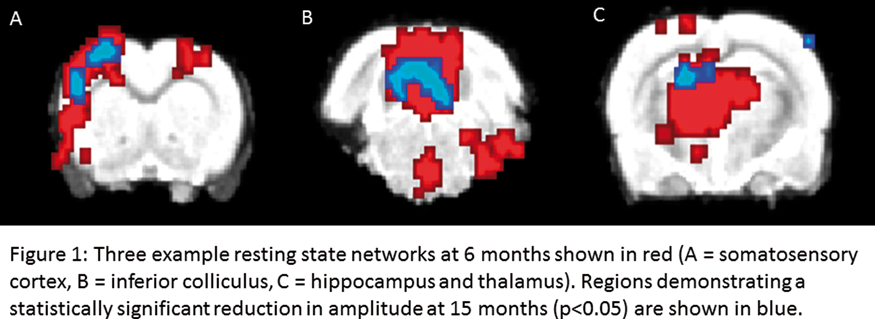
Discussion
This study demonstrates that use of propofol anaesthesia for resting-state fMRI allows the detection of eleven known resting state components, and is reproducible across subjects and over time. The reduction in amplitude of fluctuations in sensory regions5 and the hippocampus6 are consistent with observations in human studies, displaying the utility of this model for studies of healthy ageing. Network analysis using FSLNets, with the addition of the 18 month time point, will be used to investigate changes in functional connectivity between these regions. Through improved understanding of age related changes in healthy rodents, preclinical models of age-related diseases can be refined.
References
PB03-R04
Anesthesia has differential effects on optical hemodynamic measures of resting state functional connectivity in mice
1Neurovascular Research Lab, Department of Radiology, Massachusetts General Hospital, Boston, MA, USA
2Neurocritical Care and Emergency Neurology, Department of Neurology, Massachusetts General Hospital, Boston, MA, USA
3Neurophotonics Center, Boston University, Department of Biomedical Engineering, Boston, MA, USA
4Department of Neurosurgery, Yamaguchi University School of Medicine, Ube, Japan
5Athinoula A. Martinos Center for Biomedical Imaging, Department of Radiology, Massachusetts General Hospital, Charlestown, MA, USA
6Stroke Service, Department of Neurology, Massachusetts General Hospital, Boston, MA, USA
Abstract
Objectives
Resting state functional connectivity (RSFC) is an important measure of the integrity of neuronal networks in health and disease (1). Optical methods have recently been developed to determine RSFC in mice (2). However, evidence suggests that maintaining awake mice in a true resting state can be challenging (3). Therefore, assessment of RSFC under anesthesia still has a role, and we sought to determine the optimal anesthetic for optical RSFC measurement.
Methods
We assessed functional connectivity using resting state hemodynamic optical intrinsic signal imaging through exposed and unaltered full thickness mouse skull under Avertin (tribromoethanol), ketamine-xylazine, urethane, chloral hydrate, and isoflurane anesthesia. We assessed correlations between seeds (in motor, somatosensory, retrosplenial, and visual cortex) and pixels corresponding to the visible dorsal brain surface. A seed-based connectivity index (Seed CI) was calculated by averaging the averaged positive correlations for each seed. Connection variation (CV) was determined by averaging the coefficients of variation for correlation coefficients between each of the seed locations. A global connectivity index (Global CI) was calculated by averaging all possible pixel-to-pixel positive correlation coefficients. A bihemispheric connectivity index (Bihem CI) was calculated by averaging all the correlation coefficients between contralateral mirror pixels. Finally, the total power of the hemodynamic signal was determined by averaging the powers of each pixel within 0.1–0.01 Hz.
Results
Avertin and ketamine-xylazine produced the greatest amplitudes of Seed CI followed by urethane anesthesia. The CV of seed-to-seed functional connections was lowest for Avertin and ketamine-xylazine as compared to urethane, chloral hydrate, and isoflurane (Figure). Global CI was 0.35 ± 0.01, 0.32 ± 0.01, 0.29 ± 0.03, 0.22 ± 0.04, and 0.19 ± 0.02 and Bihem CI was 0.64 ± 0.01, 0.54 ± 0.03, 0.49 ± 0.08, 0.34 ± 0.08, and 0.20 ± 0.04 for Avertin, ketamine-xylazine, urethane, chloral hydrate, and isoflurane. Finally, the spontaneous hemodynamic fluctuations with Avertin, ketamine-xylazine, and urethane were greater than for chloral hydrate or isoflurane (relative averaged power was 1, 0.21, 0.65, 0.14, and 0.09, respectively).
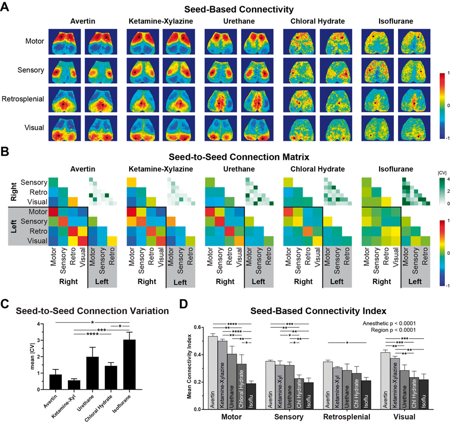
Conclusions
Avertin and ketamine-xylazine produce the most robust and consistent measures of RSFC under our experimental conditions, comparable to previous reports in awake mice. Urethane anesthesia yields weaker measures of RSFC. Chloral hydrate and isoflurane anesthesia are not suitable for experiments of this type.
References
PB03-R05
A combined cerebral blood flow method with R2 prime for calibrated fMRI in different states in animal
1iHuman Institute, ShanghaiTech University, Shanghai, China
2School of Life Science, ShanghaiTech University, Shanghai, China
3Institute of Neuroscience, CAS Center for Excellence in Brain Sciences and Intelligence Technology, Chinese Academy of Sciences, Shanghai, China
Abstract
Objectives
For basic science, neuromodulation and metabolic activity alters the baseline of brain activity, making relative measurements nearly impossible to interpret. Thus, we are focusing on developing techniques to measure absolute metabolism in the brain using MRI. “Calibrated fMRI” refers to methods to attempt to measure oxygen concentration using fMRI. We are further developing a technique called “M-Mapping” [1, 2]which, unlike other methods, does not require additional agents or gases.
Methods
To use M-Mapping, standard MRI images of relaxation (R2’) and cerebral blood flow (CBF) are collected.[1] Then, a biophysical model is applied to calculate CMRO2, the cerebral metabolic rate of oxygen use. Data recorded from C57BL/6 mice were divided into two states. One is awake by fixing the mice brain into the holder by a surgery, another is anesthetized with 2% isoflurane after finishing the awake experiment.[3] T2 is measured by a MSME(multi-slice, multi-echo) sequence while T2* is measured by a MGE(multi-slice gradient echo) scan. CBF was calculated by pCASL(pseudo-continuous arterial spin labeling) scan.[4, 5] To increase SNR we conducted a method using mean value from different individuals measured by the same method and condition.
Results
Relaxation map from MRI of the mean R2’ parameter, showing relaxation rate differences due to oxyhemoglobin/deoxyhemoglobin balance. Mean CBF map from awake, showing a weaker blood flow rate around brain. Relative cerebral metabolic rate of oxygen consumption (rCMRO2) was based on CMRO2 in the awake state divided by the anesthetized state, demonstrating awake mice has a greater value in the cortex than anesthetized mice. We are still evaluating subcortical measurements due to perfusion differences from the cortex.

Conclusions
A higher R2’ parameter and a lower CBF leading to a higher baseline in awake mice versus isoflurane. This is possibly caused by large-scale vascular dilation which has been long observed under isoflurane. This vascular dilation likely influences the hemodynamic response seen in fMRI, showing the potential of this method. For further study, medetomidine will be tested.
References
PB03-R06
Blood flow and volume measurements are critical for determining true extent of brain tumour periphery
1Cancer Research UK and Medical Research Council Oxford Institute for Radiation Oncology, Department of Oncology, University of Oxford, United Kingdom
Abstract
Objectives
When treating brain tumours, it is important that the true tumour extent is known. However, current techniques for determining tumour extent are subjective, with high inter-observer error1, and likely do not accurately capture the tumour periphery, a consequence of poor contrast and oedema2. MRI is capable of providing an array of independent measurements of brain structure and function, including information on cerebral blood flow and volume (CBF and CBV) which may allow more accurate tumour definition.
Here we use a rat brain tumour model to test more accurate mapping of tumour periphery with multimodal MRI, compared to traditional diagnosis and histological gold-standard.
Methods
Brain tumours were induced in BD-IX rats by intrastriatal injection of ENU1564 cells. All animals underwent MRI at week 3. Subsequently, animals were divided into three groups: (1) killed post-MRI, or (2) 25 Gy targeted radiotherapy or (3) sham radiotherapy followed by MRI and killing at week 4. Acquired MRI data included CBV mapping by infusion of iron oxide nanoparticles, CBF by multiphase arterial spin labelling3, apparent diffusion coefficient, T1 mapping, T2 mapping, and post-Gd T1-weighted imaging.
The gold-standard for tumour location was abnormal histological vessel staining (collagen IV), hypoxia (pimonidazole) and cellularity (cresyl violet). Vessel properties calculated for determining abnormality were density, diameter, in-plane length, individual area, and vessel area fraction. All histological images were co-registered to MRI-space using perspective transforms, combining multiple sections after elimination of damaged tissue.
Four expert observers identified tumour extent using post-Gd T1-weighted images in line with current clinical practice. Multimodal MRI was used to find tumour extent through combinations of abnormal parameters. Sensitivities and specificities for detecting tumour were calculated and tumour sizes were compared. The most important modalities contributing to tumour detection were determined.
Results
Tumour volume was found to be significantly greater by both histology and multimodal MRI than by expert observer (p < 0.01; Figure, A). Sensitivity for tumour detection was increased 45% using multimodal MRI compared to expert observer (0.81 vs. 0.58, p < 0.001), but specificity was decreased 14% (0.81 vs. 0.94, p < 0.001). All MRI modalities contributed to identification of tumour, but different classes of tumours were not primarily dependent on the same parameters. Large heterogeneous tumours were most dependent on abnormal CBF and CBV measurements, whilst smaller tumours were more dependent on post-Gd T1-weighted images and baseline T1 relaxation time (Figure, B).
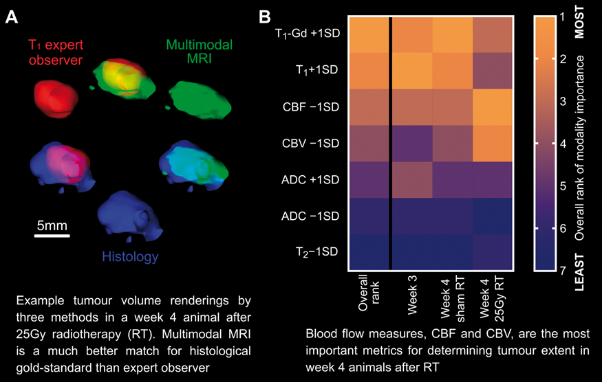
Conclusions
Blood flow and volume measurements are the most important MRI parameters for identifying late-stage tumour extent, especially with complex pathology post-radiotherapy. For smaller tumours, abnormalities in blood flow and volume still contribute to identifying tumour extent, but are not as important as more traditional metrics, such as post-Gd T1-weighted imaging. Importantly, late-stage tumours are the most problematic for determining the true extent, as artefacts from therapy can confound expert observer opinion regarding tumour location. Measurements of CBF and CBV are both feasible in a clinical setting and trials in patients are now warranted.
References
PB03-R07
Multiparametric analysis of MRI using machine learning allows stroke identification and stroke core prediction
1Department of Nuclear Medicine and Clinical Molecular Imaging, University Clinic Tuebingen, Germany
2Department of Preclinical Imaging and Radiopharmacy, University of Tuebingen, Germany
3Department of Neurology, University Clinic Tuebingen, Germany
4Department of Neuroradiology, University Clinic Tuebingen, Germany
5Institute for Pathology, University Clinic Tuebingen, Germany
Abstract
Early determination of stroke volume (SV) has great importance for understanding stroke pathophysiology, evaluation of therapies and planning of rehabilitation. Edema development in the stroke on the first few days after occlusion makes final infarct identification extremely challenging. Here, we developed a simple and easily reproducible machine learning (ML) framework using rat MRI data from 24 h after stroke-onset to identify and predict SV 1-week post-stroke induction. Additionally, we evaluated two neuroprotective therapies.
Strokes were induced using the middle cerebral artery occlusion model on 30 rats. Animals were scanned with MRI at 24 h and 1-week after stroke-onset. ADC (apparent diffusion coefficient maps) and T2 weighted images (T2WI) were acquired per time-point. Twelve rats were used as control subjects, 10 rats received whole-body hypothermia therapy and 8 rats received combined 100% O2 and whole-body hypothermia. Animals were sacrificed and histology from all animal groups was performed at 24 h (n = 7) or after 1-week (n = 8). For the ML framework, a Gaussian mixture model was applied on the combined ADC-T2WI dataset (24 h) using all control rats to find biologically distinct clusters. The cluster in agreement with stroke characteristics was then used to train a random forest classifier (RFC). Next, we applied the trained RFC on therapy datasets to identify regions with similar characteristics. To compare automated output to human prediction, an experienced user blindly drew three sets of regions of interest (ROIs) on the SV identified using only ADC images, T2WI alone and the combined ADC/T2WI datasets at 24 h. To determine final SV, a “ground truth” (GT) ROI was manually drawn by the user using the combined T2/ADC images from 1-week together with corresponding histology.
The framework produced clusters with clear radiological stroke characteristics (

We developed a simple post-processing tool for automatized SV evaluation using MRI at 24 h that can be used to predict SV 1-week after stroke-onset. The clustering output of this model identifies tissues with pronounced stroke characteristics making it a very useful tool for automatic infarct core prediction. The method still slightly overestimates SV in comparison to GT, perhaps due to a mixture of image resolution, artifacts or the conservative prediction of the model. An increment in trained data will likely improve prediction. The remarkable volume difference between the user-based approach at 24 h and the 1-week histology based ROI shows the confounding effect of edema and further denotes the necessity to use specialized tools for early SV assessment.
PB03-R08
Absolute tissue pH MRI refines ischemic penumbra mapping in an experimental model of acute ischemic stroke
1Martinos Center for Biomedical Imaging, Massachusetts General Hospital and Harvard Medical School
2Yerkes Imaging Center, Yerkes National Primate Research Center, Emory University, USA
Abstract
Objectives
Amide proton transfer (APT) MRI is sensitive to tissue pH change during acute ischemia 1. However, the commonly used analysis provides pH-weighted image with unsatisfactory pH specificity, limiting its use in the acute stroke setting 2-4. Our work aims to calibrate the recently proposed pH-specific MRI toward absolute tissue pH mapping and determine graded regional pH changes during acute stroke 5.
Methods
The experimental study has been approved by the local IACUC. Fifteen acute stroke rats underwent multi-parametric MRI and lactate MRS using a 4.7 T MRI scanner with multi-slice EPI. We collected water-suppressed single voxel point resolved spectroscopy (PRESS), T1, diffusion, APT and perfusion MRI. Two rats were excluded due to failed stroke surgery. Another group of twenty acute stroke rats underwent multi-parametric MRI (without lactate MRS).
Results
We calibrated pH-weighted MTRasym and pH-specific ΔMRAPTR indices toward tissue pH, determined from quantitative lactate MR spectroscopy (MRS). Specifically, pH was derived from the lactate content according to pH = -0.0335*[Lac] + 6.83 6,7. MTRasym and ΔMRAPTR were numerically fit with respect to pH. For MTRasym, we had pH = log10((100*MTRasym-C2)/C0)/C1, with C0, C1 and C2 being 0.46, 0.19 and −13.9 (R2 = 0.45, P < 0.05). For ΔMRAPTR, we had pH = 7.05 + log10(ΔMRAPTR/ C'0 + 1)/C1, with C'0 and C1 being 5.04 and 0.25, respectively (R2 = 0.70, P < 0.001).
Fig. 1 shows perfusion, diffusion and pH images from a representative acute stroke rat. There was noticeable lesion mismatch among perfusion (Fig. 1a), diffusion (Fig. 1b) and pH maps (Fig. 1c). Whereas the diffusion lesion core showed the worst pH drop, the peri-infarct perfusion/diffusion lesion mismatch shows heterogeneous and mild pH changes. Fig. 1d shows perfusion/pH lesion mismatch (red, benign oligemia), pH/diffusion lesion mismatch (green, metabolic penumbra) and diffusion lesion (black, ischemic core), being 21 ± 12%, 34 ± 18% and 44 ± 16% of the hypoperfusion lesion, respectively.
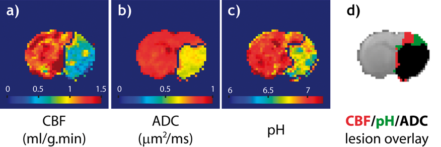
Conclusions
Our study developed fast non-invasive absolute tissue pH imaging. pH MRI provides a metabolic imaging index for stratification of heterogeneous ischemic tissue injury.
Acknowledgements
This study was supported in part by grants from R01NS083654. The author would like to thank Drs. Enfeng Wang and Takahiro Igarashi for technical support.
References
PB03-R09
Non-invasive detection of vascular normalisation in a rat brain metastasis model
1CRUK and MRC Oxford Institute for Radiation Oncology, Department of Oncology, University of Oxford, Oxford, UK
2Institute of Biomedical Engineering, Department of Engineering, University of Oxford, Oxford, UK
Abstract
Objectives
Brain metastases are the most common brain tumours found in adults and are associated with poor prognosis, typically only a few months. They are detected by magnetic resonance imaging (MRI) once blood-brain barrier breakdown has occurred, implying that these tumours are already large and likely hypoxic, a feature associated with a poor response to conventional therapies. To address this poor response, vascular normalisation (VN) is a newly employed strategy to modulate the tumour microenvironment by reshaping the tumour vasculature and allowing the immature and leaky blood vessels to mature, often reducing hypoxia. The current detection method for VN is dynamic-contrast enhanced (DCE) MRI, a technique relying on gadolinium-based contrast agents that can deposit in bone, tissue and brain, and has been linked to cases of nephrogenic systemic fibrosis.
To circumvent these issues, we propose non-invasive MRI techniques like arterial spin labelling (ASL) and amide proton transfer (APT) to detect VN in brain metastases. We aimed to compare the sensitivity of these non-invasive MRI techniques with DCE-MRI for VN detection in a rat model of brain metastasis.
Methods
Female Berlin-Druckrey IX rats were injected in the left striatum with ENU1564 cells (rat mammary carcinoma) and tumours were grown until >10 mm3 on T2-weighted anatomical images (2-3 weeks) before treatment. For treatment, rats received daily intravenous injections of cediranib (4 mg/kg), a vascular endothelial growth factor receptor 2 inhibitor, or vehicle control. 9.4 T MRI was performed on day 0 before intial cediranib injection and 2 hours post-treatment on days 1 and 2. Sequences used were multiphase pseudo-continuous ASL (8 phases, label duration = 2.4 s, post-label delay = 650 ms) [1], APT-MRI (2 s pulsed saturation, continuous wave equivalent power 0.55µT, 34 frequencies) [2] and DCE-MRI (flip angle = 5°, frame rate = 8.3 s, 60 × ), as well as T1-weighted pre- and post-gadolinium injection.
Results
Ktrans, the clinically-used vascular permeability parameter derived from DCE-MRI, was calculated to confirm VN in these tumours. The cediranib treated group showed a 65 ± 11% decrease in tumour Ktranstwo days after treatment initiation compared to a decrease of 4 ± 3% in the vehicle treated rats, indicative of VN in the cediranib-treated brain metastases (Student’s t-test, p = 0.005). Non-invasive MRI measures also differed between groups. The reduction of cerebral blood flow (CBF), as determined by ASL, was significantly less pronounced in cediranib-treated compared to vehicle-treated animals 2 days post-treatment (2-way ANOVA, Dunnett’s multiple comparison, p = 0.01). Furthermore, the APT ratio obtained from APT-MRI, showed a significant decrease over time in the vehicle-treated tumours, while no significant change was seen in the cediranib-treated metastases (Pearson’s correlation p < 0.001).
Conclusions
Non-invasive MRI can be used to detect and follow VN in brain metastases and could potentially replace DCE-MRI, particularly in patients with comorbidities involving the kidneys. Further, brain metastasis patients could be followed more closely with ASL and APT-MRI since the risk associated with gadolinium deposits accumulating in tissues is removed. A more detailed follow-up using ASL and APT-MRI may result in an improved treatment plan and potentially lead to a more tumour-tailored therapy.
References
PB03-S01
Real-time two-photon imaging study reveals increased blood-brain barrier permeability in chronically stressed mice
1Center for Neuroscience Imaging Research, Institute for Basic Science (IBS), Suwon, 16419, Republic of Korea
2Department of Biomedical Engineering, Sungkyunkwan University (SKKU), Suwon, 16419, Republic of Korea
3Biomedical Institute for Convergence at SKKU (BICS), Sungkyunkwan University (SKKU), Suwon, 16419, Republic of Korea
4Department of Biological Science, Sungkyunkwan University (SKKU), Suwon, 16419, Republic of Korea
5Samsung Advanced Institute of Health Science and Technology (SAIHST), Sungkyunkwan University (SKKU), Suwon, 16419, Republic of Korea
Abstract
Objectives
Repeated exposure to multiple stressors can alter brain architecture1. Consistent with above papers, the phenomena related to hypoperfusion were observed in chronically stressed rodents and human brain2–3. In addition, it has been reported that the blood brain barrier (BBB) is also affected4. However, according to the report of Roszkowski M et al., JCBFM in 2016, the BBB permeability is not increased in the stress model. Therefore, the relationship between increased BBB permeability and stress is not clarified yet. Our main hypothesis is that, the sustained reduction of cerebral hemodynamics can lead to hypoxic condition and hypoxic condition could turn to increased vascular endothelial growth factor (VEGF), which affects vascular plasticity. The tight junction expression is also altered in relation to vasculature changes. This sequence can lead to the increased BBB permeability. In this study, the long-term effects of chronic stress on BBB permeability are investigated using longitudinal in vivo 2p dynamic imaging.
Methods
For repeated imaging of the cerebral vasculature, a cranial window 3 mm in diameter was made in somatosensory cortex. Mice had a recovery period of 4–6 weeks and imaged using 2p microscopy before stress exposure and at the end of the 3-week stress exposure. To induce chronic stress, mice were immobilized with a plastic bag in their home cage 6 hours per day for 3 weeks. For the BBB permeability quantification, we used thinned skull window by thinning the skull to ∼30um. After surgery, animals received 70 kDa Texas-red dextran to visualize vessel structure. Then, 40 kDa FITC dextran was delivered through an intravenous catheter after a 30 sec image acquisition.
Results
We confirmed the significant reduction of vascular volume in chronically stressed model. We also confirmed increased BBB permeability at 40 kDa. But no significant changes were observed for 70 kDa dextran. We concluded that, chronic stress increased BBB permeability but did not induce BBB disruption. We were able to infer that these increases in BBB permeability could be due to the decreased expression of the claudin-5, possibly resulting from increased expression of VEGF and its receptor.

Conclusions
Prolonged exposure to stress may lead to a decrease in the vessel diameter and it could been considered as decreased blood flow, and that these changes could be a driving force for reshaping the vascular structure as well as BBB permeability.
References
PB03-S02
Real-time two-photon imaging of hippocampal neuro-vasculature and BBB permeability in chronic temporal lobe epilepsy model
1Center for Neuroscience Imaging Research (CNIR), Institute for Basic Science (IBS), Rep. of Korea
2Dept. of Biomedical Engineering, Sungkyunkwan University, Rep. of Korea
3Biomedical Institute for Convergence at SKKU (BICS), Sungkyunkwan University, Rep. of Korea
4Samsung Advanced Institute of Health Science and Technology (SAIHST), Sungkyunkwan University, Rep. of Korea
Abstract
Objectives
Blood-brain-barrier (BBB) damages have been proposed to contribute to epileptogenesis (1). However, it is still unclear how BBB damages are related with neuronal and vasculature alteration in chronic epilepsy. Pilocarpine, which is the cholinergic agonist, is common drug for experimental model of chronic temporal lobe epilepsy (TLE) (2). In TLE model, the hippocampus is known to be particularly vulnerable. Recently, direct hippocampal imaging technique was used to investigate hippocampal function real-time in vivo using two-photon microscopy (3). Using this technique, we investigated BBB damages, and neuronal and vasculature alterations in hippocampus area.
Methods
Hippocampus was imaged after the removal of cortex and the installation of imaging chamber. We confirmed the effects of surgery and installation with two-photon imaging and histological evaluation. We induced status epilepticus (SE) with pilocarpine in adult mice, and investigated vascular and cellular properties in the hippocampus at different time points, i.e., pre- and post-SE in vivo by using two-photon microscopic imaging. Calcium signal was simultaneously recorded in Thy1-GCaMP6s transgenic mice for observation of neuronal activation during SE and correlated with cellular and vascular changes.
Results
Vascular structure was imaged up to the depth of 600μm using 70kD FITC dye. BBB permeability was measured in anesthetized state with ketamine/xylazine and isoflurane. Two anesthetic agents did not produce any difference in results (5.831 x 10⁻⁴ ± 2.084 x 10⁻⁴ mm−1, 8.168 x 10⁻⁴ ± 2.084 x 10⁻⁴ mm−1; p = 0.4426). NeuN and Iba-1 counting results showed that the counting was similar in both hippocampal side. After the induction of SE, real-time two-photon imaging was carried out to investigate how SE affected BBB and neuronal and vasculature in hippocampus.
Conclusions
Imaging deep subcortical area, like hippocampus, with two-photon microscopy is powerful way to provide new information regarding the effects of SE in subcortical area with high resolution. Real-time BBB permeability, cellular calcium signal activity and vasculature of hippocampus can be detected with this technique. The results can shed new light on how SE affects hippocampus and eventually the mechanism of epileptogenesis in chronic TLE model.
References
PB03-S03
Age-related changes at the blood-brain barrier. A comparative in vivo study in Wistar rats
1Faculty of Information Technology and Bionics, Pázmány Péter Catholic University, Hungary
Abstract
Objective
Severeal articles reported increased permeability and altered morphology of the blood-brain barrier (BBB) with advanced age. Our study aimed to compare the structure of brain microvessels and function of P-glycoprotein at the BBB in young and aged rats.
Methods
Male Wistar rats 2–3 months (young) and 14–16 months (middle aged) were studied. Dual and triple-probe microdialysis techniques were used to compare BBB permeability for quinidine (QND) in young and aged rats in presence and absence of a specific P-gp inhibitor (PSC-833). Concentrations of QND were analyzed by LCMS-MS. Furthermore, comparative MR imaging of the brains was performed to study anatomical changes, and also single photon emission computed tomography (SPECT) imaging was applied for comparison P-gp functionality. For ultrastructural changes of the brain capillaries were analyzed by electronmicroscopy.
Results
The control level of QND in absence of PSC-833 was higher in aged than in young rats. However, in presence of PSC-833, the brain levels increased less in aged than in young animals suggesting lower expression level or impaired functionality of P-gp in old subjects. In MR imaging the extension of cerebral ventricles increased significantly and there were also characteristic ultrastructural changes at the BBB with aging by electronmicroscopy.

Figure
MR imaging of the brain of a representative young adult and a middle aged Wistar rats. Coronal
Conclusions
Our results indicate many differences between young adult and aged rats in the structure and function of the BBB. Our findings suggest a lower expression and/or reduced P-gp function with aging and, on the other hand, some other changes in the cerebral capillaries which are against the brain entrance of dangerous substances as it was shown by electronmicroscopy.
References
PB03-S04
The role of equilibrative nucleoside transporter-2 on the amelioration of acute lipopolysaccharide-induced breakdown of the blood-brain barrier
1School of Pharmacy, National Taiwan University, Taiwan
Abstract
Objectives
The disruption of the blood-brain barrier (BBB) can increase the permeability and may allow the entry of blood-derived proteins into the brain. Given that adenosine signaling can modulate BBB permeability (1), this study was aimed to investigate lipopolysaccharide (LPS)-induced BBB dysfunction in mice with and without the expression of equilibrative nucleoside transporters-2 (Ent2), one of the major nucleoside transporters controlling extracellular adenosine level in the brain (2).
Methods
Ent2 knockout (Ent2 -/-) mice were generated by CRISPR-Cas9 technique. Single intraperitoneal (i.p.) administration of LPS (2.5 mg/kg) was given to Ent2 -/- mice and the wild-type littermate controls. Twenty-four hours after the treatment, the expression of ZO-1 and claudin-5 at brain microvessels were examined by immunofluorescence. The BBB permeability was evaluated by brain accumulation of sodium fluorescein (10 mg; i.p.) and endogenous IgG.
Results
Acute LPS treatment significantly reduced the expression of tight junction proteins, along with an increase in the fluorescein intensity (from sodium fluorescein) and the accumulation of IgG in the brain of the littermate control mice. Conversely, Ent2 -/- mice manifested rescue of the LPS-induced BBB dysfunction.
Conclusions
These findings demonstrate that gene deletion Ent2 can protect against LPS-induced BBB dysfunction in mice. Further study is required to understand the underlying mechanism of its protective effects and to evaluate the therapeutic potential of Ent2 inhibition.
References
PB03-S05
Evidence that enolase-phosphatase 1 increases BBB disruption by interfering ADI1-MT1-MMP interaction and activation of MT1-MMP/MMP-2 axis in early ischemic damage
1The Central Laboratory, The First Affiliated Hospital of Shenzhen University, Shenzhen Second People’s Hospital, Shenzhen University School of Medicine, China
2Department of Pathophysiology, Baotou Medical College, China
Abstract
Objective
Enolase-phosphatase 1 (ENOPH1) is a newly identified enzyme associated with stress responses, which is required for the synthesis of polyamine. Our previous study found ENOPH1 mediating BBB injury under cerebral ischemia conditions, but the mechanisms underlying this role remain unknown. The aim of this study was to determine whether ENOPH1 could aggravated MT1-MMP/MMP-2 mediated early BBB disruption following ischemic injury.
Methods
Cultured brain microvascular endothelial cell lines (bEND3 cells and HBMVEC cells) were respectively transfected with ENOPH1 siRNA and CRISPR-activated plasmid, and then exposed to oxygen-glucose deprivation (OGD) condition. The MMP-2/9 of conditioned media (CM) and cellular extracts (CE) were analyzed by gelatin zymography. The level of tight junction proteins were identified by western blot assay and the interaction between ADI1 and MT1-MMP was measured by co-immunoprecipition assay. BBB disruption was assessed by endothelial cell monolayer permeability assay.
Results
(1) OGD for 6-h significantly increase MT1-MMP mRNA and protein expression in cultured brain endothelial cells; (2) Knockdown of ENOPH1 promoted downregulation of MT1-MMP protein level and decreased the activity of MMP-2 in bEND3 cells or HBMVEC cells after OGD 6-h treatment. (3) ENOPH1 appears to competitively attenuate the interaction of ADI1 and MT1-MMP by promoting ADI1 cytoplasmic translocation in endothelial cells under ischemia condition. (4) Knockdown of ENOPH1 ameliorate OGD-induced endothelial barrier disruption through increasing expression of tight junction proteins.
Conclusion
These results suggests that activation of MT1-MMP/MMP-2 axis by ENOPH1 is an important mechanism to interfere the stability of BBB after the early cerebral ischemia.
PB03-S06
CSF1R+mono/macrophages and blood brain barrier disruption after stroke
1Department of anesthesiology, Renji Hospital, school of Medicine, Shanghai Jiaotong University, Shanghai, China
Abstract
Objective
Ischemic stroke remains as one of the heaviest disease burdens worldwide due to its high incidence, high mortality and disability. Blood brain barrier (BBB) damage after ischemic stroke is an important pathological feature that may elicit hemorrhagic transformation and it is closely related to poor neurological outcomes. The peripheral mono/macrophages are highly plastic during their cross talk with the ischemic brain injury however their role on blood brain barrier damage remains elusive.
Methods
We established the transient middle artery occlusion (tMCAO) models of 2 hours in C57BL/6 male adult mice under laser speckle monitoring of cerebral blood flow. Clodronate liposomes (CLP,1 mg/200ul) was intraperitoneal injected to deplete the peripheral mono/macrophages 24 h before tMCAO. Fluorescein sodium leakage assay and crystal violet staining were performed to detect increased BBB permeability and hemorrhagic lesions. Tight junction protein integrity was examined by immunofluorescence (IF) staining and western-blot in ischemic brains 6 hours, 1 day and 3 days after tMCAO.
Results
Three days after tMCAO, the content of fluorescein sodium in the infarcted brain of CLP treated mice was significantly higher than that of PBS treated mice. Crystal violet staining showed more hemorrhagic lesions in CLP treated tMCAO mice compared to PBS treated tMCAO mice. Western-blot and IF staining showed more severe degradation of ZO-1 in CLP treated mice. The number of CSF1R+cells were increased in the infarct area after tMCAO. ELISA showed that the level of IL-34, the ligand of CSF1R was slightly increased in the plasmathe first 3 days after tMCAO. The protein level of IL-34 in the ischemic brain was decreased 6 hours after tMCAO and returned to baseline 1 day and 3 days after tMCAO. However IF showed that the distribution of IL-34 translocated from inside to outside of the ischemic neurons 3 days after tMCAO.
Conclusions
The current data suggest that ischemic neurons may interact with CSF1R+mono/macrophages, which could play a role in protecting the integrity of BBB after stroke.
PB03-S07
Surgery trauma associated macrophage migration inhibitor factor (MIF) aggravates blood brain barrier disruption following perioperative stroke
1Department of Anesthesiology Renji Hospital, School of Medicine, Shanghai Jiaotong University, Shanghai, China
Abstract
Objectives
Perioperative stroke is gaining increasing attention due to its high incidence and devastating consequences in surgical patients[1, 2]. The ischemic brain injury may be complicated by profound impacts of surgery, anesthesia, stress, pain and et al. However, the mechanisms that underlie the exacerbated ischemic brain injury in perioperative stroke remains largely unknown. Macrophage migration inhibitory factor (MIF) is a multifunctional cytokine that has been suggest to function as an important initiator of inflammatory response and a pro-apoptotic nuclease [3, 4]. In this study we sort to examine the role of MIF in the exacerbated ischemic brain injury after perioperative stroke.
Methods and results
Tibia fracture was performed 24 hours before distal middle cerebral artery occlusion (dMCAO) to induce perioperative stroke in C57BL/6 J mice. We found that the infarct volume and disruption of blood brain barrier was significantly increased 3 days after stroke in the perioperative stroke group compared to stroke alone group. Neurological assessments revealed that surgery exacerbated sensorimotor dysfunction 3–5 days after stroke. Using immunostaining, we found increased TUNEL+ endothelia cells (ECs), but not astrocytes or pericytes 3 days after perioperative stroke. The expression of PARP1 was increased in ECs, suggesting increased single strand DNA breaks in ECs in the ischemic brain after surgery. Flow cytometry demonstrated increased MIF expression in macrophages 3 days after perioperative stroke, and the increased MIF expression could not be induced separately by anesthesia, pain or stress, suggesting that surgical trauma could be more responsible for the increased MIF expression. Administration of MIF (60ug/kg) 3 hours following dMCAO increased DNA damage and TUNEL+ cells in brain CD31+ ECs, exacerbated the disruption of blood brain barrier and increased infarct volume growth. Using MIF-antagonist (ISO-1,3 mg/kg) 3 hours alleviated the exacerbated ischemic brain injury and blood brain barrier disruption after perioperative stroke.
Conclusions
Our results suggest that the blood brain barrier disruption and ischemic brain injury can be exacerbated in perioperative stroke. Increased MIF expression associated with surgical trauma may be a critical molecule to mediate brain endothelial DNA damage and blood brain barrier disruption in perioperative stroke.
References
PB03-S08
Novel PTP1B inhibitor protects blood-brain barrier function through the Akt/FoxO1 signaling pathway in brain ischemia
1Dept. of Pharmacol, Grad. Sch. Pharm. Sci., Tohoku Univ. Japan
Abstract
KY-226 is a protein tyrosine phosphatase 1B (PTP1B) inhibitor that protects neurons from cerebral ischemic injury (Brain Res 2018;1694:1–12). KY-226 restores Akt (protein kinase B) phosphorylation and extracellular signal-regulated kinase (ERK) reduction in transient middle cerebral artery occlusion (tMCAO) damage. However, the mechanisms underlying the neuroprotective effects of KY-226 are unclear. To address this, the effects of KY-226 on blood-brain barrier (BBB) dysfunction were examined in tMCAO mice. KY-226 (10 mg/kg, i.p.) was administered to ICR mice 30 min after 2 h of tMCAO. To assess Akt or ERK involvement, wortmannin (i.c.v.) or U0126 (i.v.), selective inhibitors of PI3K and ERK, respectively, were administered to mice 30 min before ischemia. BBB integrity was assessed by Evans blue leakage 24 h post-reperfusion. The levels of tight junction proteins, ZO-1 and occludin, were measured by western blotting; ZO-1 mRNA level was measured by RT-PCR. Compared to vehicle, KY-226 treatment prevented BBB breakdown and reduction in tight junction protein levels. KY-226 treatment restored ZO-1 mRNA levels post-reperfusion. Pre-administration of wortmannin or U0126 blocked the protective effects of KY-226 on ZO-1 protein and mRNA reduction in tMCAO mice. In mouse brain endothelial (bEnd.3) cells, lipopolysaccharide treatment reduced mRNA and protein levels of ZO-1, an effect rescued by KY-226 treatment. Further, KY-226 treatment restored phosphorylation of pAkt (T308) and its downstream target forkhead box protein O1 (FoxO1) (S256) in bEnd.3 cells. Collectively, we demonstrate that KY-226 protects BBB integrity by restoration of tight junction proteins, an effect partly mediated by Akt/FoxO1 pathway activation. Thus, protection of BBB integrity likely underlies KY-226-induced neuroprotection in tMCAO mice.
PB03-S09
Protective effect of targeted inhibition of TMEM16A on the blood-brain barrier integrity after ischemic stroke
1Department of Neurology, Drum Tower Hospital of Nanjing University Medical School, PR China
Abstract
Objectives
Inflammation plays a pivotal role in the blood-brain barrier (BBB) destruction after ischemic stroke1. Enhanced leukocyte adhesion to vascular endothelial cell is an essential event in inflammatory response. TMEM16A, a newly discovered protein regulating the calcium-activated chloride channels (CaCCs), is widely expressed in eukaryotes. Recently, some studies have suggested that TMEM16A is associated with stroke and inflammation2–3. In this research, we aim to further determine that targeted inhibition of TMEM16A exerts protective effects on BBB integrity after ischemic stroke and to explore the underlying molecular mechanisms. It may provide a novel clinical treatment for ischemic stroke targeting BBB destruction.
Methods
In animal studies, middle cerebral artery occlusion (MCAO) was performed in B6 mice. Some mice were subjected to the optimized dose of TMEM16A inhibitor or the same amount of PBS by caudal vein injection within 15 min after the onset of reperfusion randomly. We assessed the infarct volumes and functional outcomes at different time points after reperfusion. In addition, we performed IgG immunostaining and Evans blue extravasation to evaluate endothelial permeability. Western blot and immunofluorescence double staining were used to observe the expression of TMEM16A and the distribution of tight junction. In cell experiments, an in vitro BBB model was established by HBMEC cells. TEER, FITC-dextran and monocyte cell adhesion were used to assess the function and the structure of BBB. At the same time, ICAM-1 was tested by western blot. To further explore the underlying mechanisms, the nuclear translocation of NF-κB p65 was investigated. Co-IP was used to determine whether p65 can bind TMEM16A directly.
Results
1) TMEM16A mainly expressed in brain microvascular endothelial cells. The mRNA and protein levels of TMEM16A were increased after MCAO.
2) Compared with control groups, the infarct volumes and neurologic impairments were significantly attenuated in TMEM16A inhibition group after MCAO.
3) TMEM16A inhibition reduced BBB disruption, ICAM-1 expression and neutrophil infiltration after MCAO.
4) TMEM16A inhibition reduced in-vitro BBB model disruption, ICAM-1 expression and neutrophil infiltration after OGD treatment.
5) TMEM16A inhibition down-regulated ICAM-1 expression via NF-κB signaling pathway.
Conclusions
TMEM16A inhibition down-regulated ICAM-1 in an NF-κB -dependent manner, which could effectively prevent ischemia-induced brain injury by alleviating neutrophil infiltration, suggesting that TMEM16A is a promising therapeutic agent in ischemic stroke.
References
PB03-S10
Cognitive impairment seen in diabetes type 1 and 2 miodels paealleled blood brain barrier copromise and neuroinflammation and is reversed by poly(ADP-ribose) polymerase-1 inhibition
1Dept. of Pathology Laboratory Medicine, Lewis Katz School of Medicine Temple University, USA
Abstract
Objective
End organ injury in diabetes mellitus (DM) is driven by microvascular compromise (including diabetic retinopathy and nephropathy). Cognitive impairment is a well-known complication of DM types 1 and 2; however, its mechanisms are not known. We hypothesized that blood brain barrier (BBB) compromise plays a role in cognitive decline in DM.
Methods
In the 1st set of experiments, we used a DM type 1 model (streptozotocin injected C57BL/6 mice) and type 2 model (leptin knockout obese db/db mice), and assessed for cognitive performance, BBB permeability, gene profiling in brain microvessels, and neuroinflammation by immunohistochemistry 12 weeks after establishment of DM. In the 2nd set of experiments we treated DM type 1 and 2 animals with poly(ADP-ribose) polymerase-1 (PARP) inhibitor (olaparib) shown previously by us to be BBB protective and anti-inflammatory1, 2. After behavior assessment at 8 weeks, PARP inhibitor was continuously administered by osmotic pump. At 12 weeks, animals were assessed for cognitive performance and other parameters as in the 1st set of experiments.
Results
in the 1st set of experiments, we showed enhanced BBB permeability and memory loss (Y-maze, water maze) that were associated with hyperglycemia. Gene profiling in brain microvessels of DM type 1 and type 2 animals demonstrated deregulated expression of more than fifty genes related to angiogenesis, inflammation, vasoconstriction/vasodilation, and platelet activation pathways by at least 2-fold (including eNOS, TNFa, TGFb1, VCAM-1, E-selectin, endothelin, several chemokines and MMP9). Brain tissues from DM type 1 and 2 mice showed microglial activation, upregulated ICAM-1 expression, down regulation tight junction (TJ) proteins and diminution of pericytes coverage as compared to controls. Treatment with PARP inhibitor resulted in complete reversal of cognitive deficits by week 12 (seen DM type 1 and 2 at 8 weeks) without any effect on hyperglycemia. We are analyzing BBB permeability, gene expression, and microglia reaction, expression of TJ and pericyte markers.
Conclusions
Our findings indicate BBB compromise in DM in vivo models and its association with memory deficits, gene alterations in brain endothelium and neuroinflammation. Prevention of BBB injury by PARP inhibition may be a new therapeutic approach to prevent cognitive demise in DM.
References
PB03-T01
In vivo systemic reactions, including the morphological change of paravascular space, during water intoxication
1Dept. of Pharmacology, Keio University School of Medicine, Japan
2Dept. of Neurology, Keio University School of Medicine, Japan
Abstract
Objectives
Rapid intraperitoneal water injection causes acute hyponatremia that creates an osmotic gradient driving for water entry into the brain, leading to subsequent cerebral edema. Paravascular space (Fig.1, upper) has been suggested to participate in the fluid circulation in cerebral cortex, however, it has not been clarified whether it changes during water intoxication and the development of cerebral edema.
Methods
We have established a real-time in vivo two photon imaging with a closed cranial window (Tomita-Seylaz method) under isoflurane anesthesia (1–2%) to observe the paravascular space and astrocytes using CAG-GFP transgenic mice and intraperitoneal injection of sulforhodamine 101 (10 mM in Saline, 10 ml/kg), respectively. In addition to the morphological assessment of paravascular space and astrocytes, we simultaneously monitored electro-corticogram (ECoG), cerebral blood flow (CBF), heart rate, and arterial blood pressure to examine acute physiological responses to water intoxication. Arterial blood gas and electrolyte concentration were also analyzed before and after the water injection. Water intoxication was induced by intraperitoneal bolus injection of distilled water equal to 10% of body weight and observed the physiological responses until 40 min after the injection.
Results
We found that water intoxication induced hyponatremia and biphasic physiological reaction; the acute systemic reaction such as reduction of CBF, heart rate and arterial blood pressure, which was followed by the continuous reactions in ECoG and CBF. In the continuous reaction, heart rate and peripheral blood pressure were stable at the same level as pre-water injection. Astrocytes continuously swelled since water injection up to 140% of the volume of the pre-water injection, whereas paravascular space did not change in the acute reaction but got narrower gradually during the continuous reaction to a quarter of the pre-injection level (Fig.1).

Conclusion
Intraperitoneal water injection induced biphasic reaction in in vivo anesthetized mice. Further analysis of the biphasic reaction may lead to the better understanding of physiological function of paravascular space and pathological mechanisms of cerebral edema.
PB03-T02
Aquaporin-4 facilitator TGN-073 facilitates CNS fluid movement
1Center for Integrated Human Brain Science, Brain Research Institute, University of Niigata
2Department of Neurology, University of California, Davis
Abstract
The abundance of aquaporin-4 (AQP-4) inside the BBB strongly indicates the presence of unique water dynamics essential for brain function. Our newly developed AQP-4 facilitator TGN-073 penetrates the BBB and enters brain parenchyma. TGN-073 was originally identified in our laboratory as a potential AQP-4 ligand on the basis of previously identified physicochemical properties conserved among various AQP-4 inhibitors. We had earlier demonstrated AQP4 expressing Xenopus laevis oocyte bioassay showed 20% facilitation of the mean flux on following hypotonic shock. We subsequently assessed the effects of TGN-073 on CNS fluid movement.
Materials and Methods
We assessed the CNS fluid movement using MRI and H2[17]O, namely JJVCPE imaging. The concept of the JJVCPE imaging method and its validation studies have been published previously [1.2]. The study was approved by the Internal Review Board of University of Niigata and carried out in accordance with the Guidelines laid down by the NIH (USA) on the care and use of animals for experimental procedures. Five controls and five study adult male C57/BL6 mice, were anesthetized with an intraperitoneal administration of urethane and placed in the 7 T MRI scanner (Varian Inc, Palo Alto, Ca, USA). Rectal temperature was maintained at 37 ± 0.5°C using a customized temperature control system. 200 mg/kg of TGN-073 or equivalent amount of saline were administered intraperitoneally at 30 min before the study. For the dynamic imaging study, during 500 continuous MRI scans every 8 seconds, 0.2 ml normal saline containing 40% of H2 [17O O] was administered as an intravenous bolus injection at the 75th scan which decreased the signal intensity of images according to the concentration of H2[17]O.
Results and Discussion
A significant reduction in H2[17]O content (P < 0.05) was found in the cortex, indicating higher turnover of interstitial fluid of the cortex associated with AQP-4 facilitation. Interstitial fluid water moved to pericapillary space through the intracellular fluid space of astrocytes, and, eventually drains into the CSF. The current study showed that the AQP-4 system indeed works as an interstitial circulator, and facilitation of AQP-4 by TGN-073 increased turnover of interstitial fluid from the cortical area.

Reference
PB03-T03
Recruitment of the meningeal lymphatics by the blood-brain barrier
1Saratov State University
Abstract
The lymphatic vasculature is a part of the vascular system that is opened in the extracellular space providing reabsorption back of the filtered through capillaries plasma into the venous circulation. The situation with the brain is different due to blood-brain barrier (BBB) restriction of fluid filtration from the blood into the brain. The role of lymphatic system in the head and the neck and recently re-discovered the meningeal lymphatics in the brain drainage is actively discussed for a century. However, the anatomical pathways of the lymphatic drainage of the brain fluids and its physiological relationship with the BBB remain unknown.
Here we studied on mice the interaction between the BBB and the meningeal lymphatics. Our results clearly showed that the BBB opening by sound (a mild BBB disruption) or by photodynamic effects (PD, a strong BBB leakage) caused an increase in the diameter of the meningeal lymphatic vessels that was correlated to the rise of the BBB permeability. We also demonstrated that the opening of the BBB stimulated the accumulation of brain fluids in the cisterna magna that was more pronounced in the PD group vs. the sound group. These changes were accompanied by activation of the brain drainage and clearing from molecules, which crossed the BBB via the meningeal lymphatics.
These data suggest about the recruitment of the meningeal lymphatics by the BBB opening and homeostasis of the extracellular space in the central nervous system that sheds light on a physiology of the meningeal lymphatic drainage and the brain clearing after the BBB opening. These novel findings may call for a reassessment of basic assumption in mechanisms underlying the brain recovery after events associated with the BBB disruption such as stroke, traumatic brain injuries, tumor, dementia, diabetes, pain, hypertension, arthritis, neurodegenerative disorders.
This research was supported by grant from Russian Science Foundation 17–15-01263.
PB03-T04
Endogenous proteins reveal fluid flow patterns in the brain
1Department of Biomedical Engineering and Physics, Amsterdam University Medical Centers, Amsterdam, The Netherlands
Abstract
Objectives
Several publications suggest a brain clearance mechanism via channels around blood vessels. The glymphatic concept states that cerebrospinal fluid enters the brain along paravascular spaces around arteries, sustains bulk flow of interstitial fluid (ISF), and leaves the brain along paravascular spaces along veins1. In contrast, others suggest that ISF leaves the brain through flow along perivascular spaces around arteries in the opposite direction2. A third group concluded that ISF flow is absent and molecules disperse through the brain parenchyma by diffusion only3. The aim of the present study was to elucidate fluid flow patterns in the brain by analyzing the distribution pattern of endogenous proteins.
Methods
Previous studies relied on infusion and tracing of (mostly fluorescent) molecules and microspheres. Such data may be obscured by surgical interventions, pumping, anesthesia, and analysis at a limited number of time points. To avoid these limitations, we analyzed the distribution of two endogenous molecules that enter the brain via the choroid plexus and are carried by the cerebrospinal fluid (CSF), transthyretin (TTR) and immunoglobulin G (IgG). To this extent, 10 months old Wistar Kyoto rats (n = 10) were sacrificed under anesthesia, flushed with physiological saline solution and fixed with 4% paraformaldehyde at a perfusion pressure of 80 mmHg. Brains were removed, snap frozen, and sections were stained by immunohistochemistry (IHC) for TTR and IgG. Confocal imaging was performed on these sections and ImageJ software was used to quantify staining intensity.
Results
TTR is a 55 kDa protein, produced by the choroid plexus, which carries thyroid hormone through the brain. IHC staining and confocal imaging showed penetration of TTR from the CSF into the brain along arteries and across the pia mater. The intensity profile showed an exponential decay from the pia mater into the brain parenchyma. The half-maximal concentration was reached at a penetration depth of 419 ± 60 µm. Immunoglobulins are relatively large molecules (150 kDa). IgG staining showed a high intensity at the choroid plexus, confirming its entry into the brain at this location, and dispersion in the CSF and along arteries. We observed limited penetration of IgG from the CSF into the parenchyma. The intensity profile of IgG showed a steep exponential decay across the pia mater. The half-maximal concentration was reached at a penetration depth of 21 ± 3 µm.
Conclusions
Based on the ‘infinite’ time for distribution, molecules of different size would be expected to show a homogenous intensity profile when penetration is determined by diffusion alone. In the case of glymphatic flow, molecules of different size would also be expected to show a homogenous distribution profile in brain tissue. However, this was clearly not the case. Thus, these distribution patterns are more compatible with diffusion from the CSF into the brain parenchyma against a net outflow of ISF towards the CSF compartment, with some unknown contribution of breakdown and efflux across the blood-brain barrier.
References
PB03-T05
Elucidating brain water management in hypertension: a preclinical MRI study
1Dept. of Biomedical Engineering and Physics, University of Amsterdam, Amsterdam UMC, The Netherlands
Abstract
Objectives
Hypertension has been associated with alterations in vascular function, including endothelial dysfunction and arterial remodeling. These alterations may subsequently lead to structural and functional changes of the brain, ultimately contributing to the development of cognitive impairment and vascular dementia. However, exactly how vascular dysfunction contributes to these pathologies is still not fully understood.
Proper neuronal function is directly dependent on the composition, turnover, and amount of interstitial fluid (ISF) that bathes the cells. Most of the ISF is likely to be derived from ion and water transport across the brain capillary endothelial cells. A previous study showed increased ISF flow, altered ionic composition, and a tendency towards an increased water content of the brain in hypertensive rats1. To further elucidate differences in brain water management between normotensive and spontaneously hypertensive rats, we aimed to determine possible changes in diffusion and water exchange properties within the brain tissue by MRI.
Methods
Blood pressure and heart rate were measured in conscious spontaneously hypertensive rats (SHR, n = 10) and normotensive Wistar Kyoto rats (WKY, n = 10) of 11 months of age. MRI was performed on a 7T small animal MRI system. The MRI protocol consisted of T2w anatomical scans followed by apparent diffusion coefficient (ADC) and T1 mapping for measurements of water diffusion and water capillary exchange, respectively. After MRI, brain water content was determined by weighing the brain before and after desiccation. For image analysis, several anatomical structures were manually segmented and the volumes were measured. ADC and T1 maps were co-registered to the T2w scans, after which mean values were determined for the different brain regions.
Results
Both systolic and diastolic blood pressure, as well as heart rate, were significantly elevated in SHR. Body weight did not differ between the two strains, while the brains of SHR were lighter when compared to WKY rats. In line with the brain weights, total brain volumes were significantly lower in SHR. Notably, white matter volume was smaller in SHR, while the hippocampal volume was similar between groups. In contrast, both the third ventricle and lateral ventricles were remarkably enlarged in the hypertensive strain (Figure 1). Whereas whole brain water content was significantly higher in SHR rats, we did not find differences in hippocampal ADC values. Although we did not yet perform a detailed analysis of water exchange parameters using all dynamic T1 data, we did observe that ΔR1 values between pre- and post-contrast T1 scans tended to be lower in hypertensive rats, suggesting lower brain blood volume in this strain.
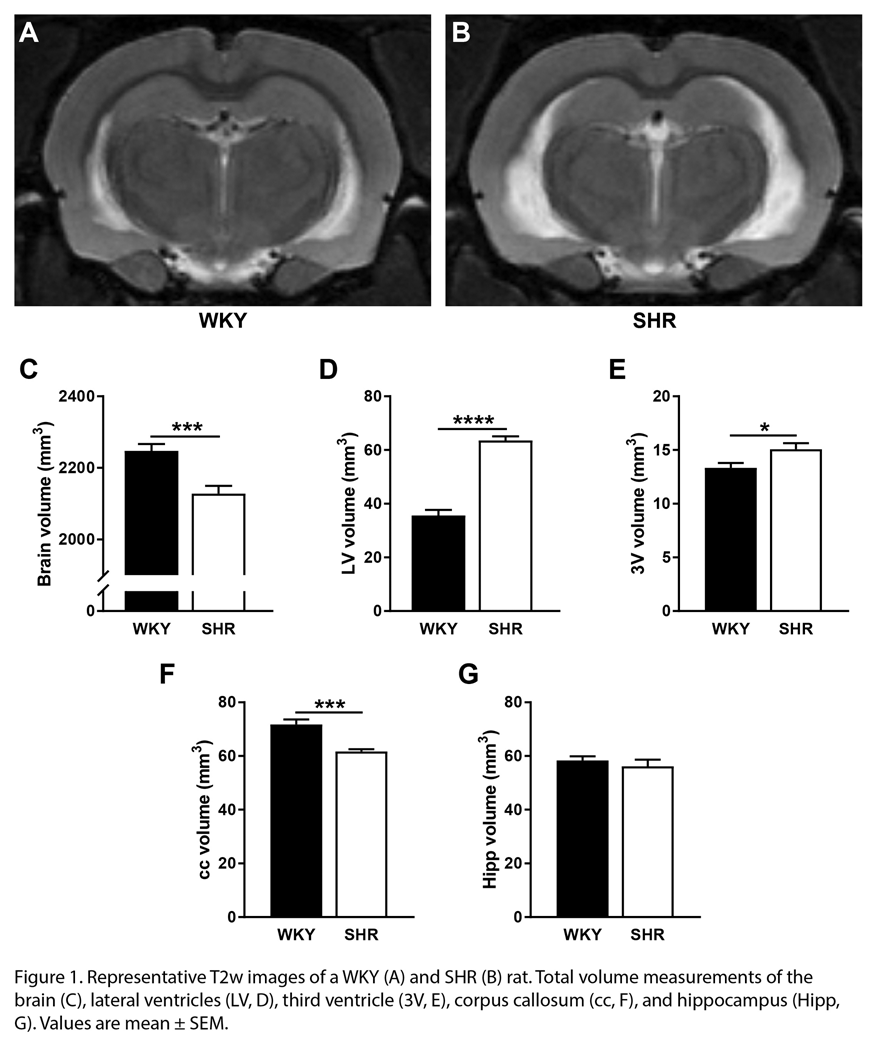
Conclusions
Brains of hypertensive rats showed increased ventricular volumes, as well as an increase in brain tissue water content. Whereas these changes indicate dysregulation of brain water management in hypertensive rats, the current limited analysis did not show significant changes in diffusion and exchange parameters explaining this imbalance. Further evaluation of brain capillary water exchange may provide further insight into the impact of hypertension on brain water management.
Reference
PB03-T06
3D diffusion-weighted MRI of perivascular fluid movement: Towards non-invasive mapping of glymphatic function
1UCL Centre for Advanced Biomedical Imaging, Division of Medicine, University College London, UK
2The Francis Crick Institute
3Neuroradiological Academic Unit, Department of Brain Repair and Rehabilitation, UCL Queen Square Institute of Neurology, London, UK
4Leonard Wolfson Experimental Neurology Centre, UCL Queen Square Institute of Neurology, Queen Square, London, UK
Abstract
Objectives
Efficient waste removal from the brain is essential to maintain normal physiology [1]. The glymphatic pathway describes a network of perivascular channels that facilitate rapid cerebrospinal fluid (CSF) transport and exchange with the brain’s interstitial fluid [2][3]. As such, perivascular channels play an important role in CSF-mediated clearance of potentially toxic molecules, such as amyloid-β [4]. Impairment to perivascular fluid movement, therefore, may lead to a build-up of deleterious proteins in the aging brain and eventually, the onset of neurodegenerative diseases such as Alzheimer’s disease. As such, the perivascular spaces (PVS) represent a promising target for non-invasive imaging biomarkers of upstream neurodegenerative processes. Recently, we have introduced the first non-invasive technique to assess the glymphatic pathway using ultra-long echo time (TE) diffusion-weighted MRI sequences to assess perivascular fluid movement in the rat brain [5]. The application of this technique, however, was limited to a single slice acquisition, confining the measurement to a relatively ‘upstream’ component of the glymphatic pathway; the PVS surrounding the MCA at the ventral aspect of the brain. In this pilot study, we use a 3D acquisition to examine more distal regions of PVS working towards whole brain mapping of perivascular function. Additionally, we investigate whether cardiac cycle-related vessel pulsatility drives perivascular fluid movement during arterial pulsation and diastole.
Methods
A fast-spin-echo 3D diffusion weighted sequence was acquired with the following sequence parameters: TR = 4s, effective TE = 133 ms, echo train length = 32, FOV = 30 mm x 30 mm x 8 mm, matrix (RO x PE x PE2) = 192 x192 x 16, 4 averages, b0 + 3 directions. Image acquisition was gated to the ECG signal to capture fluid movement in PVS during vessel pulsation and diastole. Region of interests were manually drawn around the PVS surrounding the left and right branches of the MCA at two regions. The pseudo-diffusion coefficient (D*) was calculated using methods outlined previously [5].
Results
Figure A is a schematic representation of the rat brain in basal and lateral orientations showing the anatomy of the MCA. The D* maps and graph (Figure B) show, in this animal, that perivascular fluid in both regions of the MCA has a greater D* during arterial pulsation compared to diastole. Moreover, the recorded D* values in the distal perivascular compartment are consistent with our previous characterisation of the PVS at the proximal aspect of the MCA. Here we provide preliminary evidence that we are successfully capturing PVS fluid movement at a more downstream target within the glymphatic pathway.

Conclusion
In this pilot study, we have non-invasively assessed fluid movement in a more downstream region of the glymphatic pathway in the rat brain. We have preliminary evidence to suggest that arterial pulsation may drive fluid movement in the PVS and could be an important mechanism underlying glymphatic inflow. This technique could serve as a non-invasive measure of glymphatic impairment, a potential biomarker of early neurodegenerative pathology.
References
PB03-T07
Intracranial fluids dynamics alterations and cortical thickness
1Institut de Mecanique des Fluides de Toulouse UMR 5502, Toulouse, France
2TONIC UMR 1214, INSERM, Toulouse, France
3University Hospital, Toulouse, France
4CHIMERE EA 7516, Amiens, France
Abstract
Objectives
The issue of cortical atrophy is important in normal aging and disease since it is associated with cognitive [1] and physical impairments [2]. Cortical atrophy is potentially a relevant biomarker for the early diagnosis of Alzheimer’s disease (AD) [3,4].
The vascular component is also an integral part of AD and other late-life neurodegenerative diseases. Abnormalities in blood flow appear before accumulation of abnormal proteins in AD [5–7]. The occlusion of capillaries by neutrophils are significantly higher in AD animal models than control and reduction of those occlusions with an antibody increases both blood flow and cognitive capacities [8]. Vascular alterations lead to hypoperfusion, oxidative stress and inflammation, which in turn lead to damage of neurons, glia and myelin, predominantly in the white mater [9].
Implication of vascular pathologies for gray matter remains unclear. A recent study showed that altered cerebral hemodyamics in asymptomatic carotid artery stenosis is associated with cortical thinning [10]. However there is no proven link between vascular pathologies and cortical thinning. We propose to explore brain aging with a combined biomechanical and imaging approach in order to assess both fluid dynamics alterations and brain structural modifications.
We hypothesize that there is a link between altered cerebral hemodynamics and loss of cortical thickness during brain aging.
Methods
80 patients suspected of hydrocephalus were prospectively involved. All patients complain of gait alteration, urinary difficulties, mild apathy and ventriculomegaly on brain imaging. They all underwent brain MRI with T1 weighted images to quantify cortical thickness and phase contrast images to measure arterial, venous and CSF velocities. Lumbar infusion test was also performed to gauge lumbar pressure, a surrogate marker of intracranial pressure (ICP), and CSF dynamics. The cortical volumetric segmentation was done by an automatic post-processing analysis with FREESURFER and local thicknesses were assessed with CorThiZon [3]. Venous, arterial and CSF velocities were measured from PCMRI with BIOFLOWIMAGE software. ICP and CSF dynamics were extracted form infusion tests. Pearson correlations were calculated between cortical thickness and arterial, venous and CSF velocities, but also ICP and derived indices.
Results
Mean cortical thickness is positively correlated with mean ICP (r = 0.48, p = 0.001), ICP pulse amplitude (r = 0.43, p = 0.001), arterial flow (r = 0.44, p = 0.001), aqueductal CSF flow(r = 046, p = 0.001), but negatively correlates with venous flow (r = -0.44, p = 0.001)
Conclusions
We demonstrate that cortical thickness is correlated with arterial and CSF pulsatility. The causality is more complex since it involves local microcirculation that could not be directly measured. However the association between intracranial pulsatility and gray matter thickness suggests that there is a relationship between vascular alterations at the macroscale level and the pathobiology of cortical atrophy.
References
PB03-T08
Water turnover in brain, ventricles and subarachoid spaces in normal volunteers: dynamic PET study using H215O
1Dept. of Neurosurgery, Nagoya City University Graduate School of Medical Sciences, Japan
2Dept. of Radiology, Nagoya City Rehabilitation Center, Japan
3Faculty of Health Science, Institute of Medical, Pharmaceutical and Health Sciences, Kanazawa University, Japan
4Dept. of Behavioral Neurology and Neuropsychiatry, Osaka University United Graduate School of Child Development, Japan
Abstract
Introduction
In order to clarify the origin and turnover of water molecules in CSF, dynamic PET (positron emission tomography) study was performed using radio labeled H2O.
Methods
Ten normal volunteers were included. Dynamic PET data were obtained for 15 minutes after intravenous injection of saline including H215O (500MBq). Voxels of interest (VOI) were set in the internal carotid artery (ICA), superior sagittal sinus (SSS), choroid plexus (CP), cortical gray matter (GM), white matter (WM), basal ganglia (BG), lateral ventricle (LV), Sylvian fissure (FS), and prepontine cistern (PPC). Time and relative radio activity (RAA) curves of each VOI were analyzed. To evaluate the accuracy and validity of our compensation method of dynamic PET data, dynamic PET of [18F]FDG (n = 3), [18F]DOPA (n = 3), and [11C]RAC (n = 3) were also performed.
Results
The maximum peak radio activities of GM, WM and BG were at 22.5, 50.0 and 22.5 seconds after the peak in ICA, respectively. At that time, the relative peak activities of GM, WM and BG were 53, 42 and 55% of the ICA peak activity, respectively. The activities in the whole brain structures decreased gradually. On the contrary, the activity of LV, FS and PPC increased gradually until the end of the measurement {12.9, 28.4, and 47.2% of the whole brain parenchyma (GM + WM + BG) activity at 9.5 minutes, respectively}. In dynamic PET of [18F]FDG (n = 3), [18F]DOPA (n = 3), and [11C]RAC (n = 3), no influx of radio-labeled molecules into ventricles during the same duration of measurement, which showed the accuracy of compensation method used in the present study.
Conclusions
The present study showed very fast movement of water molecules from artery to brain parenchyma and ventricular and subarachnoid CSF.
PB03-U01
Microbiota-immune cell interaction: critical role of tryptophan metabolites in stroke
1Institute for Stroke and Dementia Research, Klinikum der Universitat Muenchen, Munich, Germany
2Munich Cluster for Systems Neurology (SyNergy), Munich, Germany
3Department of Neurology, University Medical Center Hamburg-Eppendorf, Hamburg, Germany
Abstract
Introduction
Stroke is characterized by the recruitment of circulating immune cells to the injured brain. We have shown that immune cells migrate from the gut to the brain where they contribute to neuroinflammation and secondary brain injury [1,2].
There is accumulating evidence showing that metabolites derived from the gut microbiota influence brain diseases via the regulation of intestinal immune cell function. In particular, metabolites derived from the amino-acid tryptophan play a crucial role in immune tolerance. Importantly, tryptophan is the common precursor of ligands of the transcription factor aryl hydrocarbon receptor (AhR). AhR is highly expressed in immune cells and induces T cell polarization directly or via dendritic cells (DCs).
Objectives
Here, we seek to determine how the tryptophan/AhR pathway regulates peripheral immune homeostasis after stroke. Understanding how immune cells are modulated by gut metabolites may have strong therapeutic implications in stroke.
Methods
Cerebral ischemia is induced by the transient occlusion of the middle cerebral artery in wild-type C57BL6 mice or AhR-deficient-DC mice (CD11ccreAhrfx/fx). Blood and the distal small intestine are sampled for the detection of tryptophan metabolites and quantified by metabolomics (mass spectroscopy). Analysis of gut bacteria is performed by 16S rRNA sequencing from stool samples. Immune cells are isolated from the intestine and analyzed by flow cytometry.
Results
We found that bacteria from the genus Lactobacillus—known to metabolize tryptophan into AhR ligands—were significantly reduced in mice subjected to stroke. We demonstrated that tryptophan catabolism is augmented after stroke in comparison to sham surgery, shown by 1) a reduced plasma concentration of tryptophan and 2) an increase concentration in urine of indicans–the tryptophan metabolites directly derived from the microbiota metabolism–whereas the host metabolite kynurenine was not changed after stroke. These findings suggest that the gut microbiota involved in tryptophan metabolism is affected after stroke.
To test the implication of AhR on the tolerogenic function of DCs, we used AhR-deficient-DCs mice. Using a dimensionality reduction algorithm and an automated clustering analysis, we identified that the frequency of intestinal conventional DC1 was changed after stroke in an AhR-dependent manner, and correlate with an increase expression of Foxp3 in T cells.

Conclusion
These results are the first to link stroke-induced dysbiosis and the tryptophan/AhR pathway as immunomodulators of DCs and T cells. Findings derived from this study may allow for the identification of microbial metabolites-based therapies to alleviate the neuroinflammatory response to stroke.
References
PB03-U02
The role of Interleukin 10 in a murine model of stroke
1Dept. of Neurology, University Medical Center Hamburg-Eppendorf, Germany
Abstract
Objectives
The activation of the immune system has a profound impact on the fate of the ischemic tissue and the clinical outcome in stroke1. The evolving immune response in the ischemic brain is highly organized and several immune cells (e.g. γδ T cells) and pro-inflammatory mediators (Interleukin 17) are pivotal for the activation of the detrimental innate immune response2,3. The activation of the immune system is controlled by Interleukin 10 (IL-10) and regulatory T cells, which are restricting the harmful overwhelming immune response4. Even though several pro- and anti-inflammatory pathways are established, the precise interplay between neuroprotective and pro-inflammatory immune cells and cytokines are only incompletely understood. The goal of our study was to understand, whether IL-10 is able to control detrimental Interleukin-17 (IL-17) producing T cells in stroke.
Methods
We employed the transient middle cerebral artery occlusion (tMCAO) model and analyzed stroke sizes and brain atrophy in Il10-/-, WT and bone marrow chimeric mice by Magnetic Resonance Imaging (MRI) and Triphenyl-Tetrazoliumchlorid stainings (TTC). We analyzed the cellular inflammatory response in the ischemic brain and the peripheral immune compartment by flow cytometry, cell sorting and subsequent RT-qPCR. To study the effects of IL-10 and IL-17 in vivo we neutralized IL-17 in Il10-/- and WT mice and treated WT mice intracerebrally with recombinant IL-10.
Results
Gene deficiency of IL-10 resulted in significantly worsened neurological outcome and increased infarct size. The analysis of the cellular source of IL-10 in ischemic brains showed that Tregs and macrophages as well as brain resident microglia are producing IL-10. Bone marrow chimeric mice revealed that IL-10 in the peripheral immune system is responsible for the protective phenotype. Furthermore experiments in IL-10 deficient mice showed that IL-10 was able to control Th17 cells and IL-17 producing γδT cells (Figure) in the postischemic brain. Notably neutralization of IL-17 abolished the worsened neurological outcome in Il10-/- mice and intracerebral injection of IL-10 inhibited the IL-17 production in γδ T cells of WT mice.

Conclusion
Our results suggest that infiltrating IL-10-producing immune cells control the detrimental IL-17-dependent immune response in the ischemic brain tissue.
References
PB03-U03
Regulatory roles of IRF4/5 signaling in post-stroke inflammation and stroke outcomes
1Department of Neurology, University of Texas Health Science Center at Houston McGovern Medical School
Abstract
Objectives
The Inflammatory response is a fundamental pathophysiological procedure in stroke; the activation of phagocytes (microglia/monocytes) characterized by either pro- or anti-inflammatory state, is crucial in initiating and perpetuating the post-stroke inflammation [1]. Interferon regulatory factor 4 (IRF4) and IRF5 induce expression of inflammatory mediators in peripheral macrophages [2]. In the present study we tested our hypothesis that IRF4 and IRF5 signaling are key determinants of microglia/monocyte activation after stroke, and play a critical role in mediating ischemic injury.
Methods
Myeloid cell specific IRF4/5 conditional knockout (CKO) and Lenti-IRF4/5 virus treated mice were subjected to a 60-minute middle cerebral artery occlusion (MCAO) model. IRF4/5 floxed mice and Lenti-GFP treated mice served as controls. Ischemic lesion was quantified at 7d of stroke, and behavior deficits (NDS, corner test, open field, Y-maze) were evaluated at 3d and 7d. Microglia/monocyte activation and other immune cell infiltration in brains were examined by flow cytometry (FC), and phenotypes of these phagocytes were determined by cell membrane (CD68, CD206) and intracellular (IL-1b, TNF-a, iNOS, IL-4, IL-10) markers. Multiplex was performed to measure brain and blood cytokine levels at both timepoints. IRF4/5 mRNA levels were determined with RT-PCR in FC sorted microglia/monocytes.
Results
CKO of IRF4 or IRF5 resulted in higher levels of IRF5 or IRF4 mRNA respectively in FC sorted microglia and monocytes; Lenti-virus treatment also led to increased IRF mRNA levels in these phagocytes. At 3d of stroke, IRF4 CKO microglia/monocytes expressed higher levels of pro-inflammatory markers (CD68, IL-1b, TNF-a, iNOS) than the cells from controls; while increased levels of anti-inflammatory markers (CD206, IL-4, IL-10) were seen in IRF5 CKO vs floxed microglia/monocytes. However, Lenti-IRF5 and -IRF4 treatment induced higher expression of pro- and anti-inflammatory cytokines respectively in the phagocytes. Brain IL-1b and iNOS levels were significantly higher in IRF4 CKO vs control mice; IL-4 and IL-10 levels were significantly higher in IRF5 CKO vs control mice brains. Serum levels of cytokines exhibited the similar pattern as the levels of microglial cytokines, although IL-1b and IL-10 only showed a trend of the change pattern. At 7d of stroke, the pro-inflammatory profiles in IRF4 CKO and Lenti-IRF5 treated mice were quenched except the high serum levels of TNF-a; however, the up-regulated anti-inflammatory profiles persisted in IRF5 CKO and Lenti-IRF4 treated mice. Compared to the control mice, IRF4 CKO and Lenti-IRF5 treated mice had larger infarct volumes at 7d of MCAO; while smaller infarct volumes were seen in IRF5 CKO and Lenti-IRF4 treated mice. Behavior deficits showed the similar pattern at both time points.
Conclusions
IRF4 and IRF5 form a regulatory axis that counteract each other in the activation of microglia/monocytes after stroke. IRF4 signaling promotes anti-inflammatory responses and benefits stroke outcomes; whereas IRF5 signaling boosts pro-inflammatory responses and is detrimental to stroke.
References
PB03-U04
Lipid metabolism in the resolution phase of cerebral post-ischemic inflammation
1Stroke Renaissance Project, Tokyo Metropolitan Institute of Medical Science, Japan
2Precursory Research for Innovative Medical care, Japan Agency for Medical Research and Development
Abstract
Objectives
Microglia and infiltrating myeloid cells are pivotal players in the inflammation after ischemic stroke. In the acute phase of ischemic brain injury, microglia and macrophages trigger the post-ischemic inflammation; however, these immune cells change their phenotype from inflammatory to pro-resolving around several days after stroke onset. The aim of this study is to clarify the relationships between intracerebral lipid metabolism and phenotypic change of cerebral myeloid cells.
Methods
Microglia and infiltrating myeloid cells were collected from ischemic brain and were analyzed by FACS. Time-dependent changes in the phenotype of these myeloid cells were examined by microarray analysis. Since several lipid enzymes were related to the phenotypic change of myeloid cells, we investigated the time-dependent change of lipids in the ischemic brain by mass spectrometry.
Results
Infiltrating myeloid cells (macrophages/neutrophils) majorly produced inflammatory
cytokines on day 3 after ischemic stroke onset (inflammatory phase); however, microglia and infiltrating macrophages expressed high level of neurotrophic factors and scavenger receptors which efficiently removed inflammatogenic molecules from ischemic brain on day 6 after stroke onset (resolution phase). This enhanced expression of scavenger receptor in myeloid cells can be induced by the administration of retinoids to mouse model of ischemic stroke. Microarray analysis of microglia and infiltrating macrophages on day 3 and 6 after stroke onset revealed that some phospholipases were implicated in the cerebral post-ischemic inflammation. Indeed, the expression levels of pro-resolving lipid mediators in ischemic brain increased in the resolution phase of ischemic brain injury, indicating the possibility that phospholipase derived from ischemic brain had a key role in the resolution of cerebral post-ischemic inflammation.
Conclusions
Retinoids and some pro-resolving lipid mediators have important roles in the resolution of cerebral post-ischemic inflammation. Lipid enzymes such as phospholipases might be atherapeutic target of ischemic stroke.
PB03-U05
DJ-1 is a novel damage-associated molecular pattern in ischemic stroke
1Stroke Renaissance Project, Tokyo Metropolitan Institute of Medical Science
2Dept. of Computational Biology and Medical Sciences, Grad. School of Frontier Sciences, The University of Tokyo
3Precursory Research for Innovative Medical care, Japan Agency for Medical Research and Development
Abstract
Objectives
Post-ischemic inflammation is an essential step in the progression of ischemic stroke pathologies. However, the detailed molecular mechanisms underlying the activation of infiltrating immune cells which triggers sterile post-ischemic has not been sufficiently clarified. We tried to identify the previously unknown damage-associated molecular patterns (DAMPs) which were inflammatogenic self-molecules derived from damaged tissue.
Methods
Among the candidate proteins which were detected from brain lysate by mass spectrometry, recombinant proteins were generated and added to the culture of bone-marrow derived macrophage (BMM). Extracellular release of DAMPs and the anti-inflammatory effect of neutralizing antibody were examined by using a mouse model of transient middle cerebral artery occlusion (MCAO).
Results
We successfully identified DJ-1 (Park7) as a novel DAMP in brain lysate. Recombinant DJ-1 protein activated BMMs through Toll-like receptor 2 (TLR2) and TLR4. The expression of inflammatory cytokines such as TNF-α, IL-1β, and IL-23 were induced in a concentration-dependent manner in vitro. In ischemic brain, DJ-1 was rapidly released extracellularly from necrotic brain cells within 6 hours after stroke onset. Extracellular DJ-1 which increased in the infarct lesion over 24 hours after stroke onset were contact with the surface of infiltrating myeloid cells. Administration of DJ-1-neutralizing antibody suppressed the expression of inflammatory cytokines in the ischemic brain and attenuated ischemic neuronal cell death.
Conclusions
DJ-1 is the previously unknown DAMP activates infiltrating myeloid cells in ischemic brain. Thus, extracellular DJ-1 would be a therapeutic target for ischemic stroke.
PB03-U06
Myeloid specific deficiency of CD36 promotes the transition of infiltrating monocytes into microglia-like CD45low population in the stroked brain
1burke-cornell medical research institute
Abstract
Objective
Peripheral immune cells are known to play a crucial role in post stroke injury progression and repair. Particularly, Monocytes/macrophages (MMs) have been implicated in stroke-induced inflammation and injury. Although the inflammatory nature of MMs in acute stroke has been well documented, their role during the recovery phase of stroke is less clear. With emerging evidence for the involvement of scavenger receptors in innate immunity, the present study investigated a role of CD36 in the changes of infiltrated MMs subsets during the sub-acute (7 days (d)) and recovery phase (2 month (m)) of stroke.
Methods
C57 mice (M/F, 12 weeks old) were subjected to transient focal ischemia and brain immune cells were collected at 7d (n = 12) and 2 m (n = 14). CD11b+ cells were further distinguished by CD45
Results
Stroke caused the appearance of CD45
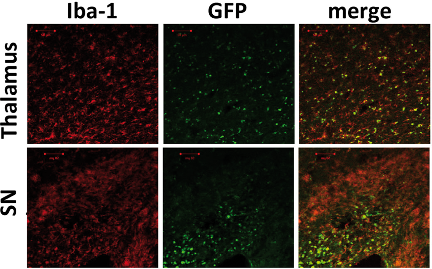
Conclusion
The present study showed that peripheral MMs continuously infiltrate into primary and secondary injured areas and that CD36 expressed in MMs sustains CD45
PB03-U07
AntagoMiR-494 reduced neutrophil infiltration and alleviated acute ischemic brain injury in experimental stroke
1Institute of Cerebrovascular Disease Research and Department of Neurology, Xuanwu Hospital, the First Clinical Medical College of Capital Medical University
Abstract
Background
Post-stroke inflammation contributes to secondary brain tissue damage leading to poor functional recovery. The objective of this study is to evaluate the role of microRNA-494 in modulation of phenotype shift and infiltration of neutrophils in experimental stroke.
Methods
Blood samples from 76 AIS patients and 52 heathy controls were collected, and miR-494 levels and phenotype markers in neutrophils were detected by quantitative real-time PCR. Overexpression or knockdown of microRNA-494 in vivo and in vitro was done in experimental ischemic stroke in C57BL-6 J mice subjected to middle cerebral artery occlusion and cortical neurons subjected to oxygen-glucose deprivation (OGD).
Results
Functional annotation analysis revealed that miR-494 target genes are involved in regulating immune response and neuronal function via four pathways including focal adhesion, TGFb signaling, amyotrophic lateral sclerosis, and glutamatergic synapse. Subsequently, we verified miR-494 levels in neutrophils of 76 AIS patients and 52 age-matched control volunteers by RT-PCR, which were all increased significantly within 6 h after cerebral ischemia. ROC analysis showed that the AUCs for miR-494 levels in neutrophils were 0.683 (P = 0.0006) indicating a diagnostic potential of miR-494 in neutrophils of AIS patients. Although the number and percentage of neutrophils of AIS patients were higher than in healthy controls, no correlation between miR-494 levels and the number and percentage of neutrophils was found. N1 and N2 are representative pro-inflammatory and anti-inflammatory subpopulations of neutrophils, which are marked by CD16 and CD206, respectively. Our results revealed that CD16 and CD206 expressions were not correlated with neutrophil miR-494 levels. However, neutrophil miR-494 levels were correlated with MMP-9 and CD11b. The in vitro experiment showed that the conditioned medium from LPS-activated neutrophils or the addition of agomir-494 treatment did not exacerbate OGD-mediated neuronal injury. Our in vivo experiment showed that both N1 and N2 subtypes of neutrophils were increased in the blood of MCAO mice, while only the N1 population was decreased by antagomir-494, subsequently reducing the N1 to N2 ratio in peripheral blood. Moreover, antagomir-494 decreased the mRNA levels of MMP-9 and CD11b in MCAO mice, as well as the infiltration of LY-6 G labeled neutrophils in the ischemic brain.
Conclusions
From basic research and clinical research, we illustrates antagomir-494 reducted neutrophil infiltration after cerebral ischemia, offering an epigenetic target as peripheral immune regulation for acute ischemic stroke.
References
PB03-U08
EZH2 suppression alleviates ischemic brain injury through blocking activation of pro-inflammatory microglia
1Department of Neurology, Drum Tower Hospital of Nanjing University Medical School, PR China
2Department of Neurology, Nanjing Drum Tower Hospital Clinical College of Nanjing Medical University, Nanjing China
Abstract
Objectives
EZH2, a histone methyltransferase, has been recognized in the regulation of the immune response in recent years, especially in tumor immune and autoimmune diseases[1–3], while its role in microglial activation and ischemic brain injury remains to be unraveled. The aim of this study is to invest the role of EZH2 in microglia-associated inflammation after ischemic stroke and further detect the influence of EZH2 suppression on ischemic brain injury, and thus reveal a novel therapeutic target for ischemic stroke.
Methods
Middle cerebral artery occlusion (MCAO) was induced in adult male mice for in vivo studies. EZH2 inhibitor DZNep was administrated to mice intravenously (0.05 mg/kg) 24 hours prior to MCAO induction and once per day after MCAO. For in vitro studies, primary microglia were exposed to oxygen glucose deprivation (OGD) and treated with DZNep (10uM). Infarct volumes and neurological impairments were evaluated by 2,3,5-triphenyltetrazolium chloride (TTC) staining and Neurological Severity Scores (NSS), rotarod test and grip strength respectively. Immunofluorescence staining (IF) and western blot were performed to detect EZH2, STAT3 and p-STAT3 expression level. Microglial activation phenotype was assessed by IF staining and flow cytometry both in vivo and in vitro. Expression levels of pro-inflammatory cytokines were quantified byreal-time polymerase chain reaction (PCR).
Results
Both in vivo ischemic/reperfusion injury (I/R) and in vitro OGD treatment induced remarkable up-regulation of EZH2 in microglia. EZH2 inhibitor DZNep administration improved behavioral function of mice and reduced infarct volumes after MCAO. EZH2 suppression increased the percentage of anti-inflammatory (CD206+) microglia while decreased the percentage of pro-inflammatory (CD86+) microglia both in vivo and in vitro. Additionally, pro-inflammatory cytokines, such as IL-1β, IL-6, TNF-α and CXCL10, were also significantly reduced by DZNep both in vivo and in vitro. EZH2 suppression by DZNep blocked the phosphorylation of STAT3, which was up-regulated by I/R injury and OGD treatment.
Conclusions
EZH2 suppression alleviates ischemic brain injury through blocking activation of pro-inflammatory microglia probably via blocking STAT3 activation. Thus, we revealed that EZH2 could be a potential therapeutic target for ischemic stroke.
References
PB03-U09
FasL mutation attenuates cytotoxicity of CD8+ T cells to neurons by a novel pathway in ischemic stroke
1Department of Neurology, Drum Tower Hospital of Nanjing University Medical School, PR China
Abstract
Objectives
CD8+ T cells play a detrimental role in ischemic stroke, one of which is the severe CD8+ T cell-mediated neuronal injury [1]. FasL has been considered one of the major molecules involved in killing the target cells by CD8+ T cells [2], but the pathophysiological role of the Fas/FasL system in cytotoxic effects of CD8+ T cells in ischemic stroke is still unknown.
Methods
Middle cerebral artery occlusion (MCAO) was conducted in adult male FasL-deficient (B6 Smn.C3H-FasL gld) mice and their wild-type (WT) control mice for 60 min. Ischemic infarct volumes and neurological function were evaluated through 2,3,5-triphenyltetrazolium chloride (TTC) staining, rotarod tests, corner tests and forepaw grip strength, respectively. Perforin, granzyme-B, TNF- α and IFN-γ of CD8+ T cells infiltrated in the infarcted lateral hemisphere of WT and gld mice after MCAO were quantified by flow cytometry. Rag1-/- mice, the immune-deficient mice, were reconstituted with CD8+ T cells from gld and WT mice with or without MCAO models to further investigate the cytotoxicity of CD8+ T cells. The cytotoxicity of CD8+ T cells was measured by CCK8, LDH, quantitative real-time polymerase chain reaction (q-PCR) in oxygen glucose deprivation (OGD) and co-cultured in vitro models. Proteomic analysis was performed after being treated by CD8+ T cells in co-culturing with OGD neurons. The q-PCR and western blotting were used for further validation.
Results
It was found that FasL mutant CD8+ T cells dramatically reduced brain injury in ischemic stroke. The infarct volumes and neurological deficits of Rag1-/- mice reconstituted with CD8+ T cells from gld mice after MCAO were alleviated compared with that from WT mice. Furthermore, the FasL mutant CD8+ T cells attenuated cytotoxicity of CD8+ T cells to neurons in vitro, which have lower apoptotic levels after co-cultured. Proteomic analysis showed that PDPK1 (3-Phosphoinositide Dependent Protein Kinase 1) plays a vital role in alleviating cytotoxicity of CD8+ T cells to neurons after FasL mutation of CD8+ T cells in ischemic stroke and cytotoxicity factors were decreased after inhibition of PDPK1.
Conclusions
Our study highlights that FasL mutation attenuates cytotoxicity of CD8+ T cells in ischemic stroke. FasL mutant CD8+ T cells reduced neuronal injury in vitro models. The cytotoxicity of CD8+ T cells was regulated by the FasL/PDPK1 pathway in ischemic stroke. A better understanding of the mechanisms underlying the neurotoxic pathways of CD8+ T cells could be an essential step in developing immune-modulatory therapies for ischemic stroke.
References
PB03-U10
The association between mucosal-associated invariant T (MAIT) cells and acute ischemic stroke
1Department of Neurology, Juntendo University School of Medicine, Japan
2Stroke Center, Jichi Medical University Hospital, Division of Neurology, Department of Medicine, Jichi Medical University
3Department of Immunology, Juntendo University School of Medicine, Japan
4Department of Neurology, Juntendo University Urayasu Hospital, Japan
Abstract
Objectives
Mucosal-associated invariant T (MAIT) cells are innate T cells restricted by MHC-related molecule 1 (MR1)1. MAIT cells have been associated with autoimmune diseases, such as multiple sclerosis and arthritis2,3,4; however, their relation to ischemic stroke remains unclear. The purpose of this study was to elucidate the association between MAIT cells and acute ischemic stroke.
Methods
We used MR1 knockout C57BL/6 (MR1-/-) mice, which are deficient in MAIT cells, and wild-type C57BL/6 (MR1+/+) littermates in our study. We performed a one-hour transient middle cerebral artery occlusion and evaluated cerebral infarct volume and motor function of the mice at 24 and 72 hours after reperfusion. Microglial activation was evaluated by immunohistochemistry with anti-Iba-1 antibody.
Results
The infarct volume tended to decrease at 24 hours after ischemia-reperfusion in MR1-/-mice. At 72 hours, we observed a significant reduction in infarct volume and a significant improvement of neurological dysfunction in MR1-/-mice group (p < 0.05) compared to wild-type littermates. There were significantly fewer microglia cells in the MR1-/-mice compared to wild-type mice at 24 hours after ischemia-reperfusion, and the same trend was seen at 72 hours.
Conclusions
These results indicate that MAIT cells may play an important role in pathological responses, and that they may regulate inflammation in acute focal cerebral ischemia.
References
PB03-U11
ATP induces neutrophil extracellular traps formation in the postischemic brain
1Department of Biomedical Sciences, Inha University School of Medicine, Republic of Korea
2Medical Research Center, Inha University School of Medicine, Republic of Korea
3Department of Anatomy, Inha University School of Medicine, Republic of Korea
Abstract
Cerebral ischemia leads to brain damages via a complicated pathological events such as excitotoxicity, peri-infarct depolarization, inflammation, and apoptosis. In the postischemic brain, inflammatory response caused brain damages, which was aggravated by infiltration of neutrophils in early stage of stroke. However, the role of neutrophils in the early period after stroke has not yet been fully understood. In permanent middle cerebral artery occlusion (pMCAO) model, we already showed recruitment of neutrophils, formation of neutrophil extracellular traps (NETs), and increase of CitH3 (NETosis marker) positive cells which were observed in cortical arteriole, microvessel and brain parenchyma. In addition, we previously showed the rapid induction of CitH3 level in PMNs in peripheral blood and CSF after pMCAO. Because cerebral ischemia is non-infectious disease, we assumed that danger associated molecular patterns (DAMPs) stimulated neutrophils to migrate to damaged brain parenchyma and form NETs in the postischemic brain. It is thought that ATP is a candidate for induction of CitH3 and NETs formation after stroke. We measured increase of ATP level in plasma and CSF in the postischemic brain. Treatment of peripheral blood PMNs with ATP or BzATP upregulated CitH3 level and showed NETs formation. Pretreatment of ATP receptor antagonist reduced CitH3 level and NETs formation in bone marrow PMNs. Furthermore administration of ATP receptor antagonist reduced CitH3 level in the postischemic brain. This study shows ATP which is released from damaged cells could induce NETosis after stroke.
PB03-U12
Post-stroke administration of melatonin improves long-term outcomes after focal cerebral ischemia/reperfusion (FI/R) via interleukin-4 (IL-4) dependent M2 microglial polarization
1Department of Neurosurgery, Yokohama City University, Japan
2Department of Neurology, University of Pittsburgh School of Medicine, USA
Abstract
Microglia represent rational but difficult therapeutic targets for stroke due to their diverse phenotypes that play dual-faced protective (M2 phenotype) and toxic (M1 phenotype) effects. Previous study has shown that subcutaneous melatonin injection increased the level of Interleukin-4 (IL-4), the best known M2 inducing cytokine, in the blood. In this study, we investigated the role of melatonin in microglia M2 polarization and its effect on long-term recovery after stroke. Focal cerebral ischemia (FCI)was induced for 60 min focal ischemia reperfusion (FIR). Animals were randomly assigned to receive either melatonin or vehicle treatment at 2 h after stroke. Brains were assessed for cerebral tissue loss at 3 and 14 days of reperfusion. Neurological performance was analyzed up to 14 days after ischemia. Markers for microglia polarization were assessed using immunofluorescent staining and RT-PCR. In vitro experiments using primary microglia and in-transwell microglia-neuron coculture were done to confirm the effect of melatonin on microglial inflammatory responses and its effect on microglia-potentiated neuronal injury upon oxygen-glucose deprivation (OGD). Melatonin significantly reduced infarct volume and attenuated sensorimotor deficits 3–14 day after FIR. IL-4 deficiency, abolished melatonin-afforded long term protection. Melatonin-treated mice showed significantly reduced expression of inflammatory cytokine and chemokines, which is accompanied by significantly increased expression of M2 markers and decreased expression of M1 markers in microglia. In primary microglial cultures, melatonin inhibited LPS (a M1 inducer)-induced production of NO and TNFα, confirming that melatonin has direct anti-inflammatory effect on microglia. Furthermore, melatonin ameliorated the neurotoxic effect of M1 microglia on OGD neurons, and this effect was absent in IL-4 deficient microglia. Melatonin may represent an innovative therapeutic strategy that shifts microglia polarization toward a protective M2 phenotype in an IL-4-dependent manner and thus enhance long-term recovery after stroke.

PB03-U13
Anti-inflammatory effect of serum bilirubin in acute stroke patients
1Dept. of Neurosurgery, Akita University, Japan
2Dept. of Surgical Neurology, Res. Inst. Brain & Blood Vessels -Akita, Japan
Abstract
Background
During the acute phase of stroke, various kind of free radicals and inflammatory cytokines are released from injured brain tissue. An appropriate activation of the internal anti-inflammatory agent at the stroke onset might provide a chance to reduce initial damage. This study aimed to investigate the influence of bilirubin which might show intrinsic anti-inflammatory effect on the pathogenesis of acute stroke.
Method
Acute stroke patients were asked to participate into this study between December 2017 and March 2018 (hemorrhagic stroke: n = 16 and ischemic stroke: n = 36). Serum bilirubin was measured in the blood samples on admission. High sensitive C-reactive protein (hsCRP) was measured as an inflammatory marker at onset. Stroke severity at onset was assessed by National Institute of Health stroke scale (NIHSS). Stroke subtypes were classified into hemorrhagic stroke and ischemic stroke. The lesion of hemorrhagic stroke was classified into lobar lesion and deep brain lesion.
Results
There was no correlation between serum bilirubin amount and the stroke severity in crude stroke patients. In patients of hemorrhagic stroke, there was no correlation between bilirubin amount and NIHSS in lobar hemorrhage. While, patients with deep brain hemorrhage showed the significant negative relations between bilirubin amount and NIHSS score (R = 0.54, p = 0.01) or hsCRP amount (R = 0.48, p = 0.03). Regarding ischemic stroke, only patients with mild deficits (NIHSS less than 10) and without any previous medication showed the significant negative relation between bilirubin serum concentration and NIHSS score (R = 0.622, p = 0.007). However, the bilirubin amount did not show any correlation with hsCRP amount in these patients.
Conclusion
Hemorrhagic stroke patients with deep brain lesions might benefit from the anti-inflammatory effect of bilirubin at the acute phase. Moreover, Serum bilirubin might be able to express the effect on ischemic stroke patients with mild damage.
PB03-V01
PAR1-agonists as regulators of neuroinflammation in vitro
1Lomonosov Moscow State University
2Pirogov Russian National Research Medical University
3Russian Cardiology Research and Production Complex
Abstract
Aim
To investigate the participation of PAR1-agonists – activated protein C (APC) and its functional analogue, the new synthetic peptide 9 (AP9), an analogue of the “tethered ligand” released from PAR1 by APC in the model of the neuroinflammation in vitro.
Methods
To create the model of neuroinflammation in vitro, mast cells (MC) and hippocampal neurons were used. The toxic effect of activated MC on neurons was estimated by using a morphological analysis of survival with the help from fluorescent dyes. The change in the cytoskeleton in the presence of PAR1 agonists was carried out by immunocytochemical staining.
Results
Previously, we showed that pre-treatment of activated MC by APC and AP9 leads to a decrease in their toxic effect on neurons when they were incubated together and a decrease in the proportion of apoptotic cells. In the present study, in the co-cultivation of hippocampal neurons with MC, it has been demonstrated that blockade by a specific antagonist (SCH79797) of PAR1 on both MC and neurons reverses the protective action of both APC and AP9. These data indicate a PAR1-mediated protector action of AP9, similar to that of APC. The main participants in the inflammatory reaction are MC; whose morphological changes are the criteria for their activation and the severity of the inflammatory process. We have shown that activation cells by Thr at high concentrations (50 nM) leads to disorganization and re-distribution of the actin cytoskelet, however, preincubation of RBL-2H3(a mast cell line) with the new peptide AP9 and APC promotes actin ordering, similar to control groups. Apparently, the protective effect of AP9 and APC is determined by their ability to order sub-membrane actin and to stabilize cell membranes, reducing secretion.
Conclusions
PAR1-agonists – new peptide (AP9) and APC exert an anti-inflammatory and protective actions, in the model of neuroinflammation in vitro.
This work was supported by RFFR №19-015-00529, RFFR №18-34-00977
PB03-V02
The role of brain regulatory T cells during the chronic phase after CNS injury
1Department of Microbiology and Immunology, Keio University School of Medicine, Japan
Abstract
In addition to maintaining immune tolerance, Foxp3+ regulatory T (Treg) cells perform specialized functions in tissue homeostasis and remodeling. However, the characteristics and functions of brain Treg cells are not well understood due to a low number of Treg cells in the brain under normal conditions. We found a massive accumulation of Tregs in the brain taht potentiates neurological recovery during the chronic phase after ischemic stroke. Although brain Tregs relate to Tregs in other tissues such as visceral adipose tissue (VAT) and muscle, brain Tregs are apparently distinct and express unique genes related to the nervous system including the serotonin receptor, HTR7. The amplification of brain Tregs is dependent on interleukin (IL)-2, IL-33, serotonin, and T cell receptor (TCR) recognition. And, the infiltration of brain Tregs into the brain is driven by chemokines, CCL1 and CCL20. Brain Tregs suppress neurotoxic astrogliosis via the production of amphiregulin (Areg), a low-affinity epidermal growth factor receptor (EGFR) ligand. Furthermore, we also discovered the existence of brain Tregs in the brain form Alzheimer disease model and EAE model. These Tregs had phenotypes of tissue Tregs by expressing Areg, ST2 (IL-33 receptor), HTR7. Our findings suggest that Tregs and their products may provide novel therapeutic opportunities for neuronal protection against stroke and other neuroinflammatory diseases.
PB03-V03
Berberine attenuates macrophages infiltration in intracranial aneurysms through FAK/Grp78/UPR Axis
1Department of Neurosurgery, Huashan Hospital, Fudan University, Shanghai, China
Abstract
Objectives
Inflammatory cells, such as macrophages, play key roles in the pathogenesis of intracranial aneurysms (IAs). Berberine (BBR), an active component of a Chinese herb Coptis chinensis French, has been shown to have anti-inflammatory properties through suppressing macrophage migration in various inflammation animal model. The goal of this study was to examine BBR’s effect on inflammation and IAs formation in a rodent aneurysm model.
Methods
Human aneurysm tissues were collected by microsurgical clipping and immunostained for phospho-Focal adhesion kinase (FAK) and CD68+ macrophages. A rodent aneurysm model was induced in 5-week-old male Sprague Dawley (SD) rats by intracranial surgery, then these rats were orally administrated 200 mg/kg/day BBR for 35 days. Then the rat brains were perfused and evaluated by immunostaining, western blot and PCR. Macrophage cell line RAW264.7 cells were treated with different inhibitors to evaluate the role of FAK/Grp78/UPR axis during macrophage infiltration.
Results
Immunostaining data showed that BBR inhibited CD68+ macrophages accumulation in IAs tissues and suppressed FAK phosphorylation. In lipopolysaccharide (LPS)-stimulated RAW264.7 cells, BBR treatment remarkably attenuated macrophages infiltration, suppressed the expression of matrix metalloproteinases (MMPs), and reduced proinflammatory cytokine secretion, including MCP-1, interleukin 1β (IL-1β), interleukin 6 (IL-6) and tumor necrosis factor-α (TNF-α). Mechanistically, BBR downregulated FAK/Grp78/Unfolded Protein Response (UPR) signaling pathway in RAW264.7 cells.
Conclusions
BBR prevents IAs formation through inhibiting FAK phosphorylation and inactivating UPR pathway in macrophages
References
PB03-V04
Intermittent fasting: a potential modifier of DNA methylation in neuroinflammation in a chronic hypoperfusion model
1Dept of Physiology, National University of Singapore, Singapore
Abstract
Vascular dementia (VaD), the second most common cause of dementia, is a progressive neurodegenerative disorder that leads to cognitive decline1. Despite not being the main contributor of dementia, addressing VaD is still of significant importance due to its rapid step-wise decline in disease progression as opposed to a gradual decline in Alzheimer’s disease (AD)2. Adding gravity to this concern, VaD has greater mortality risk as opposed to AD upon diagnosis. VaD is characterised by reduced cerebral blood flow (CBF) to the brain (cerebral hypoperfusion) and white matter hyperintensities. With age being a high-risk factor for VaD, the rapidly aging population translates to increasing VaD cases and hence the need to tackle the issue at the soonest. While increased inflammation is reported in VaD, to what extent neuroinflammation is involved, the role of epigenetics like DNA methylation in modifying the genes resulting in neuroinflammation and the profile of DNA methylation in a VaD brain remain unanswered. Additionally, intermittent fasting(IF), a form of dietary restriction is known to potentially induce epigenetic changes eliciting beneficial effects like potentially slowing the progression of cognitive decline. While there are a few studies present about DNA methylation in a chronic hypoperfusion model, the novelty of this study stems from the fact that it is conducted in mice and the presence of IF as a potential therapeutic intervention.
We therefore aim:
(1) To validate that the bilateral common carotid artery stenosis (BCAS) is a representative chronic cerebral hypoperfusion model
(2) To analyse DNA methylation profile in a chronic hypoperfusion model in relation to neuroinflammation
(3) To investigate whether IF intervention changes the DNA methylation profile: (a) overall pattern and (b) specific to neuroinflammation
The robustness of the BCAS model was affirmed through CBF and white matter lesion measurements. A significant reduction (about 30%) in CBF was observed (Pericam imaging) immediately upon insertion of the micro-coils in the mice. Luxol-fast blue staining was performed to visualise white matter lesion in the different regions of the brain including the corpus callosum.
Representative VaD chronic cerebral hypoperfusion model was established using the BCAS method that involves inserting micro-coils (inner diameter of 0.18 mm) in 6 months old C57BL/6 male mice. The coils were left in the mice for 7, 15 and 30 days respectively. The mice were further divided into ad libitum and IF groups which were then sacrificed, DNA was extracted from the cerebral cortex and sent for DNA methylation screening. The DNA methylation profile varied within the different time points especially in terms of neuroinflammation. Moreover, with IF, the overall pattern and neuroinflammation-related DNA methylation compared to the ad libitum group differed significantly. DNA methylation patterns increased in neuroinflammation-related genes. This trend difference with IF intervention further promises its potential beneficial effects.
Essentially, IF is promising as a potential modifier of DNA methylation profile in neuroinflammation-related genes in a chronic hypoperfusion model.
References
PB03-V05
Cortical spreading depression induces expression of BDNF and pro-BDNF proteins, which may impact neuroinflammation
1Institute of Neurological Sciences and Psychiatry, Hacettepe University, Turkey
2Department of Neurology, Faculty of Medicine, Hacettepe University, Turkey
3Department of Psychiatry, Faculty of Medicine, Hacettepe University, Turkey
Abstract
Objectives
Cortical spreading depression (CSD) is associated with ischemic stroke, acute brain injury, intracranial hemorrhage and migraine aura. CSD is known to trigger neuroinflammation, however, its mechanisms have not been fully understood. Brain-derived neurotrophic factor (BDNF) has neuroprotective and antiinflammatory effects in developing and mature brain. Its pro- form (proBDNF) is not only an inactive precursor, but also has opposing effects to BDNF. Here, we investigated the changes in BDNF and proBDNF levels after multiple CSDs and, their potential role in the CSD-induced neuroinflammation.
Methods
In adult Swiss albino mice, multiple CSDs (n = 9) were induced by applying 1 M KCl-soaked cotton ball over the burr hole on the frontal bone for 1 hour under urethane anesthesia. DC potential was recorded with an electrode placed over the parietal bone. In the sham group%0.9 NaCl-soaked cotton ball was applied. At 1, 2 and 6th hours; BDNF and proBDNF levels in parietal cortices were determined by Western blotting (n = 5 and n = 3 for CSD and sham groups, respectively). Brains from another group of mice (n = 3) were prepared for immunohistochemistry and, sections were labeled with anti-NeuN, anti-S100, anti-interleukin 1-beta (IL-1β), anti-tumor necrosis factor alpha (TNF-α). Also the effects of increased BDNF on CSD-induced inflammatory markers were examined after intracerebroventricular injection of human recombinant BDNF protein (5ug) via a cannula placed in the right lateral ventricle of mice (n = 3).
Results
After multiple CSDs both BDNF and proBDNF proteins significantly increased at the 6th hour in the ipsilateral CSD cortices compared to the contralateral cortices as well as ipsilateral cortices of the sham group (Mann Whitney U test, p < 0.05). Neuronal IL-1β was activated in the cytoplasm at the 1st hour, and neuronal TNF-α at the 2nd hour after multiple CSDs. Cortical BDNF levels reached to levels almost 10 fold higher compared to the contralateral hemisphere 2 hours after icv BDNF injection. The assessment of the effects of increased BDNF on CSD-induced IL-1β and TNF-α is ongoing.
Conclusions
The level of BDNF and proBDNF proteins in the cortex increases after multiple CSDs. Multiple CSDs also induce synthesis and translocation of IL-1β and TNF-α. These findings suggest that BDNF may have a role on CSD-triggered neuroinflammation.
Keywords
cortical spreading depression (CSD), brain-derived neurotrophic factor (BDNF), proBDNF, neuroinflammation, interleukin 1-beta (IL-1β), tumor necrosis factor alpha (TNF-α)
Acknowledgement
Financially supported by Hacettepe University Scientific Research Projects Coordination Unit, Project Number: THD-2018-17073.
References
PB03-V06
Detecting neuroinflammation in breast cancer patients treated with chemotherapy with simultaneous [18F]-DPA-714 PET-MR imaging
1Department of Imaging and Pathology, KU Leuven, Belgium
2Department of Nuclear Medicine and Molecular Imaging, KU Leuven, Leuven, Belgium
3Division of Nuclear Medicine, University Hospital Leuven, Leuven, Belgium
4Department of Oncology, KU Leuven, Leuven, Belgium
5Department of Surgical Oncology, University Hospital Leuven, Leuven, Belgium
6Department of Neurosciences, KU Leuven, Leuven, Belgium
7Department of Psychiatry, University Hospitals Leuven, Leuven, Belgium
8Department of Radiology, University Hospital Leuven, Leuven, Belgium
Abstract
The current dogma of chemotherapy-induced cognitive symptoms largely revolves around neuroinflammation. Several studies have shown a correlation between peripheral inflammation, as a proxy for neuroinflammation, and cognitive performance1; while none have investigated neuroinflammation directly in a clinical sample. This study aimed at investigating if neuroinflammation measured by 18F-DPA-714 TSPO PET is a candidate mechanism for neuro-cognitive impairment after chemotherapy in women treated for breast cancer.
Patients were first screened for TSPO binding affinity (high (HA), medium (MA), low (LA)), and LA binders were excluded (6 patients)2,3. Five women (48 ± 8 years old; 5 MA) with early-stage breast cancer who received adjuvant chemotherapy (C+) and 9 women (50 ± 4 years old; 2 HA and 7 MA) with breast cancer who did not receive chemotherapy (C-) underwent scanning one month (30 ± 12 days) after chemotherapy was completed or after breast-surgery, respectively. Sixty minutes dynamic 18F-DPA-714 PET measurements were performed (injected activity 144 ± 14 MBq), with simultaneous acquisition of magnetic resonance (MR) T1-weighted 3D Fast SPGR images on a GE SIGNA PET-MR system. Full kinetic modelling was applied and total volume of distribution (VT) parametric images were calculated by a plasma-input based Logan graphical analysis and skull-stripped (PMOD v3.8; Zurich, Switzerland). To explore differences between the patient groups, two-sample T-tests of the VT maps using SPM124 (C+ vs. C-) was performed with ppeak uncorrected <.05, Kext > 100 voxels and pcluster FWE-corrected <.05, with proportional global normalization to correct for differences in binding affinities.
There was a significant higher translocator protein (TSPO) distribution volume in breast cancer patients treated with chemotherapy when compared to chemotherapy-naïve patients, in a cluster covering mainly the left temporal cortex, spreading out in the parietal and frontal cortex (pcluster FWE-corrected = .013, peak cluster x = -46, y = -10, z = 38; 3223 voxels).
This is the first in-vivo study to evaluate chemotherapy-related changes in neuroinflammation, reflected by difference in TSPO binding. Increased neuroinflammation, as measured with 18F-DPA-714 PET was associated with chemotherapy treatment in breast cancer patients. This suggests activated microglia, possibly caused by released cytokines, may be partly responsible for cognitive complaints after breast cancer treatment seen in this population.

References
PB03-V07
Hypertensive stimuli promote brain inflammation in a pressure-dependent manner
1Vascular Biology and Immunopharmacology Group, Department of Physiology, Anatomy and Microbiology, La Trobe University, Melbourne, Australia
2Department of Pharmaceutical and Biomedical Sciences, Raabe College of Pharmacy, Ohio Northern University, USA
Abstract
Hypertension increases the risk for stroke and cognitive impairment, and is strongly associated with inflammation of the vasculature and kidneys, as well as cerebrovascular dysfunction and oxidative stress. However, it is unclear to what extent there is inflammation and immune cell infiltration in the brain during hypertension.
To test whether chronic administration of angiotensin II or aldosterone/salt causes brain inflammation and whether this is blood pressure-dependent.
Male C57Bl/6 mice were administered vehicle (saline; n = 22) or angiotensin II (Ang II, 0.7 mg/kg/d s.c.; n = 23) for 14 d via osmotic minipumps. A subset of mice also received hydralazine hydrochloride (50 mg/kg; n = 15) in their drinking water for 14 d after minipump implantation. Another cohort of mice were treated with vehicle (87% propylene glycol, 9% ethanol, 4% water; n = 7) or aldosterone (0.72 mg/kg/d s.c plus 0.9% NaCl for drinking; n = 8) for 14 d using osmotic minipumps. Systolic blood pressure was measured using tail-cuff plethysmography, immune cell numbers using flow cytometry and inflammatory markers using real-time PCR.
Ang II infusion caused an increase in blood pressure and promoted accumulation of leukocytes in the brain (CD45+high), including neutrophils (CD11b+ Ly6G+), monocytes (CD11b+ Ly6C+high and Ly6C+low), T cells (CD3+) and B cells (CD19+), all of which were elevated by ∼2.5-fold compared to vehicle-treated mice (n = 6–8, P < 0.05). Similarly, aldosterone/salt-induced hypertension was associated with increases in brain myeloid cells (CD11b+, ∼3.5-fold) and T cells (CD3+, ∼2-fold) (n = 7–8, P < 0.05). Co-administration of hydralazine prevented the pressor response to Ang II (163 ± 5 mmHg vs. Ang II + hydralazine, 121 ± 4 mmHg; n = 7–8, P < 0.05). Ang II-induced increases in brain neutrophils and monocytes were blunted by co-administration of hydralazine (n = 7–8, P < 0.05), however, hydralazine had no effect on T cell or B cell numbers (n = 7–8). Ang II-induced increases in brain mRNA expression of chemokine (C-C motif) receptor 2 (CCR2), chemokine (C-C motif) ligand 2 (CCL2) and CCL8 were also blunted by co-administration of hydralazine (n = 7–8, P < 0.05).
Our data indicate that immune cell infiltration and inflammation occur in the brain during both the Ang II and aldosterone/salt models of hypertension. Furthermore, the Ang II-induced brain infiltration of myeloid cells, but not lymphoid cells, appears to be pressure-dependent. Chronic brain inflammation may be a contributing factor to the increased stroke risk and cognitive impairment during hypertension and may be mitigated by blood pressure reduction.
PB03-V08
Reduced ambient temperature enhances inflammation-induced encephalopathy in endotoxemic mice – Role of phosphoinositide 3-kinase gamma
1Institute of Molecular Cell Biology, Jena University Hospital, Friedrich Schiller University, Germany
2Department of Pharmacology, University of Oslo, Oslo, Norway
3Leibniz Institute on Aging – Fritz Lipmann Institute (FLI), Jena, Germany
4Department of Anesthesiology and Intensive Care Medicine, Jena University Hospital, Friedrich Schiller University, Jena, Germany
Abstract
Background
Sepsis-associated encephalopathy is an early and frequent event of infection-induced systemic inflammatory response syndrome. Phosphoinositide 3-kinase γ is linked to neuroinflammation and inflammation-related microglial activity. In homiotherms, variations in ambient temperature outside the thermoneutral zone lead to thermoregulatory responses, mainly driven by a gradually increased sympathetic activity, and may affect disease severity. This study was aimed at determining the impact of thermoregulatory responses upon reduced ambient temperature exposition and the specific role of phosphoinositide 3-kinase γ in the pathogenesis of sepsis-associated encephalopathy.
Methods
Experiments were performed in phosphoinositide 3-kinase γ wild-type, knockout, and kinase-dead mice, which were kept at neutral (30 ± 0.5°C) or moderately lowered (26 ± 0.5°C) ambient temperature. Mice were exposed to endotoxin-induced systemic inflammatory response and monitored for thermoregulatory response and blood–brain barrier integrity. Primary microglial cells and brain tissue derived from treated mice were analyzed for inflammatory responses and related cell functions (directed migration, phagocytosis). Comparisons between groups were made with one-way or two-way analysis of variance, if appropriate. Post hoc comparisons were made with the Holm–Sidak test or t-tests with Bonferroni’s correction for adjustments of multiple comparisons. Data not following normal distribution was tested with Kruskal-Wallis test followed by Dunn’s multiple comparisons test.
Results
We found that a moderate reduction of ambient temperature led to enhanced hypothermia of mice undergoing endotoxin-induced systemic inflammatory response accompanied by aggravated sepsis-associated encephalopathy. Phosphoinositide 3-kinase γ deficiency enhances blood–brain barrier injury and upregulation of matrix metalloproteinases as well as an impairment of microglial phagocytic activity.
Conclusions
This study reveals that enhanced adaptive thermoregulatory mechanisms in response to temperatures below the thermoneutral range of ambient temperature lead to exacerbated endotoxin-induced blood–brain barrier injury and accompanied neuroinflammation. The signaling protein phosphoinositide 3-kinase γ was characterized as a critical mediator of key microglial cell functions involved in endotoxin-induced blood–brain barrier injury and accompanied neuroinflammation. Phosphoinositide 3-kinase γ serves a protective role in that it suppresses release of matrix metalloproteinases, maintains microglial motility and reinforces phagocytosis leading to improved brain tissue integrity.
PB03-V09
Modelling inflammatory brain-heart interaction in duchenne muscular dystrophy
1Department of Medical Biophysics, Schulich School of Medicine and Dentistry, University of Western Ontario, Canada
2Imaging Program, Lawson Health Research Institute, St. Joseph's Health Care London, Canada
Abstract
Objective
Duchenne muscular dystrophy (DMD) is a neuromuscular degenerative disorder and the second most commonly inherited disease worldwide1. DMD is caused by the absence or disruption of the protein dystrophin within various tissues—most notably skeletal muscles and neurons in the central nervous system. DMD is clinically characterized by progressive skeletal muscle, cardiac, and cognitive degeneration, which is accompanied by chronic inflammation, and subsequently leads to death by cardiac or respiratory failure. While endogenous and systemic inflammation are known to be associated with DMD skeletal and cardiac muscle degeneration, little is known about the impact of systemic inflammation on the brain and even less about the role of neuroinflammation in DMD—despite it being a prime proponent of other neurodegenerative diseases, such as Alzheimer’s dementia2,3. Furthermore, recent studies have shown that inflammatory responses to local myocardial damage can systemically trigger cerebral microglial activation—a hallmark of neuroinflammation4. Thus, our objectives were to firstly investigate the role of neuroinflammation in DMD, and then to delineate the inflammatory interaction between the heart and brain using an in-vivo whole-body DMD model.
Methods
Two murine models of moderate DMD severity (8 week old +/- mdx:utr) were imaged through PET scanning and associated biodistribution and autoradiography analyses using 18 F-FEPPA—a PET tracer targeting translocator protein (TSPO), overexpressed on both microglial and macrophages. After 60 minutes dynamic PET imaging, each mouse’s brain, heart, and skeletal muscle were collected for validation and quantification of TSPO presence using immunohistochemistry techniques.
Results
Preliminary results in the 8-week old moderately severe DMD mice showed elevated TSPO PET binding in the heart, brain, and skeletal muscle (Fig. 1A-C). The global increased TSPO PET heart and brain images co-localized with autoradiography and immunohistochemistry findings (Fig. 1E-G). Immunohistochemistry findings, when compared to age-matched wildtype, further validated heightened TSPO presence in DMD tissues. Exploratory biodistribution data showed differential uptake of 18F-FEPPA across different tissue types (Fig. 1D).

Conclusion
These data are the first to not only demonstrate the presence of inflammatory biomarkers in the brain during DMD, but also to model in-vivo the suspected link between cardiac- and neurodegeneration using inflammation in a dystrophic disease. Upon demonstrating the existence of this inflammatory interaction, we will next use this protocol in addition to other imaging modalities (i.e. DCECT, MRI) to map this interaction across multiple disease severities, and during DMD progression. This method illustrates the advantage of 18F-FEPPA PET as an useful tool to non-invasively detect DMD progression, increasing the possibility to propose early intervention prior to severe muscular or neurological damage. It also offers a novel approach to assessing efficacy of DMD treatments, which may now include neuroprotective or anti-inflammatory therapies.