
Abstract

NLA-1
The Niels Lassen Award Session
Mechanisms underlying negative fMRI responses in the striatum
1Center for Animal MRI, University of North Carolina at Chapel Hill, USA
2Biomedical Research Imaging Center, University of North Carolina at Chapel Hill, USA
3Department of Neurology, Univeristy of North Carolina at Chapel Hill, USA
4Departments of Psychiatry and Behavioral Sciences, Stanford University, USA
5National Institute of Environmental Health Sciences, USA
6Department of Psychiatry, University of North Carolina at Chapel Hill, USA
Abstract
Objectives
We manipulate striatal neuronal circuitry in order to better understand the origin of its negative fMRI responses, which go against traditional neurovascular coupling rules. We performed optogenetic-fMRI of 5 distinct circuits related to the striatum, recorded electrophysiological signals, measured dopamine (DA) release, manipulated neurotransmission using concurrent intracranial pharmacology and fMRI, performed multispectral fiber-photometry to assess striatal neurovascular coupling, and finally, conducted human fMRI studies.
Methods
Our investigations utilized established procedures and neuroscience tools including: optogenetic-fMRI, electrophysiology, electrical deep-brain stimulation (DBS), and fast-scan cyclic voltammetry (FSCV) in wildtype male Sprague-Dawley or TH-Cre Long-Evans rats, and multi-spectral fiber-photometry for simultaneous GCaMP and cerebral blood volume (CBV) measurements in A2A-Cre C57B/6 mice. We developed an intracranial pharmacological-fMRI system to deliver 0.5 uL/10 min drug microinfusions into the striatum via plastic cannulae. We performed a human fMRI paradigm with transcutaneous electrical nerve stimulation (TENS) and transcranial magnetic stimulation (TMS). Rat fMRI studies were performed on a Bruker 9.4-Tesla/30-cm scanner. Rats were maintained on light dexmedetomidine sedation with low dose isoflurane. Feraheme (30 mg/kg i.v.) was used for CBV-fMRI, acquired by a gradient echo-EPI sequence. fMRI data was preprocessed and analyzed using established pipelines.
Results
Optogenetic-fMRI studies (Fig.a) revealed several basal ganglia circuits that drive robust negative CBV changes in the striatum, including: stimulation of inputs via the primary motor cortex (M1), direct activation of medium spiny neurons (MSNs), and antidromic activation of D1-receptor-expressing-MSNs (D1MSNs) via the substantia nigra pars reticulata (SNr). Only DAergic neuron stimulation via the ventral tegmental area (VTA) evoked positive CBV responses. Acute electrophysiology (Fig.b) showed increased local field potential (LFP) power and predominantly excitatory single-unit activity in both striatum and respective optogenetic stimulation sites (M1, striatum, and SNr). To probe the involvement of vasoactive neurotransmitters in negative striatal CBV-fMRI, we utilized intracranial pharmacological-fMRI (Fig.c) and FSCV (Fig.d) during circuit manipulations. Compared to vehicle, only oxotremorine-M, an M4 muscarinic acetylcholine (ACh) receptor agonist, attenuated the negative CBV response. ACh itself had no effect, and neither did somatostatin or D1 + D2 receptor antagonists (Fig.c). Neither optogenetic D1MSN stimulations nor M1 DBS evoked DA release, but VTA DBS evoked increases in both local oxygen and DA concentrations (Fig.d). In awake mice, fiber-photometry measured simultaneous D2MSN activity increases and local CBV decreases, indicating that negative striatal CBV can be downstream of D1- and D2MSN activity (Fig.e). Finally, we found bilateral negative striatal fMRI responses to median nerve TENS, and ipsilateral negative striatal responses to TMS at the left posterior middle frontal gyrus in humans (Fig.f). This indicates that atypical striatal neurovascular coupling is conserved across conditions and species.

Conclusions
Our results suggest revising interpretation of positive fMRI responses in the striatum from neuronal activation to DA release, and negative fMRI responses in the striatum from neuronal inhibition to activation. These findings may apply to other brain areas with atypical coupling, and more broadly to neuroimaging techniques using vascular responses as a surrogate for neuronal activity.
NLA-2
The Niels Lassen Award Session
Systemic Inflammasome activation links post-stroke monocyte activation and T cell death
1Institute for Stroke and Dementia Research, University Medical Center Munich, 81377 Munich, Germany
2School of Life Sciences, Lanzhou University, Lanzhou, Gansu, China
3Institute of Neuropathology, University Medical Center Freiburg, 79106 Freiburg, Germany
Abstract
Background
Acute brain lesions induce a multiphasic peripheral immune response. Acute immune activation partially overlaps with a subsequent immunosuppression. This phenomenon can lead to post-stroke infections, which account substantially to in-hospital deaths and poor outcomes. However, the mechanisms potentially linking these opposing immune alterations are unknown. In this study, we investigated the soluble mediator-derived activation of the inflammasome and subsequent pyroptotic cell death as a potential explanation for subacute immune exhaustion and stroke-induced T cell death.
Methods
Acute brain ischemia was induced by transient middle cerebral artery occlusion (MCAO) in WT, RAGE−/−, MyD88−/−, ASC−/−, Casp-1−/− and GSDMD−/− mice. Immune phenotyping and inflammasome activation were studied by flow cytometry and Western blot. The role of soluble mediators was investigated in a mouse model of parabiosis. To investigate the role of cell-specific inflammasome activation, we used adaptive T cell transfer to lymphocyte-deficient mice. For analysis of the inflammasome impact we used systemic in vivo administration of anti-IL1b antibody. For confirmation in vitro, live cell imaging with bone marrow-derived macrophages (BMDMs) and T cells was used.
Results
Already 18 h after stroke a severe decrease of CD3+ T cell numbers in spleen (50 ± 5%) and whole blood (60 ± 10%) was found compared to sham-operated mice. Analysis of splenocytes revealed an increase in cleaved caspase-1 levels 12 h after stroke compared to sham mice. The same was observed also in germfree animals, indicating that stroke independent of bacterial contamination/infection can lead to systemic inflammasome activation. We used the parabiosis model for identification if brain-derived soluble mediators (i.e. alarmins) activate the inflammasome and found a decrease of T cells in blood and spleen not only in the ischemic parabionts but also in the non-operated mice. Additionally, lymphopenia was reduced in mice deficient in key alarmin receptors (RAGE−/−, MyD88−/−). To further investigate this phenomenon, BMDMs and T cells were cultured and then treated with sham or stroke serum. Stroke serum resulted in inflammasome activation and increased cell death both in BMDMs and T cells. Correspondingly, Casp-1−/− and GSDMD−/− mice were protected from stroke-induced lymphopenia. We were able to exclude a cell-autonomous inflammasome-dependent T cell death, because adoptive transfer of Casp-1−/− or ASC−/− T cells in lymphocyte-deficient (Rag-1−/−) mice did not affect post-stroke lymphopenia. In contrast, co-culture of BMDMs and T cells revealed increased T cell death, when BMDMs were pre-treated with stroke but not sham serum. Further analysis of splenic monocytes and T cells revealed high levels of activated caspase-1 in monocytes but not in T cells. While the systemic neutralization of IL-1b–the main inflammasome substrate–did not improve lymphopenia after stroke, we found an increased expression of FasL on post-stroke monocytes.
Conclusion
This study provides first evidence that stroke leads to systemic inflammasome activation. The inflammasome activation occurs in splenic monocytes by soluble mediators and is causal for subsequent T cell death. Our results identify a new pathway to target secondary inflammatory comorbidities after stroke.
NLA-3
The Niels Lassen Award Session
Longitudinal imaging of calcium functional connectivity across development in the mouse cortex
1Dept. of Radiology, Washington University in St. Louis, USA
2Dept. of Genetics, Washington University in St. Louis, USA
3Dept. of Psychiatry, Washington University in St. Louis, USA
4Dept. of Biomedical Engineering, Washington University in St. Louis, USA
5Dept. of Physics, Washington University in St. Louis, USA
Abstract
Objectives
Functional connectivity (FC) networks in human infants have been previously shown to vary in their development, with somatomotor areas showing more connectivity than association cortex in infancy.1 However, the developmental trajectory of FC changes between infancy and adulthood is not well-characterized, and study of the development of FC in neurodevelopmental disorders and genetic diseases has tended to focus on a single developmental stage or exclusively on one network. By longitudinally collecting FC data in development, we also can determine how closely FC patterns pair with genotype and behavioral changes. Here, we use longitudinal mesoscopic calcium imaging, and the greater homogeneity in environment and genotype provided in mice, to evaluate functional connectivity in the cortex across development.
Methods
Transgenic mice expressing the genetically-encoded calcium indicator GCaMP6f under the Thy1 promoter were used to collect calcium and hemoglobin resting-state data at five developmental timepoints (postnatal day 15 (P15), P22, P28, P35, and P60) proximal to developmental milestones such as eye-opening and critical periods such as that for vision (Figure 1A). Fluorescence imaging of the cortex was performed transcranially (using a chronic optical window affixed to the intact skull) and by sequential LED illumination (λ = 454 nm (GCaMP6 fluorescence excitation), 523 nm, 595 nm, 640 nm; Figure 1B) and sCMOS detection. Surgical reflection of the scalp and attachment of the chronic optical window at postnatal day 14 (P14) precedes imaging at P15 and subsequent data collection at timepoints terminating at early adulthood (P60).
Results
Seed-based functional connectivity displays characteristically high correlation for homotopic contralateral regions and a manageable signal-to-noise ratio typical of adult data collected on the fluorescence imaging system and passing quality control metrics (Figure 1C: P15 individuals’ seed-based correlation maps; Figure 1D: representative FC maps from each timepoint). Group-based analysis benefited from a within-individual image registration using vascular landmarks, while vascular and cranial bone-derived features were used to perform within-individual registration. Survival to adulthood following window placement surgery and imaging at P15 was also high (81%), and mice windowed at P14 display similar quality data at later timepoints as well. Calcium data within the delta frequency band, 0.4–4.0 Hz, produce stable, consistent FC maps with 30 s or more of imaging, as has been found in previous adult studies.3

Conclusions
Mesoscopic optical intrinsic signal imaging permits longitudinal study of cortical dynamics across development in the mouse. Chronic optical windows also permit repeated imaging during periods of development and weight gain following the peak period of cranial bone growth. The methodology shows great potential for elucidating the functional architecture typical of specific developmental timepoints both in healthy controls and disease or injury models.
References
NLA-4
The Niels Lassen Award Session
Correlation between APT-CEST and 18F-Choline PET in glioma at 3T
1Institute of Nuclear Medicine, University College Hospital, UK
2Medical Physics and Biomedical Engineering, University College Hospital, UK
3Medical School, University College Hospital, UK
4High Field Magnetic Resonance, Max Plank Institute, Germany
5Teenage Cancer Unit London, UK
6Institute of Neurology, University College London, UK
Abstract
Objective
High grade gliomas are the most common primary malignant brain tumours, and with a poor 5 year survival of 10%1, there has been much recent interest in the development of both MRI and PET for improved assessment of tumour burden. Amide Proton Transfer (APT) MRI is a powerful diagnostic tool and has been shown to correlate with glioma tumour grade2and molecular genetics3. However, the source of the APT signal and contrast remains contentious. In this study, we investigate if the APT signal is a non-invasive biomarker of Teenage and Young Adult (TYA) glioma cell proliferation through comparison with 18F-Cho PET SUV as the gold standard.
Methods
TYA patients (n = 10) referred for 18F-Cho PET-MRI with suspected glioma were recruited for APT-MRI. Studies were conducted on a Siemens mMR biograph at our institution. PET images were acquired following the administration of 18F-Cho and the Standardised Uptake Value (SUV) images were. APT-MRI was acquired with a gradient echo based snapCEST acquisition4 at B1 = 0.75, 1.25μT for B1 correction. A WASAB1 scan5 was also acquired for field homogeneity corrections. APT-MRI was calculated as the asymmetry at 3.5 ppm and corrected for field inhomogeneities6. Regions of interest (ROI) of the ‘non-enhancing’, ‘enhancing’ and ‘necrotic core’ in the tumour and healthy ‘white matter’, were segmented from T2w-FLAIR and T1w-postGd images (Figure 1ABCD) and the mean APT signal and mean SUV were extracted for each ROI. Results The strongest correlation was observed for APT versus 18F-Cho PET (Spearman ρ = 0.85, p < 0.001). The highest APT signal was seen in the enhancing and necrotic core ROIs and the lowest APT signal seen in the non-enhancing tumour. Figure 1E demonstrates that APT can separate the different tumour ROIs and in particular, the non-enhancing tumour ROI can be differentiated from normal white matter(p < 0.001). 18F-Cho PET SUV, whilst being able to separate enhancing and necrotic tumour from normal white matter(p < 0.005), could not distinguish non-enhancing tumour from white matter (p > 0.05). Conclusion This study presents the first comparison of APT-CEST and 18F-Cho PET in patients with TYA gliomas. The strong positive correlation indirectly demonstrates that APT signal may be a marker of glioma cell proliferation and further demonstrates the potential of APT in the assessment of glioma burden. The principles by which the two imaging modalities work is certainly different. While the contrast in 18F-Cho PET originates from over expression of choline kinase in tumour cell membranes, elevated APT signal has been thought to represent elevated endogenous cytosolic proteins and peptides in brain tumours. Additionally, the significantly increased APT signal but not 18F-Cho PET in non-enhancing tumour compared to normal white matter indicates that APT- could be a useful adjunct in monitoring disease activity in 18F-Cho negative non-enhancing tumour.

References
Oral Sessions
BS01-1
Translational Studies in Stroke
CD36 deficiency reduce scar formation and improve functional outcome in chronic stroke
1Burke-Cornell Medical Research Institute
Abstract
CD36 is a multifunctional receptor involved in inflammation, lipid metabolism, and innate immunity. Expressed in monocytes/macrophages, endothelial cells and activated astrocytes, CD36 is implicated in stroke-induced inflammation and anti-angiogenic response, inducing endothelial cell death and glial scar formation, respectively. In our previous acute stroke study, we reported that CD36 and GFAP expression occurs in a coordinated manner and contributes to injury-induced scar formation, evidenced by injury size-matched CD36 KO mice showing attenuated scar formation. However, the significance of CD36 in scar formation in chronic stroke and the effect on functional recovery is presently unknown. Utilizing genetic CD36 deficient mice, the current study investigated the effect of CD36 deficiency on chronic scar tissue formation and vasculature, as well as on motor and cognitive outcome.
Mice were subjected to transient MCAo for different durations – 30 min for WT and 40 min for CD36 KO (N = 13/group) – to produce similar injury sizes between genotypes. They were sacrificed at 3 months post stroke. Snap frozen brain tissues were serially sectioned (20 µm), immunostained with GFAP and CD31 antibodies to visualize glial scar and vessels. Glial scar volume from serial sections was also measured. A battery of behavior tests were performed before (baseline), and 2 weeks and 7 weeks after stroke including i) motor (rotarod, open field), ii) anxiety (plus maze, Light dark box), and iii) depression (sucrose preference, forced swim test). A Morris water maze task consisting of a training phase, probe trial, and a visible platform relearning task was performed to evaluate cognitive function at nine weeks.
Compared to WT, CD36 KO mice showed reduced glial scar volume (3.9 ± 2.3 mm3 vs 2.1 ± 0.9, p = 0.048, n = 8/group, Figure A, B). Interestingly, scar tissue volume showed significant correlation with lesion volume in the WT animals (r2 = 0.5413, p = 0.037, Figure C), but not in KO mice (ns). GFAP and CD31 densities in glial scar, the penumbra and the contralateral hemisphere were not different among groups. CD36 KO animals displayed reduced performance in rotarod and hypoactivity at baseline. In WT mice, stroke resulted in motor deficit, hyperactivity, anhedonia and reduced depressive behavior at all time points, which was attenuated in CD36 KO mice. For cognition test, performance in a water maze during training and probe trial did not show genotype differences. However, compared to WT mice, CD36 KO mice performed significantly better at the visible platform task.
Our findings demonstrate that CD36 deficiency leads to reduced glial scar formation and improved motor and cognitive function. This observation suggests CD36-mediated scar formation is detrimental for chronic functional recovery and opens up the possibility of strategies aimed at reducing glial scar to create a more permissive milieu for plasticity and functional recovery in chronic stroke.

BS01-2
Translational Studies in Stroke
DNA polymerase-beta-dependent DNA repair pathway contributes to neuronal survival and white matter protection against cerebral ischemia
1Dept. of Neurology, University of Pittsburgh
1Pittsburgh Institute of Brain Disorders & Recovery, University of Pittsburgh, Pittsburgh, PA 15213, USA
2The Dominick P. Purpura Department of Neuroscience, Albert Einstein College of Medicine, New York City, NY 10461, USA
3Geriatric Research, Educational and Clinical Center, Veterans Affairs Pittsburgh Health Care System, Pittsburgh, PA 15261, USA
Abstract
Objectives
The major hallmarks of oxidative DNA damage (ODD) after cerebral ischemia/reperfusion injury are the induction of apurinic/apyrimidinic (AP) sites and single strand breaks (SSB). Following ODD, DNA polymerase-beta (PolyB), a neuron-specific essential enzyme for the base excision-repair (BER) pathway, acts downstream of AP endonuclease-1 to repair AP sites and SSB. The coordination between these two enzymes is essential for accurately correcting damaged base pairs. Global knockout of PolyB leads to retarded neurogenesis and is embryonic lethal, indicating that the inadequate DNA repair of endogenous ODD is destructive to neuronal development. PolyB is a highly inducible gene, and its expression is increased after sublethal brain injury. However, the role of PolyB in neuroprotection against ischemic brain injury and long-term stroke outcomes are unknown.
Methods
PolyB conditional knockout (cKO) mice were generated by crossing PolyB-flox mice with CAG-CreER mice, then subjected to tamoxifen administration for 5 days. Transient middle cerebral artery occlusion (tMCAO) was induced for 45 min at 9 days after tamoxifen injections. STAIR guidelines were followed, including randomization and investigator blinding. Long-term neurological recovery and grey/white matter injury were assessed by well-established behavioral and histological methods up to 35 days after ischemia. Functional integrity of grey and white matter was assessed by electrophysiological measurement of cortical action field potentials (CAFP) and compound action potentials (CAP), respectively.
Results
tMCAO-induced sensorimotor and cognitive deficits were significantly exacerbated in PolyB cKO mice up to 35 days after ischemic injury compared to WT mice. PolyB cKO mice exhibited enlarged cortical neuronal tissue loss, extensive demyelination and oligodendrocyte loss in the CC/EC, and impaired CAFP and CAP at 35 days after tMCAO compared to WT mice. During the acute stages of ischemic injury (1–3 days after tMCAO), PolyB cKO potentiated the accumulation of AP sites and SSB, leading to PARP1 activation, NAD+depletion, necrotic neuronal cell death, and neuronal release of HMGB1. Post-tMCAO administration of either PJ34, a specific PARP-1 inhibitor, or nicotinamide mononucleotide (NMN), the precursor of the NAD metabolism, significantly improved short-term and long-term stroke outcomes in PolyB cKO mice.
Conclusions
These results provide new evidence that PolyB plays an essential role in endogenous neuroprotection against ischemic brain injury in both grey and white matter. The PolyB-dependent DNA base excision repair pathway may potentially serve as a therapeutic target for the improvement of long-term neurological recovery after stroke.
Keywords
PolyB; DNA damage; DNA base excision repair pathway; ischemic injury; white matter injury
This project was supported by NIH/NINDS grants NS036736 and NS100803.
BS01-3
Translational Studies in Stroke
Older female mice lacking triggering recepter expressed on myeloid cells-2 have worse post-stroke neurological function and enhanced pro-inflammatory responses
1Department of Neurology, University of California, San Francisco
2Department of Medicine, University of California, San Francisco
Abstract
Objectives
We previously reported that triggering receptor expressed on myeloid cells-2 (TREM2) expression on microglia/macrophage activates its phagocytic function and promotes stroke recovery1. We also found that its expression on brain resident microglia, rather than that of circulating macrophage, contribute to its phagocytic activity and stroke recovery2. Although its importance in stroke pathophysiology has been gradually clarified, it is still unclear whether there is a similar effect in females. Furthermore, TREM2 mutations have been linked to dementia. In the present study, we explored the influence of TREM2 deficiency in aged female mice subjected to middle cerebral artery occlusion (MCAO). We assessed histology, sensorimotor and cognitive functions along with post stroke immune responses.
Methods
Age matched (≥8 months) female TREM2 knockout (TREM2-KO) and Wild type (Wt) mice were subjected to permanent distal MCAO. Sensorimotor and cognitive (Y-maze test) functions and infarct size were assessed up to 14d after stroke (n = 6/each). Microglial activation (isolectin B4, IB4 & CD68), phagocytosis (oil red O stain) and pro- (inducible nitric oxide synthase; iNOS) and anti-inflammatory (arginase; Arg) markers were assessed in 1,3,7 and 14d post MCAO by immunohistochemisry.
Results
At baseline, aged female mice lacking TREM2 had similar cognitive & sensorimotor function. However, post stroke, TREM2-KO females, compared to Wt, showed worsened cognitive (p < 0.01) and neurological function (p < 0.05). Female TREM2-KO mice also had larger infarct volumes (p < 0.05) and reduced phagocytic activity (p < 0.01). Microglia/macrophage accumulation, which peaked by 7d, was significantly inhibited in TREM2-KO mice (p < 0.01). TREM2-KO mice also showed fewer CD68+ phagocytes; however, iNOS+CD68+ cells were significantly increased at days 3 & 7 (p < 0.05), while, Arg+CD68+ cells was significantly decreased at days 7 & 14 (p < 0.05) in the TREM2-KO group, compared to the Wt group and suggests an anti-inflammatory potential of TREM2.
Conclusions
Similar to male mice, TREM2 deficient female mice had larger infarct sizes and worsened neurological performance. Microglial responses also seemed to suggest that TREM2 deficiency worsens neurological outcome and promotes a pro-inflammatory phenotype.
References
BS01-4
Translational Studies in Stroke
Optogenetic induction of peri-infarct spreading depolarizations in the ET-1 rat model of focal cortical ischemia
1Sunnybrook Research Institute, Canada
Abstract
Objectives
Peri-infarct spreading depolarization (PID) frequently occur post ischemic stroke in both patients and animals, propagating from the infarct core into the penumbra(1,2) and clusters of PIDs have been associated with worsened neurological outcome in patients(3). The etiology of PIDs is not well understood, complicating the development of targeted treatments. We hypothesized that PIDs may result from frequency-specific activation of specific neuronal subpopulations.
Methods
We transfected the somatosensory cortex of 6 adult male Sprague Dawley rats with 3ul of the AAV5.CAMKII. ArchT.GFP.WPRE.SV40 or AAV5.CAG.hChR2(H134R)-mCherry to induce opsin transfection in pyramidal neurons or interneurons, respectively. Two weeks later, we induced focal cortical ischemia by cortical microinjection of 800 pmol of ET-1(4) into the primary somatosensory cortex. The neuronal activity was recorded via two electrodes ∼2 mm apart in rostro-caudal direction, with the rostral electrode placed in the close proximity of the ET-1 injection site. We thus contrasted the neuronal activity at the ‘core’ of the ischemic insult to the one in the peri-infarct zone, which coincided with the AAV5-infected area.
Results
Following ET-1 injection, we observed an average of five CSDs, with a mean amplitude of 57 mV and a mean duration of 1.2 min, propagating from the infarct core to the peri-infarct tissue. In all subjects, the CSDs subsided within the first hour after the ischemic insult. In the second hour following stroke, we stimulated peri-infarct pyramidal neurons or interneurons at different frequency-band (theta to high-gamma). Only theta band stimulation of the pyramidal neurons was found to reliably trigger PIDs. Stimulating pyramidal neurons at higher frequencies or interneurons at any frequency did not elicit any PIDs.

Conclusions
Activation of peri-infarct zone pyramidal neurons in the theta band elicits cortical PIDs. The identification of a neuronal-specific and frequency-specific trigger for PIDs provides a roadmap for development of PID-targeted treatments in the acute stage of stroke.
BS01-5
Translational Studies in Stroke
Brain functional connectivity changes in acute ischemic stroke measured with bedside diffuse optical tomography
1Department of Anesthesiology, Washington University in Saint Louis
2Department of Radiology, Washington University in Saint Louis
3Division of Biology and Biological Sciences, Washington University in Saint Louis
5Department of Anesthesiology, University of Pennsylvania
6Department of Neurology, Washington University in Saint Louis
7Department of Physics, Washington University in Saint Louis
8Department of Biomedical Engineering, Washington University in Saint Louis
Abstract
Objective
Given the enormous impact of stroke on global health, the rapid evolution of stroke during acute recovery, and known stroke related functional connectivity (fc) disruptions measured with MRI (1), this study develops a bedside imaging technique utilizing High-Density Diffuse Optical Tomography (HD-DOT) to measure clinically relevant fc changes.
Methods
The research was approved by the Human Research Protection Office at Washington University School of Medicine. All participants (healthy) or their family (stroke) provided informed consent. Data is presented for 13 first-time stroke patients scanned within 72 hours of stroke onset, and for seven healthy adults whose data has been published previously (2). Prior to patient DOT scanning, NIHSS (National Institute of Health Stroke Scale) was assessed by nursing staff.
Clinical scans were performed in patient rooms without interrupting standard clinical care with a novel DOT system. The system used 750 and 850 nm LEDs and avalanche photodiodes (APD) coupled to 82 optical fibers (48 sources and 34 detectors) in bilateral rectangular neoprene arrays, to provide 440 usable measurements per wavelength, sampling the field of view at 10 Hz (2).
Three-dimensional maps of oxyhemoglobin changes were computed using NIRFAST (3) on band-passed, log-ratio measurements after hemodynamic artifact removal (2). Excess motion was automatically detected and removed by evaluating global variance in the time domain.
Functional connectivity was calculated by correlating the time domain signal within a 5 mm radius sphere (seed) with each brain voxel. ANOVA was then used to compare average normal versus average stroke fc maps.
Finally, a similarity metric was generated by spatially correlating the patient fc maps to the average normal results. The relationship between NIHSS and the skewness of the similarity for each participant was determined through linear regression.
Results
The average fc map for the normal and the stroke subjects (top), the box and whisker plot of fc (middle), and the ANOVA of average normal to average subject fc maps reveal clear disruption in the stroke group (p < 1e-6). Further, linear regression reveals a significant relationship between disruption in brain function to disruption in behavior (p < 0.0005, R2 = 0.69) (bottom).

Conclusions
This study demonstrates the feasibility of hospital bedside fc DOT imaging. The results show expected significant disruptions in fc for stroke participants relative to a healthy population. Further, the significant negative trend of similarity skew with increasing stroke severity is a novel finding pointing towards the potential of bedside HD-DOT neuroimaging sensitivity not only to the presence of stroke but also to the magnitude of stroke’s impact on the patient. While these results are very promising, more patients, longer acquisitions, and increased stroke heterogeneity will be needed prior to this technique seeing wide clinical application.
References
BS01-6
Translational Studies in Stroke
Clinical validation of penumbra and ischemic core prediction from deep learning algorithm using baseline multimodal MRI in acute ischemic stroke patients: A multi-center study
1Radiology department, Stanford University, USA
2Neurology department, Stanford University, USA
3Electrical engineering department, Stanford University, USA
Abstract
Objective
We aim to predict penumbra and ischemic core using deep learning algorithm using baseline diffusion and perfusion imaging, and investigate the clinical relevance of the model prediction.
Methods
Acute ischemic stroke patients were selected from Imaging Collaterals in Acute Stroke study from Apr 2014 to Aug 2017 and DEFUSE2 study from Jul 2008 to Oct 2011. We included patients who underwent baseline MR imaging including perfusion-weighted and diffusion-weighted imaging; and 3–5 days follow-up MR imaging with T2-FLAIR. The ground truth was defined as the stroke lesions on follow-up T2-FLAIR, which were manually outlined by neuroradiologists blinded to clinical information. Perfusion maps such as Tmax, cerebral blood flow, cerebral blood volume and mean transient time were reconstructed by RAPID software. Patients were grouped in non-reperfused group if they did not reperfuse within 24 hours (TICI <= 2a, to train the first model to predict penumbra boundary) and reperfused group if they reperfused within 24 hours of onset (TICI >= 2b, to train the second model to predict ischemic core boundary). All images were co-registered to MNI template space. Residual U-Net model was trained with the above-mentioned 6 different contrasts as inputs, with mixed loss function of cross-entropy, Dice, L1, predicted volume, and recall (Figure A). Five-fold cross-validation was performed to evaluate the predictions. The model was comprehensively compared with conventional thresholding methods of Tmax > 6s and/or ADC < 620 using area under curve, dice score, sensitivity, specificity, and volume difference from ground truth using paired samples Wilcoxon test. The association between volume prediction from model and conventional thresholding methods and stroke outcomes (3 month modified Rankin score [mRS]) and symptom improvements (the difference between baseline and 3 month NIHSS, categorized into –42 to –5, –4 to –1, 0 to 4, 5 to 10, 10 to 15, and 16 to 42) were analyzed using ordered logistic regression.
Results
167 patients were included in the study (81 male, age 65 ± 16 years, NIHSS 15 [IQR 11–20], onset-to-treatment time 6.0 hours [IQR 4.7–8.0]), including 65 patients in non-reperfused group and 102 patients in reperfused group. The model performed better than conventional thresholding methods in terms of dice score, area under curve, and volume accuracy (Table 1&2). The volume of penumbra plus ischemic core predicted from model (OR 1.10 per 10 ml increase, 95%CI 1.04–1.16) was an independent risk factor for unfavorable outcome (3 month mRS 3–6), adjusted by baseline NIHSS (OR 1.15, 95%CI 1.06–1.25) and reperfusion rate (OR 0.88 per 10% increase, 95%CI 0.80–0.97). The penumbra volume predicted from model (OR 0.92 per 10 ml increase, 95%CI 0.87–0.99) was an independent factor for 3 month symptom improvements, adjusted by tPA use (OR 2.16, 95%CI 1.06–4.40), baseline NIHSS (OR 1.18, 95%CI 1.10–1.27), reperfusion rate (OR 1.15 per 10% increase, 95%CI 1.07–1.24), and thrombectomy treatment (OR 1.51, 95%CI 0.58–3.89). However, penumbra volume predicted by Tmax and ADC was not independently associated with symptom improvements.

Conclusions
Deep learning algorithm can accurately estimate the penumbra and ischemic core lesion. The volume prediction was independently associated with stroke outcome and symptom improvements.
Reference
BS02-1
Advanced Imaging: PET & MRI
Static and dynamic structure-function relationships across anatomical connectivity strengths in the rodent and human brain
1Biomedical MR Imaging and Spectroscopy Group, Center for Image Sciences, University Medical Center Utrecht and Utrecht University, Utrecht, The Netherlands
2Department of Pediatric Neurology, Brain Center Rudolf Magnus, University Medical Center Utrecht, Utrecht, The Netherlands
Abstract
Objectives
Resting-state functional MRI (rs-fMRI) and diffusion-weighted MRI allow non-invasive reconstruction of functional and anatomical neural network connectivity, respectively. Knowledge of the relationship between these connectivity types is critical for understanding of brain function in health and disease. Partial correspondence between functional and anatomical connectivity strength has been shown based on resting-state data averaged across several minutes of acquisition1,2. Functional connectivity is however not static, and dynamic fluctuations have been reported over shorter intervals3. Whether and how dynamic functional connectivity strength relates to anatomical connectivity strength remains unclear. We aimed to identify to what extent the brain’s structure-function relationship changes when functional connectivity is being considered dynamic rather than static, which we examined in the rat and human brain.
Methods
Functional connectivity: Whole-brain rodent functional connectivity was measured between 102 cortical Paxinos and Watson atlas regions in a rat from rs-fMRI at 9.4T under 1.5% isoflurane anesthesia (10 min; 800 volumes). Whole-brain human functional connectivity was determined between 96 cortical Harvard-Oxford atlas regions from a high-quality Midnight Scan Club rs-fMRI acquisition in an adult human subject (30 min; 818 volumes)4. Data were motion-corrected, regressed of motion parameters, and band-pass filtered (0.01–0.1 Hz). Functional connectivity was calculated as Pearson’s correlation coefficient between time-series for all pairs of regions. The full time-series (static; Figure 1B) was compared to dynamic time-series (dynamic; sliding-window 100 s; Figure 1C).
Structural data: High-resolution whole-brain diffusion-weighted imaging of a postmortem rat brain was performed at 9.4T (b-value = 3842 s/mm2; 60 unique diffusion-weighted directions). High-resolution whole-brain human anatomical connectivity was determined from the MASSIVE brain dataset acquired at 3T (5 different b-values with >1000 diffusion-weighted images)5. Anatomical connectivity between regions was computed with state-of-the-art constrained spherical deconvolution tractography. Anatomical connectivity strength was evenly divided in the categories ‘weak’, ‘moderate’ or ‘strong’. Structure-function relationships were determined for each of these categories.
Results
The relationship between static functional and anatomical connectivity was significant for the strong anatomical connections (rat: r = 0.24, p < 0.0001; human: r = 0.33, p < 0.0001; Figure 1D). The relationship was not significant for the weak and moderate anatomical connections. The structure-function relationships in weak and moderate anatomical connections were also low when considering dynamic functional connectivity (‘weak’: rat: r = 0.01 ± 0.027 (mean ± standard deviation); human: r = 0.02 ± 0.044; ‘moderate’: rat: r = 0.02 ± 0.0023; human: r = 0.04 ± 0.049). This relationship was stronger with similar variation in strong anatomical connections (rat: r = 0.23 ± 0.019; human: r = 0.19 ± 0.040) (Figure 1E).

Conclusions
Strong anatomical connections show clear structure-function correlation strengths, which was not apparent for weaker connections. However, when the dynamic variation of functional connectivity was considered, this relationship was less obvious. This implicates that the structure-function relationship is not merely determined by anatomical connectivity strength, but also depends on functional state, which may be globally determined, e.g. by arousal. Comparable results between human and rat brain indicate conservation of anatomical-functional relationships across species.
Funded by the Netherlands Organization for Scientific Research and the Dutch Brain Foundation.
References
BS02-2
Advanced Imaging: PET & MRI
[18F]MK-6240 for imaging tau in patients with Alzheimer's disease: Longitudinal evaluation in Alzheimer’s disease and cognitively normal adults at 6 and 12 months
1Biogen
Abstract
Objectives
[18F]MK-6240 is a high affinity selective tau PET tracer currently under investigation for clinical assessment in patients with Alzheimer’s disease (AD). In previous studies, [18F]MK-6240 demonstrated properties consistent with a best in class tau radiotracer, that is, low retention in cognitively normals (CN), high dynamic range of uptake in subjects with AD in brain regions associated with neurofibrillary tangles (NFT) deposition and low test-retest variability1. Here we report preliminary longitudinal data from three AD patients scanned at baseline and approximately 6 months post-baseline and four AD patients scanned at baseline and approximately 6 and 12 months post-baseline. Additionally three CN adults were also scanned at baseline and approximately 6 and 12 months post-baseline. For all subjects, baseline scans were acquired as part of a test-retest study. All seven AD patients were amyloid positive and in the mild to severe range on MMSE.
Methods
Three CN and seven AD patients underwent three high specific activity [18F]MK-6240 PET scans. The first two [18F]MK-6240 scans occurred within an average time window of 14 days. The third scan was performed 6 months (±20 days) after the first scan. Four of those AD patients and the three CN underwent a fourth high specific activity [18F]MK-6240 PET scan at 12 months (±23 days) after the first scan. All scans were acquire dynamically (0–160 minutes) from which SUVR images from 90 to 120 minutes were derived. The cerebellar grey matter (excluding the dorsal part) was used as reference region (Figure 1).

Results
SUVR values in NFT associated regions were high in AD. In contrast a low uniform distribution was consistently observed in CN. Initial quantitative assessment of [18F]MK6240 signal change over time was performed by comparing data obtained at 6 and 12 months to baseline using SUVR90–120 values computed in a whole brain cortical gray matter region of interest. Cortical grey matter SUVR90–120 showed changes of 6.4 ± 8.2% and 6.5 ± 4.5% on average at 6 and 12 month respectively. Signal changes in the CN population were within the expected TRT variability. In addition, changes in tau signal over time were also tested using a number analytical approaches that included different cortical masks and composite SUVRs in addition the used of previously defined tailored methodologies to detect tau spread2. These analyses demonstrated that specific quantitative methodologies could be more sensitive to [18F]MK6240 time changes highlighting the importance of selecting the right analytical approach to be used in interventional clinical trials to maximize the likelihood of detecting therapeutic effects.
Conclusions
Good sensitivity, low test-retest variability, and first evidence of sensitivity to the pathological longitudinal changes provide further evidences of [18F]MK-6240 value as biomarker to support the development of novel tau targeting therapies. Further characterization of the tracer and metrics of signal change are ongoing, focusing on the collection of additional longitudinal data.
References
BS02-3
Advanced Imaging: PET & MRI
Reward anticipation processing in major depressive disorder and prediction of treatment response – an fMRI study
1Neurobiology Research Unit, Copenhagen Universtity Hospital, Rigshospitalet, Copenhagen University, Denmark
2Faculty of Health and Medical Sciences, University of Copenhagen, Copenhagen, Denmark
3Psychiatric Centre of Copenhagen, Copenhagen University Hospital, Rigshospitalet, Denmark
Abstract
Objective
Major depressive disorder (MDD) is a prevalent brain disorder with profound financial and emotional burden, exacerbated by limited treatment efficacy. Identifying neural pathways altered in depressed individuals and baseline measures predictive of treatment response can facilitate improved treatment strategies. Anhedonia, diminished interest and pleasure in rewarding stimuli, is a core feature of MDD. Ventral striatum (VS), medial prefrontal cortex (mPFC) and anterior insula (AI) are key regions of reward processing. Here we assayed brain reactivity during a commonly used reward functional magnetic resonance imaging (fMRI) paradigm in depressed individuals and healthy controls (HC). We evaluated group differences in anticipatory and outcome brain responses to reward-related stimuli. Further, we assessed whether treatment response to the selective serotonin reuptake inhibitor antidepressant escitalopram was predicted by baseline reward fMRI measures.
Methods
77 MDD patients and 53 HC completed a guessing reward fMRI task modeled using a fast event-related design to estimate anticipatory and outcome brain activation. We contrasted the anticipation period where participants could win or lose money against control trials, during which money could not be won or lost. Similarly, we contrasted the outcome period where participants won or lost money and control trials. Patients completed the fMRI task prior to an eight-week treatment protocol with escitalopram (flexible dosage: 5–20 mg). Depression severity at baseline and eight weeks (subgroup, N = 59) was determined with the Hamilton Rating Scale for Depression-17 Item (HAM-D17). Anhedonia severity was determined with the Snaith-Hamilton Pleasure Scale (SHAPS). fMRI data were processed in SPM12. Task-reactivity was assessed with whole-brain, voxel-level analyses (FWE-corrected p < 0.05, voxel-wise). Group differences and associations with treatment response were evaluated within VS, mPFC and AI regions significantly responsive to task.
Results
Whole-brain, voxel-level analyses across participants revealed statistically significant activation in VS, mPFC and AI to reward anticipation and significant activation in VS during outcome-win vs. outcome-loss. ROI-analysis of VS, mPFC and AI reactivity during reward anticipation, and VS reactivity during outcome-win vs. outcome-loss did not reveal a difference in brain responses between MDD and HC groups (puncorr > 0.29). However, HAM-D17 score at eight weeks escitalopram treatment was significantly positively correlated with baseline right VS response to reward anticipation (slope: 3.08, 95% CI: [0.79, 5.37], units: HAM-D17 per right VS response, puncorr = 0.009). No associations were observed with SHAPS at eight weeks (puncorr > 0.21).

Conclusions
Although our task successfully engaged a known distributed reward circuit including VS, mPFC and AI, we were not able to identify significant differences in reward-related reactivity between depressed individuals and healthy controls. Baseline anticipatory right VS response was positively correlated with depressive symptoms after eight weeks escitalopram treatment. This finding suggests that pre-treatment reward-related brain function may represent an informative predictor of escitalopram antidepressant treatment response.
BS02-4
Advanced Imaging: PET & MRI
Human line-scanning fMRI: Initial results of ultra-high temporal and spatial resolution hemodynamic imaging
1Dept. of Radiology, University Medical Center Utrecht, The Netherlands
2Spinoza Centre for Neuroimaging Amsterdam, Royal Netherlands Academy of Arts and Sciences (KNAW), The Netherlands
3Max Planck Institute for Biological Cybernetics, Tuebingen, Germany
4Graduate Training Centre of Neuroscience, Tuebingen, Germany
Abstract
Currently there is an incomplete understanding of how (capillary) blood flow and oxygen distribute across cortical layers to meet the local metabolic demand. Increasing our knowledge on these processes is paramount to advance fundamental neuroscience on laminar information flow, but also for better understanding of microvascular pathophysiological mechanisms in a wide range of brain disorders. There is a rising impetus to identify small vessel damage (microvascular dysfunction) that underly age-related brain disorders. Noninvasive characterization of microvascular dysfunction will rely heavily on extracting hemodynamic information at very high spatial but also temporal resolution. A promising technique that has been pioneered in rodents is the line-scanning fMRI method. The line-scanning approach sacrifices volume coverage and resolution along the cortical surface in order to achieve very high resolution across the cortical depth and time using gradient-echo readouts. The very high temporal resolution, ∼50 ms rather than the typical 1–3s in fMRI, will allow filtering out microvessel responses and characterize the distribution and transit of blood flow across the cortical depth. The aim is to extract these responses at a submillimeter (∼250 um) spatial resolution across cortical depth, but also across hemispheres.
Here we report our initial results in implementing the line-scanning method for humans at 7 tesla, comparing resting-state fMRI temporal signal-to-noise ratio measurements and outer volume suppression quality in human primary motor and visual cortex for both a 32 channel head coil and 16 channel surface coil. Four healthy volunteers were scanned at 7 T (Philips) with a 32 channel receive head coil and 16 channel high density array surface coil. The following line-scanning fMRI acquisition parameters were used: line spatial resolution = 250 µm, TR = 50 ms, TE = 18 ms, 2500 timepoints, scan time 4s17min, RF spoiling scheme, flip angle = 16°, line length = 180 mm, matrix size = 720), line thickness = 2.5 mm, and fat suppression. Linescanning was enabled by turning off phase-encoding in the direction perpendicular to the line. Reconstruction was performed offline using in-house software. Signal-to-noise ratios (tSNR) and outer volume suppression quality (ratio signal along the line and signal outside the line) were computed. Our initial findings show signal suppression in phantoms and human subjects ranging between 92–96%. Signal suppression will primarily depend on local B1&B0 which can differ for different regions across the brain, where incomplete suppression can result in bleed-in of unwanted signals reducing tSNR.

Other excitation approaches, aimed at spin-echo fMRI readouts, can employ multiple 90° or 90–180° combinations to obtain a pencil-beam excitation, albeit with reduced sensitivity due to stimulated- or spin echo formation. With these planned improvements and acquisition approaches, tSNR and/or specificity will likely improve enabling task-evoked (functional and hypercapnia) fMRI. Ultimately, the aim is also to acquire line-scanning data at multiple locations along the line and across hemispheres which should be feasible in humans given the highly folded cortical ribbon in humans compared to rodents.
This technique could yield novel insights for fundamental neuroscience on laminar information flow, but also for better understanding of microvascular pathophysiological mechanisms in a wide range of brain disorders.
BS02-5
Advanced Imaging: PET & MRI
Brain temperature and metabolites- an in-vivo mr spectroscopy assessment of energy states, metabolism, neuronal maturation, and neurotransmission in infants undergoing therapeutic hypothermia for hypoxic-ischemic encephalopathy
1Fetal and Neonatal Institute, Division of Neonatology Children’s Hospital Los Angeles, Department of Pediatrics, Keck School of Medicine, University of Southern California, Los Angeles, CA, USA
2Department of Radiology, Children's Hospital Los Angeles, Keck School of Medicine, University of Southern California, Los Angeles, USA
Abstract
Objective
To determine the relationship between brain temperature and selective brain metabolites during and after therapeutic hypothermia
Methods
This is a prospective observational study. Infants with suspected hypoxic-ischemic encephalopathy (HIE) undergoing therapeutic hypothermia (TH) admitted to Children’s Hospital Los Angeles from April 2012 to Dec 2018 were enrolled. We excluded infants with gestational age <35 weeks, congenital anomalies, culture-proven sepsis, or perinatal stroke. MRI was obtained during and after TH. Brain temperatures, derived by MR spectroscopy from the chemical shift differences between the water signal and metabolites, were correlated with brain metabolite concentrations at regions of interest known to be prone to ischemic injury (left thalamus-Thal, right basal ganglia-BG, parietal occipital grey matter-GM, and white matter-WM). Brain metabolites representative of cellular energy state (phosphocreatine-PCr and creatine-Cr), membrane metabolism (total choline), and neuronal/axonal maturation (N-acetyl-aspartate-NAA), neurotransmission (glutamate-Glu), and anaerobic metabolism (lactate) were analyzed. Normality of data was determined by D’Agostino & Pearson omnibus test. Correlation between brain temperature and metabolites were analyzed using Pearson’s or Spearman correlation depending on normality.
Results
A total of 541 MR spectra from 76 (36 males) infants with mean (SD) gestation age of 38 ± 2 weeks and birth weight of 3211 ± 666 grams were analyzed. Fifty-seven infants had MR spectra both during and after TH. For MR scans performed during TH, rectal temperature (33.4 ± 0.4°C) was maintained within therapeutic range. Mean (range) regional brain temperatures during and after TH were 33.5°C (31.3–35.7) and 37°C (34.3–39.9), respectively. In terms of cellular energy state, there was a significant negative correlation between brain temperature and PCr (BG, r = –0.32; Thal, r = –0.38; GM, r = –0.36; WM, r = –0.50, all p < 0.001) and a significant positive correlation between temperature and Cr (BG, r = 0.62; Thal, r = 0.53; GM, r = 0.56; WM, r = 0.49, all p < 0.001). Additionally, brain temperature correlated significantly with the membrane metabolism marker total choline (BG, r = 0.61; Thal, r = 0.37; GM, r = 0.54; WM, r = 0.39, all p < 0.001) and the neurotransmitter Glu (BG, r = 0.5; Thal, r = 0.39; GM, r = 0.55; WM, r = 0.25, all p < 0.01). No correlation was found between temperature and NAA or lactate (p > 0.05).

Conclusion
Local brain tissue temperatures and neurochemicals were quantified by in-vivo MR spectroscopy simultaneously. A significant impact of hypothermia on the energy status, membrane metabolism, and neurotransmission was observed. On the other hand, metabolic markers for neuronal/axonal maturation and aerobic/anaerobic metabolism were not altered by temperature.
BS02-6
Advanced Imaging: PET & MRI
MR-based protocol for metabolically-based evaluation of tissue viability during recanalization therapy: initial experience
1Department of Radiology, New York University Langone Medical Center, United States
2Department of Psychiatry, New York University Langone Medical Center, United States
3Department of Neurology, New York University Langone Medical Center, United States
Abstract
Objectives
To demonstrate the development and use of an acute imaging protocol for the metabolic assessment of tissue viability during acute stroke.
Methods
The DAWN and DEFUSE 3 trials (1,2) have demonstrated that there is much to gain from the use of physiologically based guidelines to extend the use of mechanical recanalization. Literature reports provide strong data supporting the use of brain tissue sodium concentration (TSC) as a biomarker for identifying physiologically non-viable tissue during evolving brain ischemia (3,4). Testing this hypothesis in vivo, in humans, have been previously hampered by acquisition times that were long for routine clinical use. Recent developments in MRI data acquisition and hardware make it possible to acquire the data to provide the aforementioned assessment in under 5 minutes at a level of signal-to-noise ratio (SNR) and spatial resolution compatible with physiologically driven MRI scans such as diffusion weighted imaging and perfusion imaging. This was achieved using an Ultra-Short-Echo Time sequence with optimal acquisition throughput (TPI, TE/TR = 0.3/100 ms, p = 0.2). Signal excitation/reception was performed using a patient-friendly double-tuned (1H/23Na) birdcage coil (Quality Electrodynamics Inc., Mayfield Heights, Ohio). The protocol was implemented on a MAGNETOM Skyra 3 Tesla scanner at NYU’s Tisch hospital. The scanner is located adjacent (20 feet) to the neuro interventional suite where patients are recanalized. Subject’s anesthesia was maintained (FabiusMRI, DraegerInc., Telford, PA) and physiological status continuously monitored using MRI-compatible equipment (Expression MR400, Phillips Healthcare, Andover, MA).
Results
After phantom validation and healthy volunteer studies to determine the quantitative performance of the data acquisition techniques the protocol was used on post-endovascular thrombectomy subjects (n = 3), immediately upon procedure completion and under its own IRB approved protocol. During these studies, the use of the proposed methodology was found to be compatible with the clinical care of the subjects. Specifically, performing the required scans was not found to interfere with the subject’s post-recanalization care. Tissue sodium concentration data were, likewise, found to meet the required levels of SNR to provide the quantitative assessment mentioned above. A representative data set from one of these sessions is shown in figure 1. This mechanically-recanalized patient had an area of non-salvaged tissue in the left parietal lobe that is clearly depicted on the 23Na MRI scan. The TSC in this area was 76 mM at the time of the scan.

Conclusions
This work demonstrates that state-of-the-art MRI methodology can be used to provide a clinically viable imaging protocol for evaluating the use of sodium MRI as a quantitative biomarker for identifying physiologically viable tissue during evolving brain ischemia.
References
BS03-1
Neurovascular Coupling: Mechanisms
Spatial-temporal dynamics of functional hyperemia: mural cell calcium and vascular responses from the synapse to the pia
1INSERM U1128, France
Abstract
Objectives
Functional hyperemia, a regional increase of blood flow triggered by local neural activation, is used to map brain activity in health and disease. Despite its vast importance for functional imaging, the spatial-temporal dynamics of functional hyperemia, as well as its site of initiation remain unclear. Furthermore, the dynamics of mural cell (pericyte and smooth muscle cell) Ca2+ signals in different vascular compartments remain elusive.
Methods
In vivo two-photon calcium imaging of neuron, oligodendrocyte precursor cell, pericyte and smooth muscle cell responses to sensory stimulation in combination with vessel diameter and red blood cell velocity measurements in NG2-creERT2;GCaMP6f mice (both anesthetised and awake). First, by exploiting the unique neural-vascular anatomy of the olfactory bulb we are able to map out these responses along the entire vascular arbour, from juxta-synaptic capillaries back to the upstream pia. Second, these dynamics are investigated in the primary somatosensory cortex.
Results
In the olfactory bulb, we first show that activation of oligodendrocyte precursor cells is a reliable marker of synaptic input and precedes (by ∼300 ms) a synchronous Ca2+ drop in upstream pericytes and smooth muscle cells enwrapping the vessels that feed the activated synapses. Despite this simultaneous activation of mural cells, the resulting hemodynamics varied dramatically but precisely in terms of timing, amplitude and direction according to the vascular compartment. The most rapid dilation occurs with indistinguishable onset at the parenchymal arteriole and proximal first-order capillary and is paradoxically associated with a local decrease or delayed increase in blood velocity. In contrast, a slower dilation associated with a rapid velocity increase occurs in the upstream pial arteriole and downstream capillaries. Proportionally, the largest velocity increase occurs in juxta-synaptic capillaries. Interesting similarities and differences in these olfactory bulb dynamics were observed in the somatosensory cortex.
Conclusions
These results establish the precise temporal and spatial dynamics of blood volume and velocity changes essential for the interpretation of blood flow based imaging techniques such as BOLD-fMRI.
BS03-2
Neurovascular Coupling: Mechanisms
Precapillary sphincters exist in the brain and regulate blood flow to the capillary bed
1Department of Neuroscience, University of Copenhagen
Abstract
Objectives
In the brain, mural cells on the first order branches of penetrating arterioles (PA) regulate blood flow to the capillary bed. We wish to investigate whether precapillary sphincters (PS) exist in the brain. PS are mural cells encircling the very initial segment of the PA branch and are known from most textbook examples of microcirculation, but their presence and function in the brain has not been convincingly demonstrated1. We provide evidence that they do exist in the mouse brain, and that they have a significant role in blood flow regulation to the capillary bed.
Methods
We performed in vivo experiments in anaesthetized adult NG2-dsRed mice, by whisker pad stimulation and consecutive two-photon imaging. The mouse was administered FITC-dextran i.v. allowing us to identify and image branch points of PAs in layer 1–6 of the right barrel cortex. We investigated the PS function by 4D recording of whisker pad stimulation, line scanning to measure red blood cell (RBC) velocity through the PS or during cortical spreading depression. We also investigated the active and passive structure elements around the vessels by immunohistochemistry and looked for precapillary sphincters in awake mice with cranial windows, in anaesthetized mice with thinned skull and in ex vivo fixed preparations.
Results
We found that PS’s do exist in the mouse brain, as NG2-positive α-SMA containing mural cells encircling the proximal PA branches. The PS was often followed by an expanded vessel lumen only sparsely covered by NG2-positive cells which we named ‘the bulb’, where one or two endothelial nuclei resided. Importantly, the PS lumen (∼4 µm) formed a bottleneck because the diameter was narrower than the bulb (∼8 µm) and the rest of the 1st order capillary (∼6 µm). Upon whisker stimulation, the PS lumen dilated (26 ± 3%) significantly more than the rest of the 1. order capillary (16 ± 2%) and the bulb (15 ± 3%) (N = 14, linear mixed effects, Tukey post hoc test). Resonance line scanning revealed that the red blood cells (RBC) passed quickly through the PS but when they entered the larger volume of the bulb they slowed down and kept the slower pace in the 1st order capillary. During whisker pad stimulation the RBC flux and velocity through the PS and the bulb increased significantly, but not for the 1st order capillary, indicating that the sphincter and bulb work as a safety mechanism ensuring steady state RBC velocity in the capillary bed (N = 5, LME, Tukey post hoc test). During the early and late constriction phase of cortical spreading depolarization, the PS lumen narrowed to a diameter where the RBC flux in the capillary was either slowed or blocked, indicating that the PS is also a bottleneck in cortical spreading depression.

Conclusions
Precapillary sphincters do exist in the mouse brain, they are contractile, and they function as the first encounter of vascular resistance the RBC's meet in the capillary bed and can therefore actively regulate blood flow.
Reference
BS03-3
Neurovascular Coupling: Mechanisms
The contribution of specific inhibitory cortical interneurons to neurovascular coupling
1Department of Psychology, University of Sheffield, UK
Abstract
Objectives
Changes in neural activity and local cerebral blood flow (CBF) are tightly coupled through a process termed neurovascular coupling (NVC). The associated changes in blood oxygenation underlie the BOLD fMRI signal, commonly used as a surrogate measure of neural activity.
To date, the majority of NVC research has focussed on the involvement of excitatory neurons in CBF regulation. However, inhibitory interneurons are also able to induce changes in brain vessel diameter1 and inhibitory neural activity has previously been linked to the negative BOLD signal2,3. Therefore, a better understanding of the contribution of inhibitory interneurons to NVC is important not only for understanding normal brain function but also for improving the interpretation of functional imaging signals, such as BOLD fMRI.
While recent studies have investigated the contribution of inhibitory interneurons to NVC through the use of a VGAT-targeted optogenetic approach4,5,6, our objective was to use a cell-specific optogenetic approach to investigate the contribution of two specific interneuron subpopulations: those expressing either somatostatin (SST) or neuronal nitric oxide synthase (NOS1).
Methods
The use of transgenic mice expressing channelrhodopsin in either SST- or NOS1-expressing interneurons allowed cell-specific investigation of how these two subpopulations of cortical interneurons contribute to NVC. 2D optical imaging spectroscopy was applied to anaesthetised mice to record changes in cortical surface haemodynamics in response to short (2s) and long (16s) duration photostimulation (473 nm LED) or mechanical whisker stimulation. Evoked electrophysiological responses were concurrently recorded using a 16 channel Neuronexus probe.
Results
We show that specific activation of NOS1-expressing interneurons elicits an increase in blood volume in the absence of measurable alterations in neural activity. Meanwhile, activation of SST-expressing interneurons results in an increase in blood volume with a concurrent change in neuronal activity. In some SST-ChR2 mice, on activation of SST-expressing interneurons an accompanying decrease in blood volume was observed in the surrounding brain regions (a ‘negative surround’ response).
Conclusions
Specific photoactivation of either SST- or NOS1-expressing interneurons is sufficient to evoke a localised hemodynamic response, supporting the idea that these interneuron subpopulations play a role in CBF regulation1. Furthermore, our results suggest that the negative BOLD signal is associated with neuronal inhibition. Our data also indicate that the relationship between measurable vascular and neural activity may depend on the specific neurons involved. As such, care must be taken when interpreting data acquired using techniques such as BOLD fMRI.
References
BS03-4
Neurovascular Coupling: Mechanisms
Cerebral hemodynamics are differentially regulated by excitatory and inhibitory circuits
1Dept. of Radiology, Washington University in St. Louis, United States of America
2Dept. of Neurology, Washington University in St. Louis, United States of America
3Dept. of Biomedical Engineering, Washington University in St. Louis, United States of America
Abstract
As the largest subpopulation of GABAergic neurons,1 parvalbumin (PV) interneurons are crucial to many brain processes, including neuroplasticity and brain development.2,3 With optogenetics, it is possible to specifically modulate activity in PV-based circuits and explore their role in neurovascular coupling. Inhibitory neurons are metabolically active and reports have shown that driving activity in all GABAergic neurons (i.e., those expressing VGAT) results in increased cerebral blood flow despite suppression of local spontaneous neural activity.4 Other studies have reported that cells expressing PV have the ability to constrict local arterioles ex vivo.5 We combined optogenetics (Opto) and laser speckle contrast imaging (LSCI) with optical intrinsic signal (OIS) imaging to investigate these interactions in this study.
Ten PV-ChR2 and 3 Thy1-ChR2 were used for experimentation. Awake Opto-OIS determined hemoglobin concentration for both experiments. Experiment 1: In addition to Opto-OIS, LSCI was used to record cerebral blood flow (CBF) for Thy1-ChR2 (n = 3) and PV-ChR2 (n = 4) mice (
Photostimulation of Thy1- or PV-based circuits elicited robust local hemodynamics (

How activity in specific cell populations is coupled to local changes in blood flow is fundamental to understanding how the brain regulates its blood supply. The “flipped” local hemodynamic activity following PV stimulation is in accordance with what would be expected if local oxygen consumption was occurring simultaneously with vasoconstriction; an effect emphasized with concurrent WS and PS of left barrel cortex.
References
BS03-5
Neurovascular Coupling: Mechanisms
Simultaneous multispectral fiber-photometry and functional MRI reveals heterogeneous neurovascular coupling in cerebral cortex and striatum
1Center for Animal MRI, Department of Neurology/BRIC, University of North Carolina at Chapel Hill
2In Vivo Neurobiology Group, Neurobiology Laboratory, NIH/NIEHS
Abstract
Objective
Our interpretation of fMRI data is built on the assumption that neuronal and vascular responses are tightly coupled, termed neurovascular coupling. This phenomenon is best examined by measuring neural activity and cerebral hemodynamics simultaneously and can be described by a hemodynamic response function(HRF). Despite known neurovascular coupling variations among brain areas, existing methods measuring neurovascular coupling, particularly in the deep brain areas, are technically demanding, often requiring resolution of interference between measurement modalities or addressing spatial/temporal mismatch. This represents a major hurdle towards an accurate fMRI interpretation. Here, we developed a novel multispectral fiber-photometry method that allows robust, simultaneous measurement of neuronal and vascular activity over time. We compared stimulus evoked photometry-measured cerebral blood volume(CBV) against concurrent CBV-fMRI, and demonstrated a marked difference in HRF polarity between cerebral cortex and striatum.
Methods
Fig.1a shows two distinct spectra for neuronal and vascular activity measurement, which was achieved respectively by expressing genetically-encoded calcium indicator (GCaMP6f) to the targeted brain area and intravascularly injecting Rhodamine B(40 mg/kg), a red fluorescent dye. The diagram of the multispectral fiber-photometry platform is shown in Fig.1b. Sixteen rats were used in the current study. AAV9-CaMKIIα-GCaMP6f was injected in the somatosensory cortex (Group 1, n = 9, Fig.1c) or in the dorsal lateral striatum (Group 2, n = 7). Three rats from Group 1 had Channelrhodopsin-2(ChR2) expressed in the thalamus VPL for optogenetic stimulation. Group 1 rats underwent simultaneous photometry and fMRI study, where forepaw electrical stimulation or thalamic optogenetic stimulation was given. Group 2 rats received photometry recording only. All MRI data were collected in a Bruker BioSpec 9.4T animal MRI system with a custom-built surface coil. Feraheme(30 mg/kg i.v.) was used for CBV-fMRI, acquired by a gradient echo-EPI sequence.
Results
Robust fMRI-derived CBV changes were detected in the S1 area to both forepaw electrical and thalamic optogenetic stimulation (Fig.1d). Averaged time-courses of GCaMP6f, Rhodamine-CBV, and fMRI-CBV were plotted in Fig.1e. Scattered plot showed that Rhodamine-CBV and fMRI-CBV were highly correlated (Fig.1f). To obtain HRF, GCaMP6f signals were high-pass filtered and thresholded, then deconvolved from Rhodamine-CBV. The results demonstrated a well-predicted HRF shape and polarity in the S1 and were similar between different stimulation modalities (Fig.1g). Interestingly, the HRF in the striatum showed an inverted polarity (Fig.1h). This atypical neurovascular coupling warrants extra cautions when interpreting fMRI data in this brain region.

Conclusions
This work enables high fidelity measurement of neurovascular coupling in various brain areas and can be easily implemented on awake behaving animals. This technique is compatible with fMRI and can seamlessly add concurrent cellular/physiology data in spatial and temporal scales markedly different from fMRI. Given the superior signal-to-noise ratio of the photometry method, our approach also has the potential to help fMRI noise removal. We expect this platform to be an attractive means to identify the contributions of selected circuits and cellular populations to hemodynamic responses, and serve as a useful steppingstone towards a more profound understanding of neurovascular coupling.
BS03-6
Neurovascular Coupling: Mechanisms
Simultaneous mesoscopic Ca2+ imaging and fMRI: Neuroimaging spanning spatiotemporal scales
1Department of Radiology and Biomedical Imaging, Yale University, USA
2Department of Neurobiology, Yale University, USA
3Department of Neuroscience, Yale University, USA
4Kavli Institute for Neuroscience, Yale University, USA
5Program in Cellular Neuroscience, Neurodegeneration and Repair, Yale University, USA
6Department Statistics and Data Science, Yale University, USA
7Child Study Center, Yale University, USA
8Department of Biomedical Engineering, Yale University, USA
9Department of Ophthalmology and Visual Science, Yale University, USA
10Department of Neurosurgery, Yale University, USA
Abstract
Objective
We describe a novel multi-modal imaging technology which combines Ca2+ imaging of the full cortical surface with whole brain functional magnetic resonance imaging (fMRI). Novel hardware and software were developed for the collection and analysis of these data. Furthermore, we present results from data collected using transgenic mice expressing fluorescently labeled Ca2+ (GCaMP6f) within excitatory neurons (N = 6).
Simultaneous Ca2+ and blood-oxygen-level-dependent (BOLD) data have been collected before.1–5 However, previously, only one fiber was implanted to record fluorescence from a region of interest (ROI). Here, we use an array of one million fibers to record from the whole cortex. This dramatic increase in spatial coverage enables us to tackle neuroscience questions which previously could not be asked.
Methods
Through intact-skull, Ca2+ data is collected simultaneously with BOLD-fMRI data under light isoflurane anesthesia. We collect spontaneous activity and evoked responses to hind-paw stimulation.
BOLD data from the whole brain is collected every second (spatial resolution 0.4x0.4x0.4mm3). Ca2+ data are recorded at an effective rate of 10 Hz (spatial resolution of 25x25μm2). Standard pre-processing is applied.6–8 Ca2+/BOLD ROIs of evoked responses are identified using generalized linear modeling. We compute Ca2+/BOLD functional parcellations and connectivity matrices using spontaneous activity data and multi-graph k-way clustering.9
Multi-modal 2D/3D image registration is non-trivial. We solve this problem using the vasculature on the surface of the cortex, which is visible in Ca2+ data and visualized by collecting a MR-angiogram. Registration is done using software developed in-house for this purpose (www.bioimagesuite.org).
Results: (1)
From literature, larger/smaller elicited Ca2+ changes co-occur with larger/smaller BOLD changes when stimulus current/frequency is modulated.10 However, this does not realize the full potential of simultaneous imaging. Here, we hold the stimulus constant and probe for a relationship between spontaneous fluctuations in Ca2+/BOLD responses. We find individual response amplitudes are moderately correlated.
(2)
We collect 5x10mins of spontaneous activity. From these data we calculate Ca2+/BOLD parcellations which we apply to 10mins of data at a time, to calculate connectivity profiles that span the duration of our experiment (2.5 hrs.). By computing the correlation of connectivity patterns across time, we measure stability and find connectivity to be very stable.
(3)
We transpose Ca2+-parcellations onto BOLD data, and BOLD-parcellations onto Ca2+ data, and examine parcel topology/patterns in connectivity. To quantify similarity between Ca2+/BOLD, we compute the correlation between Ca2+ and BOLD connectivity. We find regions which show high/low synchrony in Ca2+ also show high/low synchrony in BOLD.

Conclusion
New hardware and software were developed to enable the collection and analysis of these data. Furthermore, we show three results which demonstrate sensitivity and stability. Our technology can be used to provide a firmer biological basis for understanding the functional organization of the brain in health and disease with a direct link to an imaging contrast available in human subjects.
BS04-1
Hemorrhagic stroke
Using zebrafish larval models of intracerebral haemorrhage to study brain injury, locomotor and neuroinflammatory outcomes
1School of Biological Sciences, University of Manchester, UK
Abstract
Objectives
Intracerebral haemorrhage (ICH) is a devastating condition accounting for 49% of 6.5 million annual stroke deaths worldwide and more than half (58%) of disability adjusted life years lost to stroke1. Our understanding of the disease mechanisms underlying ICH is incomplete partly due to the complexity of the condition and limitations associated with existing mammalian pre-clinical models. It is essential, therefore, that we continue to interrogate ICH pathophysiology using novel pre-clinical approaches. The aim of this work was to investigate the use of zebrafish larvae as an alternative model system for studying the pathological consequences of ICH.
Methods
Zebrafish larvae are transparent, allowing for easy visualisation of brain tissue in intact animals. Furthermore, genetic and chemical zebrafish models that exhibit spontaneous ICH during larval stages exist2,3. We obtained and crossed both of these ICH models onto transgenic fluorescent reporter backgrounds and through the use of non-invasive live imaging, behavioural assessment and quantitative PCR, we quantified brain injury (annexinV cell death reporter), locomotor function (swimming ability) and neuroinflammation (macrophages/neutrophils activity, pro-inflammatory cytokine expression) at various time-points post-ICH.
Results
We show that ICH in both zebrafish larval models is comparable in timing, frequency and location. ICH results in increased brain cell death and a persistent locomotor deficit. Additionally, in haemorrhaged larvae we observed a significant increase in macrophage recruitment to the site of injury. Live in vivo imaging allowed us to track active macrophage-based phagocytosis of dying brain cells 24 hours after haemorrhage. Morphological analyses and quantification indicated that an increase in overall macrophage activation occurs in the haemorrhaged brain4. Furthermore, we show that ICH-induced brain injury in zebrafish larvae is accompanied with increased expression of several pro-inflammatory cytokines, including interleukin-1b.
Conclusions
Our study shows that in zebrafish larvae, bleeding in the brain induces quantifiable phenotypic outcomes that mimic key features of human ICH. We hope that this methodology will enable the pre-clinical ICH community to adopt the zebrafish larval model as an alternative to rodents, supporting future high throughput drug screening and as a complementary approach to elucidating crucial mechanisms associated with ICH pathophysiology4. We also propose these models provide a useful platform for studying childhood-onset ICH.
References
BS04-2
Hemorrhagic stroke
Spreading depolarizations limit hematoma expansion in a model of intracortical hemorrhage: a novel protective role
1Neurovascular Research Laboratory, Department of Radiology, Massachusetts General Hospital, Harvard Medical School, Charlestown, Massachusetts, USA
2Charité – Universitätsmedizin Berlin, Berlin Institute of Health, Klinik und Hochschulambulanz für Neurologie und Centrum für Schlaganfallforschung Berlin (CSB), Berlin, Germany
3Stroke Service and Neuroscience Intensive Care Unit, Department of Neurology, Massachusetts General Hospital, Harvard Medical School, Boston, Massachusetts, USA
4Athinoula A. Martinos Center for Biomedical Imaging, Department of Radiology, Massachusetts General Hospital, Harvard Medical School, Charlestown, Massachusetts, USA
Abstract
Objectives
Cortical spreading depolarizations (CSDs) occur in all types of brain injury. While they are thought to be detrimental in ischemic stroke and subarachnoid hemorrhage, their impact in the setting of intracerebral hemorrhage (ICH) is unknown. We therefore investigated the effect of CSDs in hyperacute stages of ICH in a mouse model.
Methods
ICH was induced in CD1 mice (n = 47) using intracortical injection of bacterial collagenase VIIs. Femoral artery was cannulated to monitor systemic physiology. Intrinsic optical signals (IOS), laser speckle flowmetry (LSF) and electrocorticography were recorded for 4 hours starting immediately after collagenase injection. Brains were harvested at the end of the recording.
Results
17 out of 41 animals studied between 0–4 h developed a total of 34 spontaneous CSDs, which originated from the hematoma without exception. Spontaneous CSDs always occurred during periods of rapid hematoma growth (monitored using IOS). Conversely, hematoma expansion slowed down immediately after the occurrence of CSD (1.6 ± 1.1 vs. 1.1 ± 0.8 mm3/10 min growth before and after CSDs, respectively; p = 0.04). This observation suggested that either the naturally slowing hematoma growth did not trigger any further CSDs, or CSD occurrence slowed down the hematoma growth. To determine the direction of causality, in a separate cohort we exogenously induced 7 CSDs over 4 h at a remote site by intermittent topical KCl application and followed the hematoma growth. Exogenously induced CSDs significantly reduced hemorrhage growth rates (2.8 ± 1.0 vs. 4.8 ± 1.5 mm3/h; p = 0.03) and the final hematoma volumes (10.7 ± 4.1 vs. 18.2 ± 5.8 mm3; p = 0.03) compared with control mice that only developed spontaneous CSDs (1.7 ± 0.4 CSDs/4 h).
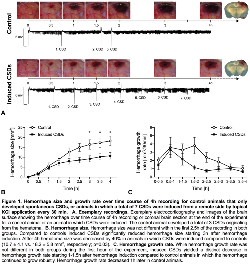
Conclusion
These data for the first time show that CSDs limit hematoma expansion, presumably by their lasting vasoconstrictive effect (post-CSD oligemia). Therefore, CSD inhibition during hyperacute ICH may have deleterious consequences.
BS04-3
Hemorrhagic stroke
Red blood cell-derived microparticles treatment attenuates intracerebral hemorrhage-induced behavioral deficits in rats
1Cerebral Vascular Disease Research Laboratories, University of Miami Miller School of Medicine, Miami, Florida 33136, USA
2Department of Neurology, University of Miami Miller School of Medicine, Miami, Florida 33136, USA
3Neuroscience Program, University of Miami Miller School of Medicine, Miami, Florida 33136, USA
4Wallace H Coulter Platelet Laboratory, Division of Hematology/Oncology, Department of Medicine, University of Miami Miller School of Medicine, Miami, Florida 33136, USA
Abstract
Objectives
Spontaneous intracerebral hemorrhage (sICH), the deadliest stroke sub-type, has no therapeutic options. Prevention of hematoma expansion is an attractive therapeutic target. We identified red blood cell-derived microparticles (RMP) as a hemostatic agent1 and observed that RMP treatment lowers post-sICH hematoma volume when measured at 24 h post-collagenase injection. The goal of this study was to evaluate the potential of RMP therapy in improving long-term outcomes in a rat model of sICH.
Methods
RMPs were prepared from human RBCs1. sICH was induced in young male rats by injecting collagenase into the right striatum. Rats were randomly assigned to vehicle, RMP (2.55x10¹⁰ particles/kg, b.w. i.v.), or recombinant Factor VIIa (rFVIIa) (positive control, 120 µg/kg, b.w. i.v.) treatment groups. RMP dose and treatment paradigms were determined based on earlier pharmacokinetic, dose response, and multiple paradigm comparing studies. Behavioral deficits were evaluated using neurological scores, ladder rung walking test, corner turn test, forelimb use asymmetry test (cylinder test), and contextual fear conditioning test up to four weeks post-sICH by an investigator blinded to experimental conditions. Rats were then euthanized to collect brains for histological assessment. After excluding significant outliers identified by Grubb’s test, statistical analyses used were one-way ANOVA followed by Tukey’s multiple comparison test and Student’s t-test, when applicable.
Results
Using a 12-point scale (12 indicating maximum impairment), the neurological scores in the control group at day 1, 3, 7, and 14 were 10.61 ± 0.23, 8.58 ± 0.67, 5.53 ± 0.66, and 3.11 ± 0.56, respectively (Figure A). The RMP-treated group scores at day 1, 3, 7, and 14 were 7.76 ± 0.77, 5.41 ± 0.65, 2.63 ± 0.41, and 1.59 ± 0.32, respectively. The rFVIIa-treated group scores at day 1, 3, 7, and 14 were 7.11 ± 0.75, 5.68 ± 0.61, 2.74 ± 0.39, and 1.53 ± 0.30, respectively. The RMP- and rFVIIa-treated groups had significantly lower scores than the control group up to 14 days post-sICH. In the ladder rung walking test, the control group had a significantly greater percentage of contralateral foot faults than the baseline up to 28 days post-sICH, and the rFVIIa group had foot faults significantly greater than the baseline up to 21 days post-sICH (Figure B). However, the foot faults in the RMP group were not significantly different than the baseline at any time point. There was no significant difference between treatments in the corner turn test, cylinder test, and contextual fear conditioning test. We are in the process of evaluating brain histopathology.

Conclusions
Our results demonstrate that RMP treatment, which limits hematoma volume in acute phase, also lowers post-sICH long-term behavioral deficits. Compared to rFVIIa, RMP therapy appears superior in lowering sICH-induced long-term behavioral deficits. Our results indicate that RMPs have the potential to lower not only hematoma growth in sICH patients, but also post-sICH behavioral deficits.
Grant support
NIH/NS094896.
Conflict of interest
RxMP Therapeutics provided the testing material for the study. Dr. Ahn, Dr. Jy, and the University of Miami have partial ownership in RxMP Therapeutics. Drs. Ahn and Jy also received grant support from RxMP Therapeutics.
Reference
BS04-4
Hemorrhagic stroke
Pro-inflammatory response promoted by Porphyromonas gingivalis lipopolysaccharide enhances the rupture of experimental intracranial aneurysms
1Department of Neurosurgery, Institute of Biomedical Sciences, Tokushima University Graduate School, Japan
Abstract
Objectives
Subarachnoid hemorrhage (SAH) is a catastrophic event that results in high morbidity and a poor prognosis. To prevent SAH its pathogenesis must be understood. In our newly established aneurysm model rats, we found that vascular inflammatory response was associated with the aneurysm rupture. Recently the evidence that periodontal disease could be a part of pathophysiology in an intracranial aneurysm was reported. We hypothesized that the inflammatory response exacerbated by periodontal pathogens affects the experimental intracranial aneurysm rupture.
Methods
Ten-week-old female Sprague-Dawley rats were subjected to aneurysmal induction surgery which consisted of estrogen deficiency, renal hypertension, and hemodynamic stress. Two weeks later, they were divided into 2 groups; rats treated with Porphyromonas gingivalis lipopolysaccharide (Pg-LPS) or saline control (VC). The VC- (n = 17) or Pg-LPS (n = 19) rats received intraperitoneal injection weekly.
Results
During 90 days observation period, 9 VC rats (53%) and 14 Pg-LPS rats (74%) had intracranial aneurysms. Especially, Pg-LPS rats (63%) had a higher incidence of ruptured aneurysm than VC rats (29%) (p < 0.05), indicating that Pg-LPS promoted the experimental aneurysmal rupture. Intriguingly, the administration of Pg-LPS increased the plasma level of IL-1β and MMP9 and the mRNA level of TLR2, IL-1β and MMP9/TIMP2 in the vascular wall on day 36 after aneurysm induction. These results suggest that the local and systemic pro-inflammatory response promoted by Pg-LPS enhances the rupture of intracranial aneurysms.
Conclusion
Our study first document that Pg-LPS enhances the intracranial aneurysm rupture. These findings warrant further investigation concerning their clinical applicability.
BS04-5
Hemorrhagic stroke
Correlates of spreading depolarization, spreading depression and negative ultraslow potential in human epidural versus subdural electrocorticography
1Center for Stroke Research Berlin, Charité Universitätsmedizin Berlin, Germany
2Department of Neurology, Charité Universitätsmedizin Berlin, Germany
3Department of Experimental Neurology, Charité Universitätsmedizin Berlin, Germany
4Department of Neurosurgery, Charité Universitätsmedizin Berlin, Germany
5Bernstein Center for Computational Neuroscience Berlin, Berlin, Germany
6Einstein Center for Neurosciences Berlin, Berlin, Germany
7UC Gardner Neuroscience Institute, University of Cincinnati (UC) College of Medicine, Cincinnati, OH, USA
8Department of Neurosurgery, University of Cincinnati (UC) College of Medicine, Cincinnati, OH, USA
Abstract
Objectives
Spreading depolarizations (SD) are characterized by abrupt, near-complete breakdown of the transmembrane ion gradients, neuronal edema, excitotoxicity and activity loss (=depression). The concept of the SD continuum is important because many SDs have intermediate characteristics, as opposed to the two extremes of SD in either severely ischemic or normal tissue. The SD extreme in ischemic tissue, termed terminal SD, is characterized by prolonged depolarization in addition to the negative ultraslow potential (NUP). The NUP is the largest bioelectrical signal ever recorded from the human brain and is thought to reflect the progressive recruitment of neurons into cell death in the wake of SDs.1 However, it is unclear whether the NUP is a field potential or results from contaminating sensitivities of platinum electrodes. In contrast to Ag/AgCl-based electrodes in animals, platinum/iridium electrodes are the gold standard for intracranial recordings in humans. Here, in an approach of reverse translation from bedside to bench, we investigated the full continuum from short-lasting SDs under normoxia to long-lasting SDs under systemic hypoxia to terminal SD under severe global ischemia using platinum/iridium electrodes in rats to determine whether the peculiar tent-like shape of the human NUP is also found in lissencephalic animals. In an approach of classic translation from bench to bedside, we then investigated the use of less invasive epidural titanium peg electrodes in patients.
Methods
Male, wild-type Wistar rats were anesthetized with thiopental-sodium intraperitoneally, tracheotomised, and artificially ventilated (n = 8). A parietal craniotomy was performed. A K+-selective, Ag/AgCl-based microelectrode was used to measure intracortical electrocorticogram and extracellular K+ concentration; two platinum/iridium plate electrodes were placed on the cortex. In seven patients with either aneurysmal subarachnoid hemorrhage or malignant hemispheric stroke, two epidural peg electrodes were placed 10 mm from a subdural strip.
Results
In the rodents, sensitivities for detecting SDs, spreading depressions or NUP were 100% for both electrode types. However, the platinum/iridium-recorded NUP was ten times smaller in rats than humans, indicating species differences. In the patients, we found that 31/67 SDs (46%) on the subdural strip were also detected epidurally. SDs that had longer negative direct current (DC) shifts and spread more widely across the subdural strip were more likely to be observed in epidural recordings. One patient displayed an SD-initiated NUP while undergoing brain death despite continued circulatory function. The NUP’s median amplitude was –150 mV subdurally and –67 mV epidurally. This suggests that the human NUP is a bioelectrical field potential rather than an artefact of platinum sensitivity to other factors, since the dura separates the epidural from the subdural compartment and the epidural microenvironment unlikely changed, given that ventilation, mean arterial pressure and peripheral oxygen saturation remained constant during the NUP.
Conclusions
Our data provide further evidence for the clinical value of invasive ECoG monitoring, highlighting important possibilities as well as limitations of less invasive recording techniques.
Reference
BS04-6
Hemorrhagic stroke
The effect of subarachnoid hemorrhage on resting state functional connectivity and behavior in mice
1Neurovascular Research Lab, Department of Radiology, Massachusetts General Hospital, Boston, MA, USA
2Neurocritical Care and Emergency Neurology, Department of Neurology, Massachusetts General Hospital, Boston, MA, USA
3Department of Pediatrics, Massachusetts General Hospital, Boston, MA, USA
4Neurophotonics Center, Boston University, Department of Biomedical Engineering, Boston, MA, USA
5Department of Neurosurgery, Yamaguchi University School of Medicine, Ube, Japan
6Athinoula A. Martinos Center for Biomedical Imaging, Department of Radiology, Massachusetts General Hospital, Charlestown, MA, USA
7Stroke Service, Department of Neurology, Massachusetts General Hospital, Boston, MA, USA
Abstract
Objectives
Subarachnoid hemorrhage (SAH) is one of the most severe forms of stroke with high rates of long-term disability. Survivors, even with relatively good outcomes, suffer from lasting cognitive deficits precluding return to work. Recent evidence in humans suggests that these cognitive deficits are associated with alterations in resting state functional connectivity (RSFC) (1). Therefore, we determined measures of RSFC, behavior, and white matter integrity over the course of 3 months in mice subjected to SAH versus sham surgery
Methods
We used functional hemodynamic optical intrinsic signal imaging (2) to determine the degree of spontaneous hemodynamic fluctuations and measures of RSFC using the prechiasmatic blood injection model in mice. We assessed behavioral outcomes after SAH on the Y-maze, open field test (OFT, 30 minutes), Morris water maze (MWM), and rotarod at early (2 weeks), intermediate (1 months), and late (3 months) time points. We assessed the effect of injury on the corpus callosum using Luxol fast blue staining.
Results
At an early (5 day) time point, we found decreased seed-based RSFC in motor, sensory, retrosplenial, and visual cortex following SAH (n = 11 mice, seed is black circle in representative correlation maps with only right-sided seeds shown) compared to sham (n = 11 mice). Representative connection matrices demonstrate different patterns of correlation coefficients between seeds with an overall decreased amplitude of correlation coefficients in SAH mice. A hemispheric map, which quantifies connectivity between mirror voxels in contralateral hemisphere, shows decreased homotopic connectivity following SAH. Finally, a power map demonstrates an overall decreased amplitude of hemodynamic (i.e. blood flow) fluctuations in post-SAH mice (Figure). These alterations in RSFC resolve by 1 month. On Y-maze testing, there is no difference between sham and SAH animals at the early time point. However, in intermediate and late time points, percent alternation was 58.2 ± 2.7% for sham and 49.2 ± 2.7% for SAH (p = 0.032, repeated measures). Open field testing revealed 34 ± 6 meters traveled for sham and 14 ± 4 meters for SAH (p = 0.020) at the early time point, but this effect was not significantly different at later time points. Morris water maze testing showed significantly increased latency to the hidden platform for SAH mice compared to sham over the course of 7 trials (p = 0.028) for early but not later timepoints. Rotarod testing revealed no differences between SAH and sham mice for all tested time points. After 3 months, mice were sacrificed and brains were prepared with the myelin stain, Luxol fast blue. There was no difference in the thickness, intensity, or variation in corpus callosum staining between SAH and sham mice.

Conclusions
SAH leads to alterations in hemodynamically-derived RSFC for at least 5 days which are associated with time-delimited long-term memory deficits and persistent working memory deficits.
References
BS05-1
Dementia and Cognition Impairment
ApoE-dependent pharmacogenetic response to rapamycin for Alzheimer's disease prevention
1Dept. of Pharmacology and Nutritional Sciences, University of Kentucky, Lexington, KY, United States
2Sanders Brown Center on Aging, University of Kentucky, Lexington, KY, United States
Abstract
Objective
The apolipoprotein E gene is the primary genetic risk factor of Alzheimer’s disease (AD), the ɛ4 allele (APOE4) could lead to 12-fold increased risk, while ɛ3 allele (APOE3) is considered as neutral. Rapamycin has been shown to extend lifespan and restore cerebrovascular functions in APOE4 mice, suggesting potential AD prevention. However, whether Rapamycin can be applied universally as preventative therapeutic remains unknown. The purpose of this study was to identify the APOE gene-dependent response to Rapamycin.
Methods
Results
Rapamycin restored the low CBF and increased water content of pre-symptomatic E4FAD mice (p = 0.007, Mean ± SEM = –0.7154 ± 0.4355, Mean ± SEM = 1.51 ± 0.412). The effects were more pronounced in female mice with FAD mutation. The restoration of CBF was associated with significantly reduced Aβ. In contrast, In E3FAD mice brains, glycolysis and carbohydrate pathways were significantly altered by Rapamycin. Specifically, the pentose phosphate pathway (ribose 1-phosphate), nucleotide sugars (UDP-galactose), and aminosugars (N-acetylneuraminate) metabolism are consistently slowdown with decrease TCA cycle intermediates.

Conclusion
Our results showed that Rapamycin restored cerebral vascular functions and facilitated A-beta clearance in asymptomatic mice expressing the human APOE4 gene whereas altered brain metabolism in APOE3 mice. The APOE3 mice displayed evidence of decreased glycolysis and carbohydrate processing and many more changes than E4. These suggest energetics of the brain are subtly different in the two genotypes, and that they are affected differently by Rapamycin treatment. Also, Rapamycin is a promising AD prevention for E4 carriers. These pharmacogenetic differences provide novel insights into differential functions of APOE gene’s influence on drug response that may facilitate modulating AD. It is important to further study the mechanisms of drug action.
BS05-2
Dementia and Cognition Impairment
The relationship between cognitive function, cortical blood flow and sub-cortical white-matter health in the elderly
1NeuroPoly Lab, Institute of Biomedical Engineering, Polytechnique Montreal, Montréal, QC, Canada
2Centre de recherche de l’Institut Universitaire de Gériatrie de Montréal (CRIUGM), Montreal, QC, Canada
3Department of Neurosciences, Faculty of Medicine, Université de Montréal, QC, Canada
4Department of biomedical Sciences, Faculty of Medicine, Université de Montréal, QC, Canada
5Institut de Recherches Cliniques de Montréal, Université de Montréal, Montréal, QC, Canada
6Montreal Heart Institute, Montreal, QC, Canada
7PERFORM Centre, Concordia University, Montréal, QC, Canada
8Department of Pharmacology and Physiology, Faculty of Medicine, Université de Montréal, QC, Canada
9Department of Medicine, Faculty of Medicine, Université de Montréal, QC, Canada
10Physics Department, Concordia University, Montréal, QC, Canada
Abstract
Objectives
Alteration of white matter integrity (WM) is commonly observed in aging, and the associated degradation in neural connectivity contributes to cognitive decline in older adults 1. No prior study has examined the direct relationship between grey matter (GM) health, white-matter (WM) integrity and cognitive function with respect to arterial stiffening. In this work, we aimed to determine whether cognitive function is associated with age, sex, cerebral blood flow (CBF) and microstructural WM integrity using state of the art microstructural modeling in healthy elderly classified according to their level of arterial stiffness.
Methods
54 healthy individuals (age = 69.91 ± 3.31years) were scanned on a 3T (MAGNETOM Prisma Fit, Siemens) to compute Neurite orientation dispersion and density (NODDI) metrics from diffusion-weighted images (DWI). CBF in the GM was measured using pseudo-continuous arterial spin labelling (pcASL). DWIs were processed using TOAD2 to compute the intracellular volume fraction (vic) and the isotropic volume fraction (v iso) corresponding respectively to the neurite density index and the orientation dispersion index3. pcASL data were processed using Neurolens 4. The trail making test part B-part A (TMTB-A) was evaluated as a measure of cognitive flexibility. Carotid-femoral pulse wave velocity (cfPWV) was measured following the Van Bortel protocol5. Fixed entry linear models were used to identify the relationship between age, sex, WM integrity and CBF. The first model (A) included age and sex as predictor variable, the second model (B) included the CBF in the GM and the third model included either vic (C1) or v iso (C2) to take into account WM microstructural integrity. The analysis have been done in the whole group as well as in group participants according to their cfPWV value.
Results
Table 1i shows that age and sex are not independently associated with TMTB-A (p > 0.05). Model B, where the contribution of CBF in the GM was added to age and sex was found to account for more variance than previous model despite non significant result (F change at p = 0.815 vs p = 0.886). Model C1, where v iso was added to age, sex and CBF, was found to account for significantly more variance than previous models (significant F change at p = 0.013). This analysis indicated an R2 of 0.121 meaning that age, sex, CBF and v iso accounted for 12.1% of the total variance in TMTB-A in the elderly. Table 1ii shows that the same trend for individuals with a cfPWV > 8.5 m/s where age, sex, CBF and v iso where found to account for 19.4 % of the total variance in TMTB-A, while, individuals with a cfWV < 8.5 m/s did not show the same trend (Table 1iii).

Conclusion
We provide a first evidence that v iso account for about 12% of the total variance in cognitive flexibility in the elderly and about 20% in individuals with higher vascular stiffness. This suggest that preserving the WM integrity may help prevent cognitive decline, in particular in individuals exhibiting early signs of vascular aging.
References
BS05-3
Dementia and Cognition Impairment
Carotid arterial stiffness and cerebral blood flow pulsatility in patients with amnestic mild cognitive impairment
1Institute for Exercise and Environmental Medicine, Texas Health Presbyterian Hospital Dallas, USA
2Dept. of Neurology and Neurotherapeutics, University of Texas Southwestern Medical Center, USA
3Human Informatics Research Institute, National Institute of Advanced Industrial Science and Technology, Japan
4Dept. of Internal Medicine, University of Texas Southwestern Medical Center, USA
Abstract
Objectives
Stiffening of central elastic arteries is associated with increased cerebral blood flow (CBF) pulsatility, a potential risk factor for cognitive impairment1. However, the relationship among arterial stiffness, pulsatile changes in arterial pressure, and CBF in patients with mild cognitive impairment (MCI) remains unknown. Therefore, this study investigated the associations among the carotid arterial stiffness, arterial pressure, and CBF pulsatility in patients with amnestic MCI, a prodromal stage of Alzheimer disease (AD).
Methods
Fifty-eight amnestic MCI patients and 22 cognitively normal subjects (NC) underwent the measurements of CBF velocity (CBFV) from the middle cerebral artery using transcranial Doppler. The common carotid artery (CCA) stiffness and pulse pressure (cPP) were measured using ultrasound and applanation tonometry. Pulsatility index (PI) was calculated from pulsatile velocity normalized by mean velocity. The β-stiffness index was calculated as an index of CCA stiffness.
Results
Higher PI was correlated with increased β-stiffness index (MCI: r = 0.411, NC: r = 0.685, P < 0.01, Figure) and cPP (MCI: r = 0.542, NC: r = 0.751, P < 0.001) in both groups. PI, β-stiffness index, and cPP did not differ between groups with independent t-test. However, MCI patients had higher β-stiffness index (P = 0.004) and cPP (P = 0.041) than NC after adjustment for age and sex. There was also a trend towards increased PI in MCI patients (P = 0.070).

Conclusions
MCI patients had higher CCA stiffness and cPP which may lead to increased CBF pulsatility when compared with cognitively normal older adults. These findings support the hypothesis that increased central arterial stiffness is an important risk factor for developing AD.
Reference
BS05-4
Dementia and Cognition Impairment
White matter hyperintensities impact on regional brain volumes and white matter microstructure: a general population study (HUNT MRI)
1Department of Neuromedicine and Movement Science, NTNU. Norway
2Department of Radiology and Nuclear Medicine, St Olavs Hospital, Trondheim University Hospital, Norway
3Department of Circulation and Medical Imaging, NTNU, Norway
4Department of Clinical Medicine, UiT The Arctic University of Norway, Norway
5PET Center, University Hospital North Norway, Norway
Abstract
Objectives
This study was motivated by evidence suggesting that WMH is a localized manifestation of a more widespread pathology. If this is correct, mere presence of WMH may affect brain volume and microstructure in a different manner than changes associated with increasing WMH load. The aim of this study was therefore to uncover effects on brain volume and microstructure driven by presence of WMH or by increasing WMH volume, or by a combination of both factors.
Methods
The study was approved by the Regional Ethics Committee and the HUNT board. Participants were between 50–65 years and drawn among previous HUNT participants with clinical and demographic data. Exclusion criteria were standard MRI safety measures. 1006 individuals were scanned on one 1.5T Signa HDx MRI scanner with an 8-channel head coil (GE). The T1-weighted 3D IR-FSPGR, transversal FLAIR and DTI scans were used. WMH were assessed semi-qualitatively with Fazekas scale by two neuroradiologists and quantitatively by manual delineated on the FLAIR images. DTI data was processed using FSL. Regional volume differences were assessed with tensor based morphometry using the ANTS toolkit. Statistical analyses were performed in R and PALM.
Results
There were 904 participants (52.7% women) included after excluding individuals with brain pathology other than WMH, and normal variants affecting brain structure/image quality. The control group was those with Fazekas 0 (54.5%) and the WMH group those with Fazekas ≥1. The WMH group was significantly older (mean difference 1.3 years) and included more women.

Conclusions
Both volumetric and DTI data demonstrated that regions in the brainstem and thalamus exhibited group differences while cortical regions and white matter near cortex were sensitive to increasing WMH volume. The findings demonstrate that effects due to presence of WMH on regional volume and FA are distinct from effects due to increasing WMH volume.
BS05-5
Dementia and Cognition Impairment
Genetic deletion of Krüppel-like transcription factor 11 aggravates the pathogenesis of vascular cognitive impairment and dementia
1Department of Neurology, University of Pittsburgh School of Medicine, Pittsburgh, USA
Abstract
Objectives
Vascular cognitive impairment and dementia (VCID) ranks the second most common cause of dementia after Alzheimer’s disease. Recent emerging evidences have suggested that targeting structural and functional abnormality in cerebral vasculature may help to prevent or delay the progression of cognitive decline or dementia, white matter injury, and neuronal loss in VCID brains. Krüppel-like factors (KLFs) are members of the zinc finger family of transcription factors and the function of the KLFs in the central nervous system and neurologic diseases is poorly explored. KLF11 is a member of the KLF family and we have previously demonstrated that genetic deletion of KLF11 aggravates ischemic cerebrovascular and brain parenchymal injury in a mouse transient focal cerebral ischemia model. In this study we sought to determine the role of KLF11 in the regulation of cerebrovascular function and the pathogenesis of vascular cognitive impairment and dementia (VCID).
Methods
Experimental VCID was induced in KLF11 knockout mice (KLF11 KO) and wild-type (WT) controls by asymmetric common carotid artery stenosis (ACAS) with an ameroid constrictor placed on the left CCA and a microcoil placed on the right CCA for five weeks. Regional cerebral blood flow was recorded with a laser speckle imaging system. Morris water maze, novel objective recognition and elevated plus maze testing were performed to assess long-term cognitive functions. Accelerating rotarod, adhesive tape removal, and corner testing were performed to assess sensorimotor functions. BBB integrity was assessed by using Evans blue dye and fluorescent BBB tracer extravasation assays. Neuronal loss was analyzed by cresyl violet stain and NeuN immunostaining in brain sections. White matter injury was examined by using luxol fast blue staining, and immunostaining with anti-MBP and anti-SMI-32 antibodies. Total RNA was extracted from brain tissues or brain microvessels, and the expression of BBB tight junctions (TJs) was detected by quantitative real-time PCR and western blotting.
Results
Mice underwent ACAS for 35 days developed remarkable cognitive impairments, myelin loss and axonal damage in corpus callosum and external capsule, and neuronal death in hippocampal CA1 region and cerebral cortex. Compared to WT controls, KLF11 KO mice exhibited significantly server cognitive and sensorimotor deficits following VCID. Genetic deficiency of KLF11 in mice also significantly aggravated neuronal loss, demyelination, and axonal injury in response to chronic cerebral hypoperfusion. Mechanistically, genetic deficiency of KLF11 in mice led to increased BBB permeability and leakage in VCID brains 3 days after ACAS, evidenced by significantly enhanced extravasation of Evans blue and BBB tracers. Also, we found KLF11 binding sites in the promoter region of major endothelial tight junction proteins (TJs) including Occludin, Claudin 5, and ZO-1. Genetic deletion of KLF11 significantly decreased cerebral TJ levels in mice with VCID.
Conclusions
The present study has demonstrated that KLF11 preserves BBB structural and functional integrity and confers brain protection in an experimental VCID model. KLF11 may be a novel therapeutic target for the treatment of VCID.
BS05-6
Dementia and Cognition Impairment
Neuropsychiatric symptoms relate to tau but not amyloid in Alzheimer's disease spectrum
1McGill Center for Studies in Aging, McGill University, Canada
2Translational Neuroimaging Laboratory, Douglas Mental Health Institute, Canada
3Douglas Hospital Research Center, Canada
Abstract
Introduction
Neuropsychiatric Inventory-Questionnaire (NPI-Q) is a survey addressed to informants to assess the participant’s neuropsychiatric symptoms (NPS) and their impact on the participant (global severity) and their informant (global distress). Thus, NPI-Q contains 2 global domains and 12 subscales: delusions, hallucinations, agitation, depression, anxiety, elation, apathy, disinhibition, irritability, motor disturbance, nighttime behavior and appetite. Although NPS has been closely related to the clinical progression to dementia in carriers of Alzheimer’s disease (AD) pathophysiology, the relationship between NPS and brain amyloid and tau pathologies is still unclear. Here we hypothesize that tau and amyloid deposition associate with different subscales of the NPI-Q, across the AD clinical spectrum.
Methods
94 individuals (CN = 74, MCI = 9, AD = 11) underwent positron emission tomography (PET) amyloid [18F]AZD4694 and tau [18F]MK-6240. [18F]AZD4694 and [18F]MK-6240 standardized uptake value ratios (SUVRs) used the cerebellum grey matter as the reference region and were calculated between 40–70 min and 90–110 min post-injection, respectively. A voxel based regression model1 evaluated the relationship between the accumulation of a biomarker, using [18F]MK6240 or [18F]AZD4694 PET scans, and the NPI-Q global scores, as well as the 12 subscales. The model’s covariates were age, gender, years of education and diagnosis of participants.
Results
We did not find association between global NPI-Q scores (distress and severity) and brain amyloid deposition. Observing the subscales, depression and hallucinations correlated with amyloid deposition in the paracentral lobule and the frontal lobe respectively. Tau deposition was however correlated with global NPI-Q distress score, but not with global severity score. The most impacted regions were the left frontal lobe and the precuneus. Additionally, delusions, hallucinations, agitation, depression, apathy, disinhibition, motor disturbance and appetite subscales correlated with tau accumulation. The strongest correlations were found with depression, correlated with tau deposition in the frontal lobe and precuneus among other regions, and hallucinations and delusions both showing high correlation with tau pathology in the cuneus.

Conclusion
These preliminary results showed a strong association between the distress caused by the patients’ NPS in their caregivers and tau aggregation in regions of brain known to be involved with self-consciousness and holistic thoughts. These regions are also recognized as being associated with NPS and AD. The findings align with the idea that tau is more closely related with cognitive and behavioral symptoms than amyloid. This study demonstrates NPS correlate with tau and it is thus important to assess them in the context of the AD clinical spectrum. Moreover, it emphasizes the importance of the informant’s perspective in the managements of the diseases, which is often overlooked.
Reference
BS06-1
Advances in Basic through Clinical Stroke
Activity dependent neuroprotection in the acute phase after stroke
1Dept. of Psychiatry, University of British Columbia, Canada
2Centre for Brain Health, University of British Columbia, Canada
Abstract
Background
Stroke represents a leading cause of death and disability worldwide. Optogenetic stimulation used to enhance stroke recovery has shown potential benefits when applied weeks after injury. However, benefits of acute brain stimulation have not been reported. Changes in gamma oscillations (20–50 Hz) have been observed in several neurological disorders but the relationship between gamma oscillations and cellular pathologies is unclear. We investigated the effect of the gamma-wave modulation in the acute phase – within 1 hr – after stroke.
Methods
We combined multimodal approaches employing optogenetics in conjunction with laser speckle imaging, two photon microscopy, electrophysiology and behavioral tasks. Transgenic VGAT-ChR2 mice were implanted with a transcranial chronic window and subjected to photothrombotic stroke while awake in the target area of somatomotor cortex.
Results
Optogenetic stimulation at 40 Hz ipsilateral to the stroke side resulted in a significantly higher increase in blood flow over the course of the first week following stroke (Stroke n = 8 vs Stroke + stimulation n = 10: p = 0.0148). Stroke area and stroke volume were significantly reduced in mice that received the stimulation (Area: Stroke n = 8 vs Stroke + stimulation n = 10, p = 0.0010; Volume: Stroke n = 8 vs Stroke + stimulation n = 10, p = 0.0249). Assessment of motor function showed a significant improvement over time in mice that received stimulation (NDS: Stroke n = 8 vs Stroke + stimulation n = 10, p < 0.0001. Tapered beam test: Stroke n = 9 vs Stroke + stimulation n = 10, Group × time effect: p < 0.0001). Microglia activation was assessed to investigate whether the stimulation at this specific frequency would induce local activation of these cells. Electrophysiological recordings in vivo showed an increase in synchronization in the areas associated to the stimulated one.
Conclusions
In this study, we describe the beneficial effects of acute, 40 Hz brain stimulation at gamma range: reduced lesion volume and improved motor function after stroke.
BS06-2
Advances in Basic through Clinical Stroke
ATN-161 reduces inflammation and blood-brain barrier dysfunction following in vitro stroke conditions in association with conservation of claudin-5 expression
1Department of Neuroscience, University of Kentucky
2Department of Neurosurgery, University of Kentucky
3Department of Neurology, University of Kentucky
Abstract
Cerebrovascular remodeling and inflammatory mechanisms significantly increase blood-brain barrier (BBB) permeability following ischemic stroke, leading to injury expansion and worsened outcomes. Previously, our laboratory has shown that inhibition of the pro-angiogenic integrin, α5β1, by the small peptide ATN-161 results in reduced infarct volumes, edema, and behavioral dysfunction through cerebrovascular stabilization and reduced leukocyte infiltration. Here, our
Methods
Mouse brain endothelial cells were grown to confluency in a monolayer and either underwent 8-hour oxygen-glucose deprivation (OGD) or inflammatory stimulation through TNF-a administration. Immediately following OGD or TNF-a treatment, the cells received PBS or the integrin α5β1 inhibitor, ATN-161, during the 24 hour reperfusion. Localization and expression of claudin-5 and integrin α5β1 was determined by immunocytochemistry. Integrin, ROCK1, β-catenin, claudin-5, TNF-α, IL-1β, and VEGF transcription and expression were determined by qPCR and western blot, respectively. Monolayer permeability was determined by FITC-dextran assay.
Results
ATN-161 administration decreased integrin α5β1 expression in both OGD and TNF-α conditions. Additionally, a decrease in the cytokines TNF-α and IL-1β was found. Furthermore, monlayer permeability was decreased under ATN-161 administration in association with conservation of claudin-5 expression and localization to the extracellular wall following OGD. This may occur in a VEGF-A independent fashion as ATN-161 administration increased VEGF production, a known factor for increased permeability through claudin-5 dysregulation. Importantly, direct regulators of claudin-5 expression and function, ROCK1 and β catenin, were increased following OGD.
Conclusions
ATN-161 reduced inflammation and BBB-like dysfunction following OGD and TNF- administration. This observation correlates with conservation of claudin-5. Though VEGF was upregulated, it appears that ATN-161 prevents claudin-5 dysregulation by increasing the regulators, ROCK1 and β-catenin. This suggests ATN-161 has a dual beneficial effect following ischemic stroke.
BS06-3
Advances in Basic through Clinical Stroke
Optogenetic functional activation expands infarct volume independent of periinfarct spreading depolarizations in focal cerebral ischemia
1Departments of Radiology, Massachusetts General Hospital, Harvard Medical School, USA
2Department of Neurosurgery, Yamaguchi University, Japan
3Departments of Neurology, Massachusetts General Hospital, Harvard Medical School, USA
4Martinos Center for Biomedical Imaging, Massachusetts General Hospital, USA
Abstract
Objectives
Peri-infarct depolarizations (PIDs) are believed to contribute to injury progression and worsen the outcome. Previous studies showed that exogenously induced spreading depolarizations (SDs) by depolarizing interventions (e.g. topical KCl) expand infarct volumes. However, such interventions are often invasive and can directly cause tissue injury. Here we employed a novel, non-invasive and non-injurious SD induction method using optogenetics in transgenic mice expressing channelrhodopsin-2 in neurons (Thy1-ChR2-YFP).
Methods
We transiently (1 h) occluded the middle cerebral artery using a microvascular clip (dMCAO) or using a filament (fMCAO) in Thy1-ChR2-YFP mice (age ∼10 weeks, male), and triggered an SD every 5 minutes using optogenetic stimulation (470 nm, 1–2 mw, 10 seconds) of non-ischemic remote ipsilateral cortex. Tissue and neurological outcomes were studied 24 hours after fMCAO and 48 hours after dMCAO by TTC staining. We also examined the effect of light on wild-type (C57BL/6) animals.
Results
Optogenetic SD group had 8.0 ± 0.6 PIDs/h (n = 6) compared with 1.5 ± 0.8 spontaneous PIDs/h (n = 6) in the control group during dMCAO. Surprisingly, this 5-fold increase in PID numbers in the optogenetic SD group did not alter the infarct volume compared with the control group (10.7 ± 2.5 vs. 12.7 ± 2.4 mm3, respectively; p = 0.576). Moreover, optogenetic SD in fMCAO did not increase the cortical infarct volumes compared with the control group (19.7 ± 8.7 vs. 29.0 ± 6.7 mm3, n = 7 each, p = 0.418), despite 3.5-fold higher PID numbers (9.1 ± 0.5 and 2.6 ± 0.7 PIDs/h, respectively). Neurological deficit scores did not differ (7.0 ± 3.0 vs. 8.7 ± 1.2, n = 7 each, in optogenetic SD vs. control groups, respectively; p = 0.201). In contrast, when SD was induced by topical KCl application (7.0 ± 0.6 vs. 0.4 ± 0.3 PIDs/h, n = 7 and 14), infarct volumes increased significantly (14.0 ± 2.4 vs. 5.7 ± 0.9 mm3, p = 0.013). These data seemed to contradict our previous finding that increasing PID frequency by functional activation enlarged infarct volumes. To explain the apparent discrepancy, we functionally activated the peri-infarct tissue by optogenetic stimulation using a stimulus protocol that did not trigger SDs (1mW, 8 Hz, 6 ms, 15sec, repeated ∼10 cycles) during dMCAO. Optogenetic functional activation in penumbra significantly increased infarct volumes (10.0 ± 1.7 vs. 5.7 ± 0.9 mm3, n = 9 and 14, p = 0.032). Activation of contralateral homotopic cortex did not alter infarct volumes (6.0 ± 1.6 mm3, n = 6, p = 0.986). In wild-type mice (i.e. ChR2-/-), optogenetic light application did not significantly affect the PID frequencies, infarct volumes or neurological deficits after dMCAO and fMCAO.
Conclusions
Our results suggest that SDs induced non-invasively and non-injuriously away from the primary injury site do not worsen tissue outcome, and that exogenous interventions that trigger peri-infarct SDs (e.g. KCl, functional activation, hypotension) directly increase infarct volumes independent of SDs. As a clinically-relevant finding, functional activation of peri-infarct tissue in acute phase can worsen the outcome of ischemic stroke.
BS06-4
Advances in Basic through Clinical Stroke
Dendritic remodeling of the spinal motor neurons in the denervated gray matter after stroke in mice
1Dept. of Neurology, West China hospital of Sichuan University, China
2Dept. of Neurology, Henry Ford Hospital, US
Abstract
Objective
We investigated morphological dendritic changes of the spinal motor neurons, the final common pathway of motor control, during recovery after stroke in adult mice.
Methods
Mice (C57BL/6 J, 2–3 month-old) were subjected to permanent right unilateral middle cerebral artery occlusion (MCAo) by advancing an 8–0 surgical nylon suture with an expanded silicone tip from the external carotid artery (ECA) into the lumen of the internal carotid artery under isoflurane anesthesia, to block the origin of the MCA. Animals were sacrificed at 7,14, 21 and 28 days after MCAo (n = 12/group), respectively. A separate group of naïve mice was used for normal control (n = 12). Four mice in each group were randomly selected to receive intramuscular injection of pseudorabies virus (PRV)-512-EGFP or AAV-retro into the left forelimb wrist extensor and flexor muscles with a 10 µl total volume (1 × 10¹⁰ viral particles) through a skin incision at 56 hours or 21 days before sacrifice, respectively. Spinal cord samples were processed for longitudinal vibratome sectioning. EGFP labeling in the spinal motor neurons were digitized using a laser-scanning confocal imaging system. Cervical cord samples form the remaining 4 mice were processed for Golgi staining to assess the dendrites and synaptic spines of the spinal motor neurons in the denervated side of the spinal gray matter. NeuronJ, a plugin of NIH software ImageJ, was used to measure dendrite length, branch number and spine density of the spinal motor neurons.
Results
Compared to normal control, significant increases of both neurite length and branch numbers were observed in animal groups of 14 days after MCAo with PRV tracing, AAV tracing and Golgi staining, as well as increased dendritic spine density in animal groups of 21 and 28 days after MCAo (P < 0.05).
Conclusion
Our data indicate the presence of anatomical remodeling of the spinal motor neurons in the denervated side of the spinal cord post stroke. This remodeling may facilitate rewiring of the stroke-impaired corticospinal innervation, and thereby promote spontaneous motor functional recovery after stroke. These data also suggest that anatomical remodeling of the spinal motor neurons represent a therapeutic target for treatment of stroke.
BS06-5
Advances in Basic through Clinical Stroke
Pre-clinical investigation of a novel thrombolytic and anti-inflammatory therapy for the treatment of acute ischaemic stroke
1Faculty of Biology, Medicine and Health, University of Manchester, UK
Abstract
Objectives
To determine the thrombolytic and anti-inflammatory efficacy of a novel, constitutively active variant of a haemostatic protease (Protease-X) in a murine model of acute ischaemic stroke. This variant has been developed to circumvent the conformational quiescence of the protease and its requirement for substrate induced activation. As such, the variant exhibits enhanced proteolytic activity against von Willebrand factor (VWF) and has fibrin(ogen)olytic activity. These properties suggest the variant may have enhanced thrombolytic potential compared to WT Protease-X and, moreover, may attenuate VWF and fibrinogen dependent pro-inflammatory pathways.
Methods
In adult male CD1 mice, thrombotic occlusion of the middle cerebral artery (MCA) was induced by application of ferric chloride to model an acute ischaemic event. Restricted cerebral blood flow (CBF) was confirmed by laser Doppler flowmetry and monitored for 1 hour. At 1 hour post-occlusion animals were injected (i.v) with either (wt)Protease-X (6 mg/kg), (ca)Protease-X (6 mg/kg), N-acetyl cysteine (400 mg/kg) or vehicle. Laser speckle contrast imaging was used to monitor CBF in the MCA territory (contralateral and ipsilateral) over a further 1 hour period. Ex vivo analyses were performed at 24 hours post-occlusion.
Results
We observed a >6 fold enhancement in the restoration of CBF between the vehicle and (ca)Protease-X treated groups (Figure 1A), with ipsilateral/contralateral flux ratio increases of 8.9 ± 7.4 and 58.4 ± 9.3 %, respectively (mean ± SEM, n=7, p<0.002). The enhanced restoration of CBF translated to a 50% decrease in infarct volume (Figure 1B). Immunohistochemistry was used to quantify the amount of VWF and fibrin(ogen) deposited in the occluded vessel and surrounding tissue. Between the vehicle and (ca)Protease-X treated groups there was an almost 30 fold decrease in VWF deposition (5.9 ± 0.92 and 0.21 ± 0.38 %, respectively) and a >8 fold decrease in fibrin(ogen) staining (4.2 ± 1.0 and 0.49 ± 0.35 %, respectively). We also observed an almost 5 fold reduction in the number of CD41+ particles within the infarct (176.5 ± 44.0 and 36.3 ± 13.8 cells/mm2, respectively). Further immunohistochemical analyses revealed a 4 fold decrease in neutrophil extravasation into the ischaemic tissue (19.6 ± 3.0 and 5.4 ± 1.7 Ly6G+ cells/mm2, respectively) and a >7 fold decrease in microglial activation in the peri-infarct region of the ipsilateral cortex (115.3 ± 15.4 and 15.7 ± 8.3 activated cells/mm2, respectively).

Conclusions
The (ca)Protease-X variant exhibited potent thrombolytic activity, significantly reducing both VWF and fibrin(ogen) deposits at the site of occlusion. Furthermore, the variant effectively reduced the number of VWF-platelet aggregates in the cerebral microvasculature, thereby restoring CBF to the infarct penumbra and reducing the infarct volume. The variant also appears to reduce neutrophil infiltration into the ischaemic tissue, which may further contribute to its neuroprotective influence. However, further in vitro experiments are required to confirm this mechanism and to fully evaluate the therapeutic time window.
References
BS06-6
Advances in Basic through Clinical Stroke
A multi-center investigation of the association of acute stroke severity and long-term outcome with acute stroke lesion topography
1Athinoula A. Martinos Center for Biomedical Imaging, Dept of Radiology, Massachusetts General Hospital, United States of America
2Harvard Medical School, United States of America
Abstract
Objectives
There is increasing awareness that long-term stroke deficits are dependent on both the location and volume of the lesion.1 Voxel-based lesion symptom mapping (VLSM) techniques2 have been used to show the association between chronic infarct location and neurocognitive deficits. Several studies have also reported on associations of acute cerebral infarct topology with outcomes,3 but these were single center studies, with limited sample size. Here, using data from a multi-center ischemic stroke imaging repository (MRI – GENetics Interface Exploration [MRI-GENIE] study4), we propose to investigate the relationship between acute infarct location and both baseline stroke severity (NIH Stroke Scale [NIHSS]) and long-term functional outcomes (modified Rankin Scale [mRS]).
Methods
DWI datasets from MRI-GENIE were retrospectively analyzed. This cohort consisted of 3301 subjects from 12 centers, with 2770 having usable DWI. Six of the 12 centers had baseline NIHSS and 3-month (60–190 day) mRS recorded as part of the Genetics of Ischaemic Stroke Functional Outcome study.5 Analysis was limited to MRI acquired within 1-week of admission. An automated lesion segmentation algorithm consisting of an ensemble of five 3D convolutional neural networks trained using DeepMedic6 with DWI from 267 subjects from MRI-GENIE was applied to segment DWI lesions for all 2770 subjects. DWI were co-registered to an atlas. Seven datasets that could not be co-registered were excluded. VLSM was performed with either NIHSS or mRS as the behavior variable. Voxels were tested only if the voxels were infarcted in at least 10 subjects. Resulting T-scores maps were thresholded voxelwise (P < 0.001), corrected for multiple comparisons based on cluster size permutation method (1000 permutations; P < 0.05).7
Results
The demographics for 903 subjects meeting inclusion criteria are: mean ± SD age 64 ± 15 y.o, 61% male, median [IQR] NIHSS 3 [2–7], mRS 1 [1–3], lesion volume 4.0 [1.0–17.7] cm3 and stroke subtype: large artery atherosclerosis (20%), small artery occlusion (17%), cardioembolic (16%), undetermined (40%), other (7%). The figure incidence maps show that most strokes were in the middle cerebral artery territory. Subjects with left hemispheric infarcts were likely to present with greater NIHSS. For long-term outcomes, lesions in regions associated with neurologic neglect in the right hemisphere and language regions in the left hemisphere had higher mRS.

Conclusion
The results confirm our hypothesis that acute stroke infarct location is an important determinant of baseline stroke severity and long-term functional outcomes. Our findings in the left hemisphere are consistent with prior studies.8 With large multi-center data sets, we had sufficient power to detect the association of right hemispheric infarcts with poor long-term outcomes, which was not found in prior smaller studies.8 We speculate that these patients likely suffered from neglect syndrome or associated cognitive impairment, which are known to hinder recovery.9 Selection strategies of which patients may benefit from aggressive post-stroke rehabilitation programs might be enhanced from integration of VLSM methods with clinical assessments.
References
BS07-1
Brain Injuries: Traumatic, Ischemic, & Hypoxic
Neurovascular dysfunction develops post-mild traumatic brain injury in a pediatric animal model
1CNRS UMR 5287, University of Bordeaux, France
2UMR 5203 CNRS – U1191 INSERM, University of Montpellier, France
3Basic Science Department, Loma Linda University School of Medicine, CA, USA
Abstract
Objectives
Vascular dysfunction is a hallmark of pediatric traumatic brain injury (TBI) and predicts poor outcome on the long term. To date little is known on neurovascular changes after pediatric mild-TBI. In this study we used the newly developed mouse model of mild pediatric TBI,
Results
Hypoperfusion occurred in the impacted regions hours post-injury returning to control levels at 1 week. Histologically, we observed morphological changes in the intraparenchymal blood vessels with significantly decreased diameters compared to sham tomato-lectin stained cerebral blood vessels at 1 day, while at 7 days they exhibited greater diamaters. Surprisingly, vessels in CHILD animals had wider diameters compared to sham vessels at 30 days post-TBI. In addition, intraparenchymal blood vessels exhbited functional impairment at 1 and 3 days after injury. At 1 day post-TBI, intraparenchymal blood vessels in the CHILD group constricted significantly more but dilated less compared to sham vessels. On the contrary, at day 3 post-mTBI, the cortical blood vessels of CHILD animals showed a greater dilation after NMDA. These vascular property changes were not associated with different expression of the thromboxane receptors (TXA2Rs) or contractile proteins (α–SMA) in the ipsilateral cortex at these timepoints. CHILD animals exhibited significant behavioral impairments at all timepoints post-TBI with long-term anxiety-like changes up to 30 days. These changes were associated with a signature of low gamma and high theta cortical EEG waves, quantified in vivo at 30 days post-mTBI.
Conclusions
We observed vascular dysfunctions after experimental mild pediatric TBI. Regional perfusion changes after injury were associated with changes in neurovascular coupling and morphological changes of the of intraparenchymal blood vessels (capillary and larger blood vessels). This vascular dysfunction can contribute to long-term EEG-behavioral modifications observed in our model.
Reference
BS07-2
Brain Injuries: Traumatic, Ischemic, & Hypoxic
Morphological and molecular changes of astrocytes following a juvenile mild traumatic brain injury
1CNRS-UMR 5287, Bordeaux University, Bordeaux, France
2UMR 1141 Inserm, Paris Diderot University, Paris, France
3Dept. of Basic Sciences, Loma Linda University, Loma Linda, California, USA
4Institut de Recherche Biomédicale des Armées, Brétigny sur Orges, France
5Dept. of Pediatrics, University of California, Irvine, California, USA
Abstract
Objectives
Traumatic brain injury (TBI) is a leading cause of disability and death among children worldwide. Mild TBI represents around 80% of all pediatric emergency visits and is associated with a higher probability to develop long-term affective and learning disorders. To date, molecular and cellular mechanisms underlying the post-TBI cognitive dysfunction are unknown. Astrocytes are involved in various physiological homeostatic functions and astrogliosis was shown to significantly alter astrocytes’ properties following brain injury. However, such alterations that may occur over time havenever been investigated in the context of juvenile mild TBI (jmTBI). We hypothesize that post-traumatic astrogliosis elicits a specific molecular signature impacting neural network organization and contribute to the long-term affective and cognitive disorders emerging after jmTBI.
Methods
A Closed-Head Injury with Long-Term Disorder (CHILD) was induced over the left-parietal cortex using an electromagnetic impactor in C57BL6 mice on postnatal day 17. In a first experiment, astrocytes were isolated from whole brain with MACS using GLAST to perform RNA-sequencing at 1 day post-injury (dpi). Levels of gene expression were analyzed by differential expression analysis and Over-representation Enrichment Analysis method on WebGestalt. In a second experiment, morphological analysis of astrocytes was performed with GFAP immunolabeling at 1, 7 and 30 dpi and compared to in vivo MRI at 30 dpi.
Results
Results from the RNA sequencing showed that a significant number of genes were differently regulated in jmTBI animals. Specific genes known to be markers of reactive astrocytes were upregulated (Lcn2, Timp1, Cxcl10, Serpina3n, Aspg), with also upregulation of one A1-specific (H2-D1) and one A2-specific (Tgm1). A broader analysis of the upregulated genes in astrocytes of jmTBI animals revealed a significant overrepresentation of genes belonging to the oxidative phosphorylation KEGG pathway, and mitochondrial respiratory chain, metabolism and extracellular matrix organization pathways. From these results we conclude that the jmTBI induces a reactive phenotype in astrocytes 1 dpi, and may dysregulate the morphology and metabolism in these cells.
According to these results, astrocyte morphology was depicted by semi-automatic skeleton analysis in the somatosensory cortex (SSC), dentate gyrus (DG), infra-limbic area of amygdala (ILA) and prefrontal cortex (PFC). The morphology of GFAP-positive cells was altered after injury in all regions. Astrocytes had significantly longer processes in the ipsilateral SSC and DG at 1 dpi, and in the ipsilateral DG, AMY and ILA at 7 dpi. At 30 dpi GFAP-positive astrocytes tended to have longer processes in the contralateral side of the brain. Therefore, our results suggest that there is a progressive spread of astrogliosis from the injury site to remote brain regions over one month after injury.
Furthermore, T2 imaging showed a decrease in values in SSC and PFC that could be interpreted as a metabolic change since T2* values can indirectly depend on oxygen saturation. This result supports the RNA sequencing data.
Conclusion
Taken together, astrocytes are early responders and may contribute to early neuronal network reorganization through changes in reactivity, morphology and metabolism, which could prime the developing brain for long-term affective and cognitive disorders after jmTBI.
Acknowledgements
We thank Marie-Line Fournier and Justine Aussudre.
BS07-3
Brain Injuries: Traumatic, Ischemic, & Hypoxic
M2 microglia-derived exosome promotes white matter repair and long term functional recovery after cerebral ischemia in mice
1Shanghai Ruijin Hospital, School of Medicine, Shanghai Jiao Tong University, China
2School of Biomedical Engineering, Shanghai Jiao Tong University, China
Abstract
Objectives
White matter injury after ischemic stroke is an important event that causes severe neurological and cognitive impairment.1 M2 microglia can promote white matter repair and oligodendrocyte precursor cells survival and differentiation.2, 3 However, the molecular mechanism is unclear. In this study, we explored the effects and mechanism of M2 microglia derived exosomes on white matter repair after cerebral ischemia in mice.
Methods
Microglia BV2 cells were stimulated by IL-4 to polarize to M2 phenotype, and exosomes were isolated from M2 microglia (M2-Exo). Adult male ICR mice (n = 72) were subjected to 90-minute middle cerebral artery occlusion. M2-Exo (100 mg) was intravenously injected once daily for 7 consecutive days. Brain atrophy volume, rotarod test, elevated body swing test, step-through and T-maze tests were examined within 28 days following middle cerebral artery occlusion. Oligodendrocyte precursor cells survival, proliferation, differentiation and white matter integrity were evaluated using immunohistochemistry and western blot analysis.
Results
M2-Exo treatment reduced brain atrophy (p < 0.05), promoted sensorimotor, cognitive and memory functions recovery within 28 days after cerebral ischemia in mice (p < 0.05). Immunostaining showed that M2-Exo enhanced the number of BrdU+/Pdgfr-α+and BrdU+/adenomatous polyposis coli + cells, increased the fluorescence-intensity of myelin basic protein in the cortex, striatum and corpus callosum after cerebral ischemia compared with the control group (p < 0.05). In vitro experiments further showed that M2-Exo enhanced oligodendrocyte precursor cell survival under oxygen-glucose deprivation (p < 0.05), and promoted the proliferation and differentiation of oligodendrocyte precursor cells (p < 0.05).

Conclusion
This study showed that M2-Exo treatment enhanced white matter repair and improved long term functional recovery after cerebral ischemia in mice, promoted oligodendrocyte precursor cell survival, proliferation and differentiation, suggesting that M2-Exo is a novel therapeutic strategy for white matter repairing after stroke.
References
BS07-4
Brain Injuries: Traumatic, Ischemic, & Hypoxic
Investigation into susceptibility of brain endothelial cells to secondary hypoxia challenge in two models of traumatic brain injury
1Trauma Research, Keenan Research Centre in the Li Ka Shing Knowledge Institute at St. Michael’s Hospital
2Institute of Medical Sciences, University of Toronto, Toronto, Ontario, Canada
3Departments of Anesthesia & Surgery, University of Toronto
4Department of Physics, Ryerson University, Toronto
Abstract
Introduction
Traumatic brain injury (TBI) consists of a primary trauma with subsequent ongoing secondary cellular cascades that exacerbate the extent of injury. While conventional brain injury arises from blunt force trauma to the head, increasing evidence points to primary shockwaves from explosive devices as contributors of brain injury. However, the effects of shockwaves on the brain vasculature are not well understood but evidence points to various responses depending on the level of injury which include altered nitric oxide signalling, changes to (blood brain barrier) BBB permeablity, vasospasm and acute reduced blood flow.
Methods
In the current study we used 2 in vitro injury models and evaluated the effects of secondary hypoxia challenge using brain endothelial cells. For in vivo studies we used the fluid percussion injury device and the Baker lab shock tube device. In vitro: We used a well-characterized stretch injury model to induce a primary mechanical trauma in a brain endothelial cell line (bEnd.3). bEnd.3 cells were grown to confluence on a silastic membrane and a ∼2.5 atm pressure pulse was applied over 50 ms to induce a mechanical stretch injury. A pulsed high intensity focused ultrasound (pHIFU) device was used to deliver a short 50 ms shockwave burst at ∼9 MPa to bEnd.3 cells. Both injury groups were then subjected to a secondary 4 hr hypoxia insult. In vivo: A 2 atm injury was applied unilaterally to adult male Sprague-Dawley rats for conventional head trauma modelling. The Baker lab shock tube device was applied to adult male Sprague-Dawley rats at incident pressure levels of 11.5 kPa and 35 kPa to deliver a subclinical shock wave exposure.
Results
Both the stretch injury and pHIFU injury were titrated such that the initial mechanical trauma was sublethal. Nuclear counts indicated no significant change in cell density at 24 hours with primary trauma alone. Following secondary hypoxia, at 6 hours-post injury, there was a ∼25% reduction in cell density in both injury groups. However, at 24 hours post-injury there was a ∼60% decline in density of cells in the stretch injury group relative to controls but little change in the pHIFU group. In vivo data indicated varied expression of occludin in response to injury type and severity. At 24 hours there was a reduction in dimerized occludin in low level blast and the presence of a unique low molecular weight isoform. In FPI tissues a distinct higher molecular weight monomeric isoform was observed. VE-cadherin expression demonstrated a unique high molecular weight product in only seen in blast tissue while an increase in normal molecular weight VE-cadherin was observed in FPI tissues.
Conclusion
The results indicate a susceptibility of brain endothelial cells to secondary hypoxic challenge follow a sublethal primary injury. Moreover, the two models of vivo trauma, which reflect varying means of force transduction to tissues demonstrate varying biological responses with respect tight junction response to injury. These findings have implications for understanding mechanisms of vascular dysfunction after brain trauma.
BS07-5
Brain Injuries: Traumatic, Ischemic, & Hypoxic
Caloric restriction confers long-term protection against grey and white matter injury after transient focal ischemia
1State Key Laboratory of Medical Neurobiology and Institutes of Brain Science, Fudan University, Shanghai, China
2Pittsburgh Institute for Brain Disease and Recovery (PIBDR) and the Department of Neurology, University of Pittsburgh, Pittsburgh PA, USA
Abstract
Objectives
Caloric restriction (CR) has been extensively examined as a preventative strategy against aging and various diseases1,2, but CR effects on cerebral ischemia are largely unknown3,4. In the present study, we sought to better define the histological and functional outcomes associated with CR-induced ischemic neuroprotection, and then, to determine whether sirtuins and/or secreted hormones associated with satiety were associated with and contributed to such ischemic neuroprotection.
Methods
C57BL6/J mice were subjected to ad libitum food access (LF) or a diet restricted to 70% of ad libitum food access (RF) for two to four weeks followed by 60 min of transient focal ischemia (tFCI). Foot fault and corner tests, and performance on the Morris water maze task was performed to determine the long term neurological deficits induced by transient focal ischemia. Infarct and atrophy volume was respectively calculated by TTC and MAP2 staining at 2 or 28 days after ischemia. White matter integrity was assessed by double labeling of MBP/SMI32, and compound action potential of corpus callosum was recorded up to 28 days after surgery. Plasma content of leptin, resistin and adiponectin was examined in both LF and RF mice after four-week calorie restriction. Sirt1 knockdown, sirt3 knockout and adiponectin knockout mice were used to investigate the underlying mechanism of calorie restriction in cerebral ischemia.
Results
Our result showed that RF for four weeks protected against subsequent tFCI-induced infarct. RF improved sensorimotor function after stroke in the foot fault and corner tests, as well as performance in the Morris water maze test. In addition, RF preserved ischemic white matter tract integrity assessed by histology and compound action potential. Sirt1 and Sirt3 were both upregulated in RF ischemic brain, but heterozygous deletion of Sirt1 or knockout of Sirt3 did not alter the protection induced by RF against ischemic injury. RF induced significant release of adiponectin, a hormone related to glucose metabolism, but not leptin. Knockout of adiponectin decreased RF-induced protection after tFCI. The expression of adiponectin receptor in matured oligodendrocytes further support the potential effect of adiponectin in white matter integrity.
Conclusions
These data demonstrate the novel finding that white matter, as well as neurons, benefit from CR prior to cerebral ischemic injury, and that adiponectin may contribute to these protective effects.
References
BS07-6
Brain Injuries: Traumatic, Ischemic, & Hypoxic
Changes in sensorimotor function after traumatic brain injury are related to alterations in white matter integrity as shown with MRI in rats
1Biomedical MR Imaging and Spectroscopy Group, Center for Image Sciences, University Medical Center Utrecht and Utrecht University, Utrecht, The Netherlands
2Department of Pediatric Neurology, Brain Center Rudolf Magnus, University Medical Center Utrecht, Utrecht, The Netherlands
Abstract
Objectives
Traumatic brain injury (TBI) is the leading cause of acquired disability and death among people up to 45 years of age1. Diffuse axonal injury is believed to be a significant cause of behavioral impairment. This type of injury is unfortunately hard to detect with conventional imaging methods, and little is known about its longitudinal characteristics and relationship with behavioral outcome. Natural variance, comorbidities and lack of pre-traumatic data in human populations complicate the study of axonal damage and behavior after TBI. The aim of the present study was to characterize the spatiotemporal pattern of white matter injury with diffusion MRI, and to elucidate its relationship to changes in sensorimotor function at different levels of traumatic brain injury.
Methods
We induced mild or moderate TBI in 52 adult male Sprague-Dawley rats using the Marmarou weight-drop-model under isoflurane anesthesia2. Fourteen animals underwent a sham procedure. Diffusion-weighted MRI (4.7T; 2D multislice EPI; isotropic 0.5-mm voxels; b = 1282 s/mm2; 60 diffusion-weighting directions) was done under isoflurane anesthesia prior to injury, and at 1 h, 24 h, one week, one month and three to four months after injury. The experimental protocol is shown in
Results
Sensorimotor deficits were apparent at 24 h and one week after moderate TBI, followed by normalization at later time-points (

Conclusions
We found clear indications of early white matter injury after moderate TBI, but not after mild TBI. Sensorimotor deficits correlated with loss of white matter integrity, highlighting the importance of intact axonal structure for good functional outcome after TBI. Normalization of diffusion parameters at chronic time-points after TBI may reflect resolution of acute damage and/or structural plasticity, which contribute to functional recovery. Our data suggest that diffusion MRI can be a valuable tool in the diagnosis of TBI.
Keywords
Traumatic Brain Injury; Diffuse Axonal Injury; Animal model; Behavior; Diffusion MRI.
Funding
Funded by QNRF grant no. NPRP 7–1648-3-420.
References
BS08-1
Cerebrovascular Regulation: Experimental and Clinical
Membrane lipid-KIR2.x channel interactions enable hemodynamic sensing in cerebral arteries
1Robarts Research Institute, Department of Physiology and Pharmacology, University of Western Ontario, London, Ontario, Canada N6A 5B7
2Department of Neuroscience, Translational Neurobiology, University of Copenhagen, Blegdamsvej 3, DK-2220 Copenhagen, Denmark
3Faculty of Science, Health, Education and Engineering, University of the Sunshine Coast, Maroochydore DC, Queensland 4558, Australia
4Department of Physics and Astronomy, University of Western Ontario, London, Ontario, Canada N6A 3K7
5Department of Physiology and Pharmacology. University of Calgary, Calgary, Alberta, Canada T2N 4N1
Abstract
Inward rectifying (KIR) K+ channels are expressed in both smooth muscle and endothelial cells of the rat cerebral circulation. This tandem arrangement is unusual for a presumptive background current and suggests a dynamic yet undiscovered role for this channel in tone development. This study defined whether distinct pools of cerebral arterial KIR channels were uniquely modulated by membrane lipids and hemodynamic stimuli. A Ba2+-sensitive KIR current was isolated in smooth muscle and endothelial cells of rat cerebral arteries; molecular analyses subsequently confirmed KIR2.1/KIR2.2 mRNA and protein expression in both cells. Patch clamp electrophysiology next demonstrated that each population of KIR channels were sensitive to key membrane lipids and hemodynamic stimuli. In this regard, endothelial KIR was sensitive to phosphatidylinositol 4,5-bisphosphate (PIP2) content, with depletion impairing the ability of laminar flow to activate this channel pool. In contrast, smooth muscle KIR was sensitive to membrane cholesterol content, with sequestration blocking the ability of pressure to inhibit channel activity. The idea that membrane lipids help confer flow and pressure sensitivity of KIR channels was confirmed in intact arteries using myography. Virtual models integrating structural/electrical observations reconceptualised KIR as a dynamic regulator of membrane potential working in concert with other currents to set basal tone across a range of intravascular flow and pressure. The data show for the first time that specific membrane lipid-KIR interactions enable unique channel populations to sense hemodynamic stimuli and drive vasomotor responses to set basal perfusion in the cerebral circulation. Funding CIHR (DGW) and Rorabeck Chair in Molecular Neuroscience (DGW).
BS08-2
Cerebrovascular Regulation: Experimental and Clinical
Vascular and metabolic dysfunctions in APP/PS1 mouse model of Alzheimer's disease
1Department of Bioengineering, University of Pittsburgh, USA
2Department of Radiology, University of Pittsburgh, USA
3Department of Psychiatry, University of Pittsburgh, USA
Abstract
Objectives
The brain has no significant energy storage, and neural activity increases the blood flow to deliver oxygen and glucose1. Growing evidence points at brain hypoperfusion and impaired metabolism as independent contributors to Alzheimer’s disease (AD)1 and precede dementia onset3. The initial vascular dysfunctions slow the clearance and escalate proteinopathies4. The Aβ accumulated in the arterial vessel walls in turn leads to the death of smooth muscle and deepens further the inadequate oxygen supply1. Teasing apart tightly coupled neuronal, vascular and metabolic events presents a challenge for determining their separate contributions to AD. We conducted a multimodal in vivo study of the events that develop synergistically in the vessel and the tissue with amyloid accumulation. The emerging hypothesis is that neurodegeneration and vascular deficits not only co-exist but also interact to exacerbate cognitive decline. Our goal is to reveal which events lay at the origin of brain dysfunction in order to provide early diagnostic markers and suitable intervention targets.
Methods
We imaged the amyloid label Methoxy-X04 in vivo with wide-field fluorescence and two-photon microscopy in APP/PS1 mouse model of AD. In order to evaluate the cerebrovascular health independent from the neuronal activity we used hypercapnia by changing the inspired gas from air to 10% CO2 for 120 sec. Tissue oxygenation was measured using polarographic oxygen microelectrodes and nicotinamide adenine dinucleotide (NADH) as an intrinsic marker for cellular metabolic states4. We combined this information with multispectral hemoglobin-based intrinsic signal to evaluate blood oxygenation, total blood volume, and vessel diameter changes during hypercapnia and resting state. Finally, mean transit time of intravascular dye through the vascular bed was computed from the peak difference between the arterial entry and the venous output.
Results
The hypercapnia-evoked vascular reactivity was impaired in the AD mice compare to controls with significantly higher heterogeneity across brain regions. The oxygenation was significantly lower in the AD mice and this manifested before vascular amyloid appearance. Reduction of oxygen supply from hyperoxia to normoxia produced no detectable changes in controls, however AD mice showed characteristic NADH pattern, indicative of chronic tissue hypoxia. The reactive changes in vascular diameter were lower in AD animals. Overall the old AD had significantly longer transit time than young AD and controls indicating vascular deficits of brain perfusion.
Conclusions
Cerebrovascular and metabolic deficiencies in AD mice appear to be synergistic with the decreased neuronal activity. We found that cerebrovascular response to hypercapnia in AD mice is impaired independently from the direct neuronal control. The longer transit time through the brain circulatory system at rest indicates a basal vascular deficiency in AD mice. Our results reveal that the tissue hypoxia is more prominent in areas further from arterial oxygen supply, precede plaques appearance and are not directly dependent on the amyloid location in the arterial wall. This work can lead to new strategies that target vascular and metabolic pathways to halt AD progression.
References
BS08-3
Cerebrovascular Regulation: Experimental and Clinical
Regulation of cerebral blood flow and neurovascular coupling by microglia
1Laboratory of Neuroimmunology, Institute of Experimental Medicine, Hungarian Academy of Sciences
Abstract
Objectives
Microglia are the main immunocompetent cells of the brain with emerging roles that extend beyond immune-related functions. Despite the broad implication of microglial actions in shaping neuronal function in health and disease, their role in cerebral blood flow (CBF) remained largely unclear to date. The aim of this study was to investigate whether microglia contribute to cortical perfusion in mice and to explore the mechanisms involved.
Methods
Formation of contacts between microglia and other cell types in the neurovascular unit was assessed by in vivo two-photon imaging and high resolution anatomy (confocal microscopy, immunoelectron microscopy and electron tomography), using the microglia-specific marker, P2Y12R (Hickman et al., 2013). CBF changes in the microcirculation were measured by Laser Speckle Contrast Imaging (LSCI) through the intact skull bone, in real time. The role of microglial actions were investigated via selective elimination of microglia by PLX5622 (Szalay et al., 2016), by using P2Y12R-/-, Cx3Cr1-/- and IL-1α/ß-/- mice or blocking P2Y12R signalling by PSB-0739 injected into the cisterna magna. Cortical hypoperfusion was induced by repeated, transient unilateral common carotid artery (CCA) occlusions and whisker stimulation was used to investigate neurovascular coupling in the barrel cortex. In a separate set of studies, perivascular macrophages were selectively eliminated by intracerebroventricular administration of clodronate. Real time modulation of microglial activity was performed by using a newly developed chemogenetic approach (Cx3Cr1CreER2-hM3Dq DREADD mice).
Results
Confocal and ultrastructural analysis revealed that P2Y12R-positive microglia form direct contact with the main cell types of the neurovascular unit known to shape cerebral perfusion, including endothelial cells, pericytes and astrocytes. We demonstrate that elimination of microglia by PLX5622 markedly impairs cortical perfusion and blood flow redistribution through actions that differ from those exerted by perivascular macrophages. After CCA occlusion, microglial control of CBF takes place partially via P2Y12R-mediated manner, while endothelin-1 signaling and interleukin-1 are dispensable for this process. In addition, we show that an absence of microglia, blockade of P2Y12R signalling or chemogenetic modulation of microglial activity alters CBF responses to whisker stimulation in the barrel cortex.
Conclusions
These comprehensive studies identify microglia as key regulators of CBF under both physiological conditions and during cortical hypoperfusion, in mice. Since microglial activity and function are profoundly altered in the vast majority of brain disorders, our studies could have broad implications for the pathophysiology and potential treatment of cerebrovascular and neurodegenerative diseases.
References
BS08-4
Cerebrovascular Regulation: Experimental and Clinical
Derivation of an intracranial pressure index by the waveform analysis of cerebral blood flow measured non-invasively using fast diffuse correlation spectroscopy
1HemoPhotonics S.L., Castelldefels (Barcelona), Spain
2ICFO-Institut de Ciències Fotòniques, The Barcelona Institute of Science and Technology, Barcelona, Spain
3Neurotraumatology and Neurosurgery Research Unit (UNINN), Vall d'Hebron Research Institute (VHIR), Universitat Autònoma de Barcelona, Barcelona, Spain
4Synthetic Perceptive, Emotive and Cognitive Systems (SPECS), Institute for Bioengineering of Catalonia (IBEC), Barcelona Institute of Science and Technology (BIST), Barcelona, Spain
5Department of Neurosurgery, Vall d'Hebron University Hospital, Universitat Autònoma de Barcelona, Barcelona, Spain
6Institució Catalana de Reserca i Estudis Avançats (ICREA), Barcelona, Spain
Abstract
Objectives
Typically, intracranial pressure (ICP) is measured invasively by implanting an intracranial sensor through a bur hole, which is associated with risk of severe complications. Despite a 40-year history of research on non-invasive ICP, no current method is accurate enough for clinics.1 However, non-invasive ICP monitoring is especially important in children or in patients affected by certain pathologies such as acute liver failure. We propose a method that analyses the waveform of pulsatile, microvascular cerebral blood flow (CBF) and compares the information with the results of invasive continuous ICP monitoring. Recent developments in diffuse correlation spectroscopy (DCS) allow for fast measurements (∼30–100 Hz) that can resolve the pulsatile behavior of blood flow in a continuous and non-invasive manner and were used to estimate the ICP and the critical closing pressure by correlating to continuous blood pressure.2,3 We present a proof of concept for predicting ICP values in pediatric patients based on a direct analysis of the pulsatile blood flow waveform using machine learning to link the complex relationship between pulsatile CBF and ICP given a limited dataset to train on.
Methods
Fast DCS data (>30 min.) was acquired from the frontal lobes of five patients (ages 7–55 months) suffering from benign enlargement of subarachnoid spaces. ICP was monitored synchronously using an epidural sensor, which may overestimate the true ICP and will therefore bias the results of this study.4 The DCS signal was fed into a recurrent neural network, cross-validated with number of recurrent cells and number of hidden features, to analyze the waveform and predict absolute ICP values. The neural network was trained and bootstrapped by randomly sampling 50% of the training dataset.
Results
Figure 1a) shows the results of the correlation analysis of the predicted mean ICP values versus the measured mean ICP values including all subjects, with a slope of 0.98 (concordance correlation, ρc = 0.98). Figure 1b) shows the results of the Bland Altman analysis. The epidural ICP values can be predicted with an accuracy of ±1.95 mmHg, i.e. two standard deviations from the mean.

Conclusions
As a proof of concept, we have demonstrated on a small number of cases the feasibility of predicting absolute ICP values based on a waveform analysis of pulsatile CBF by machine learning algorithms with a reasonable accuracy. Further studies are being carried on as more subjects are recruited to improve the dataset that the algorithm is trained on.
Acknowledgements & disclosures
European Union’s Horizon 2020 project “BitMap: Brain injury and trauma monitoring using advanced photonics” (No. 675332), Fundació CELLEX Barcelona, the Obra social “laCaixa” Foundation (LlumMedBcn), KidsBrainIT (ERA-NET NEURON) and FIS/ERDF PI18/00468. We disclose the following. UW is the CEO, has equity ownership in HemoPhotonics S.L. and UW, JF are employees. Their role has been defined by the BitMap project and was reviewed by the European Commission.
References
BS08-5
Cerebrovascular Regulation: Experimental and Clinical
Haemodynamic impairments in asymptomatic unilateral carotid artery stenosis are most pronounced within individual watershed areas
1Department of Neuroradiology, Technical University of Munich, Munich, Germany
2MRRC, Yale University, New Haven, CT, United States
3Clinic for Radiology, Technical University of Munich, Munich, Germany
4PET center, Institute of Radiopharmaceutical Cancer Research, Helmholtz-Zentrum Dresden-Rossendorf, Dresden, Germany
5Institute of Clinical Medicine, Aarhus University, Aarhus, Denmark
6Philips Healthcare, Hamburg, Germany
7Clinic for Neurology, Technical University of Munich, Munich, Germany
Abstract
Objectives
Severe internal carotid-artery stenosis (ICAS) is a major public health issue, as it accounts for approximately 10% of all strokes.1 Despite several studies,2–5 mechanisms of related haemodynamic impairments are still not well understood, which limits the currently insufficient treatment guidelines6. To improve diagnostic significance, we propose a multimodal-MRI protocol to better characterise haemodynamic impairments in asymptomatic ICAS. Since perfusion impairments arise first in the highly variableborder zones7 between perfusion territories,8 we hypothesize to be most sensitive to ICAS-impairments within subject’s individual watershed areas (iWSAs)7.
Methods
Fifty-nine participants (29 asymptomatic, unilateral ICAS-patients, age = 70.1 ± 4.8y and 30 age-matched HC, age = 70.3 ± 7.3y) underwent MRI on a Philips 3T Ingenia with written informed consent. Imaging yielded maps of cerebrovascular reactivity (CVR)9 by breathhold-fMRI;10 cerebral blood flow (CBF) by pCASL;11 relative oxygen extraction fraction (rOEF) by multiparametric-quantitative BOLD (mq-BOLD);12 relative cerebral blood volume (rCBV), capillary transit-time heterogeneity (CTH), and oxygen extraction capacity (OEC) by parametric modeling13 of dynamic susceptibility contrast (DSC) data14 (Fig.1C-H). Based on DSC-derived time-to-peak (TTP) maps, iWSAs were defined for each participant (Fig.1A).7 Mean haemodynamic parameter values within each hemisphere were compared between ICAS-patients vs. HC and inside vs. outside iWSAs (Fig.1B) within GM and WM.
Results
We found statistically significant lateralisation of CBF, CVR, rCBV, CTH and OEC for ICAS-patients, whereas no significant rOEF lateralisation was found (Fig.1I). Inside iWSAs, lateralisation was significantly enhanced for CBF and CVR (t-test, p < 0.05), with a strong trend for rCBV. Overall, lateralisation was stronger within WM than GM (Fig.1I). Contrary, OEC and CTH were indeed lateralised, but comparable inside vs. outside iWSAs (Fig.1I). For HC, all parameters were symmetrical between hemispheres (data not shown).

Discussion
The multimodal MRI-protocol is sensitive to haemodynamic impairments in unilateral-ICAS. Specificity was affirmed by symmetrical HC results. As hypothesized, impairments of CBF, CVR and rCBV were stronger within iWSAs (Fig.1I). Pronounced effects in WM-iWSA fit with the different blood supply in GM/WM. Ipsilaterally decreased CBF agrees with recent studies.2 Decreased CVR, along with increased rCBV, indicates chronic vasodilation.15 Consistent with current literature,2 no rOEF lateralisation was found on group level. Observed ΔCBF vs. ΔrOEF mismatch could imply variable oxygen diffusivity16– potentially moderated by CTH17,18. Increased CTH in ICAS agrees with previous studies.18 Interestingly, we found CTH and OEC lateralisation independent of iWSA-locations, which coincides with previous CTH and Tmax comparisons.19,20 This indicates different CTH and TTP sensitivities to macrovascular effects and microcapillary flow heterogeneity.18
Conclusion
We successfully analyzed haemodynamic impairments in unilateral-ICAS and found lateralisation in accordance with current literature. Application of iWSA confirmed increased sensitivity to CBF, CVR and rCBV changes. Interestingly, CTH and OEC increases are independent of iWSA-locations.
References
BS08-6
Cerebrovascular Regulation: Experimental and Clinical
Uncoupling of cerebral blood flow and oxidative metabolism in patients with asymptomatic high-grade carotid artery stenosis assessed by multi-modal MRI
1Dept. of Neuroradiology, Klinikum rechts der Isar, Technical University Munich, Germany
2Dept. of Radiology, Klinikum rechts der Isar, Technical University Munich, Germany
3Dept.of Radiology & Biomedical Imaging, Magnetic Resonance Research Center, Yale University, New Haven, CT, USA
4Clinic for Neurology, Klinikum rechts der Isar, Technical University Munich, Germany
Abstract
Objectives
Oxygen extraction (OEF), oxidative metabolism (CMRO2), cerebral blood flow (CBF) and flow-metabolism coupling are fundamental to brain’s health and function. These hemodynamic and metabolic parameters may be chronically altered in patients with steno-occlusive disease of the internal carotid artery due to a reduction of perfusion pressure in the vasculature distal to the stenosis,1 which may cause progressive cognitive impairment.2–4 Identifying carotid stenosis patients with dysfunctional cerebral hemodynamics and oxidative metabolism is thus crucial as these impairments are potentially reversible by surgical treatment.5–7
Here we used multi-modal MRI in patients with one-sided high-grade carotid stenosis to compare respective parameters and flow-metabolism coupling between cerebral territories of the affected and unaffected carotid arteries as well as between the right and left hemisphere in controls.
Methods
Twenty-nine patients with asymptomatic, unilateral high-grade carotid stenosis (69.9 ± 7.5y, 10 female) and 30 age-matched healthy controls (70.1 ± 5.0y, 15 female) underwent an MRI protocol on a 3T Philips Ingenia scanner including pseudo-continuous arterial spin labeling (pCASL), multi-parametric quantitative BOLD (mqBOLD), and dynamic susceptibility contrast (DSC). pCASL yielded absolute CBF, whereas mqBOLD derived relative OEF (rOEF). Relative CMRO2 was calculated according to rCMRO2 = Ca x rOEF x CBF with Ca being the arterial oxygen content. All parameter maps were spatially normalized to MNI space. Mean values were extracted within each hemisphere’s anterior circulation8 and compared between the affected vs. unaffected side in patients and the right vs. left side in controls, respectively. To estimate interhemispheric flow-metabolism coupling, we linearly fitted mean rCMRO2 vs. CBF values across Brodmann-like areas in each hemisphere of each participant and calculated individual slope ratios between the affected vs. the unaffected side in patients and the right vs. the left side in controls, respectively.
Results
Both CBF and rCMRO2 were systematically reduced in the territory of the stenosed carotid artery compared to the contralateral side (each paired t-test: p < 0.001), while OEF was evenly distributed across both hemispheres (p = 0.427; Fig.1A). In controls, no significant side differences were observed for all parameters. Individual interhemispheric flow-metabolism coupling ratios were different between patients and controls, clearly deviating from unity in patients (p = 0.04; Fig.1B).

Discussion
We found relative reductions of CBF and rCMRO2 in the affected versus the unaffected perfusion territories of the carotid arteries in patients, whereas rOEF was unchanged, being in line with 9. Critically, interhemispheric flow-metabolism coupling was disrupted in patients. Such flow-metabolism uncoupling might result in an inability to regulate oxygen delivery in relation to the oxidative metabolic requirements of the nerve cells and lead to metabolic dysfunction and ultimately neuronal death.10 Restoration of normal flow-metabolism coupling might consequently provide a potential therapeutic target to prevent further brain damage.
Conclusion
These results provide first evidence that high-grade carotid stenoses produce subtle but measurable effects on ipsilateral blood flow, oxidative metabolism and their coupling, even in patients without obvious neurological symptoms.
References
BS09-1
Neurovascular Coupling: Clinical and Pathophysiological aspects
Effects of intracranial pressure on neurovascular coupling
1Dept of Biomedical Engineering, Carnegie Mellon University, Pittsburgh, PA 15213
2Department of Ophthalmology, University of Pittsburgh, Eye and Ear Institute, Pittsburgh, PA 15213
Abstract
Objectives
Neurovascular coupling defines the underlying interplay between neuronal activity and vascular changes in the brain. This coupling is known to be impaired due to vascular or structural changes in the brain, as seen in a variety of diseases. Measuring such impairment has the potential to be used for diagnosis and monitoring of disease. In disorders which lead to elevated intracranial pressures (ICP) such as Hydrocephalus, Traumatic Brain Injury, or Stroke, changes in neuronal activity have been observed in conjunction with vascular changes, indicating impaired neurovascular coupling.1 While it is known that elevated ICP is correlated with impaired neurovascular coupling, the mechanisms are not well understood. We therefore evaluate the influence of ICP on neuronal activity as well as vascular changes, as seen during functional activation tasks. The goal of the study is to evaluate characteristic changes both in hemodynamic response as well as functional activity as a function of ICP baseline.
Methods
ICP baseline changes from 1 to 20 mm Hg were induced in anesthetized non-human primates (Macaca mulatta). Anesthesia was maintained with a mixture of isoflurane and fentanyl. Changes in ICP were achieved by changing cerebral spinal fluid (CSF) pressure through an adjustable fluid column attached to a ventricular catheter. ICP was measured using a fiber-optic strain relief gauge implanted in the parenchyma of the brain. We measured neuronal activity with electroencephalography (EEG) at multiple locations across the scalp while presenting flashed checkerboard stimuli to produce a visually evoked potential (VEP). Simultaneously, we measured hemoglobin concentration changes using Near Infrared Spectroscopy (NIRS) to estimate cerebral blood volume and blood oxygenation changes.
Results
We found that the shape and magnitude of the VEP displayed characteristic changes as ICP baseline was increased. In particular, the magnitude of the early negative response in the VEP was modulated as we changed ICP. This effect was most prominent in occipital electrodes but shifts in the VEP could be seen at various other locations on the scalp.
We further saw characteristic burst suppressions in EEG activity, which we found to be correlated with a transient increase in ICP as well as transient vasoconstriction, as measured with NIRS. Using such changes in hemodynamics, we were able to reconstruct a characteristic hemodynamic response function, which we evaluated as a function of baseline ICP.
Conclusions
We found a correlation between ICP baseline changes and neuronal activation, as measured by visual evoked potentials and burst suppressions. We further found that the reconstructed hemodynamic response function suggests a strong correlation between ICP changes and neuronal activity. Our data suggests an impairment of neurovascular coupling which is correlated with ICP baseline.
Reference
BS09-2
Neurovascular Coupling: Clinical and Pathophysiological aspects
Crippled capillary-to-arteriole electrical signaling impairs functional hyperemia in a mouse model of chronic hypertension
1Department of Pharmacology, University of Vermont, College of Medicine, VT, USA
2Department of Anesthesiology, University of Colorado, Anschutz Medical Campus, CO, USA
3Department of Pharmacology, University of Colorado, Anschutz Medical Campus, CO, USA
4Department of Physiology, School of Medicine, University of Maryland, MD, USA
5Department of Pharmacology and Toxicology, Michigan State University, MI, USA
6Institute of Cardiovascular Sciences, University of Manchester, Manchester, UK
Abstract
Objectives
Functional hyperemia is the process underlying moment-to-moment adjustments in local blood flow required to deliver adequate amounts of oxygen and nutrients to match the ever-changing neuronal activity within the brain. Recently, we have demonstrated the involvement of a novel signaling pathway, capillary-to-arteriole electrical signaling, in functional hyperemia; whereby neural activity initiates an electrical (hyperpolarizing) signal in capillary endothelial cells via activation of inward-rectifier K+ (Kir) channels, which rapidly propagates upstream causing vasodilation of feed arterioles, resulting in the local enhancement of cerebral blood flow (CBF) to the site of signal initiation. The impact of chronic hypertension, the leading risk factor for cardiovascular and cerebrovascular diseases, on this process is unknown. Our goal in this study was to examine the impact of chronic hypertension on functional hyperemia, specifically capillary-to-arteriole electrical signaling, using a murine model of polygenic hypertension (BPH mice).
Methods
BPH (hypertensive) mice were compared to normotensive BPN mice at three ages: 5 weeks old, 4 months old and 8 months old. Using laser Doppler flowmetry, functional hyperemia was evaluated as CBF changes in response to whisker stimulation-induced neuronal activity in anesthetized mice. An ex vivo preparation with a parenchymal arteriole having intact capillary branches was used to study capillary-to-arteriole electrical signaling and patch clamp electrophysiology was utilized to measure Kir currents in isolated capillary endothelial cells.
Results
Systemic mean arterial blood pressure was significantly higher in BPH mice (152 ± 10 mmHg) compared to normotensive BPN mice (104 ± 2 mmHg) at the age of 5 weeks and remained elevated in BPH mice through the age of 8 months (BPH: 150 ± 3 mmHg, BPN: 105 ± 6 mmHg). Functional hyperemia, measured as a percent increase in CBF in response to whisker stimulation, was unaffected by age in normotensive BPN mice (28.1 ± 2.0, 25.9 ± 2.3, 29.1 ± 3.7% at 5 weeks, 4 months and 8 months old, respectively). In marked contrast, functional hyperemia was progressively blunted in aging BPH mice (20.5 ± 0.8, 17.1 ± 3.2, 12.8 ± 1.6% at 5 weeks, 4 months and 8 months old, respectively). As with functional hyperemia, ex vivo capillary-to-arteriole electrical signaling also exhibited an age-dependent attenuation in BPH mice, but was unchanged in BPN mice. Consistent with the attenuation of capillary-to-arteriole electrical signaling, Kir currents were also significantly decreased in capillary endothelial cells obtained from BPH mice. Interestingly, 5-month antihypertensive treatment with amlodipine, an L-type voltage-dependent Ca2 + channel blocker, partially restored impaired in vivo functional hyperemia, ex vivo capillary-to-arteriole electrical signaling, as well as Kir currents in capillary endothelial cells in BPH mice.
Conclusions
Here, we provide the first demonstration of progressive impairment of functional hyperemia, caused by the dysfunction of capillary-to-arteriole electrical signaling, in a mouse model of chronic hypertension. Further, our data suggest that antihypertensive treatment with amlodipine imparts beneficial effects to maintain functional hyperemia.
BS09-3
Neurovascular Coupling: Clinical and Pathophysiological aspects
Reduced tissue plasminogen activator underlies neurovascular dysfunction induced by amyloid-β peptides
1Feil Family Brain and Mind Research Institute, Weill Cornell Medicine, USA
Abstract
Objective
The amyloid-β peptide (Aβ) cleaved from amyloid precursor protein (APP), a key pathogenic factor in Alzheimer’s disease, has potent vascular effects, which may contribute to the brain dysfunction underlying the dementia (Neuron, 96:17, 2017). Aβ dampens the increase in cerebral blood flow (CBF) evoked by neural activity (functional hyperemia), a vital homeostatic response coupling brain activity to the local delivery of nutrients through CBF (Circ Res, 121:258, 2017). Since the protease tissue plasminogen activator (tPA) regulates the NMDA receptor-dependent component of functional hyperemia (PNAS, 105:1073, 2008), we tested whether reduced tPA activity contributes to the neurovascular effects of Aβ.
Methods
Cerebral blood flow (CBF) was measured by laser-Doppler flowmetry in the somatosensory cortex of urethane-chloralose anesthetized male mice: mice overexpressing the APP (tg2576), tPA-null, the tPA inhibitor-1 (PAI-1)-null, and wild-type (WT) littermates (age 3–4 months; n = 5/group). The mRNA, total level, and activity of tPA and PAI-1 were measured with RT-PCR, ELISA, and/or in situ zymography, respectively.
Results
In tg2576, the increase in CBF produced by neural activity (whisker stimulation; WS) or by topical application of the endothelium-dependent vasodilator acetylcholine (ACh) was attenuated compared to WT littermates (WS: –37%; ACh, –35%; p < 0.05; mean ± SE). Neocortical application of the NMDA receptor inhibitor MK-801 (10 µM) attenuated functional hyperemia in WT (–58%; p < 0.05), but not tg2576 mice (p > 0.05 from vehicle), pointing to suppression of the NMDA receptor-dependent component of the response in tg2576 mice. Supporting this hypothesis, the CBF increase elicited by neocortical application of NMDA (40 µM) was smaller in tg2576 mice (11 ± 2%) than in WT littermates (36 ± 3%; p < 0.05), while responses to kainate (10 µM) or AMPA (10 µM) were comparable (p > 0.05 from WT). The attenuation in the CBF increase produced by WS and NMDA in tg2576 mice was associated with reduced tPA activity, assessed by ELISA and in situ zymography (–47%; p < 0.05 from WT mice), but not tPA mRNA and protein levels (p > 0.05). Neocortical application of exogenous tPA (20 µg/ml) in tg2576 mice increased local tPA activity and rescued the attenuation in functional hyperemia (p > 0.05 from WT). Neocortical application of Aβ1–40(5 µM) failed to attenuate functional hyperemia in tPA-null mice and in mice lacking the tPA inhibitor PAI-1 (p > 0.05). Furthermore, neocortical superfusion of the PAI-1 inhibitor PAI-039 (30 µM) prevented the Aβ-induced attenuation of functional hyperemia in WT mice and rescued neurovascular function in tg2576 mice, effects associated with increased PAI-1 activity (WT, 0.02 ng/mg; tg2576, 0.07 ng/mg; p < 0.05), but not PAI-1 mRNA and protein levels (p > 0.05).
Conclusions
The data suggest that the neurovascular dysfunction induced by Aβ is mediated by a deficit in tPA activity, which, in turn, suppresses the NMDA-dependent component of functional hyperemia. The reduction in tPA activity produced by Aβ can be attributed to PAI-1 upregulation. Although the mechanisms of PAI-1 upregulation remain to be established, targeted inhibition of PAI-1 activity may be a therapeutic strategy to counteract the deleterious effects of Aβ on neurovascular function.
BS09-4
Neurovascular Coupling: Clinical and Pathophysiological aspects
Nicotinamide mononucleotide supplementation rescues cerebromicrovascular endothelial function and neurovascular coupling responses, improving cognitive function in aged mice: role of sirtuin-mediated attenuation of mitochondrial oxidative stress
1Reynolds Oklahoma Center on Aging/Dept. of Geriatric Medicine, University of Oklahoma HSC, OK, USA
2Dept. of Medical Physics and Informatics, University of Szeged, Hungary
Abstract
Objectives
Adjustment of cerebral blood flow (CBF) to neuronal activity via neurovascular coupling (NVC) has an essential role in maintenance of healthy cognitive function. In aging increased oxidative stress and cerebromicrovascular endothelial dysfunction impair NVC, contributing to cognitive decline. There is increasing evidence showing that a decrease in NAD+ availability with age plays a critical role in a range of age-related cellular impairments but its role in impaired NVC responses remains unexplored. The present study was designed to test the hypothesis that rescue of NAD+ biosynthesis may exert beneficial effects on NVC responses in aging.
Methods
To test this hypothesis 24 month old C57BL/6 mice were treated with nicotinamide mononucleotide (NMN), a key NAD+ intermediate, for 2 weeks. NVC was assessed by measuring CBF responses (by laser Doppler flowmetry; above the whisker barrel somatosensory cortex) evoked by contralateral whisker stimulation. Vascular relaxation was assessed using wire myography. qPCR was used to analyze mRNA expression. Selected reaction monitoring/tandem mass spectrometry was used for targeted proteomics. Electron microscopy and mtDNA quantitation was used to assess changes in mitochondrial content. Seahorse respirometry and flow cytometry (mitoSox) was used to assess mitochondrial respiration and mitochondrial oxidative stress in cultured cerebromicrovascular endothelial cells (CMVECs) derived from young and aged animals. Radial arm water maze and elevated plus maze tests and computerized gait analysis were used for behavioral characterization.
Results
We found that NVC responses were significantly impaired in aged mice. NMN supplementation rescued NVC responses by increasing endothelial NO-mediated vasodilation, which was associated with significantly improved spatial working memory and gait coordination. These findings are paralleled by the sirtuin-dependent protective effects of NMN on mitochondrial production of reactive oxygen species and mitochondrial bioenergetics in cultured cerebromicrovascular endothelial cells derived from aged animals. NMN treatment did not increase cellular mitochondrial content or expression of antioxidant enzymes.
Conclusions
Thus, a decrease in NAD+ availability contributes to age-related cerebromicrovascular endothelial dysfunction and NVC impairment, exacerbating cognitive decline. The cerebromicrovascular protective effects of NMN highlight the preventive and therapeutic potential of NAD+ intermediates as effective interventions in patients at risk for vascular cognitive impairment (VCI).
BS09-5
Neurovascular Coupling: Clinical and Pathophysiological aspects
Derive hemodynamic response function from resting-state activity using multispectral fiber photometry
1Center for Animal MRI, The University of North Carolina at Chapel Hill, USA
Abstract
Objectives
This work aims to reveal hemodynamic response function (HRF) by using resting-state or spontaneous neurovascular activity. We establish a multispectral fiber-photometry platform for simultaneously measuring a) genetically encoded calcium indicators (GCaMP) for neuronal activity, and b) cerebral blood volume (CBV) using fiber-photometry. We also implemented this technique in MRI environment and performed simultaneous CBV-fMRI acquisition. We first compared two independent, yet concurrently measured CBV (photometry versus fMRI) under somatosensory forepaw stimulation. We then model the HRF (via resting-state GCaMP and photometry-CBV) in S1 using a training dataset and validate it on a separate experimental dataset. Next, we showed distinct HRFs between S1 and striatum without applying any stimulations (spontaneous activity). Lastly, we applied chemogenetic modulation on S1 excitatory neurons and showed HRFs change with different neurophysiological states.
Methods
The photometry platform is shown in Figure A. Adult SD rats (300–350 g) were used. Group 1 (n = 4) received injection of AAV9-CaMKIIα-GCaMP6f-WPRE-SV40 mixed with AAV8-CaMKIIα-hM3Dq-mcherry into the S1 (Figure B, G & I), and Group 2 (n = 5) received injection of AAV9-CaMKIIα-GCaMP6f-WPRE-SV40 into the striatum (Figure H), both groups had optic fiber implanted 0.3 mm above the injection sites. Rhodamine B isothiocyanate–Dextran was injected via tail vein (40 mg/kg) for CBV measurement. All recordings were started 3 weeks after the surgery.
Group 1 subjects underwent forepaw stimulation (9 Hz, 0.5 ms pulse-width, 3mA) during simultaneous fMRI and fiber-photometry recording. Then HRF was examined with a 100-min recording, with clozapine (0.5 mg/kg, i.v.) injected at 40 min after recording onset to activate hM3Dq+ excitatory neurons. Group 2 subjects underwent a 5-minute-long resting-state recording for comparing the baseline HRF with Group 1 subjects.
HRFs were modeled by GCaMP and photometry-CBV as shown in Figure D.
Results and Conclusion
The photometry-CBV and fMRI-CBV during forepaw stimulation were significantly correlated (Figure B & C, p < .05), showing the reliability of photometry-CBV method. We derive HRF from training data using pipeline shown in Figure D. By convolving this HRF with a forepaw stimulus-evoked GCaMP data, we observed significant correlations with the measured CBV (Figure C, p < .05). In Figure E, we predicted CBV response by convolving the HRF with resting GCaMP data (green trace) that was not included in HRF training. The photometry-measured CBV (red trace) and GCaMP-predicted CBV (back trace overlaid on red) showed excellent consistency.
Figure F shows the amount of data required to derive a reliable HRF. An exponential fit shows that HRF can be reliably measured with 2 min of photometry data. We then applied the established techniques to examine HRFs derived from resting cortical and striatal data and observed significant difference in shape and polarity (Figure G & H), providing the notion that HRFs could be markedly different among brain regions. Next, we elevated spontaneous firing of S1 excitatory neurons by chemogenetics. We observed enhanced local GCaMP and CBV signals, but attenuated HRF, likely due to the activity-induced saturation of CBV dynamic range (Figure K & L). This highlights that HRF of the same brain area in the same subject could vary according to neurophysiological state.

BS09-6
Neurovascular Coupling: Clinical and Pathophysiological aspects
Arterial spin labeling underestimates cerebral blood flow in regions with fast arrival times: a simultaneous [15O] PET/MRI study with acetazolamide challenge
1Department of Radiology, Stanford University, United States
2Department of Neurosurgery, Tokyo Medial and Dental University, Japan
3Center for Stroke Research Berlin (CSB), Charité – Universitätsmedizin Berlin, Germany
4Department of Bioengineering, University of California Riverside, United States
5Global Applied Science Lab, GE Healthcare, United States
6Department of Neurosurgery, Stanford University, United States
Abstract
Introduction
Arterial spin labeling (ASL) MRI is a promising cerebral blood flow (CBF) measurement method that does not require contrast agents. The accuracy of CBF quantification with ASL needs an appropriate post labeling delay (PLD). While multi-delay ASL has been reported to improve perfusion assessment in patients with cerebrovascular stenosis and long arterial transit time (ATT),1 the quantitative capability of ASL in brain regions with short ATT is still unclear. Acetazolamide (ACZ) is a pharmacological vasodilator that increases CBF and also leads to decreased ATT.
In this study, we investigated the effects of ATT reduction after ACZ on the quantitative accuracy of ASL acquisitions. We compared ASL to concurrent [15O]-PET reference scans in healthy controls and Moyamoya patients as a model of cerebrovascular stenosis.
Methods
Data were acquired from 11 healthy controls (HC) and 16 Moyamoya patients (MM) on a hybrid PET/MRI system (Signa, GE Healthcare, WI). Each subject was scanned twice: at baseline and after administration of ACZ (15 mg/kg) scans. Subjects received intravenous bolus injections of [15O]-water (863 ± 112 MBq). Standard ASL (PLD: 2025 ms) and Hadamard encoded multi-delay (3PLDs, spaced evenly between 300–4800 ms) ASL scans were acquired with pseudo-continuous labeling. The multi-delay ASL enabled measurement of and correction for ATT in CBF values. For a reference standard, we scaled static PET scans to absolute CBF maps using global CBF values from Phase-contrast (PC) MRI.2
ASL CBF for each subject group was compared with the [15O]-PET reference by voxel-wise parametric testing, using SPM12. ATT distributions were evaluated in brain regions identified with CBF over- or under-estimation by ASL compared to [15O]-PET.
Results
The average ATT reduction (ms) was 259 ± 83 in HC and 127 ± 77 in MM after ACZ administration. Statistical parametric mapping in HC and MM showed that both standard and multi-delay ASL underestimated baseline CBF in the basal ganglia, where ATT is shorter than 800 ms (
From histogram analysis, voxels where ASL underestimated CBF in HCs were distributed with short ATTs, with peak around 800 ms in both standard and multi-delay ASL. Standard ASL also underestimated CBF in voxels with pathologically long ATT in MM, with peak around 1450 ms. While multi-delay acquisition mitigated long ATT effects, both ASL sequences showed CBF underestimation in voxels with short ATT.

Conclusion
Administration of ACZ shortened ATT and extended the region where CBF was underestimated by both ASL acquisitions compared to the simultaneous [15O]-water PET reference. Consideration of short ATT is critical for accurate ASL-CBF and for quantification of cerebrovascular reactivity.
References
BS10-1
Blood-Brain Barrier & the Neurovascular Unit
MicroRNA-98 preserves the blood brain barrier (BBB) in cerebral ischemia/reperfusion
1Department of Pathology & Laboratory Medicine, Lewis Katz School of Medicine, Temple University
2Center for Substance Abuse Research, Lewis Katz School of Medicine, Temple University
Abstract
Objective
Blood-brain barrier (BBB) permeability occurs in a variety of neurological conditions and systemic inflammatory responses, such as multiple sclerosis, infectious encephalitis, vascular dementia, epilepsy, and stroke1. Inflammation plays a critical role in pathogenesis of ischemic stroke followed by reperfusion (IS/R), that provokes activation and recruitment of inflammatory cells into the brain and attenuation of inflammatory responses, diminishes infarct size and prevents neurological deficits2,3. Despite stroke’s high incidence, morbidity and mortality, treatment options remain limited. Lately, microRNAs (miRNAs) have emerged as key regulators of gene expression in a variety of inflammatory conditions in brain microvascular endothelial cells (BMVEC) that comprise the BBB. However, miRNAs’ role during cerebral ischemia/reperfusion is still underexplored.
Methods
In the present study we utilized both in vivo and in vitro IS/R models, a mouse transient middle cerebral artery occlusion (tMCAO) and oxygen-glucose deprivation (OGD) followed by reperfusion, utilizing human primary BMVEC, respectively.
Results
We have recently identified a highly modified miRNA, known as miR-98, which is critically involved in regulating endothelial maintenance and in mediating inflammatory responses4 and its presence has been postulated to support recovery. Endothelial levels of miR-98 are significantly altered following ischemia/reperfusion insults. Overexpression of miR-98 reduced the infarct size after tMCAO. Further, miR-98 lessened proinflammatory Ly6CHI leukocyte infiltration into the brain following stroke and diminished the prevalence of activated microglia (M1 type) within the impacted area. miR-98 attenuated BBB permeability, as confirmed by changes to fluorescently-labeled dextran penetration in vivo and superior transendothelial electrical resistance (TEER) in vitro.
Conclusion
We demonstrated that post-stroke treatment with miR-98 may recuperate the integrity of tight junctions within the vasculature of the cortex. This study postulates identification and functional assessment of miRNAs in brain endothelium and lays the groundwork for improving therapeutic approaches for patients suffering from ischemic attacks.
References
BS10-2
Blood-Brain Barrier & the Neurovascular Unit
Brain microvascular complications occur in high-fat fed hyperglycemic and not euglycemic mice: role of the gut microbiome
1Department of Medicine, University of Washington
2VA Puget Sound Health Care System
Abstract
Type II diabetes, as well as obesity and consumption of Western diets, are known vascular risk factors for cognitive impairment and increased risk of dementia. Emerging clinical evidence and studies in animal models of metabolic syndrome suggest that alterations in the integrity of the cerebrovascular blood-brain barrier (BBB) are associated with cognitive decline. The BBB provides a structural and functional barrier, which impedes and regulates the influx of compounds from the blood into the brain. There is growing evidence suggesting that the disruption of the BBB is an early hallmark of CNS dysfunction. However, the mechanisms by which diet or metabolic syndrome induces dysfunction of the BBB are not completely understood.
In this study, outbred male CD-1 mice were fed 60% high-fat diet for 16 weeks. On this diet regimen, mice separated into two separate groups based on blood glucose; a group that developed hyperglycemia (non-fasted blood glucose was ≥2.5 standard deviation from low-fat fed controls) and a group that remained euglycemic. High-fat fed hyperglycemic mice developed hypothalamic and hippocampal permeability to radiolabeled-sucrose (342 Da) and whole brain permeability to radiolabeled-albumin (65 kDa). High-fat fed euglycemic mice demonstrated no permeability to radiolabeled sucrose or albumin. Hyperglycemia promotes BBB dysfunction in type II diabetes, and its absence keeps BBB integrity intact. Oxidative stress and tight junction protein expression was examined as mechanisms involved in maintaining BBB integrity.
Current evidence suggests a possible role of the gut microbiota in the pathogenesis of obesity and its concomitant diseases, including type II diabetes. In addition, there is evidence that the gut microbiome contributes to central nervous system (CNS) function and that gut dysbiosis may be a causal factor in a wide range of CNS diseases. The BBB, being a predominant interface for communication between the CNS and the periphery, is a palpable site at which signals from the microbiome may be transmitted to the CNS. To understand why some mice on high-fat diet developed hyperglycemia and subsequent microvascular complications, fecal samples were isolated from high-fat diet fed hyperglycemic and euglycemic mice and sent for microbiome analysis using 16 S rRNA gene sequencing methods. Findings from this analysis aided in our understanding of how microvascular complications arise in type II diabetes. These findings will contribute to our understanding of how the interactions between the BBB and the microbiome influence brain health and contribute to CNS disease, and open up possibilities for new therapeutic strategies to combat CNS disorders.
BS10-3
Blood-Brain Barrier & the Neurovascular Unit
CD36 on perivascular macrophages mediates neurovascular dysfunction, cerebral amyloid angiopathy, and cognitive deficits in mice overexpressing Alzheimer’s amyloid-β peptides
1Feil Family Brain and Mind Research Institute, Weill Cornell Medicine, USA
Abstract
Objective
Amyloid-β (Aβ) exerts deleterious effects on the cerebral microcirculation, which may play a role in Alzheimer’s disease and mixed dementias. In mouse models of amyloid precursor protein (APP) overexpression, the vascular effects of Aβ require the innate immunity receptor CD36 in perivascular macrophages (PVM), immune cells located in the Virchow-Robin space surrounding penetrating cerebral vessels. PVM, in turn, induce vascular oxidative stress and dysfunction via NADPH oxidase (Park et al., Circ Res 2017). However, whether CD36 in PVM is involved in the cognitive impairment induced by Aβ remains unknown.
Results
In Tg2576 mice transplanted with WT BM (Wt to Tg), CBF response induced by whisker stimulation (WS) or neocortical superfusion of the endothelium-dependent vasodilator acetylcholine (ACh) was attenuated compared to WT mice receiving WT BM (Wt to Wt) (WS, –35%; ACh, –36%; p < 0.05). CD36-/-BM transplantation in Tg2576 mice (CD36-/- to Tg) prevented the cerebrovascular dysfunction (CBF increases: WS: Wt to Tg, 9 ± 1% vs. CD36-/- to Tg, 21 ± 2%; ACh: Wt to Tg, 9 ± 1% vs CD36-/- to Tg, 17 ± 2%; p < 0.05). In addition, CD36-/- to Tg reduced SDS-soluble and -insoluble forms of Aβ1–40, not Aβ1–42 (Fig. A), which was associated with less cerebrovascular amyloid deposition (Fig. B & arrows in Fig. C image), but not amyloid plaques (Fig. C & asterisks in Fig. C images). Wt to Tg mice spent more time finding the escape hole at the Barnes maze test, indicating impairment in spatial memory (Wt to Wt, 965 ± 166 sec; Wt to Tg, 1807 ± 166 sec; p < 0.05), and showed impaired nest building ability (nesting score: Wt to Wt, 4.6 ± 0.2; Wt to Tg, 1.8 ± 0.2; p < 0.05). These cognitive deficits were ameliorated in CD36-/- to Tg mice (Barnes maze: 1217 ± 136 sec; nesting score: 2.7 ± 0.3; p < 0.05 fromWt to Tg).

Conclusion
We conclude that CD36 in PVM is critically involved in the cerebrovascular and cognitive dysfunction induced by Aβ, and in cerebral amyloid angiopathy. CD36 in PVM may be a new therapeutic target to counteract the detrimental neurovascular and cognitive effects of amyloid pathology.
Supported by R01-NS100447, R01-NS37853 & R01-NS97805
BS10-4
Blood-Brain Barrier & the Neurovascular Unit
Prion-like propagation of soluble tau aggregates to brain microvascular endothelial cells promotes cellular senescence and blocks eNOS activation
1Dept. of Cellular and Integrative Physiology, University of Texas Health Science Center San Antonio, TX
2Barshop Institute, University of Texas Health Science Center, San Antonio, TX
3South Texas Veterans Health Care System, San Antonio, TX
4Dept. of Neurology, Neuroscience, and Cell Biology, University of Texas Medical Branch at Galveston, TX
5Mitchell Center for Neurodegenerative Diseases, University of Texas Medical Branch at Galveston, TX
6Sealy Center for Vaccine Development, University of Texas Medical Branch at Galveston, TX
7The Glenn Biggs Institute for Alzheimer's & Neurodegenerative Diseases
Abstract
Extracellular amyloid plaques and intracellular neurofibrillary tau tangles are the two major hallmarks of Alzheimer’s disease. Tau is a microtubule stabilizing protein that when hyperphosphorylated, disassociates from microtubules and forms soluble tau aggregates, which can further aggregate into neurofibrillary tangles. It has been proposed that hyperphosphorylated, soluble aggregated pathogenic tau species propagate between neurons in a prion-like fashion, causing the aggregation of native tau in target cells, a model that is consistent with the progression of tau pathology in Alzheimer’s brain. Further, recent studies from our laboratory provided the first demonstration of tau deposition in microvasculature of Alzheimer’s and progressive supranuclear palsy brains, suggesting that soluble tau aggregates released from neurons may be transmitted to microvascular cells.
To test the hypothesis that pathogenic soluble tau aggregates are transmitted to brain microvascular endothelial cells and cause endothelial cell dysfunction, we exposed primary human brain microvascular endothelial cells (HBECs), which express tau endogenously, to soluble tau aggregates. We found that soluble pathogenic tau is internalized by primary HBEC, promoting phosphorylation of endogenous tau and a concomitant decrease in microtubule density and stability. Endothelial nitric oxide synthase (eNOS), like other NOS enzymes, is transported to the cell membrane for optimal activity. We found that eNOS activation was decreased in HBEC treated with soluble tau aggregates. Furthermore, transmission of soluble aggregated tau triggered cellular senescence and the senescence-associated secretory phenotype (SASP) in HBEC. Consistent with these observations, P301S (PS19) mice modeling tauopathy showed abundant microvascular pathogenic tau accumulation, microtubule instability, decreased eNOS phosphorylation, increased markers of cellular senescence in microvasculature, and profound impairments in endothelium-dependent, eNOS-mediated vascular reactivity.
Our studies indicate that soluble tau aggregates can be internalized by brain microvascular endothelial cells to cause phosphorylation and misfolding of endogenous tau protein, destabilizing the microtubule cytoskeleton and blunting the activation of eNOS. Our studies suggest that cerebrovascular tau may be a novel therapeutic target in Alzheimer’s disease.
BS10-5
Blood-Brain Barrier & the Neurovascular Unit
Organic anion transporting polypeptide (OATP)-mediated transport at the blood-brain barrier is required for atorvastatin-induced neuroprotection in experimental ischemic stroke
1Dept. of Pharmacology, College of Medicine, University of Arizona, Tucson, AZ, USA
Abstract
Objectives
Ischemic stroke is a significant public health concern in the United States as evidenced by approximately 795,000 new incidences each year. Current therapeutic approaches for stroke include reperfusion therapies (i.e., thrombolysis with recombinant tissue plasminogen activator (r-tPA), endovascular thrombectomy) but many patients still experience considerable disability despite these interventions. To date, discovery of new drugs for ischemic stroke treatment has been very challenging as indicated by poor translatability of such compounds from preclinical studies to successful Phase III clinical trials. In contrast, 3-hydroxy-3-methylglutaryl coenzyme A (HMG-CoA) reductase inhibitors (i.e., statins) are routinely given to stroke patients because they are known to improve post-stroke outcomes; however, statin use in stroke patients is limited to patients that can swallow since statins are formulated for oral administration only. Indeed, neuroprotective effectiveness of statins requires efficient delivery across the blood-brain barrier (BBB). Our laboratory has shown, in vivo, that the endogenous BBB uptake transporter Oatp1a4 facilitates blood-to-brain transport of currently marketed statins (i.e., atorvastatin). The objective of this study was to show that neuroprotective effectiveness of atorvastatin in experimental stroke requires Oatp-mediated uptake transport at the BBB. Additionally, this study is designed to demonstrate that intravenous atorvastatin administration can improve post-stroke outcomes when injected at early time points following successful reperfusion.
Methods
Male Sprague-Dawley rats (200–250 g) were subjected to transient middle cerebral artery occlusion (tMCAO) for 90 minutes followed by 22.5 h reperfusion. Sham-operated animals that underwent the same surgical procedure except that the intraluminal suture was not inserted were used as controls. Atorvastatin (20 mg/kg) was injected intravenously 2 h following removal of the intraluminal suture (i.e., reperfusion). The role of Oatp-mediated transport was determined using the pharmacological Oatp inhibitor fexofenadine (3.2 mg/kg) injected intravenously at the same time as atorvastatin. Following tMCAO, infarction volume and brain edema ratios were calculated from TTC-stained brain tissue slices. Post-stroke outcomes were assessed after tMCAO via measurement of neurological deficit scores and by the adhesive removal test, a measure of sensorimotor function.
Results
In tMCAO animals, atorvastatin significantly reduced both infarction volume and the brain edema ratio. Atorvastatin also improved post-stroke outcomes as determined by neurological deficit scores and sensorimotor performance as measured by the adhesive removal test. In the presence of fexofenadine, atorvastatin had no effect on infarction volume or the brain edema ratio. Similarly, positive effects of atorvastatin on post-stroke outcomes were attenuated by fexofenadine.
Conclusions
Our data indicate that pharmacological inhibition of BBB Oatp-mediated transport prevents atorvastatin from exerting neuroprotective effects following tMCAO. Of particular significance, our results suggest that intravenous atorvastatin administered at an early time point following reperfusion (i.e., 2 h) can provide effective neuroprotection in male Sprague-Dawley rats subjected to experimental ischemic stroke. Studies are ongoing in the laboratory to rigorously study regulation and functional expression of Oatp isoforms at the BBB in the setting of stroke.
This work was supported by grants from the National Institute of Neurological Diseases and Stroke (R01-NS084941) and the Arizona Biomedical Research Commission (ABRC Grant #ADHS16-162406) to PTR.
BS10-6
Blood-Brain Barrier & the Neurovascular Unit
Two-photon fluorescence imaging: a platform to investigate cell-penetrating peptides and liposome-based nanocarriers for drug delivery across the heterogeneous BBB in vivo
1Institute of Neuroscience, University of Copenhagen, Denmark
2Department of Pharmacy, University of Copenhagen, Denmark
3Department of Micro- and Nanotechnology, Technical University of Denmark, Denmark
4Glostrup Hospital, Copenhagen, Denmark
Abstract
Introduction
Successful development of new trans-BBB drug delivery systems to the brain relies on the feedback from in vitro models. These, however, do not represent the complexity of the brain with all its structural and functional constituents relevant to the BBB, e.g. blood flow, glycocalyx or perivascular space. Furthermore, the properties of the BBB may differ in distinct regions of the vascular tree, i.e. in pial, penetrating, pre- and capillary vessels. Consequently, even promising carrier candidates fail in in vivo trials. The in vivo assessments are necessary, but so far are either post-mortem or limited to crude imaging methods with insufficient spatio-temporal resolution to resolve the fate of tested compounds at the BBB interface.
Methods
We employed two-photon laser scanning fluorescence microscopy in living mice to characterize in vivo two distinct trans-BBB drug delivery approaches: (a) cell-penetrating peptide (CPP) Tat-based delivery system reported to be successful in clinical trials and (b) fluorescently labeled antibody-conjugated (i.e. targeted) liposome nanocarriers. The imaging data was supplemented with real-time functional readout from concurrent in vivo electrophysiology, i.e. systemic blood pressure, exhaled CO2 and neuronal activity.
Results
We show that:
(a) Fluorescently labeled Tat successfully penetrates the BBB and accumulates in the brain, with slower kinetics of accumulation and clearance of the compound from the blood when conjugated to a therapeutic drug. Regardless of the presence and type of cargo a significant fraction of CPP becomes trapped within specific elements of the BBB. Depending on location in the vascular tree, the main barriers are endothelium of pial and penetrating arteries and pericytes at the level of precapillary vessels and capillaries, with no sign of accumulation in veins. Our imaging data suggest that astrocyte endfeet may be of importance for CPPs penetration to the brain.
(b) We formulated a novel class of fluorescently labeled nanoliposomes to track the fate of a single drug carrier nanoparticle at the BBB interface in the living brain. On example of two classes of endothelium transferrin receptor antibody-targeted fluorescent liposomes, we show that nanoparticles densely and selectively associate over time at the BBB interface of post-capillary veins and capillaries, where they exhibit a high degree of motility. Although more stable in the blood than Tat-based compounds, a small fraction of liposomes becomes partially scavenged by circulating leukocytes. Our real-time imaging is first to show crossing of single carrier nanoparticles from the blood via BBB to the brain parenchyma.

Summary
Understanding how drug delivery systems interact with the BBB, whether these interactions differ within the vascular tree, and characterizing dynamic properties of these interactions is of vital importance for the development of novel drug delivery systems to the brain. High spatiotemporal resolution two-photon imaging in vivo may be instrumental for optimization of existing approaches and may help to identify new strategies for trans-BBB drug delivery in vivo.
BS11-1
Novel Preclinical Imaging Applications
Mesoscopic and microscopic imaging of sensory responses in the same animal
1INSERM – U1128, France
2CEA – Neurospin, France
Abstract
Objectives
Imaging techniques based on blood flow dynamics are commonly used to study sensory processing and higher cognitive function in the human brain. However, these techniques measure functional hyperemia, a delayed increase of blow flow resulting from neurovascular coupling, which is made of a series of steps involving neurons, glial and vascular mural cells1. Several studies have suggested that, in the brain, local activation of neurons generates a back-propagating signal from capillaries to upstream arterioles2–6, and this likely accounts for enlarging the vascular volume in the process of functional hyperaemia3,7,8. Therefore, functional hyperemia has a larger “point spread function” than the neuronal responses driving it. The olfactory bulb (OB) is an ideal model to test such a hypothesis due to its unique neuronal and vascular anatomy9–11 and its widespread use for investigating neurovascular coupling at both the microscopic and mesoscopic levels.
Methods
We developed a specialized chronic olfactory bulb preparation in which sequential imaging of the same mouse is reliable and allows quantitative comparison of odor responses, imaged at both microscopic (with Two-Photon Laser Scanning Microscopy (TPLSM)) and mesoscopic scales (with functional Ultrasound (fUS) and BOLD fMRI).
Results
Odour stimulation revealed that there is no threshold of neuronal activation below which functional hyperemia is not triggered, warranting measurement of blood flow dynamics to detect the lowest levels of brain activation. FUS CBV responses to odor spanned over several orders of concentration and were detected at the level of single voxels in all OB layers, with fUS activation map varying according to blood velocity. Neuronal and vascular responses increased non-linearly as a function of odor concentration, whereas both microscopic and mesoscopic vascular responses were linearly correlated to local neuronal calcium.
Conclusions
These data establish strengths and limits of mesoscopic imaging techniques to report neural activity.
References
BS11-2
Novel Preclinical Imaging Applications
Significantly reduced in vivo transmembrane sodium gradient in cancer
1Dept. of Biomedical Engineering, Yale University, USA
2Dept. of Radiology and Biomedical Imaging, Yale University, USA
Abstract
Objectives
The need to quantify and monitor the transmembrane sodium gradient(ΔNa+) is imperative; it is intimately involved in many bodily processes1,2 and weakened in various pathologies like cancer, due to elevated intracellular sodium.3 Nuclear magnetic resonance(NMR) can detect the 100%-abundant sodium-23(23Na) nucleus by spectroscopy. While traditional 23Na-NMR has been used to quantify total free sodium,4 the transmembrane sodium gradient cannot be quantified because all 23Na nuclei – both extracellular and intracellular – resonate at the same frequency. Our objective was to discriminate between the extracellular and intracellular 23Na signals (respectively, Na+eand Na+i) using exogenous shift reagents (SRs) allowing ΔNa+to be detected and quantified in vivo. These SRs reside in the extracellular compartment, thereby inducing a shift in the Na+esignal relative to the Na+isignal by electrostatic interaction of Na+ions and SR, e.g., thulium(Tm3+) conjugates of 1,4,7,10-tetraazacyclododecane-1,4,7,10-tetrakis(methylenephosphonate)(TmDOTP5-) and 1,4,7,10-tetraazacyclododecane-1,4,7,10-tetramethyl-1,4,7,10-tetraacetate(TmDOTMA-); their effects on 23Na shifts were rigorously quantified in terms of pH, SR concentration and SR charge in vitro prior to in vivo administration.
Methods
In Vitro Experiments
Dual-concentric NMR tubes were used in vitro as 2-compartment phantoms to quantify 23Na shift. Experiments were conducted on a Bruker vertical-bore spectrometer at 11.7T. Samples in one compartment were run at varying pH (5.5–8.5) and agent concentrations (1–10 mM) against the other compartment held at constant agent-free conditions; charge was varied by employing different agents (TmDOTP5-, TmDOTMA-).
In Vivo Experiments
Rat models with glioblastoma (GBM) were imaged using 23Na chemical shift imaging (CSI) to characterize the intratumoral peak separations upon infusion of TmDOTP5- (1 mmol/kg). Animals were injected with tumor cells intracranially and tumor growth was monitored using 1H-MRI. MRI and CSI were performed on a 9.4T-horizontal-bore Bruker system using a 23Na/1H double-tuned-coil with optimized parameters and localization over the entire brain. Rats were anesthetized via inhalation of isoflurane (1.5–2%) with a cannulated tail vein for administration of SR. Both 1H-MRI and 23Na-CSI were performed before/after SR infusion.
Results
In Vitro Results: There were prominent effects on 23Na shift by changing pH, concentration, and charge. Under biological conditions (pH6–8), pH has the least pronounced effect. The 23Na shift is crucially dependent on both SR charge and concentration (3 ppm/mM TmDOTP5-, 0.311 ppm/mM TmDOTMA- assuming 150 mM Na+).
In Vivo Results: 23Na-CSI revealed the presence of two peaks in rat brain in vivo following TmDOTP5- administration at 1-μL resolution (Figure 1). This held for both rat- and human-derived cell lines. ΔNa+is significantly reduced in the tumor compared to normal brain.

Conclusions
23Na-CSI has been used to map the transmembrane sodium gradient in in vivo rat brain models with GBM. The data reveal ΔNa+ weakening within tumors compared to normal tissue, due to increased intracellular sodium, agreeing with prior findings.
References
BS11-3
Novel Preclinical Imaging Applications
Brain tissue oxygenation is modulated by voluntary exercise in AD awake mice
1Dept. Electrical Engineering, Polytechnique Montreal, Canada
2Research Center, Montreal Heart Institute, Canada
3Amirkabir University of Technology, Tehran, Iran
4Athinoula A. Martinos Ctr. for Biomedical Imaging, Massachusetts General Hospital, USA
5Université de Montréal, Department of Surgery and Pharmacology, Faculty of Medicine, Montreal, Canada
Abstract
Objectives
Although neurovascular coupling is known to be compromised in Alzheimer’s Disease (AD), its impact onpotential tissue oxygenation alterations remains unexplored. This paper aims to investigate local changes of brain tissue oxygenationwith the progression of AD and its modulation by exercise in the barrel cortex of awake transgenic ADmice.
Methods
Brain tissue PO2 measures were performed on male wild type mice at 3 and 6 month old (WT3: n = 8, WT6: n = 8), transgenic Amyloid Precursor Protein Presenilin-1 (APP/PS1) mice at 3 and 6 month old (AD3: n = 8, AD6: n = 8), and APP/PS1 mice at 6 month old for which voluntary exercise was initiated at 3 month old (AD6&EX: n = 7). All measurements were performed awake following head-fixation training. A laser-scanning two-photon microscope combined with O2 sensitivephosphorescent dye PtPC343 was built to record imaging data. PO2 grid measurements were performed over four 200*200 μm adjacent planes at different depths (30–40 μm intervals, up to 350 μm deep) for each animal.1 We further measured tissue PO2 near arterioles (less than 100 μm from an arteriole), near venules (less than 100μm from a venule) and in the capillary bed (larger than 100 μm from closest arteriole or venule). Finally, in animals undergoing exercise, we quantified the effect ofrunningdistance on PO2.
Results
We found that the average tissue PO2 valueswere 46.4 ± 11.8 mmHg, 44.2 ± 12.3 mmHg, 41.4 ± 10.4 mmHg, 36.7 ± 9.9 mmHg and45.3 ± 9.3 mmHg in the WT3, AD3, WT6, AD6 and AD6&EX mice groups, respectively. The average tissue PO2 was significantly higher inAD6&EX group than that in the AD6 group. We also observed that regions with low PO2 values, having hypoxic potential,were more frequent in AD groups (AD3 and AD6) than those in non-AD groups (WT3 and WT6, see Figure 1). Exercise reversed this observation (AD6&EX). In animals undergoing exercise, strong correlations were observed between PO2 and running distance near arterioles, venules and in the capillary bed, with R

Conclusions
We measured for the first time changes in brain tissue oxygenation with AD and exercise and showed cerebral tissue PO2decreased with AD and was modulated by exercise. Brain tissue hypoxic potential was increased with AD and reduced by exercise. We also demonstratedthat exercise dose, assessed by running distance, correlated with brain oxygenation and blood flow.
BS11-4
Novel Preclinical Imaging Applications
Simultaneous fMRI and fast-scan cyclic voltammetry: in vitro dopamine sensing and in vivo oxygen detection at multiple spatial scales
1Center for Animal MRI, University of North Carolina at Chapel Hill, USA
2Biomedical Research Imaging Center, University of North Carolina at Chapel Hill, USA
3Department of Neurology, University of North Carolina at Chapel Hill, USA
4Department of Chemistry, University of North Carolina at Chapel Hill, USA
Abstract
Objectives
We seek to compare whole-brain and local evoked oxygen changes using simultaneous evoked blood oxygenation level-dependent (BOLD) fMRI and fast-scan cyclic voltammetry (FSCV). We design a multimodal platform to perform simultaneous FSCV-fMRI, establish feasibility in vitro using a flow-cell, characterize in vivo evoked oxygen, and compare FSCV oxygen against BOLD fMRI data.
Methods
Carbon-fiber microelectrodes were fabricated using fused-silica/polyimide capillaries (Fig.1A,B). To optimize setup for simultaneous fMRI, boluses of dopamine in phosphate/saline buffer (pH = 7.4) were flowed through an MR-compatible flow-cell to a microelectrode. FCSV used a dopamine-sensing waveform (–0.4 V to +1.3 V, then to –0.4 V; 400 V/s) through High-Definition Cyclic Voltammetry software and instrumentation. FSCV data collection was synchronized with per-slice MR TTLs, delayed to avoid MR gradient-encoding artifacts, filtered, and background-subtracted. Principal component analysis extracted quantitative analyte currents.
In vivo experiments used a tungsten stimulating electrode implanted near the ventral tegmental area (VTA) to evoke oxygen changes (4 s at 60 Hz, pulse width = 2 ms). An FSCV microelectrode and an Ag/AgCl reference were implanted in the ipsilateral nucleus accumbens (NAc) and contralateral cortex, respectively. An oxygen-sensing waveform (0 to +0.8 V, to –1.4 V, then back to 0 V; 200 V/s) was applied to the microelectrode at 5 Hz. BOLD fMRI was acquired simultaneously (TR/TE = 1000/15 ms, matrix = 80x80, FOV = 2.56cm2, 5 slices, thickness = 1.0 mm). FSCV time-courses were decimated to match BOLD time-scales and assess the correlation between evoked measurements. Contrast-to-noise ratios were calculated as [(Oxygen Peak Amplitude-Average Baseline)/Standard Deviation]x100.
Results
Enabled MR gradient amplifiers, despite not sending gradient pulses, create disruptive electrical artifacts during FSCV. These artifacts are removed by analog 2 kHz low-pass and digital Bessel filters (Fig.1C). To avoid gradient pulse artifacts, we triggered FSCV recordings using MR TTL outputs with a 50 ms delay (Fig.1D). Interleaving the MR encoding gradient and FSCV waveforms eliminated the active gradient interference.
We used flow-cell analysis with dopamine boluses to verify that physiologically relevant concentrations of electroactive neurotransmitters could be detected inside an MR bore. FSCV detected concentrations ≥50 nM (Fig.1E), and calibration constants were consistent with the sensitivity of electrodes in a shielded environment (>10nA/µM).
With timing and filtering optimized, we electrically stimulated the VTA to evoke oxygen changes in the NAc (Fig.1F,G). In addition to having 5x higher temporal resolution, FSCV had superior contrast-to-noise ratios (Fig.1H). Using an unbiased voxel-wise correlation analysis, we found significant correlations between BOLD and downsampled FSCV time-courses from near the microelectrode, the nearby NAc, and ventral pallidum (Fig.1I). A temporal offset between modalities’ oxygen detection was observed, likely due to differences in oxygen diffusion time-scales. The oxygen amplitudes significantly correlated when the shift was corrected (Fig.1J).

Conclusion
We optimized a simultaneous FSCV/fMRI experimental setup for use in vitro and in vivo. FSCV is faster, more sensitive, and can detect electroactive neurotransmitters, but BOLD provides unbiased, brain-wide measurements. This multimodality holds great promise for BOLD noise removal and improving our current understanding of fMRI data with neurochemical context at multiple temporal and spatial scales.
BS11-5
Novel Preclinical Imaging Applications
Visualising the pulsing brain; a feasibility study combining MRI with transcranial tissue doppler (TCTD) ultrasound measurements
1Department of Cardiovasculr Sciences, University of Leicester, UK
2University Hospitals of Leicester NHS Trust, UK
3Nihon Kohden, Japan
4Xinapse Systems, UK
5OtoMetrix Medical Technologies, USA
Abstract
Objectives
Previous studies of brain tissue motion have used either MRI,1,2 or a phased-array ultrasound probe3,4 to describe and quantify motion of the brain over the cardiac cycle. The brain is found to exhibit an initial sudden rostral movement after cardiac ejection followed by a longer period of caudal motion towards the brain centre. In 2018, Terem et al. introduced a new amplified MRI (aMRI) brain motion visualisation method, based on amplifying cardiac-gated MR images using a phase-based algorithm.2 This healthy subject feasibility study is the first to present complementary qualitative and quantitative information about brain tissue pulsation by combining T1-weighted imaging and aMRI with transcranial Tissue Doppler (TCTD) ultrasound measurements.
Method
MRI scans included a 3-plane localiser, 3-D T1-weighted imaging, aMRI cine acquisitions,2 and time-of-flight MR angiography. Pairs of fluid-filled markers were attached to the surface of the scalp to aid positioning of the ultrasound probe and visualisation of the path of the ultrasound beam using a 3D multi-planar reconstruction tool (Xinapse Systems, UK). Brain pulsation measurements were obtained using a prototype TCTD system developed in collaboration with Nihon Kohden (Japan). By combining anatomical information from MRI with pulsation waveforms measured using TCTD we were able to confidently visualise brain tissue motion over the cardiac cycle in relation to anatomy.
Results
MRI and TCTD measurements were successfully obtained from 5 subjects. Measured brain tissue pulsations confirmed similar displacement amplitudes as reported in previous ultrasound and MR studies [1, 4]. Comparison of brain tissue pulsations with anatomy revealed brain tissue regions with high levels of correlation separated by discontinuities between anatomical structures. Propagation of pulses from the centre of the brain to peripheral tissue was observed with peak displacement of tissue reached sooner at the ventricles (213 ± 33 ms) than at the frontal lobe (347 ± 60 ms).
Conclusion
This feasibility study is the first to combine anatomical and motion imaging from MRI with real-time quantitative assessment of brain tissue pulsations from TCTD. This revealed pronounced regional variations in pulsation waveforms associated with different anatomical structures within the brain and provides qualitative insights into the motion of healthy brains over the cardiac cycle. This information may be useful for guiding future studies comparing healthy brain motion with pathological brain motion in neurological disease states, such as traumatic brain injury, stroke, Chiari I malformation, hydrocephalus, and dementia.
References
BS11-6
Novel Preclinical Imaging Applications
Tau protein accumulation and neurodegeneration in tauopathy model mice detected by PET and MRS
1National Institutes for Quantum and Radiological Science and Technology, Dept of Functional Brain Imaging Research
2National Institutes for Quantum and Radiological Science and Technology, Dept. of Molecular Imaging and Theranostics
Abstract
Objectives
Amyloid beta protein (Aβ) and tau proteins are two major hallmarks of the pathology of Alzheimer’s disease (AD). While Aβ load detected by positron emission tomography (PET) is reported to be correlated with the amount of myoinositol (1), which is considered to be a potential glial marker of AD, the association of tau with brain metabolites has yet to be clarified. In this study, to elucidate the relationship between tau and brain metabolites, we performed tau PET imaging and magnetic resonance spectroscopy (MRS) of a tauopathy mouse model (rTg4510) (2).
Methods
Five rTg4510 mice and five wild-type mice anesthetized with 1–2% isoflurane were scanned at the age of 4 and 6 months by MRS as well as tau PET which enables to detect tau in vivo. MRS experiments were done using a 7 T spectrometer (Biospec, AVANCE-III, Bruker Biospin) with a cooled surface coil (cryoprobe©, Bruker Biospin) using a PRESS sequence (TR/TE = 4000/20 ms). Volumes of interest (VOIs) were localized in frontal cortex and hippocampus. Using water signal as a reference, absolute metabolite concentrations were calculated using LCModel. PET scans were performed using a microPET Focus 220 animal scanner (Siemens Medical Solutions) immediately after intravenous injection of tau PET tracer. Summation images from 30 to 60 min after the tracer injection were generated. VOIs were placed on hippocampus, brain cortex, and cerebellum (as a reference), using PMOD image analysis software (PMOD Technologies). To investigate the correlation between tau load detected by PET and brain metabolites (tNAA, myoinositol, glutamate (Glu), glutamine (Gln), GABA, and taurine), Pearson’s correlation tests were performed. A t-test was used to compare the concentration of taurine in the VOIs of hippocampus and frontal cortex between the rTg4510 mice and wild-type mice. Immunostaining was performed on scanned mice by using AT8 for tau staining.
Results
The correlations were found between metabolites and tau load. While tau load detected by tau PET had correlations with the concentrations of taurine (r = −0.706), tNAA (r = −0.58), and Glu (r = −0.52), there were no significant correlations with myoinositol, Gln, and GABA (Fig.1). There was a significant difference in taurine concentrations (p < 0.05), which had the strongest correlation with tau load among the metabolites, between rTg4510 and wild-type mice at the age of 6 months. Immunostaining by AT8 showed abundant tau in the cortex and hippocampus of rTg4510 mice.

Conclusions
This is the first study which investigated the association between tau load detected by PET and brain metabolites using tauopathy model mice in vivo. Among brain metabolites, taurine had the strongest negative correlation with tau accumulation. Given that taurine showed a therapeutic effect on APP/PS1 transgenic mouse model (3), it may be worthwhile to investigate taurine’s therapeutic effect for tauopathy model mice in future. The amount of tau load also had negative correlations with tNAA as well as Glu, indicating that tau might reduce those metabolites selectively and caused neural impairment.
References
BS12-1
Neuroinflammation
Paracrine IL-6 as a driving force of post-stroke regeneration
1Charité – Universitätsmedizin Berlin, corporate member of Freie Universität Berlin, Humboldt Universität zu Berlin, and Berlin Institute of Health, Department of Neurology and Department of Experimental Neurology, Neurocure Cluster of Excellence, Berlin, Germany
2Berlin-Brandenburg Center for Regenerative Therapies (BCRT), Charité – Universitätsmedizin Berlin, Berlin, Germany
3Center for Stroke Research, Charité – Universitätsmedizin Berlin, Berlin, Germany
4QUEST – Center for Transforming Biomedical Research, Berlin Institute of Health (BIH)
5Berlin Institute of Health (BIH), Berlin, Germany
6Charité Core Facility 7T Experimental MRIs, Charité – Universitätsmedizin Berlin, Berlin, Germany
7Max Delbrück Center for Molecular Medicine, BIH Core Facility Proteomics, Berlin, Germany
8German Center for Neurodegenerative Diseases (DZNE), Berlin, Germany
9German Center for Cardiovascular Research (DZHK), Berlin, Germany
Abstract
Objectives
In an aging society, treatment of stroke plays a major role, as it is among the most frequent causes for death and disability. There is an urgent need for stroke therapeutics, because state-of-the-art treatment is limited to a narrow time window1. In this project, we explore mechanisms in post-stroke regeneration. We hypothesize, that local inflammatory signaling has beneficial effects in the post-ischemic regenerative process. Interleukin-6 (IL-6) signaling affects angiogenesis and plays an important role in stroke pathophysiology2,3. Using a mouse model for inducible, brain-specific IL-6 overexpression we want to shape the subacute recovery phase and improve restoration of motor function. Concluding, we explore effects of IL-6 after stroke to open new treatment options for patients, enabling a broadened treatment window for more patients compared to state-of the-art care.
Methods
To examine the role of IL-6 signaling in the subacute phase of stroke, we applied an in vivo stroke model to genetically engineered mice. Our mouse line allows induction of IL-6 secretion by astrocytes at any time point. For modeling stroke, we used transient intraluminal occlusion of the middle cerebral artery (45 minute MCAo). We activated local IL-6 overexpression 48 h after stroke, far beyond the time window of current treatment options. Following, we measured recovery by different behavioral tests for general health and motor ability read-out. Analyzing tissue on a cellular and molecular level, we aimed to find molecular changes driving regeneration. We investigated possible contributing factors such as angiogenesis, neuronal survival, and proteome or connectivity changes of the corticospinal tract. To this end we used immunohistology, viral tracing of neurons and fiber tracts, laser capture microdissection, and proteomics to get an in depth understanding of molecular changes leading to improved recovery induced by local IL6.
Results
An increase in astrocytic IL-6 secretion resulted in a significant improvement in post-stroke regeneration. The staircase test revealed that motor function of the paretic forepaw was restored faster and to a higher degree than in controls. On a cellular level, we detected major alterations of protein expression in the corticospinal tract. We aim to isolate protein networks to reveal major factors leading to better motor performance.
Conclusions
Local overexpression of IL-6 in the brain after the acute phase of stroke has beneficial effects on post-stroke regeneration. This appears to be mediated by changes in the corticospinal tract proteome leading to improved neuroplasticity. Additional effects such as increased neuronal survival, angiogenesis, and development of compensatory crossing fibers are currently under investigation. In future projects, the differentiation between IL-6 classic- and trans-signaling by soluble receptors is an interesting new target, enabling fast translation into clinic due to current clinical trials evaluating IL-6 trans-signaling inhibitors.
References
BS12-2
Neuroinflammation
M2 microglial derived exosome improves functional recovery via reducing glial scar formation after ischemic stroke in mice
1School of Biomedical Engineering, Shanghai Jiao Tong University, China
2Shanghai Ruijin Hospital, School of Medicine, Shanghai Jiao Tong University, China
Abstract
Objective
Glia scar is a major obstacle for neuronal regeneration after stroke. Approach to promote the degradation and formation of glial scar is beneficial for the functional recovery after ischemic stroke. Increasing evidence showed the interaction of microglia and astrocyte during glial scar formation after ischemic brain injury.1 However, whether microglia could affect glial scar formation was unclear. Here, we explored the mechanism of M2 microglial exosome in glial scar formation after ischemic stroke in mice.
Methods
M2 microglia was induced by IL-4 stimulation, and M2 microglia derived exosomes (M2-exo) were isolated through ultracentrifugation. Administrated of M2-exo to ischemic mouse via tail vein injection following one day of transient middle cerebral artery occlusion (tMCAO). In vivo GFAP+ glial scar was examined by fluorescence immunostaining after 14 days in the ischemic mouse. Exosomal miR-124 expression was detected by real-time PCR according to the previous miRNA array. Finally, miR-124/STAT3 pathway analysis was performed by Western blot following 7 days of tMCAO.
Results
We successfully isolated the M2-exo. Immunostaining demonstrated that M2-exo injection reduced the area of GFAP+ cells after MCAO in mice compared to the control group (p < 0.05). Exosomal miR-124 was highly expressed in brain after ischemia; and it was related to the inhibition of glial scar associated factor STAT3 phosphorylation (p < 0.05); and paralleled with reduction of GFAP expression at the same time (p < 0.05). Blocking miR124 exosome derived from M2 microglia showed an inhibition of exosome effect on the glial scar formation as well as p-STAT3 and GFAP over-expression after MCAO.

Conclusion
Microglia regulated glial scar formation is via exosome translating signals between cells, indicating that M2 microglial exosome plays critical role for the regulating astrocyte phonocytes.
Reference
BS12-3
Neuroinflammation
Microglia-leukocyte crosstalk during neurotropic viral infection in the brain
1Institute of Experimental Sciences Hungarian Academy of Science, Budapest, Hungary
2Pathophysiology and Imaging of Neurological Disorders, Normandy University, Caen, France
3Faculty of Biology, Medicine and Health, University of Manchester, United Kingdom
Abstract
Introduction
Understanding immune mechanisms that are induced in response to viral infection in the CNS is essential for the development of appropriate therapies in infectious diseases such as herpes simplex enchephalitis or viral meningitis. In our previous study, we demonstrated that microglia control the spread of neurotropic viral infection in the brain and regulate the trafficking of monocytes (Fekete et al., 2018). Since in this experimental model, infection reaches the brain exclusively through synaptically linked neuronal circuits from the periphery, we could study the recruitment of different immune cell populations to affected neurons without any manipulation of the brain tissue. Here, we designed a set of studies to investigate the mechanisms of microglia-leukocyte crosstalk during neurotropic viral infection.
Materials and methods
To investigate the role of microglia in leukocyte recruitment to infected neurons in the brain, retrograde trans-synaptic infection was induced by peripheral injection of recombinant pseudorabies virus (PRV) strains into wild type, microglia-depleted and CX3CR1 KO mice. To study whether the major proinflammatory cytokine interleukin-1 (IL-1) is involved in this process, infection was induced in microglia-specific IL-1α or IL-1ß KO mice as well as in brain endothelial-specific IL-1R1 KO mice to assess the role of vascular IL-1R1 signalling. Cytometric bead array, flow cytometry, and immunofluorescence were used to monitor immune cell recruitment and cytokine/chemokine production in both the CNS and the periphery.
Results
Infection-induced leukocyte recruitment into the CNS was profoundly reduced in the absence of microglia, independently of the extent of blood-brain barrier injury or major changes in peripheral immune cell populations. Monocyte recruitment was found to be strongly microglia-dependent, while granulocyte responses were not influenced by the elimination of microglia. Interestingly, neurological outcome was profoundly impaired in the absence of microglia, although microglia depletion markedly reduced the levels of inflammatory mediators including IL-1α, CXCL1 and MCP-1. Blockade of IL-1-mediated actions reduced leukocyte recruitment to infected neurons, but did not alter the spread of virus infection.
Conclusion
We identify microglia as a key cell type regulating leukocyte responses during neurotropic virus infection in the brain. Understanding the communication between microglia and peripheral immune cells could help the development of targeted therapies in different neuroinflammatory diseases.
Reference
BS12-4
Neuroinflammation
Neuroinflammation-induced lymphangiogenesis near the cribriform plate contributes to drainage of CNS-derived antigens and immune cells
1Neuroscience Training Program, University of Wisconsin, Madison
Abstract
There are no conventional lymphatic vessels within the CNS parenchyma, although it has been hypothesized that lymphatics near the cribriform plate or dura maintain fluid homeostasis and immune surveillance during steady-state conditions. However, the role of these lymphatic vessels during neuroinflammation is not well understood. We report that lymphatic vessels near the cribriform plate undergo lymphangiogenesis in a VEGFC – VEGFR3 dependent manner during experimental autoimmune encephalomyelitis (EAE) and drain both CSF and cells that were once in the CNS parenchyma. Lymphangiogenesis also contributes to the drainage of CNS derived antigens that leads to antigen specific T cell proliferation in the draining lymph nodes during EAE. In contrast, meningeal lymphatics do not undergo lymphangiogenesis during EAE, suggesting heterogeneity in CNS lymphatics. We conclude that increased lymphangiogenesis near the cribriform plate can contribute to the management of neuroinflammation-induced fluid accumulation and immune surveillance.
BS12-5
Neuroinflammation
Cannabinoid receptor 2 agonists protected blood barrier, decreased neuroinflammation and altered immune responses in HIV infection in humanized mouse model and model of encephalitis
1Dept. of Pathology Laboratory Medicine, Lewis Katz School of Medicine Temple University, USA
2Center for Substance Abuse Research, Lewis Katz School of Medicine, Temple University
3Roche Innovation Center Basel, F. Hoffmann-La Roche Ltd., Basel, Switzerland
4Intramural Program, NIAAA, Bethesda USA
5Molecular Physiology, Leiden University,Leiden, The Netherlands
Abstract
Objectives
Despite effective control of HIV replication by antiretroviral therapies, a significant number of patients develop HIV-associated neurologic disorders (HAND). HAND is attributed to chronic immune activation, ongoing neuroinflammation and blood brain barrier (BBB) compromise. We have recently shown that factors implicated in HAND pathogenesis (chronic neuroinflammation, secretion of pro-inflammatory factors and BBB impairment) could be mitigated by cannabinoid receptor 2 (CB2) stimulation.1,2
Methods
We have tested the effectiveness of three novel non-toxic and orally bioavailable CB2 agonists provided by Roche Pharmaceuticals, in chronic HIV infection using the ‘humanized’ NSG mouse model. Using non-forceful feeding as the method of daily administration3, we studied effects on HIV infection up to 12 weeks. In parallel, we evaluated BBB protective effects of agonists in models of aseptic encephalitis4 and systemic inflammation.
Results
We demonstrate that HIV-infected humanized huNSG mice are a reliable and relevant model for longitudinal studies of HIV infection. CB2 agonists attenuated immune activation markers in the blood; however, none had an effect on HIV viral loads per se. The CB2 agonists diminished immune activation in the spleen and normalized the cytokine profile in the blood. CB2 agonists dampened microglial activation and improved expression of occludin that stabilizes the BBB. To further assess CB2 agonists on the BBB, utilizing non-forceful feeding in a model of aseptic encephalitis and in vivo microscopy, we demonstrated high efficacy of the new agonists to diminish leukocyte adhesion to and migration across the BBB. Agonists were able to offset BBB permeability in an in vivo model of systemic inflammatory response (LPS-induced). CB2 agonists down regulated expression of the adhesion molecules, VCAM-1 and ICAM-1, as well as attenuated de-regulation of a variety of genes involved in inflammation and endothelial injury responses in microvessels isolated from LPS-treated mice.
Conclusion
Our studies indicate that novel orally bioavailable CB2 agonists are BBB-protective and anti-inflammatory in a model of aseptic encephalitis and a systemic inflammation model. Further, they have potential in suppression of excessive immune activation in chronic HIV infection, both systemic and in the brain, and warrant further investigation as candidates to be included in a HIV treatment regimen.
References
BS12-6
Neuroinflammation
Investigating neuroinflammation in small vessel disease with [11C]PBR28 PET imaging and post-mortem tissue validation
1Department of Neuroimaging, IoPPN, King’s College London, London, UK
2Imanova Centre for Imaging Science, London, UK
3University of Exeter Medical School, Exeter, UK
4Division of Neuroscience and Experimental Psychology, Faculty of Biology, Medicine & Health, University of Manchester, UK
5University of Queensland Centre for Clinical Research, Brisbane, Australia
Abstract
Objective
The 18 kDa translocator protein (TSPO) represents the main target for the in vivoquantification of neuroinflammation with positron emission tomography (PET).1 This study aims to investigate the spatial pattern of TSPO binding in relation to the distribution of white matter lesions (WML) in cerebral small vessel disease (SVD).
Methods
Eleven patients with severe small vessel disease (Age: 72.9 ± 12.5 years, 5F/6M, 9/2 TSPO High-affinity/Medium-affinity binders) were scanned for 90 minutes after the administration of ∼300 MBq [11C]PBR28. In parallel to PET acquisition, dynamic arterial blood sampling was performed. All subjects underwent structural MRI allowing delineation of WML. MRI and PET images were co-registered to allow for evaluation of binding in and out of visible lesions. TSPO binding was evaluated by a two-compartment model that included vascular and parenchymal binding.2 To validate PET results, we studied post-mortem tissue of five independent subjects with severe SVD with immunostains for a pan microglial marker (Iba1) and TSPO.3
Results
Kinetic modelling showed that both tracer volume of distribution (VT) and blood volume fraction (Vb) were lower in WML than normal-appearing white matter (VT t(10) = 5.76, p < .001; Vb t(10) = 6.39, p < .001; paired t-test). However, the vascular tracer binding constant was higher (Kb) in WML than in normal-appearing tissue (t(10) = –3.24, p < 0.01). Immunostains showed florid microglial reaction in white matter but only a fraction of Iba1-positive cells expressed TSPO confirming the low signal in WMLs. TSPO was reduced in vessels with fibrotic walls but expressed in perivascular macrophages.
Conclusion
Activated microglia in severe SVD expressed much lower TSPO levels than in brains of older subjects with only mild SVD. Tissue analysis did not fully explain the increase binding in the vascular compartment. Increased tracer binding in the vascular compartment without evidence of increased TSPO expression in tissue could alternatively be explained by a much slower transit of [11C]PBR28 through fibrotic vessel walls compared to normal vessels. Notably, investigation of cellular and neurochemical alterations at the neurovascular interface is an important avenue to pursue to enhance knowledge of vascular contributions to dementia.
References
BS13-1
Neuroprotection revisited- novel mechanisms and applications
Gut dysbiosis promotes lipopolysaccharide-induced neuroinflammation after stroke
1Department of Neurology, Juntendo University School of Medicine, Tokyo, Japan
2Division of Neurology, Department of Internal Medicine, Jichi Medical University, Tochigi, Japan
3Department of Neurology, Juntendo University Urayasu Hospital, Chiba, Japan
4Probiotics Research Laboratory, Juntendo University Graduate School of Medicine, Tokyo, Japan
Abstract
Background and Aims
Gut dysbiosis is an important cause of host inflammation. Inflammatory processes contribute to the pathophysiology of stroke. However, little is known about the effect of gut dysbiosis on the outcome of stroke. Interestingly, gut dysbiosis causes translocation of bacteria-derived toxin lipopolysaccharide (LPS) from the gut in type 2 diabetes. LPS is a major component of the outer membrane of Gram-negative bacteria and is a potent inflammatory antigen. Therefore, we hypothesized that LPS may contribute to neuroinflammation following cerebral ischemia and investigated the effects of gut dysbiosis by inducing an experimental stroke in a murine model of diabetes as a disease model of gut dysbiosis.
Methods
We used 12-weeks-old leptin receptor-deficient (db/db) mice exhibiting features of type 2 diabetes and phenotypically lean, normal littermates (db/+ mice). In the polymyxin B (PL-B, a non-absorbable antibiotic)-treatment group (db/db + PL-B), db/db mice received PL-B once daily by oral gavage for one week. Fecal bacteria were analyzed by 16S and 23S rRNA-targeted quantitative reverse transcription-polymerase chain reaction. The outcome of ischemic stroke (middle cerebral artery occlusion; MCAO) was examined in db/+, db/db, and db/db + PL-B groups. To assess whether LPS can translocate from the gut into the ischemic brain, we fed FITC-conjugated E. coli LPS to mice by oral gavage before inducing MCAO. Histopathological findings were also examined in post-mortem brains of patients with ischemic stroke.
Results
Fecal bacterial counts of Gram-negative bacteria Enterobacteriaceae, gut permeability, and plasma levels of LPS and glucose were significantly higher in db/db mice than in db/+ mice. Oral administration of PL-B attenuated fecal Enterobacteriaceae counts, reduced gut permeability, and decreased plasma LPS without affecting plasma glucose in db/db mice. PL-B was not detected in the blood in these mice. Furthermore, oral administration of PL-B prior to MCAO significantly decreased infarct volume and microglia count in the peri-infarct area, and improved neurological function 24 h after MCAO and 7-day survival in db/db mice. These results were associated with decreased expression of LPS, toll-like receptor (TLR) 4, inflammatory cytokines, and matrix metalloproteinase 9 (MMP-9) in the ischemic brain. Orally administered FITC-conjugated LPS was detected in the peri-infarct areas and was significantly higher in db/db mice than in db/+ and PL-B-treated db/db mice. LPS and TLR4 expressions were also detected in the post-mortem brains of patients with ischemic stroke.

Conclusions
The present study demonstrated that gut dysbiosis in diabetic mice had a negative impact on the stroke outcome. Gut dysbiosis was characterized by enrichment of Enterobacteriaceae and was associated with increased gut permeability and circulating levels of LPS. Furthermore, it was associated with increased levels of LPS, TLR4, inflammatory cytokines, and MMP9 in the ischemic brain after acute cerebral ischemia (Figure). These findings signify a novel gut microbiota–brain interaction, in which a leaky gut caused by gut dysbiosis increases LPS translocation and promotes neuroinflammation after stroke. Modulation of the gut microbiota may present a therapeutic strategy for improving the outcome of stroke.
BS13-2
Neuroprotection revisited- novel mechanisms and applications
DNA hydroxymethylation protects the brain after stroke via global regulation of neuroprotective genes
1Dept of Neurological Surgery, University of Wisconsin-Madison, USA
Abstract
Objectives
Over the last two decades advancements in epigenetics have revolutionized our understanding into the mechanisms involved in the pathophysiology of stroke injury1. A recently discovered epigenetic modification known as 5-hydroxymethylcytosine (5hmC) is highly enriched in the brain and is correlated with promoting gene expression2,3. In the present study, we evaluated the role of the 5hmC producing enzymes, ten-eleven translocases (TET1–3), in regulating transcriptomic and pathogenic mechanisms following focal ischemic injury.
Methods
C57BL/6 J male and female mice were subjected to middle cerebral artery occlusion (MCAO) for 60 min (young adult mice) or 45 min (aged mice), and the peri-infarct area of the cortex was assessed at 6 h to 24 h of reperfusion. TET enzymes were knocked down by intracerebral injection using siRNA 48 h prior to MCAO induction. Ascorbate, a direct activator of TET catalytic activity, was injected intraperitoneally to induce TET activity.
Dot blot analysis was used to assess DNA cytosine modifications and gene expression was evaluated with real-time PCR, western blotting and immunohistochemistry. Genomic locations of 5hmC was examined by genome-wide sequencing analysis of differentially hydroxymethylated regions (DhMRs). Infarct volume was estimated in serial brain sections by staining with cresyl violet and tetrazolium chloride. Rotarod and beam walk behavioral tests were used to assess motor function from 1 to 7 days of reperfusion.
Results
Focal ischemia rapidly induced the activity of the 5hmC-producing TET enzymes and increased 5hmC staining in neuronal and astrocytic cells in the peri-infarct region of the ischemic cortex. The TET3 isoform was preferentially increased, while TET1 and TET2 expression remained unchanged. Levels of 5hmC were increased in a TET3-dependent manner, and knockdown of TET3 led to wide-scale reductions in the post-ischemic expression of neuroprotective genes involved in antioxidant defense and DNA repair. TET3 knockdown in adult male and female mice further increased brain degeneration and mortality following focal ischemia, demonstrating a role for TET3 and 5hmC in endogenous protection against stroke. Genome-wide sequencing analysis of DhMRs revealed that TET3 modulated 5hmC localized at the promoters of thousands of genes, many of which were in involved in neuroprotection such as pro-survival pathways, angiogenesis, neurogenesis, and metabolism (Fig. 1A-C). Moreover, TET3 activation by ascorbate provided both robust protection against ischemic injury (Fig. 1D,E) and improvements in functional recovery in young and aged mice of both sexes.

Conclusion
These results demonstrate that TET3 is a major regulator of DNA hydroxymethylation and provides endogenous neuroprotection after cerebral ischemia. Furthermore, ascorbate can promote neuroprotection after stroke by inducing TET3 and 5hmC in beneficial genes, indicating the potential of TET3 and 5hmC as novel stroke therapeutic targets.
References
BS13-3
Neuroprotection revisited- novel mechanisms and applications
Neuroprotective effects of microglia via P2Y1 receptors against ischemic neuronal injury
1Department of Neurosurgery, University of Yamanashi, Japan
2Department of Neuropharmacology, University of Yamanashi, Japan
Abstract
Objectives
Extracellular ATP, which is released from damaged cells after ischemia, activates P2 receptors. P2Y1receptors (P2Y1R) have received considerable attention, especially in astrocytes, because their activation plays a central role in the regulation of neuron-to-glia communication. However, the functions or even existence of P2Y1R in microglia remain unknown, despite the fact that many microglial P2 receptors are involved in several brain diseases. Herein, we demonstrate the presence and functional capability of microglial P2Y1R to provide neuroprotective effects following ischemic stress.
Methods
In vivo (transient forebrain ischemia) and in vitro (oxygen-glucose deprivation (OGD)) ischemia models were used in this study. Male P2Y1R knockout (KO) mice and their wild-type (WT) littermates were subjected to transient forebrain ischemia by 20-minute bilateral common carotid artery occlusion. Three days after ischemia, P2Y1R-positive signal was investigated by immunohistochemical analysis, and histological injury was assessed using terminal deoxynucleotidyl transferase-mediated uridine 5'-triphosphate-biotin nick end labeling (TUNEL) staining. Organotypic hippocampal cultures obtained from WT, KO and transgenic mice whose P2Y1Rs are ectopically expressed only in microglia (tetO-P2Y1RmOE) were prepared. Slice cultures were exposed to OGD for 40-minute. After 48 hours, cultures were examined propidium iodide (PI) fluorescence with a fluorescent microscope as an index of cell death. Microglia were also isolated using a magnetic cell separation method. Isolated microglia were examined using a flow cytometry and reverse transcription-polymerase chain reaction (RT-PCR) method to prove the validity of these models.
Results
We found that P2Y1R-positive signal was present and colocalized with the microglial marker Iba1 by immunohistochemical analysis of the hippocampus section, and was dramatically increased 3 days after ischemia. We performed flow cytometry analysis for quantitative investigation of P2Y1R-expressing microglia in sham-operated and post-ischemic mice in vivo. In sham-operated mice, 6.54% of microglia expressed P2Y1R, whereas 11.41% of post-ischemic microglia expressed P2Y1R, which is significantly higher than sham-operated mice (n = 3, P < 0.05). The number of apoptotic cells labeled with TUNEL was significantly higher in the hippocampus of the P2Y1R KO mice than that of the WT mice (n = 9, p < 0.001). Forty-eight hours after 40-minute OGD, the PI fluorescent signals in the P2Y1R KO slices were significantly higher than those of the WT slices (P < 0.001), which were rescued by ectopic expression of the P2Y1R in microglia (n = 30, p < 0.001).
Conclusion
Microglia express functional P2Y1R, which was accentuated in the pathophysiological condition such as brain ischemia. Furthermore, our data strongly suggest that microglial P2Y1R mediate neuroprotective effects against ischemic stress and OGD insult.
References
BS13-4
Neuroprotection revisited- novel mechanisms and applications
Repurpose of exendin-4 for the treatment of preterm neonatal brain injury
1Centre of Perinatal Medicine and Health, Institute of Clinical Sciences, Department of Obstetrics and Gynecology, Sahlgrenska Academy, University of Gothenburg, Sweden
2UCL School of Pharmacy, University College London, UK
Abstract
Objectives
Neonatal mortality accounts globally for 46% of total deaths in children under the age of 5. The most common risk factors are prematurity, birth asphyxia and infections. Survivors are at a high risk of developing lifelong neurological neurodisabilities, including cerebral palsy1. There are no treatments available for preterm birth and subsequent brain haemorrhage. Exendin-4 is a drug currently used for treating Type 2 diabetes mellitus and has shown neuroprotective aspects with current ongoing clinical trials for Alzheimer’s and Parkinson’s diseases. We have successfully shown that a 4-dose regimen (12 h interval) intraperitoneal administration of exendin-4 (0.5 µg/g) started either immediately or within 2 h after hypoxia-ischemia significantly reduces brain damage2. Therefore, the potential repurpose of exendin-4 as a protective therapeutic agent for neonates born preterm is highly promising.
Methods
Postnatal day 5 (P5) rats, equivalent to preterm infants, receive a single dose of collagenase (0.3U) into the medial striatum to induce germinal matrix haemorrhage (GMH), which produces grey and white matter injury accompanied by neurologic impairment. To test the efficacy of exendin-4, pups were treated with a 4-dose regimen (12 h interval) intraperitoneal administration of exendin-4 (0.5 µg/g) or the equivalent volume of saline (control) started immediately after GMH induction. Experimental groups were assessed at different time-points: A) Blood glucose levels (mmol/l) were measured using a blood glucose monitor in naïve controls or following a single exendin-4 or saline dose administration. Samples were collected at 0.5 h, 1 h, 2 h, 4 h and 6 h post-exendin-4 injection; B) 48 h survival for assessment of tissue loss (MAP-2); C) longer-term survival measurements (P10 and P16) to ascertain long-term neuroprotection of exendin-4 via measurements of anatomical and functional recovery. Naïve littermates were used as baseline controls. Body weight was recorded daily. Statistical analyses were performed using unpaired t test or Kruskal-Wallis followed by post-hoc Dunn’s.
Results
Within 48 h there is already a significant decrease in tissue loss as observed in the hippocampus (p < 0.05) and striatum (p = 0.05) brain regions (Fig.1). Negative geotaxis behaviour test showed minimal deficit in coordination in exendin-4-treated rats when compared to saline-treated controls by P10 (p < 0.05). There was no difference between exendin-4 and naïve groups. Additionally, by P15 all exendin-4 treated rats had opened their eyes, whereas saline-treated pups showed a 1d eye opening latency (P16). There was an acute initial increase in blood glucose in the exendin-4 group (0.5–1 h), which normalised by 4 h post-administration. Observed initial reduced weight gain was recovered following treatment termination.

Conclusions
Our results show that exendin-4 treatment appears to protect the preterm brain. There is a significant reduction in tissue loss at the level of the hippocampus observed 48 h post-GMH. This neuroprotection appears to be maintained over time, as shown by the significantly improved motor coordination of exendin-4-treated rats detected in the negative geotaxis test, as well as in the eye-opening latency in the saline-treated group when compared to exendin-4 treatment. Exendin-4 treatment was well tolerated.
Reference
BS13-5
Neuroprotection revisited- novel mechanisms and applications
Neuroprotection with polynitroxylated PEGylated hemoglobin as a macromolecular superoxide dismutase/catalase mimetic drug for resuscitation after traumatic brain injury combined with hemorrhage shock
1Department of Anesthesiology and Critical Care Medicine, Johns Hopkins University, Baltimore, MD, USA
2AntiRadical Therapeutics, Sioux Falls, SD, USA
3Division of Traumatology, Research Institute, National Defense Medical College, Saitama, Japan
Abstract
Objectives
In polynitroxylated PEGylated hemoglobin (PNPH aka SanFlow), polynitroxylation of hemoglobin confers superoxide dismutase/catalase (SOD/CAT) mimetic activities that may directly protect the brain from oxidative stress in addition to facilitating O2 transport. PEGylation of cell-free hemoglobin also limits extravasation and renal clearance and increases its oncotic pressure, thereby allowing it to be used as a small volume resuscitation fluid in the field. Furthermore, stabilization of PNPH with bound CO prevents metHb formation during storage and permits it to serve as a CO donor that is converted to an oxygen carrier after infusion. We determined whether transfusion of a 4% solution PNPH with bound CO is neuroprotective after traumatic brain injury (TBI) + hemorrhagic shock (HS) as a model of polytrauma.
Methods
Guinea pigs were used because, like humans, they do not synthesize their own ascorbic acid, which is important in reducing metHb. TBI was produced by a 5-mm diameter cortical impactor that briefly displaced the dura 3 mm. TBI was followed by 20 mL/kg hemorrhage to a mean arterial pressure (MAP) of 40 mmHg. At 90 min after HS, the guinea pigs were resuscitated with 20 mL/kg lactated Ringer's solution (LR) or 10 mL/kg PNPH over a 90-min period. Additional doses were infused one and two days later.
Results
Compared with animals that received LR, resuscitation with PNPH significantly augmented the early recovery of MAP after HS by 10–18 mmHg (n = 12; P <0.001). After infusion of PNPH, total blood metHb and COHb increased by 2% as CO from PNPH equilibrated with the larger erythrocyte hemoglobin pool. At 9 days of recovery, there was no statistical difference in lesion volume or the total volume of the underlying hippocampus among the sham and the two TBI + HS groups. However, accompanying the loss of cerebral cortex, ventricular enlargement as much as three-fold occurred. In those receiving PNPH, ventricular enlargement was attenuated by 23% (n = 12; P <0.05). Most importantly, unbiased stereology analysis revealed that PNPH infusion increased the number of viable neurons in the hippocampus CA1 + 2 region (Naïve + sham groups = 271,000 ± 39,000; TBI + HS + LR = 159,000 ± 28,000; TBI + HS + PNPH = 235,000 ± 49,000 viable neurons; n = 12/group; P <0.05 LR vs PNPH and LR vs Naïve/sham; 6 naïve and 6 sham-operated guinea pigs did not differ and were combined).
Conclusions
Resuscitation with PNPH after TBI+HS in guinea pigs provides significant neuroprotection in hippocampus and attenuation of hydrocephalus. These effects may be attributed, in part, to its ability to augment MAP as a hyperoncotic, small volume resuscitation fluid, by its SOD/CAT mimetic activities, and by serving as a CO donor, which at low CO concentrations exerts anti-apoptotic and anti-inflammatory effects. PNPH may serve as a multi-potential resuscitation fluid for polytrauma involving TBI and HS. Support: the US Army Medical Research and Materiel Command under Contract No. W81XWH-17-C-0223 to CJCH.
BS13-6
Neuroprotection revisited- novel mechanisms and applications
Blocking pro-inflammatory platelet-activating factor-receptors and activating cell-survival pathways: a novel therapeutic strategy in experimental ischemic stroke
1Neuroscience Center of Excellence, LSUHSC, USA
2Dept. of Pediatrics, University California Irvine, USA
3Dept. of Chemistry, University of Southern California, USA
Abstract
Objectives
Acute ischemic stroke triggers complex neurovascular, neuroinflammatory, and synaptic alterations. Our study aimed to test the prediction that blocking pro-inflammatory platelet-activating factor-receptors (PAF-Rs) plus administering selected docosanoids after middle cerebral artery occlusion (MCAo) would lead to sustained neurological recovery. Two different types of bioactive small molecules were investigated. The first was LAU-0901, an antagonist of PAF-R that blocks activated pro-inflammatory signaling and has been shown to have promising efficacy in a stroke model. The second, products of DHA, a novel synthetic docosanoid (Aspirin-triggered neuroprotectin D1 methyl-ester; AT-NPD1-ME), which activates cell-survival pathways and possesses potent anti-inflammatory and neuroprotective activity in the brain.
Methods
Sprague-Dawley rats were anesthetized with isoflurane/nitrous oxide and received 2 h MCAo by intraluminal suture. Neurological status was evaluated at 3 h and 4 h, and on days 1, 2, and 3; a grading scale of 0–12 was employed. Animals were treated with LAU-0901 (i.p. 60 mg/kg, 2 h after onset of stroke), AT-NPD1-ME (i.v. 333 mg/kg, 3 h after onset of stroke) and vehicles (cyclodextran and saline). There were four groups: LAU-0901+AT-NPD1; LAU-0901+saline; Cyclodextran+AT-NPD1; and cyclodextran+saline. On day 3, ex vivo MRI of the brain was conducted using 11.7 T MRI.
Results
LAU-0901 and AT-NPD1 treatments alone improved behavioral scores compared to vehicle groups by 22–32% (panel A). The neuroprotective effect was enhanced using the LAU-0901+AT-NPD1, which resulted in improved behavioral scores up to 50% on day 3) panel A). Total lesion volumes, which were computed using T2WI, were significantly reduced by 80% with LAU-0901+AT-NPD1 treatment compared to vehicle-treated groups (panels B and C).

Conclusion
Combination treatment of the PAF-R antagonist, LAU-0901, plus AT-NPD1-ME affords synergistic neuroprotection in the post-ischemic brain and might provide the basis for future therapeutics in patients suffering from ischemic stroke. We are currently exploring the molecular mechanisms involved.
BS14-1
Cerebral Ischemia: Reperfusion
Using contrast-enhanced ultrasound to measure blood flow kinetics in the ischaemic brain
1School of Medicine, College of Health and Medicine, University of Tasmania, Hobart, Australia
2Institute for Physical Activity and Nutrition (IPAN), School of Exercise and Nutrition Sciences, Deakin University, Victoria, Australia
Abstract
Objectives
Developing novel ways to interrogate and measure cerebral blood flow is key to improving our understanding of cerebral hemodynamics in health and how these are altered in disease. Contrast-enhanced ultrasound (CEU) uses an intravenous infusion of inert, acoustically responsive phospholipid microbubbles that are capable of traversing the entire vascular bed to directly visualise vascular flow in real-time1. This research aims to use CEU, a novel, non-invasive technique to assess cerebral blood flow kinetics in the rat brain. In addition, we aim to use a model of stroke to show that CEU can accurately measure cerebral blood flow changes associated with ischaemia and reperfusion.
Methods
Six male Sprague-Dawley rats were anesthetised with urethane i.p. (1.3 g/kg) and received an intravenous infusion of microbubbles that enables quantification of region specific changes in blood volume, velocity and vascular perfusion in the brain using CEU. Imaging was performed using an iU22 ultrasound machine (Philips Medical Systems) over individual hemispheres with a sagittal orientation and the parameters for gain and microbubble infusion were optimised. The intraluminal filament middle cerebral artery occlusion (MCAO) model was conducted by advancing a filament (Doccol Corporation) up the internal carotid artery to block the middle cerebral artery2. After 60 minutes MCAO, the filament was withdrawn to initiate reperfusion.
Results
CEU enables quantification of blood volume and velocity to give an estimate of vascular perfusion in different regions of the brain (Fig. 1A). After 10 minutes of MCAO, CEU imaging showed that there is a significant (p < 0.05) decrease in blood volume (72% ± 15% of baseline), blood velocity (63% ± 6%) and overall vascular perfusion (46% ± 10%) in the entire ipsilateral hemisphere relative to baseline. When we perform a regional analysis of just the ipsilateral cortex, these decreases are more pronounced (p < 0.01) (blood volume 45% ± 5% of baseline, Fig. 1B; blood velocity 40% ± 3%, Fig. 1C; and vascular perfusion 18% ± 2%, Fig. 1D). After 10 minutes of reperfusion of the ipsilateral hemisphere, blood velocity returns close to baseline levels (96% ± 9% of baseline) but blood volume (137% ± 15%) and vascular perfusion (130% ± 14%) are restored to a significantly (p < 0.05) greater extent than baseline. This is indicative of hyperperfusion and is even more marked (p < 0.01) specifically in the ipsilateral cortex (blood volume 218% ± 45%; blood velocity 103% ± 11%; and vascular perfusion 220% ± 42%). There were no marked changes in the contralateral hemisphere.

Conclusions
Overall, we demonstrate the novel application of CEU imaging for non-invasive, real-time assessment of cerebral blood flow in the rat brain. We show that CEU can accurately measure substantial reductions in cerebral blood flow during ischaemia and pronounced hyperperfusion in the cortex associated with reperfusion. This non-invasive technique is suitable for longitudinal, real-time cerebral blood flow imaging and will help improve our understanding of cerebral hemodynamics in health and disease.
References
BS14-2
Cerebral Ischemia: Reperfusion
Reversible mitochondrial stabilisation with mitochondrial-targeted S-nitrosothiol (MitoSNO) acutely at reperfusion as a possible neuroprotective therapy in transient ischaemic stroke
1Institute of Cardiovascular and Medical Sciences, University of Glasgow
2MRC Mitochondrial Biology Unit, University of Cambridge
3WestChem School of Chemistry, University of Glasgow
4Institute of Neuroscience and Psychology, University of Glasgow
Abstract
Mitochondria are thought to have a central role in the damage caused by reintroduction of blood into ischaemic area but our knowledge about this role, especially in the brain, remains incomplete. A mitochondrial-targeted S-nitrosothiol (MitoSNO) was generated to drive mitochondrial accumulation of a nitric oxide (NO) donor. In this study, temporal changes in mitochondrial H2O2 were examined acutely post-transient middle cerebral artery occlusion (tMCAO) with a novel mitochondria-targeted mass spectrometric probe, MitoB. Furthermore, mitochondrial glycolysis, biogenesis and mitophagy were evaluated post-stroke. Additionally, by measuring functional recovery and neurological outcome in spontaneously hypertensive stroke-prone (SHRSP) rats post-tMCAO it was investigated if MitoSNO, offered neuroprotection.
SHRSP rats (18–22 weeks, 270–329 g) were subjected to a 30min-tMCAO. In MitoB study, the probe was administered intra-arterially prior to occlusion. Animals were recovered, and tissue analysed using LC-MS/MS. Gene expression was measured from SHRSP brain samples post-stroke (n = 6) or sham surgery (n = 3). Specific probes for mitophagy (PINK1 and PARKIN) mitochondrial biogenesis (Nrf2, PPRGC1a, Tfam) and mitochondrial glycolysis (HK2) allowed determination of expression levels in isolated RNA (results shown as RQ + RQmax). For dose-escalation study, 200 (n = 5), 500 (n = 4) and 750 (n = 6) ng/kg MitoSNO and saline (n = 4) groups were studied (interim analysis unblinding stage) with more animals planned to complete the study. Animals were recovered to 10d post-tMCAO. A 30-point neurological test was determined from a battery of 9 tests. Animals were assessed in tapered beam and sticky label test. All tests were performed pre-operatively and post-operatively on days 1 (neuroscore only), 3, 7 and 10. Groups were compared using t-test and one-way ANOVA.
Compared to sham animals, markers for biogenesis; PPARGC1 (infarct:0.43 + 0.05, p = 0.0006) and tfam (peri-infarct: 0.68 + 0.08, p = 0.03) as well as mitophagy; PINK1 (infarct: 0.34 + 0.05,p = 0.0007, peri-infarct: 0.52 + 0.09, p = 0.001) and PARKIN (infarct: 0.65 + 0.07, p = 0.03, peri-infarct: 0.52 + 0.07, p = 0.008) were significantly reduced across ipsilateral but not contralateral hemisphere, at 24 h post-stroke. Mitochondrial glycolysis marker was significantly increased across infarct (2.53 + 0.58, p = 0.01) and peri-infarct region (2.18 + 0.53, p = 0.02) at 24 h. Mitochondrial H2O2 was significantly increased in peri-infarct region at 180 min post-stroke as determined by a shift in the MitoB:MitoP ratio (mean + sd;Sham:0.011 + 0.002 vs. tMCAO:0.054 + 0.009, p = 0.0086). In the intervention study, a trend towards improved removal of right (affected side) sticky label at day 10 in 500 ng/kg MitoSNO compared to control was observed (mean + sem;MitoSNO:15.3 s + 6.8 and saline:38.5 s + 17.7) with similar trends at day 3 on the tapered beam measured as %footfaults (mean + sem; MitoSNO:25.9 + 9.1 and saline: 28.2 + 10.7). All treatment groups scored higher median neuroscore at day 10, compared to control (MitoSNO; 750 ng/kg:26, 500 ng/kg:26.5, 200 ng/kg:27.0 vs. control:25.0).
MitoB will allow measurement of direct effects of drug interventions on mitochondrial oxidative stress post-stroke. The altered mitochondrial biogenesis, energetics and mitophagy elucidate mechanisms of impairment caused by ischaemia-reperfusion. Interim analysis reveals a trend towards improved functional recovery with MitoSNO compared to control. Further studies on effects of MitoSNO are on-going to increase group size and to characterise lesion volume and extent of cellular damage. The effects will be further characterised by combining MitoB with MitoSNO to determine if treatment will reduce oxidative stress after experimental stroke acutely in vivo.
BS14-3
Cerebral Ischemia: Reperfusion
Phosphorylation of heat shock protein 27 leads to activation of pentose phosphate pathway and protection from cerebral ischemia-reperfusion injury
1Department of Neurosurgery, Kobe University Graduate School of Medicine
2Department of Neurosurgery, Kobe City Nishi-Kobe Medical Center
3Department of Neurosurgery, Hyogo Brain and Heart Center at Himeji
4Department of Neurosurgery, Kita-harima Medical Center
5Division of Evidence-based Laboratory Medicine, Kobe University Graduate School of Medicine
6The Integrated Center for Mass Spectrometry, Kobe University Graduate School of Medicine
Abstract
Objectives
Based on metabolomics with gas chromatography-mass spectrometry, we showed that pentose phosphate pathway (PPP) is activated and contributes to anti-oxidative effect during cerebral ischemia-reperfusion in rat cerebral cortex.1,2 Especially, heat shock protein 27 (HSP27) phosphorylation induced by ataxia telangiectasia mutated kinase seems to play an important role in activating glucose 6-phosphate dehydrogenase (G6PD), the rate-limiting enzyme in PPP. We aimed to establish whether activating this endogenous anti-oxidative system could reduce cerebral ischemia-reperfusion injury.
Methods
First, we conducted the pathway activity profiling (PAPi) based on the metabolomics data to confirm that PPP is especially associated with cerebral reperfusion injury. Next, we intended to induce HSP27 expression by Geranylgeranylacetone (GGA) to evaluate its protective effect against ischemia-reperfusion injury. An intracerebroventricular (ICV) injection of 10μL of GGA or dimethyl sulfoxide (DMSO) for sham group was performed 3-h prior to intraluminal middle cerebral artery occlusion (MCAO) in male Wister rat. The rats were assigned to 4 groups: 1-h MCAO and 24-h reperfusion (MCAO-R) after GGA-ICV (group G), MCAO-R after DMSO-ICV (group D), sham MCAO-R after GGA-ICV (group GS), and sham MCAO-R after DMSO-ICV (group DS) (n = 5 each except for the measurement of infarct size by group G and D, n = 7 each). An immunoblotting, G6PD activity, and infarct size were assessed.
Results
PAPi indicated that PPP and taurine-hypotaurine metabolism pathway were the most reperfusion-related among 139 metabolic pathways. Western blot analysis showed significant increase of phosphorylated HSP27 in group G compared with other groups (p < 0.01). HSP27 was also increased in group G significantly compared with group GS/DS, although the difference was not significant between group G and group D. An upregulation of G6PD expression was not detected in Western blot analysis; however, G6PD activity tended to be higher in Group G compared with group D (mean 4.6 vs 3.6 nmol/min/mg protein, p = 0.38). The difference was significant between group G and group GS/DS (p < 0.01). Finally, Group G showed significant reduction of the infarct size compared to group D (19.9% vs 31.3%, p < 0.01).

Conclusions
These findings indicate that GGA could be a potential therapeutic drug for ischemic stroke by activating G6PD with HSP27 phosphorylation. Further studies are required prior to clinical application, including investigation that use more convenient route of administration and that evaluate the effect of GGA administration after MCAO.
References
BS14-4
Cerebral Ischemia: Reperfusion
Watershed areas in patients with asymptomatic unilateral internal carotid-artery stenosis: stable spatial extent after revascularization therapy
1Department of Neuroradiology, Technical University of Munich, Munich, Germany
2MRRC, Yale University, New Haven, CT, United States
3Clinic for Radiology, Technical University of Munich, Munich, Germany
4Clinic for Neurology, Technical University of Munich, Munich, Germany
Abstract
Objectives
Roughly 10% of strokes are associated with reduced perfusion pressure,1,2 often caused by an internal carotid artery stenosis (ICAS). Watershed areas (WSA) are known to be particularly vulnerable to hypoperfusion.3 Yet, perfusion territories vary in healthy people4 and even more in stenosis patients.5 In a previous study, we confirmed this observation using WSAs based on time-to-peak (TTP) maps derived from dynamic susceptibility contrast (DSC) magnetic resonance imaging (MRI).6 While revascularization treatment is known to significantly alter cerebral hemodynamics,7 it is unclear whether WSA localization changes. To answer this question, we conducted a follow up study in patients after stenosis treatment and healthy controls.
Methods
Sixteen patients (age 73.0 ± 6.1y, five females) and 17 healthy elderly (age 72.3 ± 5.5y, eleven females) were examined twice with the same MRI protocol on a clinical 3T Philips Ingenia (mean follow-up time: 14.6 ± 4.6months) including DSC-imaging (TR = 1513 ms) in both scans. Patients had undergone successful carotid endarterectomy or carotid artery stenting; controls did not have interventions.
As described previously,6 WSAs were defined semi-automatically based on TTP-maps (Fig.1A).
Every subject’s WSA mask was normalized to MNI-space and voxelwise probability maps were calculated for each group. Probability maps depict the likelihood for every voxel to belong to a WSA across subjects. Spatial overlap of each subject’s watershed mask of both scans was assessed by the Dice coefficient (DC) and compared between groups using a two-sample t-test. DC ranges from 0 (no overlap) to 1 (complete congruence).
Mean TTP values (normalized by white matter TTP) were extracted within each individual WSA, and compared between hemispheres.
Results
WSAs could be reliably segmented in both groups, with a slightly increased variance for posterior WSAs (Fig.1B). The mean DC for the WSA comparison between scan 1 and 2 was similar (≈0.6) in controls and patients (non-significant difference, two-sample t-test, p = 0.879, Fig.1C). In patients and within WSAs, relative TTP (rTTP) differed significantly between hemispheres pre-interventionally (p < 0.001) but not post-interventionally (p = 0.322, Fig.1D).

Discussion
Similar DCs in patients and controls (≈0.6, Fig.1C) and normalized post-interventional rTTP-values in patients (Fig.1D) suggest that WSAs did not show significant spatial changes after therapy despite of altered cerebral hemodynamics. Since alterations in flow territories have been reported after treatment of symptomatic ICAS patients,5 further studies are needed to investigate whether our finding is genuine for asymptomatic ICAS patients or due to a potentially limited sensitivity of TTP-based WSA definition. The latter might be due to difficulties of WSA definition within the posterior circulation (Fig.1B), where generally elevated TTP values increase uncertainty for delineation of edges.
Conclusion
Although a systematic deviation of the posterior WSA was found, the congruency between controls’ and patients’ Dice-coefficients indicates that changed hemodynamics do not seem to significantly influence WSA localization in patients with asymptomatic ICAS.
References
BS14-5
Cerebral Ischemia: Reperfusion
Towards in silico experiments on ischaemic stroke in humans
1Institute of Biomedical Engineering, Department of Engineering Science, University of Oxford, Parks Road, Oxford OX1 3PJ, UK
Abstract
Objectives
The long-term goal of our research is to develop an in silico model of blood flow and metabolism in the human brain. Such a model could be used, for instance, to investigate micro-infarcts caused by thrombus fragments and to conduct virtual clinical trials of ischaemic stroke treatment, i.e. thrombolysis and thrombectomy. This work aims to prepare the ground for a human brain perfusion model by introducing a computationally efficient model of blood flow in the entire capillary bed.
Methods
Blood flow in the capillaries is estimated using a porous media based on Darcy’s law because of the high vessel density in the microcirculation This simplification has been justified previously by homogenisation of the capillaries [1] and in cardiovascular modelling.2 The venous network is replaced by sinks. The governing partial differential equations are discretised using the finite element method on a tetrahedral mesh. For the simulations an anatomically accurate three-dimensional human brain is considered [3] incorporating both grey and white matters as shown in Figure A. On the pial surface, arterial blood pressure is prescribed, whereas a Neumann boundary condition enforces no blood flow through the surface of the ventricles.
Results
In order to demonstrate the capabilities of the developed model a healthy and an ischaemic scenario is considered. In the healthy case a uniform 75 mmHg arterial blood pressure is set on the pial surface. The ischaemic case models a middle cerebral artery occlusion; therefore, blood flow is blocked in the connected pial surface region. The infarcted region caused by the blockage in the middle cerebral artery can be visualised based on the change in perfusion computed from the capillary velocities in the healthy and ischaemic cases (Figure B). Preliminary results show satisfactory agreement between the model and clinical data (Figures B & C).

Figure description
A) Tetrahedral mesh along a transverse plane highlighting the grey and white matters of the in silico model. B) Relative change of tissue perfusion as an indicator of infarction. C) T1-weighted MRI image of a middle cerebral artery infarction.4
Conclusions
The presented model marks an important step towards a multi-scale whole-organ human brain model. Future work will include adding another porous compartment to handle blood flow in the penetrating arterioles. Finally, the porous models will be coupled to a one-dimensional network model capturing blood flow in superficial and cerebral arteries as well as the circle of Willis. The model will then enable comprehensive in silico investigation of cerebral blood flow, its disorders and treatments.
References
BS14-6
Cerebral Ischemia: Reperfusion
The predictive value of THRIVE score for outcomes of patients with acute basilar artery occlusion treated by thrombectomy
1Clinical Medical College of Yangzhou University, Yangzhou, Jiangsu, China
2Department of Neurology, Northern Jiangsu People's Hospital, Yangzhou, Jiangsu, China
3Dalian Medical University, Dalian, Liaoning, China
Abstract
Objectives
Patients with basilar artery occlusion (BAO) still have poor outcomes after revascularization. Based on the previous trials the Totaled Health Risks in Vascular Events (THRIVE) score has been shown to predict outcomes after endovascular stent therapy (EST) in acute stroke patients. We attempted to evaluate the value of THRIVE score in predicting outcomes of acute stroke patients with basilar artery occlusion.
Methods
We retrospected the patients with cerebral infarction of the basilar artery occlusion from May 2014 to August 2018. All patients had good revascularization (Thrombolysis in Cerebral Infarction (TICI) score =2b or 3) after treatment of thrombectomy by Solitaire AB stent. According to the functional outcomes of patients at 90 days, the patients were divided into two groups with good outcomes (Modified Rankin Score (mRS) ≤ 2) or poor outcomes (mRS = 3–6) at 90 days. We compared the baseline status during hospitalization between the two groups. We focused on the assessment of the correlation between THRIVE score with intracranial hemorrhage during hospitalization and outcomes of the patients.
Results
A total of 68 patients were included in our study. Patients with good functional outcomes had lower median admission NIHSS score 20 (IQR, 9–35) and THRIVE score 4 (IQR, 3–5) than did patients with poor functional outcomes. Multivariate regression analysis showed that THRIVE score was associated with poor functional outcomes (OR = 5.858, 95% CI: 2.283 ∼ 14.908, P < 0.001) and mortality at 90 days (OR = 2.397, 95% CI:1.323∼4.343, P = 0.004). But THRIVE score was not associated with intracranial hemorrhage (OR = 1.648, 95% CI: 0.679∼1.940, P = 0.607). The ROC curve was drawn to compare THRIVE score with NIHSS score. For prediction of poor functional outcome at 3-months, the AUC for THRIVE score was 0.913, compared with 0.748 for NIHSS score (P = 0.0069). For prediction of all-cause mortality at 90 days, the AUC for THRIVE score was 0.768, compared with 0.606 for NIHSS score (P = 0.0175). The THRIVE score had a similar AUC in comparison with NIHSS score for prediction of intracranial hemorrhage (AUC = 0.616 vs AUC = 0.612, P = 0.9568).

Conclusions
High level of THRIVE score predicted patients with poor long-term outcome and mortality even got good recanalization after endovascular stroke treatment. The THRIVE score was candidate for use as a patient selection with basilar artery occlusion for acute treatment strategy.
BPS01-1
Neurotransmission & Psychiatry
Lower nicotinic and muscarinic receptor occupancy in regions with high acetylcholine concentration: towards an index of regional acetylcholine balance in the human brain
1Yale PET Center, Yale School of Medicine, USA
2Department of Radiology and Biomedical Imaging, Yale School of Medicine, USA
3Department of Internal Medicine, Section of Pulmonary, Critical Care, and Sleep Medicine, Yale School of Medicine, USA
4Department of Psychiatry, Yale School of Medicine, USA
Abstract
Objective
Regionally specific changes in acetylcholine (ACh) signaling are linked to disorders including dementia, depression, and addiction. Notably, ACh in the striatum is released and regulated differently than in the rest of the brain and can have opposing behavioural effects to systemic cholinergic modulation1. Striatal ACh concentration is 2–3x higher than in other regions2,3. This ratio can vary; for example, ACh may be decreased in the cortex but elevated in the striatum in Parkinson’s disease4.
In PET studies of receptor occupancy, radiotracer displacement by a competitor drug can be influenced by the presence of an endogenous ligand. Higher regional ACh levels could reduce drug occupancy estimates at a given dose through increased competition for binding sites. The objective of this work was to assess regional differences in ACh receptor occupancy as a potential indicator of relative ACh levels across the brain.
Methods
Baseline and blocking scans from two ACh receptor tracers were analyzed: the M1 muscarinic receptor ligand [11C]LSN31721765 with 1.5 mg scopolamine transdermal (n = 4) and the α4β2* nicotinic receptor ligand (–)-[18F]flubatine with 8 mg/mL or 36 mg/mL nicotine administered via e-cigarette as previously described6 (n = 11). VT values were calculated for nine extrastriatal and four striatal regions using a two-scan protocol and one-tissue compartment model for [11C]LSN3172176, and a single-scan bolus/infusion protocol with equilibrium analysis for (–)-[18F]flubatine (baseline 90–120mins post-injection, blocking 180–210mins post-injection/60mins post-challenge). Receptor occupancy was estimated separately in striatal and extrastriatal regions using maximum likelihood estimation methods, which can reduce bias and variance7. Striatal and extrastriatal nicotine dose-occupancy curves were determined for (–)-[18F]flubatine/nicotine scans.
Results
Lassen plots showed differential tracer displacement in striatal and extrastriatal regions in [11C]LSN3172176/scopolamine scans (figure). For both tracers, receptor occupancy estimates were lower in the striatum. This effect was most consistent with scopolamine (mean extrastriatal occupancy 44 ± 6.7%; mean striatal occupancy 31 ± 7.7%) and 8 mg/mL nicotine (extrastriatal 64 ± 14%, striatal 54 ± 12%). Regional differences were smaller with 36 mg/mL e-cigarettes (extrastriatal 83 ± 7.2%, striatal 77 ± 5.7%). The nicotine dose-occupancy curve in the striatum showed a rightward shift (extrastriatal IC50 2.1 s.e. 0.76 ng/mL, striatal IC50 2.9 s.e. 1.3 ng/mL), consistent with possible ACh competition.

Conclusions
Across tracers, drug occupancy at ACh receptors was lower in striatal regions, consistent with increased competition from endogenous ACh. This is an important consideration in interpreting occupancy studies with cholinergic drugs. Work is ongoing to refine a quantification approach to measure relative ACh concentrations across the brain based on regional occupancy differences. This could potentially be applied to assess regional imbalance or alterations in ACh levels.
References
BPS01-2
Neurotransmission & Psychiatry
PET imaging of [11C]BIIB104, an AMPA positive modulator, in the living NHP brain
1Biogen, Cambridge, MA, USA
2Karolinska Institutet, Department of Clinical Neuroscience, Stockholm, Sweden
Abstract
Objectives
α-amino-3-hydroxy-5-methyl-4-isoxazolepropionic acid (AMPA) receptors mediate fast glutamatergic excitatory neurotransmission in the central nervous system, a process which can be disrupted in a number of neurological and psychiatric diseases. BIIB104 (formerly PF-04958242) is a first in class AMPA receptor potentiator, currently in phase 2 clinical development for cognitive impairment associated with schizophrenia. Tools to directly measure AMPA receptor engagement are lacking therefore the direct radiolabelling of drug candidate with carbon-11 was performed and a brain bio-distribution study was executed to evaluate BIIB104 brain kinetics in the non-human primate. Free brain exposure was estimated following a single dose of compound dosed subcutaneously. Futhermore, [11C]BIIB104 specific binding was examined in vitro by means of brain sections autoradiography experiments.
Methods
Radiolabeling was achieved by three-steps via nucleophilic [11C]cyanation reaction. Dynamic PET scans were performed in rhesus monkeys (n = 2) at baseline and after a single dose of BIIB104 (0.032 mg/kg s.c.). The plasma input function corrected for radio-metabolites was measured for each scan. The total volume of distribution (VT) which is equivalent to the equilibrium partition coefficient for the drug between brain and plasma were calculated for a selection of regions and compared to VTs obtained from in vitro equilibrium dialysis (eq. plasma fP over tissue fND drug free ratio)
Results
[11C]BIIB104 was produced and isolated in high radioactivity and radiochemical purity (>99%). The distribution of [11C]BIIB104 in the primate brain was homogenous and the whole brain partition coefficient (VT) was ∼3.7. A mild regional heterogeneity for BIIB104 in the thalamus (VT = ∼4.7) was observed. VT estimates were in broad agreement with fP/fND ratio (5.8), consistent with transport by passive diffusion across the BBB. A scenario that provides the ability to estimate free brain concentration (CFT) from the free plasma concentration (CFP) at equilibrium. Free brain concentration was estimated at 0.91 nM at 2 hours post-injection, i.e. a concentration within (>3-fold) range, shown previously to elicit a pharmacodynamic response (reduced ketamine-mediated working memory impairments) in rats, NHPs and Humans (1, 2). The lack of conclusive evidence for a change in regional VT values following a pharmacological dose and the absence of significant blocking effect of BIIB104 10 µM on tissue sections strongly suggest that any specific binding component of BIIB104 is negligible compared to the free and non-specific components in the living brain.
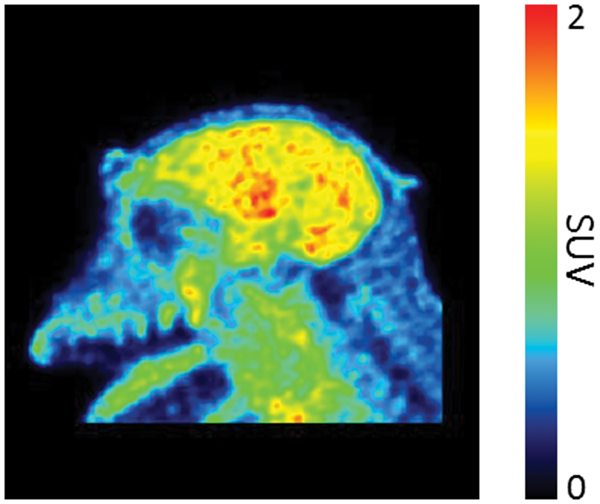
Conclusions
This study demonstrated good brain uptake. Although there was minor variability in BIIB104 distribution across various brain regions, BIIB104 kinetics were consistent with passive diffusion and the free concentration and non-specific binding being the dominating components.
References
BPS01-3
Neurotransmission & Psychiatry
Task-sensitivity of functional [18F]FDG-PET in comparison to functional MRI and arterial spin labeling
1Department of Psychiatry and Psychotherapy, Medical University of Vienna, Vienna, Austria
2Department of Biomedical Imaging and Image-guided Therapy, Division of Nuclear Medicine, Medical University of Vienna, Vienna, Austria
3Center for Biomarker Research in Medicine (CBmed), Graz, Austria
Abstract
Objectives
The investigation of task-specific changes in neuronal activation is of major interest in human neuroscience. Recently, the implementation of a constant infusion protocol of [18F]FDG in combination with a general linear model (GLM) offered the quantification of task-specific glucose metabolism in a single measurement.1,2 Since previous studies employed simple tasks targeting primary visual and motor areas only, it is still unknown if the approach is sufficiently sensitive to identify changes in higher order cognitive areas or subtle variations in cognitive loads. Therefore, we used a challenging visuo-spatial task with two difficulty levels, targeting complex areas, and compared the results to changes in cerebral blood flow and oxygen demand.
Methods
Twenty-two healthy subjects underwent one measurement on a fully-integrated PET/MRI system (Siemens mMR). To track task-specific changes in glucose metabolism, the radiotracer [18F]FDG was administered as bolus followed by constant infusion.3 Subjects played an adapted version of the video game Tetris®, comprising of two “easy” and two “difficult” task blocks (pseudo-randomized order). The task blocks lasted for 6 minutes, separated by baseline conditions of 5 minutes. For absolute quantification, arterial blood samples were drawn in the first few minutes and between the task blocks.3 Simultaneously, a pseudo-continuous Arterial Spin Labeling (pcASL) sequence was recorded. Following the functional PET (fPET) acquisition, functional MRI data (fMRI) was recorded while subjects completed “easy”, “difficult” and “control” blocks of 30 seconds.
The fPET data was analyzed using a GLM including one regressor for each difficulty level. Absolute task-specific changes in glucose metabolism were estimated with the Patlak plot. The fMRI and pcASL data were analyzed as reported previously.4,5 A conjunction map was created from significant changes in the “difficult” condition of all 3 imaging modalities (p < 0.05, FWE cluster level corrected). Paired t-tests were conducted between the difficulty levels for the regions of the conjunction map. Similarity between the modalities was determined by the Dice coefficient.
Results
All three imaging modalities exhibited spatially similar task activations. The conjunction map showed a good overlap of activations in the supplementary motor area (SMA), frontal eye field, intraparietal sulcus (IPS) and occipital cortex (Figure 1, Dice coefficient ranging from 0.49 to 0.55). Significant changes between difficulty levels were found for all modalities in all regions, except for pcASL in the SMA and the IPS. No significant correlations were observed between imaging parameters.

Conclusions
Complex task changes in glucose metabolism, cerebral blood flow and oxygen demand were robustly detected with a good regional overlap. More importantly, changes induced by different difficulty levels were found with all three modalities in most regions. Although the activation depicts qualitatively similar patterns the lack of significant quantitative correlations indicates that these imaging modalities cover distinct neuronal processes. In summary, this work demonstrates that fPET is a promising tool to detect changes in glucose metabolism of paradigms with subtle variation in task loads in higher order cognitive areas.
References
BPS01-4
Neurotransmission & Psychiatry
Patients with major depressive disorder have lower cerebral serotonin 4 receptor PET binding than healthy controls
1Neurobiology Research Unit, Rigshospitalet, Copenhagen, Denmark
2Faculty of Health and Medical Sciences, University of Copenhagen, Copenhagen, Denmark
3Department of Psychiatry, Psychiatric Centre Copenhagen, Copenhagen, Denmark
4Department of Clinical Pharmacology, H. Lundbeck A/S, Valby, Denmark
5Department of Pharmacology, University of Copenhagen, Denmark
Abstract
Objectives
Conventional medical treatment for patients with Major Depressive Disorder (MDD) is predominantly based on intervention with serotonergic drugs but 30–50% of the patients do not respond sufficiently to treatment. We have previously shown that familial risk for MDD is associated with lower striatal serotonin 4 receptor (5-HT4R) binding as measured with [11C]SB207145. The aim of this study is to investigate the 5-HT4R binding in patients with MDD as compared to healthy controls, to determine if low cerebral 5-HT4R binding can be seen as a trait marker for risk for MDD that persists throughout the depressive episode.
Methods
Un-medicated patients with moderate to severe MDD and healthy controls were included; planned inclusion is 100x2 subjects. 5-HT4R binding potential was assessed with a 120 minutes dynamic PET scan using the radioligand [11C]SB207145. We calculated the 5-HT4R non-displaceable binding potential (BPND) with the SRTM in gray matter voxels of the whole brain and primary volumes of interest: neocortex, nucleus caudate, pallidostriatum and hippocampus. ANCOVA was used for statistical analysis adjusted for age, gender, BMI and injected mass/kg.
Results
Data based on 51 patients and 72 controls showed that MDD patients had significantly lower 5-HT4R BPND compared to controls; this included whole-brain BPND was 0.688 ± 0.098 (mean ± SD) in MDD patients and 0.745 ± 0.090 in controls (p = 0.0003, ŋp2 = 0.1). Corresponding patterns was observed in all primary volumes of interest (Table 1). Whole-brain 5-HT4R BPND was not significantly associated with the severity of the depression (p = 0.07).

Conclusion
We find that patients with un-medicated moderate/severe MDD have about 8% lower whole-brain 5-HT4R binding compared to healthy individuals. Our data suggest that low striatal 5-HT4R binding is a trait marker for being at risk for MDD and that this trait persists when undergoing a depressive episode. Given the preclinical and clinical evidence of an inverse relation between 5-HT4R binding and cerebral serotonin levels1, we speculate that people with MDD have an increased cerebral serotonergic tone, perhaps in an attempt to counteract a depressive propensity. In this study, we did not directly compare people at-risk for MDD to patients with MDD, but we find lower BPND globally in the brain in MDD patients, not only in striatum, suggesting that already prior to medical treatment, the patients have increased serotonergic tone. Alternatively, or in combination, direct effects of low capacity for 5-HT4R agonism may also be implicated in the depressed state. We are currently investigating if 5-HT4R binding may serve as a predictor of antidepressant response to 5-HT-acting pharmacological treatments or facilitate relevant subtyping of depressed patient groups who may need individualized clinical care strategies.
References
BPS01-5
Neurotransmission & Psychiatry
Regional correlation between glucose metabolism and synaptic density in healthy subjects
1Division of Nuclear Medicine, Department of Imaging and Pathology, University Hospitals Leuven and KU Leuven, Belgium
2Laboratory for Cognitive Neurology, Department of Neurosciences, KU Leuven, Belgium
Abstract
Objectives
Synaptic vesicle glycoprotein 2A (SV2A) is ubiquitously and homogenously expressed in the presynaptic vesicle membrane across the brain. The high affinity and specificity SV2A PET radioligand 11C-UCB-J has been proposed as proxy for synaptic density1. 18F-FDG has been used for decades as a measure for neuronal activity, that is primarily driven by synaptic activity and astroglial glutamate transport2. Therefore, it has been debated that the signal of both tracers might represent some underlying physiological similarities, but no direct comparisons have been published in vivo. To test this hypothesis, we investigated intersubject regional correlation between 18F-FDG and 11C-UCB-J in screened healthy young subjects.
Methods
Fifteen screened healthy subjects (15 women; 29.4 ± 8.9 years) were included in a dual tracer study on a GE Signa 3T PET-MR. 11C-UCB-J and 18F-FDG were injected sequentially in each subject under standard circumstances in a single-day protocol. 11C-UCB-J data were acquired for 30 minutes, starting 1 hour post-injection (injected activity: 285 ± 42 MBq). Two hours after 11C-UCB-J injection, 18F-FDG was administered (118 ± 14 MBq) and acquired for 30 minutes at 30 minutes post-injection. Simultaneously, T1-weighted and FLAIR MR images were acquired and subject-specific attenuation correction was done using a validated ZTE sequence to delineate bone.
Parametric binding potential (BPND) images were generated for 11C-UCB-J with the centrum semiovale as reference region3. Absolute regional cerebral metabolic rate of glucose (rCMRGlu) was calculated according to the Hunter method with venous samples4. For relative quantification, 18F-FDG and 11C -UCB-J data were normalised to whole-brain grey matter (GM) uptake. Paired correlations were determined both on a volume-of-interest (VOI) and voxelwise basis. For the VOI approach, composite bilateral VOIs were defined (PMOD v4.0; N30R83 Hammers probabilistic atlas): frontal, temporal, parietal, occipital, insular and cingulate cortex, hippocampus, striatum, thalamus, cerebellum and brainstem. Pearson correlation coefficients were calculated (two-tailed, P < 0.05). Furthermore, voxel-wise paired correlation analysis (two-tailed, P < 0.005) limited to a GM mask, was performed using an in-house Matlab routine.
Results
No significant correlations were found between absolute 18F-FDG (rCMRGlu) and 11C-UCB-J (BPND) values. However, relative tracer uptake was strongly correlated in the entire cortex, including cingulum and insula, striatum (in particular caudate nucleus) and cerebellum (Figure 1), with correlation coefficients between 0.34 to 0.94 (higher in the cortex). This was confirmed VOI-based, with most significant positive correlations in the frontal (R = 0.77, P < 0.001) and temporal cortex (R = 0.86, P < 0.001), and striatum (R = 0.74, P = 0.002).

Conclusions
Regional cerebral distribution of glucose metabolism as measured by 18F-FDG and synaptic density measured by 11C -UCB-J PET in young healthy subjects shows a high intersubject correlation especially in the neocortex and striatum. This relation is lacking in thalamus, putamen, brainstem and hippocampus, likely due to differences in regional neurotransmitter activity with differential influence on neuroenergetics. It is therefore unlikely that FDG and SV2A PET provide similar information, even in healthy brain.
References
BPS01-6
Neurotransmission & Psychiatry
Cerebral adenosine receptor availability after sleep deprivation is associated with specific dynamic connectivity states determined by resting state fMRI
1Institute of Neuroscience and Medicine (INM-2), Forschungszentrum Jülich, 52425 Jülich, Germany
2Department of Psychiatry and Psychotherapy, Rheinische Friedrich-Wilhelms-University Bonn, Bonn, Germany
3Institute of Aerospace Medicine, German Aerospace Center, Cologne, Germany
4Division of Sleep Medicine, Harvard Medical School, Boston 02115, MA, USA
5Department of Neurology, Medical Faculty, Heinrich-Heine-University Düsseldorf, 40225 Düsseldorf, Germany
Abstract
Objectives
Adenosine is a neuromodulator directly linked to the energy metabolism by ATP breakdown and a mediator of sleep homeostasis. The net effect of adenosine is inhibitory, reducing neuronal activity. Sleep deprivation leads to increased adenosine concentrations and A1 adenosine receptor availability. On the other hand, it has been shown that sleep deprivation could amplify the temporal fluctuation in functional connectivity of spontaneous brain activities revealing connectivity patterns which reflect different arousal levels. A dynamic functional connectivity approach allows for estimating the covariance metrics across a series of sliding temporal windows and for generating corresponding brain states from fMRI resting state recordings.
The objective of the current study was to investigate whether adenosine A1 receptor availability in humans measured by PET is related to specific dynamic connectivity patterns induced by sleep deprivation, as determined by fMRI.
Methods
12 healthy male volunteers (mean ± SD age: 26.9 ± 5.2 years) participated in a dynamic 18F CPFPX bolus/infusions-PET study with blood sampling and metabolite correction and a resting state fMRI scan, after 52 hours sleep deprivation and after 14 hours of recovery sleep.
For the PET neuroreceptor data, regional distribution volumes (VT) were determined by calculating the tissue to plasma ratio during the steady state phase. For the neuronal activity fMRI data, 34 functional components were identified using an independent component analysis (ICA) approach (Group ICA Of fMRI Toolbox GIFT). A sliding window of 22 images (TRs) with a step of 1 TR was used to calculate covariance metrics. The number of clusters was determined using the elbow criteria, and brain clusters (states) were calculated with a k-means algorithm. Mean dwell time, fraction of states and number of transitions of specified connectivity states were determined.
Adenosine receptor availability in atlas based brain regions known to be relevant in sleep wake regulation were correlated with these dynamic functional connectivity state parameters.
Results
Three brain network states were identified in the fMRI data of which one state was present only after sleep deprivation.
The mean dwell time and fraction of this state (#2) of the total resting state time period were significantly increased after recovery sleep whereas the connectivity pattern of another state was significantly reduced (#3). Previously we have shown that absolute changes in A1 adenosine receptor availability between sleep deprivation and recovery could identify sleep deprivation sensitive subjects in psychomotor and memory performance tasks.
Correlational analysis between changes in A1 adenosine receptor availability and mean dwell time and fraction of these states revealed significant correlations (Pearson’s product moment correlational analyses p < 0.05) to fraction spent in state 3 and mean dwell time of state 2 for several brain regions including thalamus, hippocampus and striatum (r values in the range of 0.6–0.7).
Conclusions
Our findings suggest an individual functional relationship between A1 adenosine receptor availability and the occurrence of specific dynamic functional connectivity brain states after sleep deprivation.
BPS02-1
Novel imaging agents and preclinical imaging: I
Evaluation of the first 18F-labeled PET radiotracer 18F-LY2459989 for imaging kappa opioid receptor in humans
1PET Center, Department of Radiology and Biomedical Imaging, Yale University, USA
Abstract
Objectives
The kappa opioid receptor (KOR) is implicated in various neuropsychiatric conditions. Our group has previously evaluated in humans the KOR PET tracers 11C-GR103545 (agonist) [1] and 11C-LY2795050 (antagonist).2 We report here the kinetic and binding properties of the first 18F-labeled KOR antagonist radiotracer 18F-LY2459989 in humans and contrast with 11C-LY2795050.
Methods
Four healthy male subjects completed 18F-LY2459989 test-retest PET scans of 120 min each on the high resolution research tomograph (HRRT) scanner on two separate days with arterial blood sampling and metabolite analysis during each scan. Fourteen regions of interest (ROIs) were defined on the brain atlas and individual subject’s MR images for generation of regional time-activity curves (TACs). Regional TACs were fitted by one- and two-tissue compartment models (1TC, 2TC), as well as the multilinear analysis-1 (MA1) method to derive regional distribution volumes (VT). Time-stability of VT values was assessed by comparing VT values from shortened scan periods to the 120-min VT values. Test-retest reproducibility was evaluated using the absolute test-retest variability (aTRV, |test–retest|/{(test + retest)/2}). Specific binding of 18F-LY2459989 was compared with that of 11C-LY2795050 using the Guo plot.5
Results
Injected activity dose of 18F-LY2459989 was 159 ± 88 MBq, with injected mass of 1.5 ± 3.1 μg (n = 8). Plasma free fraction (fP) was 3.2 ± 0.9% (cf. 0.77 ± 0.16% for 11C-LY2795050 [2]). The 1TC model yielded mean K1 values for 18F-LY2459989 similar to those for 11C-LY2795050. Regional. TACs for both 18F-LY2459989 and 11C-LY2795050 were fitted well with the 2TC model. Lack of fit was seen in all regions with the 1TC. Fitting with MA1 (t* = 30 min) was good. There was an excellent correlation between 2TC and MA1 VT values (VT (MA1) = 0.99 × VT (2TC) – 0.01, R2 = 0.99) when unreliable parameters in a few regions were excluded. Thus MA1 was judged as the model of choice for derivation of kinetic parameters for 18F-LY2459989. Mean MA1 VT values (mL/cm3) ranged from 2.8 (cerebellum) to 8.3 (amygdala) for 18F-LY2459989: highest in the amygdala, insula, and cingulate cortex, and lowest in the thalamus and centrum semiovale. Minimum scan time for stable VT measurement was 90 min. Test-retest reproducibility in VT was excellent across all ROIs (mean aTRV of ∼7%). In comparison, 11C-LY2795050 VT values ranged from 2.0 to 4.0 and aTRV was ≤10% except for the amygdala (12%), with a minimum scan time of 70 min.2,6 Compared to 11C-LY2795050, 18F-LY2459989 shows higher regional VT and BPND values (BPND (18F-LY2459989) = 4.6 × BPND (11C-LY2795050)).

Conclusion
18F-LY2459989 displays favorable kinetic and binding characteristics and can be used for PET imaging and quantification of KOR in the human brain, with higher levels of specific binding signal than 11C-LY2795050.
Research support
NIH R21MH092664 and R33MH092664
References
BPS02-2
Novel imaging agents and preclinical imaging: I
Kinetic analysis and test-retest variability of [18F] LSN3316612, a novel PET radioligand for imaging O-GlcNAcase in human brain
1Molecular Imaging Branch NIMH/NIH
2University of Virginia
3Eli Lilly & Co
4Houston Methodist Research Institute
Abstract
Objectives
Neurofibrillary tangles (NFTs) containing hyper-phosphorylated tau protein are linked to the pathology of Alzheimer’s disease (AD).1 The O-linked N-acetyl-glucosamine (O-GlcNAc) modification reduces the aggregation of proteins, such as tau.2 As a result, inhibiting the O-linked N-acetyl-glucosamine hydrolase (O-GlcNAcase) enzyme that deglycosylates intracellular proteins, has become a key strategy for treating AD and other related tauopathies.3 A previous study reported the pharmacokinetics of [18F]LSN3316612, a novel positron emission tomography (PET) radioligand for imaging O-GlcNAcase in monkeys and mice.4 Here, we present the kinetic evaluation and test-retest variability of [18F]LSN3316612 in healthy humans.
Methods
Ten healthy human subjects each received two dynamic PET scans using a Siemens mCT scanner; the time between scans varied from one week to five months. All scans began after injection of approximately 5 mCi of [18F]LSN3316612 and lasted for three hours. Continuous blood sampling was performed using an automated sampler for the initial 10 minutes followed by discrete manual sampling for the rest of the scanning period to obtain a metabolite-corrected input function. Volumes of interest parcellated from individual T1-weighted MRI were applied to co-registered PET images to generate regional brain time activity curves. Distribution volume (VT) was calculated using an unconstrained two-tissue compartment model (2TCM). Test-retest variability was measured by intraclass correlation coefficient (ICC). The correlation between VT and area under the time activity curve (AUC) was also assessed for the possibility of bloodless quantification.
Results
[18F]LSN3316612 showed high brain penetration with moderate washout—faster than had been observed in monkeys. Peak standardized uptake value (SUV) was about 5, with highest uptake seen in the amygdala and hippocampus followed by the cortices. The lowest uptake was seen in the thalamus and cerebellum (Fig. 1A). Mean VT ranged from 12–20 mL · ccm−1 (n = 10, Fig. 1B & 1C). For all regions, VT values at 120 minutes were within 5% of their terminal value at three hours (Fig. 1D) with no indication of radiometabolite accumulation, consistent with previous findings in monkeys and rodents. Plasma parent free fraction ranged from 1.9%–5.9% with no significant difference between test and retest scans (p = 0.6). ICCs ranged from 0.69–0.96 for various regions, with an overall reproducibility of 12%. Correlation coefficients between regional VT‘s and AUCs (150–180 minutes) were high within subjects (0.76–0.98 for all except one subject), but poor across subjects with or without blood normalization.

Conclusions
[18F]LSN3316612 has favorable properties for imaging O-GlcNAcase in humans, including high brain uptake, good time stability, and reliable quantification. Test-retest reproducibility will likely improve with shorter lapses between scans (8% with 6 subjects acquired < one month apart). Intra-subject quantification without arterial blood is possible but requires further validation.
References
BPS02-3
Novel imaging agents and preclinical imaging: I
Evaluation of a radiofluorinated benzazepine-1,7-diol analogue for imaging the GluN2B subunits of the N-Methyl-D-Aspartate receptor
1Institute of Pharmaceutical Sciences, ETH Zurich, Switzerland
2Department of Nuclear Medicine, University Hospital Zurich, Switzerland
3Institute of Pharmaceutical and Medicinal Chemistry, University of Münster, Germany
Abstract
Objectives
Currently, no clinically approved GluN2B PET tracer exists despite several years of strenuous research efforts. Recently, we successfully developed a carbon-11 labeled GluN2B PET tracer, [11C]-Me-NB11, that is currently being validated in a first-in-man PET study. As part of our program to develop a fluorine-18 labeled GluN2B ligand, we have prepared a fluorinated analogue of Me-NB1, separated the enantiomers and evaluated the R-enantiomer designated (R)-OF-NB1. Previous results have shown that the R-enantiomers of this structural class of benzazepines selectively target the GluN2B subunits, whereas the S-enantiomers exhibit off-target binding mainly to sigma-1 receptors.
Methods
Racemic OF-NB1 was synthesized in a multi-step reaction sequence followed by chiral HPLC separation of the enantiomers. Binding affinities of (R)-OF-NB1 towards GluN2B subunits and off-target sigma-1 receptors were determined using rat brain homogenates. A two-step radiosynthesis was employed for the preparation of (R)-[18F]OF-NB1. In vitro autoradiography, PET imaging experiments in rodents, biodistribution and metabolite studies were undertaken to assess the suitability of (R)-[18F]OF-NB1 for imaging the GluN2B receptors. Receptor occupancy study was performed using CP-101606, a GluN2B antagonist with reported clinical efficacy.
Results
(R)-OF-NB1 exhibited a three-fold higher nanomolar affinity towards GluN2B compared to Me-NB1 and more than fifty-fold selectivity over sigma-1 receptors. (R)-[18F]OF-NB1 was synthesized via copper-mediated radiofluorination in good radiochemical yields of 12–25% and high molar activities ranging from 61–257 GBq/μmol. In autoradiographic studies, (R)-[18F]OF-NB1 demonstrated a heterogeneous binding profile on rodent brain tissue sections with high accumulation in GluN2B-rich regions such as the cortex, striatum, thalamus and hippocampus and low accumulation in GluN2B-poor cerebellum region. Radioactivity accumulation was specifically blocked with 1 μM solutions of GluN2B specific ligands, CP-101,606 and CERC-301, but not with fluspidine and SA4503, sigma-1 receptor blocking agents. PET imaging studies in Wistar rats revealed high in vivo specific binding to GluN2B binding using CP-101,606 (10 mg/kg) as a blocker. Further PET imaging with wild-type and sigma-1 receptor knock-out mice excluded in vivo off-target binding to sigma-1 receptors. Metabolite analysis showed intact (R)-[18F]OF-NB1 in the brain 60 min postinjection. A dose-dependent reduction of tracer accumulation was observed with escalating doses of CP-101,606 in vivo, showcasing the utility of (R)-[18F]OF-NB1 in receptor occupancy studies.
Conclusion
(R)-[18F]OF-NB1 is a promising radioligand for the in vivo imaging of GluN2B-containing NMDA receptors and receptor occupancy studies. This radioligand has high potential for clinical translation.
Acknowledgments
This project was supported by the Swiss National Science Foundation Grant Nr. 310030E-160403/1.
Reference
BPS02-4
Novel imaging agents and preclinical imaging: I
Development and initial in vivo evaluation in monkey brain of [11C]T-1650, a novel PET ligand for phosphodiesterase-4 subtype D
1National Institute of Health, USA
2Dept of Radiology, Hyogo medical University, Japan
3Tetra Discovery Partners Inc, USA
Abstract
Objective
Phosphodiesterase-4 (PDE4) metabolizes and thereby terminates the actions of the second messenger cyclic adenosine monophosphate (cAMP). Binding of the PDE4 inhibitor [11C](R)-rolipram was previously found to be globally decreased in unmedicated subjects with major depressive disorder (MDD) compared to controls,1 a finding consistent with the ‘cAMP theory of depression’, which posits that low cAMP signaling causes depression. However, rolipram inhibits all four PDE4 subtypes (4A, 4B, 4C, and 4D). PDE4D is related to cognitive function.2 This study used positron emission tomography (PET) to assess the newly developed PDE4D-selective radioligand [11C]T-1650 in rhesus monkeys.
Methods
Brain scans were obtained in one monkey for 90–120 minutes in 21–25 frames following a one-minute intravenous bolus injection of [11C]T-1650. To measure enzyme-specific uptake, scans were also performed after injection with selective PDE4 inhibitor, rolipram, which were injected 10 minutes before [11C]T-1650. Concurrent arterial samples were obtained to measure parent radioligand concentrations.
Results
After [11C]T-1650 injection, the tracer readily entered brain and showed widespread distribution, with lower binding in cerebellum. In the rolipram scans, radioactivity concentrations in whole brain peaked at 2.65 SUV (standardized uptake value units) at baseline and at 4.37 SUV after rolipram administration (1 mg/kg). The plasma parent peaked at 12.6 SUV and 11.8 SUV, respectively, and the plasma parent free fraction was estimated at 6.99% and 6.23%, respectively. In the pharmacokinetic analysis, target enzyme density was quantified as total distribution volume (VT) using the two-tissue compartment model as well as serial concentrations of parent radioligand in arterial plasma. VT was well identified in all examined brain regions, and VT calculated from the first 90 minutes of scan data fell well within 10%. The extent to which PDE4D was occupied by a blocking agent was estimated graphically using a Lassen plot for several brain regions. PDE4D occupancy was 92.8% for rolipram. Nondisplaceable distribution volume (VND; the X intercept when Y = 0) was estimated as 3.50 mL/cm3 in the rolipram (Fig.1).

Figure 1. Radioactivity concentrations in Plasma parent and whole brain of baseline and pre-blocked with rolipram (1 mg/kg) scans (panel A, B). The plasma parent peaked at 12.6 SUV (standardized uptake value units) and 11.8 SUV, respectively, and the plasma parent free fraction was estimated at 6.99% and 6.23%, respectively. Whole brain concentrations peaked at 2.65 SUV at baseline and at 4.37 SUV after rolipram administration (1 mg/kg). Lassen plot of baseline and pre-blocked with rolipram (1 mg/kg) scans (panel C). Occupancy by rolipram was about 92.8% and VND was 3.50.
Conclusions
[11C]T-1650 was able to successfully image and quantify PDE4D in monkey brain. Further investigation into the ability of this agent to image PDE4D is warranted.
References
BPS02-5
Novel imaging agents and preclinical imaging: I
18F-SDM-8 is an excellent PET radiotracer for imaging the synaptic vesicle glycoprotein 2A: First experience in humans
1PET Center, Department of Radiology and Biomedical Imaging, Yale University, New Haven, CT, United States
Abstract
Objectives
The ability to investigate the integrity and density of synapses in vivo will facilitate our understanding of disease pathophysiology and development of effective therapies, as Alzheimer’s disease has long been thought to be a “synaptic disease”, and synaptic abnormalities are implicated in many other neurodegenerative and psychiatric disorders. We have previously developed 11C-UCB-J as a radiotracer for the synaptic vesicle glycoprotein 2A (SV2A), and validated SV2A PET as an imaging biomarker for synaptic density (1, 2). We have further developed a novel 18F-labeled SV2A radiotracer 18F-SDM-8 and demonstrated its outstanding imaging properties in non-human primates (3). The objective of this study was to assess the pharmacokinetic and imaging characteristics of 18F-SDM-8 in humans in comparison with 11C-UCB-J.
Methods
Test-retest PET scans of 120 min each were conducted with 18F-SDM-8 on three healthy human subjects in two separate days using an HRRT scanner. Single 90-min scans with 11C-UCB-J were also performed on the same subjects. Arterial blood samples were taken for analysis of radioactive metabolites and generation of the arterial input function. Regions of interest (ROIs) were drawn on an brain atlas and individual subject’s MR images to derive regional time-activity curves (TACs). TACs were fitted with 1- and 2-tissue compartment (1TC, 2TC) models to determine volume of distribution (VT). Non-displaceable binding potential (BPND) was calculated using the centrum semiovale (CS) as reference region. Absolute test-retest variability (aTRV) of regional VT was calculated. The minimum scan time required for stable VT measurement was determined from the 120 min scan data.
Results
18F-SDM-8 was given in activity dose of 180.3 ± 6.9 MBq and mass dose of 0.23 ± 0.11 µg (n = 6). Metabolism of 18F-SDM-8 was fast, with 23 ± 5% parent fraction in plasma at 30 min following radiotracer administration (n = 6). Plasma free fraction was high, at 31 ± 1% (n = 6). In the human brain 18F-SDM-8 displayed very high uptake and fast kinetics, with peak SUV of ∼10 at 10 min post-injection. The 1TC model fitted the regional TACs quite well, while the 2TC model produced unreliable VT estimates in some regions. Mean 1TC VT values ranged from 3.5 to 19.7 mL/cm3 and were very similar to those from 11C-UCB-J except in CS, where VT was lower for 18F-SDM-8 (3.5 vs. 4.3 for 11C-UCB-J). Mean BPND values of 18F-SDM-8 were 4.62, 4.63, 4.48, 4.38, 4.34, 4.01, 4.06, 3.39, 2.92, and 2.84, respectively, for the insula, temporal cortex, putamen, parietal cortex, occipital cortex, frontal cortex, amygdala, caudate, thalamus, and hippocampus (n = 6), compared with 3.57, 3.61, 3.69, 3.37, 3.37, 3.15, 2.95, 2.58, 2.22, and 2.18 for 11C-UCB-J (n = 3) in the same regions and subjects. On average 18F-SDM-8 BPND values were ∼33% higher than those of 11C-UCB-J. Test-retest reproducibility in 18F-SDM-8 VT estimates was excellent, with mean aTRV of 5% across all brain regions. Minimum scan time for stable VT measurement was 60 min.

Conclusions
Our first imaging results in humans prove that 18F-SDM-8 is a highly promising SV2A radiotracer with superb kinetic and imaging properties: fast and high uptake in the brain, appropriate tissue kinetics, high specific binding signals, and excellent test-retest reproducibility in kinetic parameters.
References
BPS02-6
Novel imaging agents and preclinical imaging: I
Evaluation of [11C](R)-NR2B-Me, a novel PET RADIOligand for imaging The NR2B subunit of the NMDAR complex in rat and monkey brain
1Molecular Imaging Branch, National Institute of Mental Health, National Institutes of Health, Bethesda, Maryland, USA
Abstract
Objectives
The overexpression and hyperfunction of NR2B-containing N-methyl-D-aspartate (NMDA) receptors appears to contribute to neuropsychiatric and neurodegenerative disorders [1, 2]. In this context, therapies targeting NR2B show promise for the treatment of Parkinson’s disease, Alzheimer’s disease, psychiatric disorders, and brain injury [3–5]. To develop such therapeutics, positron emission tomography (PET) with effective NR2B radioligands is a necessary tool that could help elucidate both pharmacological specificity and target engagement in vivo. To date, no effective radioligands have been developed for such quantification [6, 7]. This study evaluated the novel PET radioligand (R)-7-methoxy-3-(4-(4-methylphenyl)butyl)-2,3,4,5-tetrahydro-1H-benzo[d]azepin-1-ol ((R)-NR2B-Me) for the NR2B receptor subunit in rats and monkeys.
Methods
Dynamic [11C](R)-NR2B-Me PET scans were acquired in rats (N = 34) and monkeys (N = 2) at baseline and after intravenous administration of either an NR2B-selective antagonist (RO256981 or CO101244), a sigma-1 selective antagonist (FTC146 or BD1047), a sigma-1 selective agonist (TC1 or SA4503), or cold parent ligand for self-blocking. Concurrent arterial blood samples were collected during all monkey studies. Brain uptake was characterized by standardized uptake value (SUV) for rats and by distribution volume (VT) using a two-tissue compartment model for monkeys. Receptor occupancy, non-displaceable distribution volume (VND), and non-displaceable binding potential (BPND) were determined via Lassen plot.
Results
High brain uptake followed by moderate washout over 90 minutes was observed in all baseline scans of both rats and monkeys. In rats, pre-treatment with RO256981 (0.05 mg/kg and higher) reduced brain uptake in all regions. As expected, FTC146 and BD1047 showed no blocking effects. However, both TC1 and SA4503 showed blocking effects at doses of 0.05 mg/kg and higher. In monkeys, where VND was determined via occupancy plot, whole-brain BPND for self-blocking was 4.5, which was slightly higher than the 3.8 observed for CO101244; blocking effects were about the same (97–98%).

Conclusions
[11C](R)-NR2B-Me is a promising radioligand for in vivo imaging of NR2B subunit within NMDA receptors. This radioligand demonstrated high brain uptake at baseline that could be significantly blocked by NR2B-selective antagonists in both rats and monkeys. However, more studies are needed to explore the blocking effects of sigma-1 agonists such as TC1 and SA4503, and to establish the selectivity of [11C](R)-NR2B-Me.
Research Support
Intramural Research Program of the National Institute of Mental Health, National Institutes of Health (NIMH-NIH).
References
BPS03-1
Novel imaging agents and preclinical imaging: II
Head-to-head comparison of two colony stimulating factor-1 receptor PET ligands, [11C]GW2580 and [11C]CPPC, for imaging of acute and chronic neuroinflammatory changes in mice
1National Institute of Radiological Sciences, National Institutes for Quantum and Radiological Science and Technology, 4–9-1 Anagawa, Inage-ku, Chiba 263–8555, Japan
2Laboratory for Proteolytic Neuroscience, RIKEN Brain Science Institute, Wako, Japan
Abstract
Objectives
Colony stimulating factor-1 receptor (CSF1R) has been pursued as a potential therapeutic target to mitigate proinflammatory cascades provoked by aberrant microglial activation. However, changes in the CSF1R availability under various inflammatory conditions in the living brain remains unknown. To investigate this issue, we developed two novel PET ligands, [11C]CPPC (recently published by Horti et al., PNAS, 2019) and [11C]GW2580, and applied them to imaging of CSF1R in mouse models of acute and chronic neuroinflammation.
Methods
An acute neuroinflammation model was created by unilateral injection of lipopolysaccharide into the striatum. Dynamic PET scans over 90 min after intravenous injection of the CSF1R radioligands were conducted 1–2 weeks after lipopolysaccharide treatment. In addition, PET imaging of translocator protein (TSPO) was taken to determine the location and status of neuroinflammation. After the final scans, brain tissues of these animals were immunostained for IBA1 to examine microglial activation. Tracer distribution and specific binding were also studied by ex-vivo autoradiography and blocking experiments. Furthermore, PET scans of CSF1R with the two radioligands were performed in mouse models of chronic neuroinflammation including tau transgenic (rTg4510) and APP-knock-in mice along with TSPO-PET. Moreover, an in-vitro assay of [11C]GW2580 binding in a microglial cell line, Ra2, and its blockade by a nonradioactive compound were determined.
Results
Both [11C]GW2580 and [11C]CPPC exhibited increased uptake in lesioned striata in the lipopolysaccharide model. TSPO-PET and IBA1 immunostaining demonstrated that elevated uptake of the CSF1R radioligands was concurrent with microglial activation. However, [11C]CPPC captured inflammatory changes less sensitively than [11C]GW2580, as ipsilateral-to-contralateral ratios of retentions of [11C]CPPC and [11C]GW2580 quantified in the same animals (n = 3) were 1.08 ± 0.02 and 1.20 ± 0.06, respectively 11 days after lipopolysaccharide injection. In addition, subtle neuroinflammatory alternation detectable by [11C]GW2580 with an ipsilateral-to-contralateral ratio around 1.10 was invisible by [11C]CPPC. [11C]GW2580 uptake in the lesioned striatum increased by 28 ± 3% (n = 7), 20 ± 6% (n = 3), and 9 ± 1% (n = 3) relative to the contralateral striatum at 7, 11, and 14 days after lipopolysaccharide treatment, respectively, and this time course was in line with temporal changes of TSPO. The ipsilateral-to-contralateral difference at 2 weeks was completely abolished by unlabeled tracer, while the uptake in the ipsilateral stratum in the same blocking condition remained 12–14% higher than the contralateral striatum at 1 week for both [11C]GW2580 and [11C]CPPC. rTg4510 and APP-knock-in mice modeling chronic neuroinflammation exhibited higher [11C]GW2580 uptake than non-transgenic littermates in both PET and ex-vivo autoradiography, whereas no significant difference in [11C]CPPC uptake was observed between these models and controls. Assessments of the specificity of this increased [11C]GW2580 uptake for CSF1R associated with microglial activation is underway. Finally, binding of GW2580 could be quantified using our in-vitro ligand screening system with IC50 being 42 nM (n = 3) and specific binding accounting for 40% of total binding in Ra2 cells.

Conclusions
[11C]GW2580-PET is able to capture changes of CSF1R in the brains of living animals modeling neuroinflammation with higher sensitivity than [11C]CPPC, and potentially provides an imaging-based biomarker for proinflammatory microgliosis.
BPS03-2
Novel imaging agents and preclinical imaging: II
Evaluation of the demyelination PET radiotracer [18F]3F4AP in rhesus macaque
1Gordon Center for Medical Imaging, Department of Radiology, Massachusetts General Hospital, Harvard Medical School, United States
2AP-HP, Department of Nuclear Medicine, Pitié-Salpêtrière Hospital, Sorbonne University, UPMC Paris 06, CNRS UMR 7371, INSERM U1146, France
Abstract
Objectives
4-aminopyridine (4AP) is a potassium (K+) channel blocker approved for treatment of multiple sclerosis. Axonal K+ channels are normally located beneath the myelin sheath, but become exposed and increase in expression upon demyelination. It was recently demonstrated that [18F]3-fluoro-4-aminopyridine, [18F]3F4AP, binds to K+ channels in demyelinated neurons and can detect demyelinated lesions in a rodent model of multiple sclerosis. The goal of the current work was to further characterize [18F]3F4AP in non-human primate.
Methods
Dynamic PET imaging with arterial blood sampling was performed on one rhesus macaque. Four scans were performed with different doses of cold 3F4AP (0, 0.75, 1.25, 2.5 mg/kg). Radioactivity concentration was measured in whole blood and plasma. Selected plasma samples underwent radiometabolite analysis using column-switching radio-HPLC. Plasma free fraction was measured by ultracentrifugation. Dynamic PET images were analyzed by compartmental modeling and Logan graphical analysis using the metabolite corrected arterial plasma input functions.
Results
[18F]3F4AP was metabolized slowly (>90% parent at 180 min) and exhibited low plasma protein binding (free fraction >90%). The tracer rapidly penetrated the brain (peak SUV ∼5 at 5 min) and washed out quickly. Data were fit best by a two-tissue compartment model (2TCM). VT estimates were stable while truncating data sets from 180 down to 60 min. Logan graphical analysis yielded similar outcomes and generated high quality parametric maps. No significant change in VT values were observed with the cold doses administered, however enhanced uptake (∼30% greater VT than surrounding tissue) and distinct pharmacokinetics were observed in a focal area of the brain where the animal sustained a minor injury during a craniotomy procedure performed three years prior to imaging. This change could not be attributed to changes in perfusion or permeability as K1 was not increased compared to other regions. Furthermore, no abnormal binding or retention patterns have been observed in the craniotomy site when the same animal was imaged with other tracers, notably including [18F]FDG.

Conclusion
This study showed that [18F]3F4AP has good properties for imaging the brain (e.g., high BBB permeability, high metabolic stability and high plasma free fraction) and that it yields reproducible results in primates. Blocking studies performed to date have not shown substantial displacement likely because the doses used were below the dose required to block a significant fraction of receptors, which would elicit severe side effects (namely fatal seizures) if a critical threshold were reached. Nevertheless, increased VT in the craniotomy site adds to the body of evidence that 3F4AP binds K+ channels in demyelinated fibers in vivo, which includes previous studies showing enhancement of action potential conduction in demyelinated fibers, colocalization of the PET signal with lesions in several unrelated animal models of demyelination, lack of correlation between the PET signal with other changes such BBB damage or inflammation. In summary, the findings from this study demonstrate that [18F]3F4AP can be quantified using a 2TCM or Logan graphical analysis and further investigation in demyelinating conditions is warranted.
Acknowledgements
NIH R00EB020075, P41EB022544, T32EB013180, S10OD018035
BPS03-3
Novel imaging agents and preclinical imaging: II
The new PET tracer 18F-SDM-8 effectively detects reduced SV2a binding in a rodent model of Alzheimer’s disease
1PET Center, Department of Radiology and Biomedical Imaging, Yale University, New Haven
2Cellular Neuroscience, Neurodegeneration, and Repair, Departments of Neurology and Neuroscience, Yale University, New Haven, CT
Abstract
Objectives
As the lead synaptic vesicle glycoprotein 2A (SV2A) PET tracer, 11C-UCB-J,1 showed promising results distinguishing Mild Cognitive Impairment(MCI) from Alzheimer’s Disease(AD) patients from cognitively normal controls,2 we have developed novel 18F-labeled SV2A PET tracers with comparable imaging profiles for future multicenter clinical trials to validate SV2A imaging for early diagnosis of AD. One such tracer, 18F-SDM-8, has demonstrated outstanding imaging properties in non-human primates.3 In this study we tested the sensitivity of 18F-SDM-8 to detect synapse deficit measured by SV2A in the amyloid precursor protein and presenilin 1 double transgenic (APP/PS1) mouse model, which recapitulates a key and early pathological feature of AD, i.e., the hippocampal synapse loss, using an optimized quantification method.
Methods
The SV2A PET tracer 18F-SDM-8 was administered intravenously to APP/PS1 mice and their wild-type (WT) littermates. A total of 23 APP/PS1 and 23 WT mice (56 ± 4.5 weeks) underwent baseline PET scans, and emission data were acquired from 0–90 min post-injection (p.i.) or from 30–90 min (p.i.) on an Inveon small animal PET/CT scanner. Images were reconstructed using OSEM-3D-MAP algorithm with attenuation and scatter corrections. The mouse M. Mirrione template was manually applied to PET images to generate time activity curves (TACs) for different regions of interest (ROIs). In 90-min scans, the simplified reference tissue model (SRTM) was used to estimate the non-displaceable binding potential (BPND) using brainstem (BS) as the reference region. Standardized uptake value (SUVs) of brain region averaged from 30–60 min and 60–90 min p.i. were normalized with BS to generate SUV ratio (SUVR). SUVR-1(BS) values averaged from different time windows were correlated with BPND values to determine the best static imaging window. Unpaired, two-tailed t-test was used for statistical analysis.
Results
The SV2A PET tracer 18F-SDM-8 entered the mouse brain quickly, reaching maximal brain uptake at about 5 min p.i., followed by steady wash-out and reached equilibrium (constant SUVR) around 30 min p.i. (Fig. 1A,B). From six 90-min scans, the SUVR-1(BS) values averaged from 30–60 min p.i. were found to correlate well with BPND values derived from 90 min of scan data across all brain regions (Fig. 1C. y = 1.06 x + 0.037, R2 = 0.96, p = 0.02). Given these results, further scan data were acquired from 30–60 min p.i. and used to calculate hippocampal SUVR-1(BS) values, which were 10% lower in APP/PS1 mice than WT mice (Fig. 1D. 0.719 ± 0.009 for APP/PS1 mice, n = 23, v.s. 0.800 ± 0.008, for WT mice, n = 23, p = 0.005).

Conclusion
Using 18F-SDM-8 PET and the optimized quantification method, we successfully detected the hippocampal synapse deficit in APP/PS1 mice as compared to their WT controls, supporting further evaluation and validation of this tracer in relevant disease models and populations. 18F-SDM-8 has been translated to first-in-human studies at Yale PET Center to evaluate its suitability as a PET tracer to quantify SV2A in humans.
References
BPS03-4
Novel imaging agents and preclinical imaging: II
PET imaging of colony stimulating factor 1 receptor in rat brains
1Department of Clinical and Experimental Neuroimaging, National Center for Geriatrics and Gerontology, Obu, Japan
2Department of Chemistry and Biomolecular Science, Faculty of Engineering, Gifu University, Gifu, Japan
Abstract
Objective
Activated microglia are involved in the pathophysiology of neurodegenerative diseases such as Alzheimer’s disease, and these glial cells have recently been recognized as a promising target for the treatment of neurodegenerative diseases. Here, positron emission tomography (PET) imaging of specific molecules for microglia could provide useful information needed for developing drugs that target these cells.
We have been developing a PET ligand for imaging the colony stimulating factor 1 receptor (CSF1R). CSF1R is expressed exclusively on microglia. In this study, we evaluated the potential of this novel PET ligand for imaging CSF1R in rat brains using a pilocarpine-induced status epilepticus model of brain inflammation.
Methods
[11C]NCGG401, our PET ligand, which is modified from a specific CSF1R inhibitor BLZ945, was synthesized with a high radiochemical purity (>99%). We conducted 120-min dynamic PET scans with radioactive metabolite analyses to evaluate this ligand’s kinetics in the brain after bolus intravenous administration of [11C]NCGG401 (∼30 MBq) in four healthy rats (Crl:CD (SD), male. 8-week old) with or without pre-administration of unlabeled NCGG401 (1.0 mg/kg body weight) as a target binding blocker. The total distribution volume (VT) was calculated with the Logan graphical analysis. Subsequently, we conducted 60-min dynamic PET scans in two status epilepticus model rats (Crl:CD (SD), male, 8-week old, 10 days after 400 mg/kg pilocarpine i.p.) to evaluate the ligand’s kinetics in the microglia activated brain.
Results
[11C]NCGG401 brain radioactivity showed a fast initial peak (SUV 1.0) and good washout thereafter in the healthy rats. Pre-administration of unlabeled NCGG401 increased the rate of washout significantly suggesting that there is specific binding of the ligand to CSF1R in healthy rat brains. The whole brain VT of [11C]NCGG401 in rats was ∼3.0 mL/cm3 without and ∼1.0 mL/cm3 with blocking. The status epilepticus model rats showed a higher uptake with a slower washout than did the healthy rats, suggesting that the increased binding of the ligand in the microglia activated brain.
Conclusions
[11C]NCGG401 appears to have a favorable kinetics allowing for the quantification of specific binding of the ligand to CSF1R in the rat brain, and its binding increases with microglia activated. Thus, [11C]NCGG401 is a promising PET ligand for imaging CSF1R in the rat brain.
Reference
BPS03-5
Novel imaging agents and preclinical imaging: II
PET-CT shows distinct brain uptake and retention for a nanobody and PAMAM dendrimer after intra-arterial delivery
1The Russell H. Morgan Department of Radiology and Radiological Science, Johns Hopkins University School of Medicine, Baltimore, MD, USA
2Institute for Cell Engineering, Johns Hopkins University School of Medicine, Baltimore, MD, USA
3Department of Neurosurgery, School of Medicine, University of Warmia and Mazury, Olsztyn, Poland
4NeuroRepair Department, Mossakowski Medical Research Center, Polish Academy of Science, Warsaw, Poland
Abstract
Objectives
Poor blood-brain barrier (BBB) permeability of therapeutics is a major obstacle limiting efficacy in treatment of brain tumors. It has been demonstrated that single intra-arterial (IA) infusion of anti-VEGF monoclonal antibody bevacizumab (BV) provides superior therapeutic outcomes in comparison to intravenous (IV) administrations.1 In our recent studies using PET-CT and 89ZrBV, we demonstrated that intracerebral delivery of BV using an intra-arterial (IA) infusion is more effective than intravenous administration.2 While antibodies are quickly emerging as therapeutics, their disadvantages such as large size, production logistics and immunogenicity motivate search for alternatives. Thus we have studied brain uptake of nanobody and polyamidoamine (PAMAM) dendrimer, which are quickly emerging as novel therapeutic agents and drug delivery nanoplatforms, respectively.
Methods
Nanobody was conjugated with deferoxamine (DFO) to generate NB(DFO)2. Generation-4 PAMAM dendrimer was conjugated with DFO and subsequently its primary amines were substituted with 110 butane-1,2-diol functionalities to generate G4(DFO)3(Bdiol)110. Resulting conjugates were radiolabeled with 89Zr. Uptake of 89ZrG4(DFO)3(Bdiol)110 and 89ZrNB(DFO)2 in the mouse brain upon carotid artery and tail vein infusions with intact BBB (BBBI) or osmotic BBB opening (OBBBO) with mannitol was monitored by dynamic positron emission tomography (PET) over 30 minutes to detect brain uptake and early clearance. This was followed by whole body PET-CT imaging at 1 h and 24 h post-infusion (pi) to capture their retention in the brain with subsequent verification by ex vivo biodistribution analysis.
Results
Intravenous administration of 89ZrNB(DFO)2 and 89ZrG4(DFO)3(Bdiol)110 resulted in their negligible brain accumulation regardless of BBB status. Intra-arterial (IA) administration of 89ZrNB(DFO)2 dramatically increased its brain uptake (25.79 ± 15.79%ID/cc), which was further enhanced by prior OBBBO (60.66 ± 35.41%ID/cc P < 0.05). Half of the initial brain uptake was retained after 24 h in all groups. In contrast, IA infusion of 89ZrG4(DFO)3(Bdiol)110 resulted in poor initial accumulation in the brain and there was no difference in its peak concentration in the ipsilateral hemisphere for infusion with intact BBB (3.29 ± 1.31%ID/cc) and OBBBO (3.20 ± 1.47%ID/cc). 89ZrG4(DFO)3(Bdiol)110 did not retain in the brain with complete clearance within 1 h of administration.

with complete clearance from the brain within 1 h of administration. Biodistribution results reflected those on PET-CT.
Conclusions
IA delivery of nanobodies might be an attractive therapeutic platform for CNS disorders where prolonged intracranial retention is necessary. While here we tested generation-4 hydroxy terminated PAMAM dendrimer, the same dendrimer with different surface modifications and ligands for specific brain targets can potentially exhibit higher brain retention. Our study may serve as a benchmark for quantitative performance of dendrimer-based diagnostics and therapeutics in the CNS diseases.
References
BPS03-6
Novel imaging agents and preclinical imaging: II
Characterization of two mGluR4 radiotracers [18F]KALB001 and [11C]KALB012 in non-human primates
1Massachusetts General Hospital, Harvard Medical School, USA
Abstract
Objective
Since mGluRs have neuromodulatory role in the control of both glutamatergic and GABAergic neurotransmission, there has been much interest to develop mGluR ligands for therapeutic purposes of a variety of neurological conditions including Parkinson’s disease. To enable testing of the drug candidates we have developed several PET ligands. Here we compare suitability of two mGluR4 tracers, [18F]KALB001 and [11C]KALB012 for translational studies using macaques. These tracers were previously characterized by in-vitro and rodent studies (1,2).
Methods
Ten scans were performed in four macaques; 5 with [18F]KALB001 and 5 with [11C]KALB012. Two scans (one for each tracer) used unlabeled radiotracer as a competitor at 1 mg/kg i.v. The remaining experiments were baseline scans that provided reference against which blocking data were compared. PET emission data collection began immediately prior to the start of radiotracer injection and was continued for 90 min. Arterial blood samples were collected to measure concentrations in whole-blood and plasma. One sample was drawn prior to radiotracer injection to determine plasma protein binding. Radiometabolite analysis was performed on selected plasma samples. PET images were registered to a template to allow delineation of regions of interest for extraction of time activity curves (TACs). TACs were analyzed using the metabolite corrected arterial plasma input function and standard compartmental models with varying complexity. Graphical analysis techniques were also investigated.
Results
Arterial blood showed relatively rapid clearance for both radiotracers. [18F]KALB001 and [11C]KALB012 both exhibited fast metabolism with ≈50% parent fraction in plasma by 8–10 min. Plasma protein binding was ≈85% for both radiotracers. [18F]KALB001 exhibited good brain penetration (SUVpeak ≈ 1.9), while [11C]KALB012 showed even higher uptake (SUVpeak ≈ 4) with enhanced heterogeneity between brain regions. For both radiotracers, the preferred compartmental model was a reversible two-tissue model with the blood contribution included as a model parameter. Regional volume of distribution (VT) ranged from 2.0 to 4.9 ml.cm−3 for [18F]KALB001 and from 4.4 to 12.6 ml.cm−3 for [11C]KALB012 across brain regions and baseline studies. Graphical analysis plots linearized by t* = 35 min for both radiotracers. VT from the Logan graphical analysis underestimated those from compartmental analysis (<10%) but aside from this bias, agreement between the two methods was very good (R2 > 0.85). Experiments utilizing co-injection of unlabeled radiotracer showed limited reduction (10–20%) in VT.
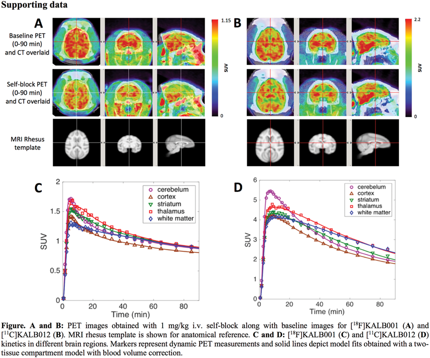
Conclusion
[18F]KALB001 and [11C]KALB012 exhibit good brain penetration and can be readily quantified by standard tracer kinetic modeling methods. The small reductions in VT observed in blocking experiments suggest that both tracers have relatively low specific binding compared to non-specific uptake. Therefore, our results indicate that neither mGluR4 expression nor receptor occupancy can be reliably measured with these tracers in the current conditions.
Acknowledgements
R01EB021708, R01NS100164, MJFF9243.01
References
BPS04-1
Brain Imaging Methodology: I
TauIQ – a quantitative algorithm for tau PET imaging in clinical trials
1Invicro
2Imperial College London
Abstract
Objectives
Tau PET promises to be a valuable biomarker in clinical trials of novel Alzheimer’s Disease (AD) drugs. To date, the field has used SUVR approaches, but this does not harness the increased power of canonical image based quantification methods recently introduced for amyloid (AmyloidIQ)1 that are able to quantify static scans with increased power. Building on these developments, we introduce TauIQ as an algorithm with increased power for clinical trials.
Methods
Data for 234 subjects (88 healthy controls (HC), 40 significant memory concern, 58 early mild cognitive impairment [MCI], 16 MCI and 30 late MCI) were downloaded from the ADNI database with each subject having undergone [18F]Flortaucipir PET, [18F]Florbetapir PET, structural MRI, MMSE, ADAS-COG and CSF total Tau assessments.
Tau canonical images for tau carrying capacity (K), and non-specific binding (NS) were initially constructed as follows – amyloid scans were analysed using AmyloidIQ to derive a subject-specific time in the disease trajectory allowing for spatiotemporal modelling of the temporally aligned tau data with a regression model (Figure A). The derived [18F]Flortaucipir canonical images successfully partitioned background (NS: non-specific and off target binding) and tau (K: stereotypical tau distribution in AD) signals.
The TauIQ algorithm (Figure B) takes as its input a T1-weighted MR image and corresponding static Tau PET image. Initially, DARTEL2 is used to spatially normalise the Tau PET image into MNI152 space using the associated T1-MR image. Following this the Tau PET image is modelled as the linear combination of the K and NS canonical images to enable estimation of the level of non-specific binding and global tau load (TauL). Statistical analysis of the residual image then allows for an estimation of any additional subject specific local tau accumulation that deviates from the characteristic spatial distribution observed at the population level.
TauIQ was applied to all 234 [18F]Flortaucipir scans and SUVR was also calculated for a Braak I/II meta ROI. The relationship of these outcome measures with clinical scores and CSF tau measures was investigated using a Pearson’s correlation co-efficient.
Results
All data were successfully quantified with both TauIQ (TauL: 35 +/- 29%, 45% of cases exhibited a local tau component) and SUVR (1.07 +/- 0.1) (See Figure C for example subjects). The correlation between TauL and clinical scores (MMSE: r = –0.59, ADAS-COG: r = 0.60) was stronger than with SUVR (MMSE: r = –0.47, ADAS-COG: r = 0.49). TauL was also more highly correlated with CSF tau (r = 0.53) than SUVR (r = 0.45).

Conclusions
TauIQ provides an algorithm that is able to quantify global and local tau accumulation in AD and demonstrates a stronger relationship with cognitive and CSF assessments than existing analysis approaches. This approach promises to increase the utility of Tau PET as an effective biomarker of tau pathology in clinical trials.
References
BPS04-2
Brain Imaging Methodology: I
Joint pattern analysis applied to PET DAT and VMAT2 imaging reveals new insights into Parkinson’s disease induced presynaptic alterations
1Department of Physics and Astronomy, University of British Columbia, Canada
2Division of Neurology, Department of Medicine, University of British Columbia, Vancouver, BC, Canada
3Djavad Mowafaghian Centre for Brain Health, Pacific Parkinson's Research Centre, University of British Columbia & Vancouver Coastal Health, Vancouver, BC, Canada
Abstract
Objectives
Most neurodegenerative diseases affect several aspects of brain function, including neurotransmitter systems, metabolic and functional connectivity. Diseases are generally characterized by common clinical characteristics, but there are also significant inter-subject variations. It is thus expected that clinical behaviors will be related to an overall multi-system pattern of disease-induced alterations and additional brain system-specific abnormalities; these system-specific abnormalities may indicate a possible unique system response to disease or subject-specific propensity to a specific clinical progression. Hence we introduce and validate the use of a joint pattern analysis approach, canonical correlation analysis (CCA) and orthogonal signal correction (OSC), to analyze multi-tracer PET data to identify common and unique information provided by each tracer/target. We apply the method to [11C]-DTBZ (VMAT2 marker) and [11C]-MP (dopamine transporter (DAT) marker) data from early Parkinson’s disease (PD) subjects; behavior of these two tracers/targets is well characterized providing robust reference information for the method’s outcome.
Methods
15 early PD subjects (disease duration 5634 months) were scanned with [11C]-DTBZ and [11C]-MP on High Resolution Research Tomograph (HRRT) and anatomical MRI scan. Non-displaceable binding potential (BPND) values were calculated within 22 striatal and extrastriatal ROIs using Logan plot [1] with occipital cortex as reference region. CCA [2] was used to extract highly correlated subject profiles. CCA loadings were used to visualize the spatial patterns along each canonical variate. OSC [3] was applied to CCA residuals to extract unique information specific to each tracer/target. Permutation tests and leave-one-out cross validation were used to examine the robustness of the patterns.
Results
Highly significant common subject profiles (p < 0.05, R2 > 0.85) were identified that decomposed the characteristic dopaminergic changes into three distinct orthogonal spatial patterns: 1) disease-induced asymmetry between less and more affected dorsal striatum; 2) disease-induced gradient with caudate and ventral striatum being relatively spared compared to putamen; 3) progressive loss in less affected striatum, which correlated significantly with disease duration (p < 0.01 for DTBZ, p < 0.05 for MP). Information unique to each tracer revealed a residual striatal asymmetry for VMAT2, consistent with the notion that VMAT2 density is highly related to terminal degeneration [4]; and a residual DAT disease-induced gradient in striatum with relative DAT preservation in substantia nigra. This finding may be indicative either of possible DAT specific early disease compensation and/or related to disease origin.5

Conclusions
These results demonstrate the applicability and relevance of the joint pattern analysis approach to multi-tracer datasets; this data driven method, while recapitulating known aspects of PD-induced tracer/target behaviour, was found to be statistically more robust and provided additional information on (i) correlated behaviors of the two systems, identified as orthogonal patterns, possibly reflecting different mechanisms underlying disease initiation or progression and (ii) system specific effects of disease. It is thus expected that this approach will be very well suited to the analysis of multi-tracer and/or multi-modality data and to relating outcomes to different aspects of disease.
References
BPS04-3
Brain Imaging Methodology: I
Likelihood estimation of drug occupancy – generalization to multiple occupancy measurements
1Neurobiology Research Unit, Copenhagen University Hospital, Copenhagen, Denmark
2Department of Psychiatry, Columbia University, New York, NY, USA
3Molecular Imaging and Neuropathology Division, New York State Psychiatric Institute, New York, NY, USA
4Department of Biostatistics, Mailman School of Public Health, Columbia University, NY, USA
Abstract
Objective
Neuroimaging with PET can be used to measure in vivo the occupancy (Δ) by a drug. For radioligands for which no brain reference region exists, the only established method for estimation of Δ is the Lassen plot1, which is based on linear regression. Simple linear regression is, however, not ideal for analyzing this type of data for several reasons, including regression attenuation and correlated errors2.
We have recently proposed to use Likelihood Estimation of Occupancy (LEO)2 instead. LEO estimates the values of VND and Δ that maximize the likelihood of VT values observed in multiple ROIs. In the scenario in which each occupancy experiment consists of one baseline and one block measurement (i.e., taken after administration of a competitor), it was shown that LEO provides improved estimation relative to the Lassen plot.
The objective of the current study was to generalize LEO into Likelihood Estimation of Multiple Occupancies (LEMO), to accommodate an arbitrary number of blocking scans, where a different value of Δ is to be estimated based on each block measurement, while VND is assumed to be the same across all scans.
Methods
Consider the case where a subject has undergone M + 1 PET measurements. Baseline VT values are modeled as
LEO and LEMO take into account the covariance matrix of the errors (Σ), which can be estimated from a separate cohort of test-retest data2.
To evaluate the performance of LEMO, 2 datasets ([11C]DASB and [11C]Cimbi-36) were simulated (N = 1000). In addition to the baseline scan, each simulated subject had 3 blocking scans, with simulated 25%, 50%, and 75% occupancy, respectively. For each blocking scan, Δ and VND were calculated using Lassen plot and LEO via comparison to the baseline scan. In addition, LEMO was used to calculate all 3 occupancies and VND in a one-step procedure. For LEO and LEMO, 10 test-retest subjects were simulated in parallel to the baseline-block data, and used to estimate Σ. All simulations were based on real test-retest data to create realistic covariance structures across ROIs2.
Results
The results are compiled in Figure 1. Only results for the lowest (25%) and highest (75%) simulated occupancy studies are shown.

Conclusion
Using all available PET measurements to estimate VND and all occupancies, LEMO appears to provide increased accuracy and precision for both [11C]Cimbi-36 and [11C]DASB. In particular, the estimates of VND and occupancy are superior using LEMO for the lowest simulated occupancy case.
References
BPS04-4
Brain Imaging Methodology: I
Resting-state networks revealed based on synaptic density PET
1Dept. of Radiology and Biomedical Imaging, Yale University, USA
2Dept. of Psychiatry, Yale University, USA
Abstract
Resting-state networks (RSN) are systems of brain regions anatomically separated but functionally connected during rest.1 First identified in fMRI studies based on blood-oxygen-level-dependent (BOLD) signal, RSNs have also been found with 18F-FDG.2,3 RSNs may represent intrinsic brain connectivity networks which reflect global functional organization during active as well as rest states. 11C-UCB-J is a PET tracer that binds to and images synaptic vesicle protein 2A (SV2A). It is proposed as a measure of synaptic density and thus, holds promise for investigating RSNs through examination of regional covariance in synaptic density that might be expected to result from coherent patterns of neural activity.
11C-UCB-J was synthesized as described previously.4 Healthy subjects (n = 72, 27 female/45 male, 45 ± 17y) were administered an i.v. bolus injection of 11C-UCB-J over one min (536.77 ± 180.60 MBq, SA: 109.24 ± 36.75 MBq/nmol) and received one dynamic PET scan (HRRT, Siemens) with reconstructed image resolution of 3 mm. MR scans were collected (3T Trio, Siemens) for PET coregistration and subsequent registration into Montreal Neurological Institute (MNI) space. Parametric volume of distribution (VT) images were generated with a 1T compartment model using the metabolite-corrected arterial plasma curve. Based on the VT image, BPnd maps were calculated with centrum semiovale as reference region (i.e. (VTvoxel/VTref)-1), then smoothed with a Gaussian kernel of 8 mm FWHM. Independent component analysis (ICA) was used to identify RSNs.5 ICA extracts maximally independent components (IC) from an unknown linear mixing matrix of random non-Gaussian vectors containing independent source signals that generate the observed signals (x). ICA solves for y = Wx, i.e., the un-mixing matrix (W) of source maps (y) and is often used to identify RSNs in fMRI. For exploratory ICA of 11C-UCB-J, parametric images were loaded into the source-based morphometry (SBM) toolbox (GIFTv4.0b). BPnd images were thresholded with a relative mean threshold of 0.7. To identify RSNs, exploratory ICA was performed with an IC number of 15, similar to previous fMRI analyses. Component spatial loading values were z-transformed and visualized using a z score threshold of 2 to determine the most prominent structures. Resulting maps were visually inspected and compared to previous resting-state ICA findings for functional classification.
Of the 15 ICs, 8 appeared spatially consistent with established RSNs. These can be characterized as primary visual (posterior), default-mode network, secondary visual, cerebellar, executive control, basal ganglia, mesial parietal/prefrontal, and primary visual (anterior). Preliminary analysis found significant age and gender effect with a few ICs, however these results require cross-validation to avoid Type I error.
With an ICA-based approach, we demonstrate extraction and identification of RSNs from 11C-UCB-J PET data in a healthy population. These data suggest activity in functional brain networks may be related to coherent, network-based changes in synaptic density, though further multimodal validation with fMRI data is warranted.
References
BPS04-5
Brain Imaging Methodology: I
Image reconstruction methods affect computer-aided assessment of patient pathologies of flutemetamol and FDG brain PET examinations in patients with neurodegenerative diseases
1Uppsala University, Sweden
2Uppsala University Hospital, Sweden
3Skåne University Hospital, Sweden
4Lund University, Sweden
Abstract
Objectives
New developments in neuro-PET, such as digital silicon photomultiplier detectors, time-of-flight (TOF), point-spread-function (PSF) modelling and regularized reconstruction methods have resulted in improved spatial resolution and signal-to-noise ratio in reconstructed images. The aim was to investigate how some of the improved PET image reconstructions affect quantitative measures and computer-aided assessment of patient pathologies.
Methods
PET data from 70 subjects acquired on a digital TOF PET/CT scanner were included. The subjects were grouped into four cohorts: Alzheimer’s disease patients (n = 16), healthy controls (n = 20), neurodegenerative disease patients (n = 20), and a control cohort comprising melanoma patients without brain involvement (n = 14). The first two cohorts received a 20-min scan with 18F-flutemetamol and the last two cohorts received a 10-min and 2-min scan, respectively, with 18F-FDG. Reconstructed images were obtained by ordered-subsets expectation maximization (OSEM; 3 iterations, 16/34 subsets, 3/5-mm gaussian postprocessing filter, ±TOF, ±PSF modelling) and block-sequential regularized expectation maximization (BSREM; TOF, PSF, β-value 75, 150, 225, 300). Each reconstruction was anatomically normalized into template space (Cortex ID Suite, GE Healthcare). Automated analysis of tracer uptake in regions of interest and comparison with the corresponding tracer uptake in a database of normal subjects in terms of standardized uptake value ratio (SUVR) and z-score was performed. Cerebellar gray matter or pons were used as reference regions in the first two cohorts, and whole cerebellum or whole brain were used in the last two cohorts.
Results
The control cohorts of both flutemetamol and FDG yielded comparable results to the normal database when using OSEM 3i/16 s 5-mm reconstruction, without TOF and PSF. TOF, PSF modelling and BSREM either increased or decreased the relative uptake difference to the normal subjects’ database within the software, depending on the tracer and chosen reference area, i.e. resulting in increased absolute z-scores.
In the flutemetamol patient cohort with normalization to cerebellar gray matter, there were no significant differences in SUVR between reconstructions in any target region. In the control cohort however, mean SUVR decreased with addition of TOF and PSF to OSEM as well as BSREM in the precuneus, with corresponding decreased z-scores (control cohort z-scores in the right precuneus: –0.02 with OSEM 3i/16 s 5 mm, and –0.66 with BSREM β 150).
In both FDG cohorts with normalization to whole cerebellum, a larger filter size resulted in increased SUVR whereas both 16 and 34 subsets resulted in similar values. Adding TOF and PSF to OSEM gradually increased SUVR with corresponding increased z-scores, and overall, BSREM resulted in the highest SUVR (patient cohort z-scores in the right prefrontal region: –1.13 with OSEM 3i/16 s 5 mm, versus –0.55 with BSREM β 150).
Using pons and whole brain for normalization changed the results in all cohorts and induced more variation to the outcome measures with the addition of TOF, PSF and BSREM.

Conclusions
Computer-aided assessment of patient pathologies was affected by image reconstruction methods and should be used with caution when employing other image reconstruction methods than those used for acquisition of the normal database. Neither pons nor whole brain should be used as reference regions.
BPS04-6
Brain Imaging Methodology: I
Towards a gold standard for validation of quantification methods for PET neuroreceptor imaging
1Lady Davis Institute, Jewish General Hospital, McGill University, Montreal, Canada
2McGill Center for Integrative Neuroscience, Montreal Neurological Institute, McGill University, Montreal Canada
3Institute of Neuroscience and Medicine INM-1, Research Centre Jülich, Jülich, Germany
4Department of Psychiatry, Psychotherapy and Psychosomatics, Medical Faculty, RWTH Aachen University, Aachen
Abstract
Introduction
Validating new algorithms for the analysis of invivo PET images in humans is challenging because the true tissue radiotracer concentrations are unknown. PET images represent 3D estimates of tissue concentrations degraded by the imaging process. Here we propose a strategy to simulate PET datasets of neuroreceptor images from an ultra-high (20μm) resolution 3D volume of 2D coronal autoradiographs of 20 different neurotransmitter binding site densities obtained from a serially sectioned post-mortem human brain. We use Monte-Carlo simulation to create a simulated 18-F-Flumazenil PET image from 3D reconstructions of 3D GABA-A receptor distribution.
Methods
The brain from a donor was extracted within 24 h post-mortem, cut into 6 slabs of 2–3 cm, shock frozen (–40°C), and serially sectioned (20μm) with a cryostat microtome.1 Alternating sections (2242 in total) were incubated with radioligands for 1 of 20 different neurotransmitter binding sites, including receptors for glutamate, GABA, serotonin, noradrenalin, dopamine, acteylcholine. Radiolabelled sections were exposed against tritium-sensitive films and autoradiographs digitized to 20x20μm resolution images. Several processing steps were used to align autoradiographs to the donor’s post-mortem MRI.2
Ligand binding densities were estimated for positions between autoradiographs acquired for a particular receptor type. A binary gray-matter (GM) image, i.e., mask, was extracted from the donor’s MRI.3 The GM mask was digitally sliced in the coronal plane. Coronal GM mask slices in the same position as corresponding autoradiographs of the target ligand were aligned to their neighboring GM mask slices with 2D affine transformations (Fig.1.A/B). These transformations were applied to the autoradiographs to transform them to match the morphology of GM mask slices between the acquired autoradiographs. Missing receptor densities were estimated by taking the distance-weighted average of the autoradiographs transformed from the rostral and caudal directions, respectively. This method was used to construct a single volume of flumazenil binding from acquired autoradiographs within 1 brain slab.
A single slice of estimated ligand binding density was used to produce a 30 s simulated PET image with GATE(v7.1).4 Radioactivity concentrations (Bq/mm2) for the PET simulation template were assigned from the voxel intensities of the ligand binding density slice. The spatial coordinates of decay events were binned into 2 volumes with 1x1x1mm voxels: 1 with all decay events and 1 with only those coincidences detected by the simulated PET system.
Results
The estimation of missing ligand binding density shows good agreement with the underlying anatomy of the donor’s brain (Fig.1.C). The Monte-Carlo simulation indicates that even under idealized condition of 100% sensitivity or perfect reconstruction, partial-volume effects nonetheless significantly degrade PET resolution (Fig.1.D)

Discussion
2D autoradiographs sampled at least 400μm from one another were reconstructed into a 3D volume by estimating ligand binding density. Further refinements in the reconstruction algorithms, e.g., non-linear warping of GM masks, will help improve ligand density estimation. The aim of future work is to create whole-brain 3D volumes for each of the 20 neurotransmitter receptors and use GATE to create a database of highly realistic simulated PET images that can serve to validate PET software for neuroreceptor quantification.
BPS05-1
Brain Imaging Methodology: II
Simulating the effect of cerebral blood flow changes on quantification of [18F]flutemetamol and [18F]florbetaben studies
1Amsterdam UMC, Vrije Universiteit Amsterdam, Radiology and Nuclear Medicine, Amsterdam Neuroscience, De Boelelaan 1117, Amsterdam, Netherlands
2Wallenberg Centre for Molecular and Translational Medicine and the Department of Psychiatry and Neurochemistry, University of Gothenburg, Gothenburg, Sweden
3Barcelonaβeta Brain Research Centre, Pasqual Maragall Foundation, Barcelona, Spain
4Life Molecular Imaging GmbH
5GE Healthcare, Amersham, United Kingdom
Abstract
Objectives
Cerebral blood flow (CBF) fluctuates during the course of the day and declines with increasing age1. Additional reductions have been reported in APOE4-carriers with elevated levels of amyloid in the brain and in patients suffering from Alzheimer’s dementia1. This may compromise longitudinal assessments of amyloid load, as bias may be sensitive to changes in CBF2. The purpose of this study was to assess the effect of global and local CBF changes on quantification of [18F]flutemetamol and [18F]florbetaben scans, through computer simulation.
Methods
For [18F]flutemetamol and [18F]florbetaben, cortical and cerebellar grey matter time-activity curves (TACs) of 110 minutes were simulated using a two tissue compartment (2T4k_Vb) model together with a metabolite corrected plasma input function and kinetic parameters established previously for a range of amyloid load levels (DVR from 1.02–1.77&1.02–2.04, for both tracers respectively).3,4 Subsequently, CBF changes were simulated by changing tracer delivery (K1) (+25% to –25%, while keeping the K1/k2 ratio constant)5 in both cortical and cerebellar regions (global changes), or by changing K1 only in the cortical region (local changes). For each condition, a single, noise free TAC was simulated. Next, amyloid load was quantified using SUVr (90–110 minutes post injection) and DVR(=BPND+1), the latter derived using the 2T4k_Vb model, the simplified reference tissue model (SRTM) and reference Logan (RLogan) 70–110 minutes, unfixed k2’ and results were compared with the simulated amyloid load (DVR).
Results
2T4k_Vb derived DVR showed minimal bias (<1%) for both tracers. In contrast, SRTM derived DVR showed an exponential increase in bias for local K1 changes only, mainly at low amyloid levels (Fig.1a,g). After inspection, this behaviour was determined by the second term of the SRTM equation5 {k2 -R1k2/(1 + BPND)}, approaching zero. Furthermore, for [18F]flutemetamol, RLogan derived DVR underestimated amyloid load, in line with results for other tracers (Fig.1h,k)6. For [18F]florbetaben, RLogan derived DVR overestimated and underestimated amyloid load for low and high levels of simulated amyloid load, respectively (Fig.1b,e). The global flow dependency of SUVr increased for higher levels of amyloid load, for both tracers (Fig.1.c,f,i,l) In contrast, for [18F]flutemetamol the local flow dependency decreased for higher amyloid load levels (Fig.1i,l).

Conclusion
All simplified models provided a CBF dependent, biased estimate of amyloid load, while the 2T4k_Vb model showed nearly perfect estimates, emphasizing the importance of verifying accuracy and precision of simplified approaches against plasma input. Overall, bias in reference Logan derived DVR was lowest and relatively constant, in particular for low amyloid load. Therefore, this method seems most suitable for longitudinal studies. SRTM provided only reasonable estimates of amyloid load for global CBF changes.
References
BPS05-2
Brain Imaging Methodology: II
The white matter reference region used in amyloid PET is susceptible to flow and input function fluctuation: a consideration of kinetics
1Dept. of Diagnostic Radiology, Tokyo Metropolitan Geriatric Hospital and Institute of Gerontology
2Research Team for Neuroimaging,Tokyo Metropolitan Institute of Gerontology
Abstract
Many studies have demonstrated the superiority of white matter (WM) reference region in amyloid PET studies in comparison to cerebellum reference region. However, the principle behind its improved stability is yet to be elucidated.
We conducted simulations using Python 3.6, varying K1 and input function, and adding statistical noise.
Our simulation revealed that WM reference region was more susceptible to cerebral blood flow (CBF) variation and input function fluctuation than cerebellar reference region. The stability of values calculated based on a white matter reference region were only as stable as those determined from a cerebellum reference region when the statistical noise in the cerebellum was 2.4 times higher.
Our study suggests that a second scan with the cerebellum in the axial center of a 3-dimensional (3D) PET may improve the stability of a longitudinal study.

BPS05-3
Brain Imaging Methodology: II
Alzheimer’s disease pattern derived from pharmacokinetic modeling of 11C-Pittsburgh compound B PET scans
1Department of Nuclear Medicine and Molecular Imaging, University Medical Center Groningen, University of Groningen, The Netherlands
2Department of Neuroscience, University Medical Center Groningen, University of Groningen, The Netherlands
3Department of Neurology, Alzheimer Research Centre, University Medical Center Groningen, University of Groningen, Groningen, Groningen, The Netherlands
4Institute Born-Bunge, Laboratory of Neurochemistry and Behaviour, University of Antwerp, Antwerp, Antwerp, Belgium
Abstract
Objectives
Disease patterns (DP) derived from the scaled subprofile model, a principal component analysis based method (SSM/PCA), have been used to successfully identify Alzheimer’s disease (AD) patients based on 18F-FDG PET scans.1 In the current study, a similar approach was explored with parametric images derived from pharmacokinetic analysis of 11C-Pittsbough Compound B (PIB) to generate DPs based on different imaging characteristics of the disease.
Methods
Pharmacokinetic modeling using the simplified reference tissue model 2 was applied to the dynamic PIB-PET scans of 30 subjects (15 AD and 15 healthy controls). Parametric images of binding potential (BPND) and relative influx of tracer (R1) were used to construct metric-specific DPs. Then Z-scores were assigned to the images based on its similarities with these DPs. Moreover, the subjects were classified based on a specific threshold derived from receiver-operator characteristic curves. Finally, a set of 32 different subjects diagnosed with AD (n = 4), Lewy body dementia (DLB) (n = 5), frontal temporal dementia (FTD) (n = 5), or mild cognitive impairment (MCI) with (+) (n = 11) or without (-) (n = 7) deposits of amyloid-β plaques, were tested against the DPs.
Results
Visual inspection of the DPs generated for each method were in line with previous studies: a higher binding (BPND images)2 and hypoperfusion (R1)3 were observed in multiple gray matter regions of AD, as compared to healthy subjects. The Z-score threshold for classifying AD patients based on BPND and R1 parametric maps were, respectively, 4.3 (area under the curve (AUC) = 1) and 1.3 (AUC = 0.9). Figure 1 shows the distribution of the Z-scores of the testing subjects. The AD group, as expected, presented an average value above the threshold, of 6.9 ± 6.8 (mean ± SD) for the BPND and 2.0 ± 1.7 for the R1. Meanwhile, the MCI- and DLB groups both presented mean Z-scores smaller than the thresholds (for the DLB group, BPND was of 0.7 ± 1.0 and R1 was 1.0 ± 0.8, and for the MCI-, 0.7 ± 3.1 and 0.8 ± 1.2 respectively). The MCI+ group presented a similar BPND pattern as the AD subjects, with a mean of 14.4 ± 7.2, but the same did not apply to the R1, with a mean of 0.6 ± 0.6. Interestingly, an opposite effect was observed in the FTD group, with high R1 Z-scores (2.1 ± 1.4), and low BPND (–0.4 ± 0.2).

Conclusions
Pharmacokinetic modeling of dynamic PIB-PET scans provide high-quality parametric maps, such as BPND and R1, that provide complimentary information. These images can be used as input for a SSM/PCA analysis, resulting in DPs that demonstrate different characteristic of AD patients when compared to healthy subjects. The multiparametric combination of these parametric images showed to be effective for a better discrimination of AD from other dementias.
References
BPS05-4
Brain Imaging Methodology: II
Characterization of [18F]BCPP-EF, [11C]SA-4503 and [11C]UCB-J for the quantification of mitochondrial and synaptic function in the healthy human brain
1Invicro LLC
2Abbvie Inc
3Neuroepidemiology and Ageing Research Unit, Imperial College London, UK
4Division of Brain Sciences, Imperial College London, UK
5King’s College London, UK
6Hamamatsu Photonics
7MINDMAPS Consortium
Abstract
Objectives
Mitochondrial complex 1 (MC1), sigma 1 receptor (s1R) and synaptic vesicle protein 2A (SV2A) are involved in regulating the brains bioenergetic status or maintaining neuronal integrity, which are altered in neurodegenerative diseases,1.2 Here we characterise the kinetic behaviour of three PET radioligands[18F]BCPP-EF, [11C]SA-4503, [11C]UCB-J, which bind to MC1, s1R and SV2A respectively, for their application in future studies as markers of neurodegenerative disease progression. We also test whether MC1, s1R or SV2A are altered with healthy ageing.
Methods
As part of the MIND MAPS programme,3 12 healthy human subjects underwent a structural MRI scan and 3 90-minute dynamic PET scans with each of the radioligands, including arterial blood sampling. Tissue time activity curves (TACs) were fitted with a one-tissue compartment model (1TC), a two-tissue compartment model (2TC) and multilinear analysis 1 (MA1) to identify the optimal kinetic analysis method for each radioligand. Time-stability analyses were performed for each radioligand to enable the identification of a suitable acquisition duration. Linear regression was performed to estimate the dependence of VT on age.
Results
All three radioligands readily entered the brain and yielded heterogeneous uptake consistent with the known distribution of their targets (Figure 1A). The optimal models for determination of regional estimates of VT were MA1 for [18F]BCPP-EF and [11C]SA-4503, and both 1TC and MA1 for [11C]UCB-J (Figure 1B). Acquisition times of 70, 80 and 60 minutes for [18F]BCPP-EF, [11C]SA-4503, [11C]UCB-J, respectively (Figure 1C), were found to provide good estimates of regional VT values. We observed a moderate negative correlation of [18F]BCPP-EF and [11C]UCB-J VT binding with age for most grey matter regions, reaching statistical significance for [11C]UCB-J, but not [18F]BCPP-EF. Importantly the dependence on age we observed in regional VT estimates was also seen for ROI volumes, suggesting that partial volume correction will be required to confirm the effects of age. The exception to this was the age-related reduction of [11C]UCB-J VT in the caudate (1.29%/yr, r = 0.77, p < 0.005) which was ∼1.7 fold greater than that the volume reduction (0.75%/yr, r = 0.66, p < 0.05).
Conclusion
We established a set of optimal tracer kinetic quantification models for [18F]BCPP-EF, [11C]SA-4503 and [11C]UCB-J in the healthy human brain. MA1 better estimates MC1 and s1R density, using [18F]BCPP-EF and [11C]SA-4503 respectively, while either MA1 or 1TC are both suitable models for quantifying [11C]UCB-J. Time-stability data indicated that all three radioligands can be used within clinically feasible scan durations, making their use more practical for a wide range of clinical populations. Lastly, we showed a moderate effect of healthy aging on [11C]UCB-J signal in the caudate, suggesting that [11C]UCB-J PET may serve as a more sensitive marker of age-related synaptic loss in that structure than does regional volume.

BPS05-5
Brain Imaging Methodology: II
Non-invasive quantification of tau accumulation in dementia using simultaneous 18F-PI-2620 PET/MRI
1Department of Radiology, Stanford University, Stanford CA, USA
2Department of Neurology, Stanford University, Stanford CA, USA
Abstract
Objectives
18F-PI-2620 is a novel tau tracer with high specificity to enable in vivo visualization of tau accumulation in Alzheimer’s disease1. This study utilizes simultaneous PET/MRI to quantify volume of distribution (VT) of 18F-PI-2620 without invasive arterial blood sampling and assess tau distribution in patients with Alzheimer’s disease dementia relative to healthy controls.
Methods
Simultaneous, time-of-flight PET/MRI (Signa, GE Healthcare) was performed in 13 individuals (5 Alzheimer’s disease, 3 mild cognitive impairment, mean age 67.8 years, 4 female; and 5 healthy controls, mean age 66.3 years, 3 female). Subjects underwent dynamic PET scans (during manual injection of 5–10 mCi of 18F-PI-2620) up to 90 minutes post-injection. The concurrent MRI protocol included neck MRA for image-derived input function (IDIF)2 and zero-echo time imaging (ZTE) for attenuation correction3. IDIFs were created within cervical arteries, correcting for spill-over effects with the true arterial volume (segmented on MR angiogram, Figure 1a)2. Using the reconstructed IDIFs and time activity curves of pre-selected cortical regions, VT was fit using PMOD software using Logan analysis and compared to relative standardized uptake value (SUVR) at 60–90 min, referenced to the cerebellum. Arterial spin labeling MRI with post-label delay of 2 seconds was also acquired to assess cerebral blood flow (CBF).
Results
PET/MRI provided high-quality, time-matched IDIFs and reliable fits with Logan plots of the 18F-PI-2620 tracer (

In healthy subjects, relatively uniform tau uptake in the cortex was observed on both SUVR and VT assessment, except in known areas of off-target binding such as the sagittal sinus. In dementia patients compared to healthy volunteers, VT calculated from IDIF was higher in across multiple cortical association areas such as the superior parietal cortex (5.3 ± 2 ml/ccm, corrected P-value <1e-4) and posterior temporal lobe (4.9 ± 2 ml/ccm, P = 0.001); but not in control regions (pre-central and post-central gyrus, 4.0 ± 2 ml/ccm) (
Conclusions
Simultaneous PET/MRI enables quantification of critical parameters including VT to characterize tau distribution of patients in vivo, without the need for arterial blood sampling. Hybrid imaging enables concurrent assessment of perfusion with ASL, which in future work could inform K1 estimates in 18F-PI-2620 compartment modeling to reduce scan time4, or allow investigation of the vascular contribution to dementia in the same study.
References
BPS05-6
Brain Imaging Methodology: II
Impact of image processing on [11C]PIB amyloid quantification
1Medical Imaging Center, University Medical Center Groningen, University of Groningen, The Netherlands
2Dept. of Radiology and Nuclear Medicine, VU Medical Center, Amsterdam University Medical Centers, The Netherlands
Abstract
Objectives
Amyloid PET quantification can be affected by differences in image processing. For example, brain tissue segmentations can be performed in the subject- or standard-space (e.g. MNI), which might have an impact in regional and parametric analysis of the images. The aim of this study was to assess the effects of using different non-rigid image deformations (to standard-space) on quantitative PET metrics. Additionally, effects of using different grey matter masks, derived from population- or subject-based segmentations, were compared.
Methods
Alzheimer’s Disease (AD, n = 12) and healthy (HC, n = 16) subjects underwent 70 min dynamic [11C]PiB PET scans and T1-weighted MRI scan. The 40–60 min of the [11C]PiB image were averaged and used for the registration to the individual MR scan (subject-space). Following, the MRI was used to calculate the transformation from subject-to-standard space using three different
Metrics were extracted for all the combinations of space, tissue segmentation, and transformation matrix, within grey matter. [11C]PiB SUV ratios (SUVR) were calculated using the cerebellum as reference. Two intensity metrics were extracted, one using the uptake in all grey matter (SUVRmeanall) and another including only amyloid-beta positive (Aβ+) voxels, with a SUVR≥1.5 threshold (SUVRmeanAβ+). Moreover, two volume-based metrics were defined: amyloid fractional volume (AFV, percentage of Aβ+ volume) and total amyloid burden (TAB, SUVRmeanAβ+ times Aβ+ volume).
All image data were processed with SPM12, and the analysis was performed in R Studio (v1.1.456; R v3.5.1). Mean differences and standard deviations are shown.
Results
When using the same spatial normalisation methodology and grey matter definition, small variations in the intensity metrics were found (0.4% ± 0.7%) between the subject- and standard-space results. However, within a specific space, grey matter definition had a greater impact in SUVRmeanall (5.67% ± 2.8%), but not on SUVRmeanAβ+ (0.7% ± 1.5%). Overall, normalisation methods AMOUNT and CONCENTRATION provided similar intensity metrics (0.7% ± 0.6%). Meanwhile, TPM was different than the other methods (7.5% ± 7.4%).
Volumetric measurements showed larger differences between spaces when the spatial normalisation and grey matter were the same (AFV: 4.6% ± 6.8%; TAB: 15%±10%). Within the same space and normalisation, large differences were observed for AFV and TAB depending of the grey matter segmentation method (4.6% ± 15% and 33% ± 14%, respectively). For spatial normalisation methods, CONCENTRATION and AMOUNT provided similar volume-based metrics (12% ± 9.5%), while TPM showed larger differences to the other methods (27% ± 21%).

Conclusions
Intensity metrics were less affected by differences in PET image processing procedures while volumetric measurements depended more strongly on both space and spatial normalisation method applied and may thus only be valid when derived in subject-space. Therefore, only when SUVR are quantitative image analysis may be performed in either subject- or template-space, while volumetric measures should be performed in subject-space only. (This project is funded by European Union’s Horizon 2020 research and innovation programme under the Marie Skłodowska-Curie grant agreement No 764458.)
BPS06-1
Quantitative brain imaging using integrated PET/MRI
Increased tau aggregration in young traumatic brain injury and post-concussion syndrome patients
1Department of Surgical Sciences/Radiology, Uppsala University, Uppsala, Sweden
2Department of Neuroscience, Uppsala University, Uppsala, Sweden
3Department of Medicinal Chemistry, Uppsala University, Uppsala, Sweden
Abstract
Objectives
Traumatic brain injury (TBI) and post-concussion syndrome (PCS) due to repeated sports-related injury have been suggested to be associated with aggregation of neurofibrillary tangles (tau) and neuroinflammation. The aim of the present work was to simultaneously study structural brain damage and both tau aggregation and neuroinflammation (TSPO) in TBI and PCS patients in-vivo using PET-MRI
Methods
27 subjects (6 TBI and 12 PCS in chronic phase, 9 age-matched HC; mean age 27, range 19–43) underwent two 60-min dynamic PET-MRI scans on the same day after bolus injection of 18F-THK5317 and 11C-PK11195 (Signa PET/MR, GE Healthcare). Simultaneous MRI consisted of T1, T2, susceptibility-weighted imaging (SWI), arterial spin labelling (ASL), diffusion tensor imaging (DTI), MR-spectroscopy (MRS) and resting state fMRI. PET images were reconstructed using zero echo time-based attenuation correction. Parametric images of 18F-THK5317 DVR-1 and 11C-PK11195 BPND were made using the reference Logan method and basis function implementation of the simplified reference tissue model (SRTM2), respectively. Cerebellar gray matter (tau) and a supervised clustering based region (TSPO) were used as reference tissue. All images were spatially normalised to MNI space using SPM12, and differences between groups were assessed using t-tests at the voxel level and using a probabilistic volume of interest (VOI) template.
Results
Diffuse axonal injury and microhemorrhages were seen on MRI in TBI, whereas no structural abnormalities were found for PCS. In TBI, voxel clusters with significantly increased 18F-THK5317 and 11C-PK11195 binding were found mainly in thalamus, temporal lobe white matter and midbrain (tau only), see figure. In PCS, 18F-THK5317 binding was increased in corpus callosum, whereas 11C-PK11195 binding was increased in hippocampus. VOI-based analysis did not show any significant differences in binding between HC, TBI and PCS. ASL showed decreased overall grey matter CBF in both TBI and PCS by about 20%.

Conclusion
The results suggest co-localised tau aggregation and neuroinflammation in thalamus and temporal lobe white matter, as well as tau aggregation in midbrain, in TBI, and tau aggregation in corpus callosum and neuroinflammation in hippocampus in PCS. Individual variations in TBI and PCS groups need to be studied in more detail.
BPS06-2
Quantitative brain imaging using integrated PET/MRI
Investigating the optimal method to generalize an ultra-low-dose amyloid PET/MRI deep learning network across scanner models
1Dept. of Radiology, Stanford University, Stanford, CA, United States
2Dept. of Nuclear Medicine, University of Leipzig, Leipzig, Germany
3Subtle Medical Inc., Menlo Park, CA, United States
Abstract
Objectives
To increase the utility (e.g. in diagnosis or multi-center trials) of a previously-trained ultra-low-dose amyloid PET/MRI deep learning network1, we aim to generalize this network (training data from a time-of-flight PET/MRI scanner: Signa PET/MRI, GE Healthcare; “Scanner 1”) to data acquired on different scanner models (mMR, Siemens Healthineers; “Scanner 2”) and with different scan protocols. Training data from the two scanners as well as transfer learning2 techniques were utilized in various combinations for investigating the optimal method for network generalization.
Methods
Data Acquisition: 40 datasets (39 participants, 19 female; 67 ± 8 years) were recruited for simultaneous amyloid PET/MRI scanning (PET: 18F-florbetaben amyloid tracer, data acquired 90–110 minutes post-injection; MRI: T1-, T2-, T2 FLAIR-weighted images) on Scanner 1 while 40 other participants (23 female, 64 ± 11 years) were scanned on Scanner 2 (T2 FLAIR images not acquired). List-mode PET data were reconstructed for the full-dose image and was also either randomly undersampled by a factor of 100 (Scanner 1) or framed for 1 minute from the start of PET acquisition (Scanner 2) for reconstruction to produce low-dose (1% and ∼5% dose respectively) PET images.
U-net Implementation: A convolutional neural network ("U-Net3,4, ” architecture from Chen et al.1) was trained with Scanner 1 data, with the three MR images and the low-dose PET image as inputs.
To determine the optimal method for network generalization to Scanner 2 data (T1-weighted images replaced the missing T2 FLAIR acquisitions), the trained network was either (A) directly applied to Scanner 2 data or (B) fine-tuned for 100 epochs with Scanner 2 data (learning rate = 0.0001). Networks were also trained from scratch based on (C) Scanner 2 data only or (D) all data from both scanners (hyperparameters also based on Chen et al.1). 5-fold cross-validation was used during network training.
Data Analysis: For comparing images to their full-dose counterparts, three image quality metrics peak signal-to-noise ratio (PSNR), structural similarity (SSIM), and root mean square error (RMSE) were calculated. The metrics derived from different image types were compared using paired t-tests at p = 0.05/3 (Bonferroni correction). A certified physician read all images and rated the image quality (5-point scale) as well as the amyloid uptake status (positive/negative).
Results
All synthesized images showed marked improvement in noise reduction. For scanner 2 data, image quality improved the most using method B (Figure 1; p < 0.01 for all metric comparisons involving method B). After transfer learning, the image quality metrics also showed improvement (compared to the low-dose image) at a level slightly greater than using the original trained network on Scanner 1 data (p < 0.03 for all comparisons). All synthesized image types yielded high diagnostic accuracy (90% compared to full-dose image readings); the reader preferred images synthesized using methods B-D more than the full-dose images (Figure 1).

Conclusions
A network trained on PET/MRI data acquired from one scanner and fine-tuned on data from another was the optimal method for the generalization of deep-learning-based ultra-low-dose PET/MRI imaging across scanners, overcoming bias from the source of training data.
References
BPS06-3
Quantitative brain imaging using integrated PET/MRI
H2O15-PET and BOLD fMRI during galvanic vestibular stimulation – not entirely the same thing
1Institute of Basic Medical Sciences, University of Oslo, Norway
2Department of Neurology, Ludwig-Maximilians-Universität in Munich, Germany
3German Center for Vertigo and Balance Disorders (DSGZLMU), Ludwig-Maximilians-Universität in Munich, Germany
4Department of Nuclear Medicine, Ludwig-Maximilians-Universität in Munich, Germany
5Department of Nuclear Medicine, Technical University Munich, Germany
6Munich Cluster of Systems Neurology – SyNergy, Munich, Germany
Abstract
Objectives
BOLD fMRI has replaced H2O15-PET in studies of brain activation. Application of vestibular stimulation in different imaging methods has revealed convergences and differences in the cortical activation patterns [1–2]. The aim is to use integrated PET-MRI scanner using H2O15 to validate BOLD fMRI responses in a well-established model of galvanic vestibular stimulation (GVS).
Methods
Eight (n = 10) or six (n = 9) simultaneous PET-fMRI scans were acquired from 19 healthy volunteers in a hybrid scanner (Siemens mMR Biograph) during rest/AC-GVS stimulation at 1 Hz with eyes closed in a block design (30 s blocks, 4 repetitions per run).3 List mode PET started with the semibolus injection of 750MBq H2O15 simultaneously to fMRI acquisition. PET data was OSEM-reconstructed for the 30 seconds following the start of the first stimulation block (1.00122x1.00122x2.03125 mm, 256x256x127 voxels). Functional MRI data was acquired using EPI sequences with a combined 16-channel head-neck coil (101 consecutive image volumes per run, TR 2.5 s, 3.1x3.1x3mm, 64x64x39 voxels). Data preprocessing involved motion correction, spatial normalization to MNI, and smoothing (12 mm FWHM). Statistical analysis was performed using the General Linear Model and a random effects approach. Group-level SPMs were generated for the contrasts PET, MR-Onset (events at stimulus onset), MR-Block, and the respective negative contrasts (One-sample t-tests, SPM12, p < 0.05 FDR).
Results
The PET data revealed activation of the multisensory vestibular cortical network with a predominance in the right hemisphere and ocular motor areas bilaterally, as well as deactivations in occipital, temporal, and parietal areas, e.g. in precuneus, hippocampus, and postcentral gyrus (fig. 1).

MR-Onset exhibited related, but bilateral and more widespread activations in the vestibular and ocular motor networks, and no deactivations were identified. MR-Block showed also a bilateral, but less widespread activation pattern than MR-Onset, and resembled closer the pattern found in PET, with activation of vestibular and ocular motor areas, and negative signal changes in occipital, temporal, and parietal areas. Both MRI contrasts revealed bilateral activation clusters in middle temporal gyrus that were not seen in PET.
Conclusions
Both PET and fMRI detected activation patterns conform with the vestibular processing network, however, the fMRI patterns were more widespread and bilateral than PET, and MRI-Onset demonstrated no deactivations. Whereas H2O15-PET maps cerebral blood flow, BOLD MRI is the result of complex interactions between blood flow, volume, oxygenation, and oxygen extraction. Beside the non-identical physiological processes mapped by the modalities, some of the differences may arise from processing steps. It is e.g. required to apply some form of global scaling to PET data, whereas fMRI performs a baseline correction. The study demonstrates the importance of validating methods before results may be extrapolated across different modalities. Future work could focus on processing parameters and/or other fMRI sequences and their consequences on the similarity/dissimilarity of the resulting maps.
References
BPS06-4
Quantitative brain imaging using integrated PET/MRI
Validation of resting-state connectivity in the dopaminergic system using a simultaneously acquired [11C]raclopride PET/BOLD fMRI protocol
1Department for Preclinical Imaging and Radiopharmacy, Werner Siemens Imaging Center, Eberhard Karls University Tuebingen, Germany
Abstract
Objectives
Resting-state functional connectivity acquired by BOLD-fMRI is widely used to investigate functional changes in the brain. [11C]Raclopride ([11C]Rac) is a well-established positron emission tomography (PET) tracer binding to the dopamine-D2-receptor which may offer a complementary approach to acquire functional connectivity in dopaminergic regions. In our study, we aimed to compute functional molecular-level connectivity from [11C]Rac binding potentials in the rat brain, performed reliability analyses and compared it to the output of the simultaneously acquired fMRI.
Methods
Eight rats underwent two simultaneous [11C]Rac-PET/BOLD-resting-state-fMRI measurements with a 17 week retest interval. Rats were anesthetized with 1.3% isoflurane in air and [11C]Rac was applied i.v. as a fast bolus (30 s) followed by a constant infusion over 100 minutes using a Kbol-value of 34 minutes. The data were reconstructed in 1-minute frames using OSEM2D. BOLD- resting-state -fMRI data were acquired simultaneously using a T2* weighted gradient echo planar imaging sequence (repetition time: 2500 ms, echo time: 18 ms, voxel size: 0.27x0.27x0.80mm3). Following preprocessing using SPM12, regions of the dopaminergic system exhibiting a binding potential higher than 0.5 were selected for further analysis including nucleus accumbens, caudate putamen, cingulate cortex, medial prefrontal cortex, superior colliculus, midbrain and inferior colliculus. Data analyses were performed after [11C]Rac PET reached steady state. Resting-state functional connectivity was computed pairwise in each subject for both [11C]Rac-PET and fMRI using Pearson’s r and group-level mean correlation matrices were generated. Mean correlation matrices over the whole steady state period were used to perform between-scan (test retest) as well as between-modality calculations. To calculate the within-scan reliability, the matrices were split into an early (31–60 minutes) and a late time period (61–90 minutes). Pearson’s r was used to calculate between-scan and within-scan reliability; dice coefficient was used to compare [11C]Rac PET and fMRI outputs applying sparsity thresholding (10–20%).
Results
[11C]Rac PET derived resting-state functional connectivity revealed correlations between caudate putamen and several posterior and anterior dopaminergic regions (e.g.: cingulate cortex, inferior colliculus, thalamus). The output exhibited excellent group-level reliability (between-scan 0.79, within-scan 0.80). fMRI showed Pearson’s r of 0.93 for between-scan and of 0.98 within-scan reliability.
Comparison between PET and fMRI output showed similar clusters of connectivity between caudate putamen and nucleus accumbens as well as cingulate cortex and prefrontal cortex. This was quantified by a dice overlap of 0.54–0.59 depending on the sparsity threshold.
Conclusions
Resting-state functional connectivity of [11C]Rac PET revealed stable and reliable results for test retest measurements and between different scan periods. To determine the added value of this novel approach to the more established fMRI derived functional connectivity, future studies involving pharmacological and pathological alterations of functional connectivity must be performed.
BPS06-5
Quantitative brain imaging using integrated PET/MRI
Simultaneous PET-MR-EEG to detect dopamine release during absence seizures
1Centre for Medical Engineering, School of Biomedical Engineering and Imaging Sciences, King's College London, London SE1 7EH, UK
2King's College London & Guy's and St Thomas' PET Centre, St Thomas' Hospital, London SE1 7EH, UK
3Univ Lyon, CarMeN Laboratory, INSERM, INRA, INSA Lyon, Université Claude Bernard Lyon 1, Lyon 69000, France
4Siemens Healthcare SAS, Saint-Denis 92310, France
5Department of Biomedical Engineering, Centre for Medical Engineering, School of Biomedical Engineering and Imaging Sciences, King’s College London, London, SE1 7EH, UK
6Department of Clinical Neurophysiology and Epilepsy, St Thomas' Hospital, Guy's and St Thomas' NHS Foundation Trust, London SE1 7EH, UK
Abstract
Objectives
PET-MR-EEG offers the potential to image neurotransmitter release and cerebral blood flow (CBF) during epileptic seizures captured by electroencephalography (EEG). Our previous investigations revealed that a 32-channel MR-compatible EEG cap causes minimal photon attenuation1. Dopamine receptor binding is reduced at rest in epilepsy2, but it is not known if modulation of dopamine release occurs during seizures. We tested the hypothesis that increased dopamine release occurs during hyperventilation-induced seizures in humans as an endogenous anti-seizure mechanism.
Methods
Four participants with generalised epilepsy and absence seizures (median ± interquartile range age 27.5 ± 23.5 years), and 11 healthy controls (31.0 ± 7.5 years) were injected with 237.5 ± 3.2 Megabecquerels of [18F]fallypride, a D2/D3 selective radiotracer that is sensitive to dopamine release3. Emission data were acquired between –1–50, 70–120, and 180–240 minutes post-injection (p.i.) using a Siemens Biograph mMR. Magnetic resonance (MR) images, including 3D T1-weighted MP-RAGE sequences, were acquired throughout, together with 32-channel EEG during the final hour.
At 204 minutes p.i., participants performed auditory-cued hyperventilation (HV) at a rate of 30 breaths per minute, as per clinical EEG practice (three blocks of three-minute HV, each followed by two minutes of free breathing). Pseudo-continuous arterial spin labelling data was acquired from four minutes before until eight minutes after the task.
The emission data were reconstructed in one-minute frames (3 iterations, 21 subsets, 4 millimetre Gaussian smoothing), using pseudo-CTs that were generated from the T1-weighted images for attenuation correction4. The median radioactivity concentration was computed from realigned emission images in basal ganglia, thalami, and cerebella (used as a reference) delineated by anatomical segmentation of the T1-weighted image via MAPER5. The time-activity curves (TACs) for 180–240 minutes were fit to a single exponential function, omitting peri-ictal frames.
Results
Good compliance with the HV task was observed, with marked EEG slowing and global reduction of CBF observed for all participants (Debatisse J et al, Brain 2019 conference 2019). Generalised spike-and-wave discharges (GSWs) typical of absence seizures were observed during scanning for two of the four participants with epilepsy. Four instances of GSW were observed for one (two during HV); and two instances of unprovoked GSW was observed for the other (Figure). For each of these two participants, both of whom reported disorientation during the HV, there was no significant deviation in thalamic TACs for pre-ictal, ictal, post-ictal (p ≥ 0.20 and p0 ≥ 0.09 Kruskall-Wallis vs resting frames).

Conclusions
These preliminary analyses do not provide evidence for increased dopamine release during absence seizures. We will apply the optimized lp-ntPET model6 to a larger patient sample in order to quantify dopaminergic modulation at GSW onset within individual thalamic nuclei, segmented using diffusion-based probabilistic tractography. Our approach might prove fruitful in other paroxysmal disorders, e.g. migraine.
First GSW discharges at 192.5 mins p.i. (upper panel) and corresponding TACs for 180–240 minutes p.i. (lower panel; logarithmic y-axis).
References
BPS07-1
Imaging in Neurological Diseases
Amyloid-dependent and amyloid-independent effects of tau in Alzheimer's disease
1Department of Neurology & Neurosurgery, McGill University, Montreal, Canada
Abstract
Background
Recent tau-PET studies have emphasized that tau NFTs are more closely linked to atrophy, regional hypometabolism, and domain-specific cognitive dysfunction than Aβ deposition. However, animal models have suggested that the interaction between Aβ and tau pathologies may better explain cognitive function as compared to the additive effects of both pathological hallmarks of Alzheimer’s Disease. Here, we test whether the interaction between amyloid and tau pathologies is related to clinical function beyond their individual additive effects.
Methods
We first examined cognitively normal (n = 87), Mild Cognitive Impairment (n = 16) and Alzheimer’s disease (n = 25) participants who underwent tau PET with [18F]MK6240 and amyloid-b PET with [18F]AZD4694. We also assessed an independent sample of cognitively normal (n = 120), Mild Cognitive Impairment (n = 63) and Alzheimer’s disease (n = 24) individuals who underwent tau PET with [18F]AV1451 and amyloid-b PET with [18F]Florbetapir from ADNI. Global cognitive status was assessed using the CDR Sum of Boxes (CDR-SoB), which is obtained summing scores of tests assessing memory, orientation, calculation, and problem solving. A voxel-wise interaction model was built to assess the main and interactive effects of Amyloid PET and Tau PET SUVRs on clinical status. Age and years of education were employed as covariates in each model.
Results
Voxel-wise analyses revealed dissociated regional effects for amyloid-PET, tau-PET, and the synergistic interaction between the two. In ADNI, a significant synergistic effect of amyloid-β and tau SUVR was associated with worse clinical status in the precuneus, anterior and posterior cingulate, lateral temporal, medial prefrontal, orbitofrontal, frontal pole, and basal forebrain cortices was associated with worse clinical status across the Alzheimer’s disease spectrum. In the TRIAD cohort, the synergistic effect of amyloid-β and tau SUVR was associated with worse clinical status in the posterior cingulate/precuneus, inferior parietal cortices and medial prefrontal cortex.

Conclusions
This study suggests that the combination of amyloid and tau pathologies have detrimental effects on clinical function that are not captured by their independent effects. The voxel-wise associations are also related to the typical distribution of each pathology, which overlap in hubs of the brain’s default mode network (DMN). These results suggest that combinations of amyloid and tau therapies may be most effective for Alzheimer’s disease. Furthermore, our results do not support a phase of Alzheimer’s disease that is amyloid-independent.
BPS07-2
Imaging in Neurological Diseases
Failure of remyelination in periventricular white matter lesions in Multiple Sclerosis
1Institut du Cerveau et de la Moelle épinière (ICM), Inserm U 1127, CNRS UMR 7225, Sorbonne Universités, UPMC Univ Paris 06 UMR S 1127
2Neurology Department, Pitie-Salpetriere Hospital, APHP, Paris, France
3IMIV, CEA, Inserm, CNRS, Univ. Paris-Sud, Université Paris Saclay, CEA-SHFJ
4Neurology Department, St Antoine Hospital, APHP, Paris, France
Abstract
Objectives
Spontaneous remyelination mostly fails in white matter (WM) lesions of patients with multiple sclerosis (MS). Neuropathological studies1 suggest that the efficacy of this process might depend on lesion location, being particularly reduced in the proximity of ventricular cerebrospinal fluid (vCSF). However, to date this evidence has not been confirmed by in-vivo data. In a recent study2 we have demonstrated that it is possible to measure remyelination in vivo with PET using [11C]PiB. Here, we used the same technique to explore the relationship between the extent of remyelination and the distance from vCSF in MS.
Methods
Nineteen patients with MS and eight healthy controls (HC) underwent a 90 min dynamic [11C]PiB PET exam at baseline and after 2–4 months. [11C]PiB distribution volume ratio (DVR) maps were obtained with Logan graphical analysis based on reference region extracted using a supervised clustering algorithm2. In patients, each lesional voxel was classified as demyelinated if its DVR value was more than one standard deviation below the mean DVR value of all the voxels in HC that were localized at the same distance from vCSF2. Individual maps of demyelinated voxels at baseline, as well as maps of dynamically demyelinating voxels (voxels classified as normally myelinated at baseline and demyelinated at follow-up), and of remyelinating voxels (voxels classified as demyelinated at baseline and normally myelinated at follow-up), were generated.
At the group level, we computed the following probability maps A) demyelinated voxels at baseline, B) demyelinating and C) remyelinating voxels at follow-up. At the single subject level, we modelled the relationship between the probability of remyelination and the distance from ventricles using a segmented model. The relationship between the probability to remyelinate near vCSF and cortical thickness was measured with Pearson correlation coefficient.
Results
Both the probability of voxels to be demyelinated at baseline and to demyelinate over the follow-up were higher close to vCSF compared to more distant regions (Figure 1A-B). The opposite effect was observed for the probability of voxel to remyelinate (Figure 1C). The mean probability of voxels to remyelinate, which was 0.08 ± 0.09 close to the ventricles, increased of 0.04 ± 0.03 (p < 0.001) for each mm of distance from vCSF up to a distance of 14.12 ± 7.00 mm, where the gradient almost reached a plateau. A higher probability of voxels to remyelinate close to the vCSF significantly correlated with a higher cortical thickness (r = 0.56, p = 0.013).

Conclusions
We demonstrated for the first time in vivo that spontaneous remyelination is significantly reduced in periventricular WM lesions in MS, and becomes progressively more extensive with increasing distance from ventricles. These data suggest that the presence of potentially cytotoxic soluble mediators in the CSF could exert an inhibitory effect on the remyelination process around the ventricles3. This hypothesis is further supported by the positive correlation we found between periventricular remyelination and cortical thickness, suggesting that similar mediators could influence tissue damage and repair in areas close to the CSF both in WM and in cortex.
References
BPS07-3
Imaging in Neurological Diseases
On regional variations of SUVR values and off-target binding of tau-imaging tracers in cognitively normal older subjects: Indirect comparisons of [18F]AV1451 to [18F]RO-948
1Dept. of Radiology, Johns Hopkins University, USA
2Laboratory of Behavioral Neuroscience, National Institute on Aging, USA
3Pharma Research and Early Development, Hoffmann-La Roche, Switzerland
Abstract
Objectives
Recently, we introduced a new tau-imaging tracer, [18F]RO-948 that appear to have little noticeable off-target binding in the brain except in the substantia nigra (SN). The tracer showed low but consistent regional variations of SUVR values across cognitively normal elderly subjects. Similar trends were reported for the currently most widely used tracer [18F]AV1451, excluding regions with off-target binding. The aims of this work are to re-examine off-target binding of [18F]AV1451 by contrasting to [18F]RO-948, and to evaluate whether [18F]AV1451 and [18F]RO-948 share similar regional variations in normal older adults.
Methods
Ten cognitively normal subjects were studied with [18F]RO-948 (age: 62 ± 8; 5 males) or [18F]AV1451 (age: 65 ± 5; 2 females/3 males). All subjects were confirmed amyloid-β negative with [18F]AV45 or [11C]PIB scans. For tau-scans, regional SUVR values were obtained using frames obtained from 60 to 90 min post-injection for [18F]RO-948 or from 80 to 110 min for [18F]AV1451 (reference region: the cerebellum, Cb). The Freesurfer-derived volumes of interest (VOIs) were manually edited for inaccuracies, as needed. Voxels within superior 6 mm of Cb VOIs were removed to avoid contamination from the supra-tentorial regions. VOIs for SN and the choroid plexus (CP), along the hippocampus (Hp) were manually defined. Thus, a total of 43 left-right merged VOIs were used for SUVR calculation.
Results
In a scatter plot of mean regional SUVR data of [18F]AV1451 (=y) versus [18F]RO-948 (Figure A), data points of 8 regions (listed in the legend) deviated from the identity line (above y = 1.3·x) while data points of the remaining 35 regions (including SN) scattered along the identity line (y = 0.95 x + 0.49; R2 = 0.685). The relationships were maintained in individual subjects for the 8 regions (except the hippocampus, Hp which had data below y = 1.3·x in 3 subjects), and for the 35 regions (R2: 0.256–0.78; p < 0.002 in all subjects). Among the 8 regions, Hp alone showed a significant correlation with CP (Figure B; p = 0.02) in scans of [18F]AV1451. Because of a relatively wider range of SUVR values in CP with [18F]AV1451 (1.2–3.1, compared to 0.9–1.3 of [18F]RO-948) and proximity of the regions, Hp for [18F]AV1451 appears to be more vulnerable than [18F]RO-948 to CP tracer retention.

Conclusion
This study suggested that [18F]AV1451 had additional binding compared to [18F]RO-948 in the caudate nucleus and amygdala as well as 6 other commonly reported off-target binding regions. Second, SUVR values of [18F]AV1451 in Hp were strongly influenced by the level of tracer retention in CP. Third, [18F]AV1451 showed similar regional variations of SUVR values in the 35 brain regions that were consistent across subjects. The findings suggested that the use of normal regional variations (e.g., region-specific cut-off values) could improve identification of tau-positive scans for both tau-imaging tracers. The finding from this preliminary study should be confirmed with larger numbers of subjects and head-to-head comparisons of both tracers within the same subjects.
Supported in part by the Intramural Research Program, National Institute on Aging, NIH.
BPS07-4
Imaging in Neurological Diseases
[11C]JNJ717 P2X7 receptor PET as a novel neuroinflammation target: ex vivo and in vivo comparison with [18F]DPA714 in human ALS
1Division of Nuclear Medicine and Department of Imaging and pathology, University Hospitals Leuven and KU Leuven, Belgium
2Department of Neurology, University Hospitals Leuven and Laboratory of Neurobiology, Center for Brain & Disease Research KU Leuven and VIB, Belgium
3Laboratory for Radiopharmaceutical Research, Department of Pharmaceutical and Pharmacological Sciences, KU Leuven, Campus Gasthuisberg, O&N2, Herestraat 49, Box 821, 3000 Leuven, Belgium
4Department of Neuroscience – Laboratory for Neuropathology, KU-Leuven, Leuven, Belgium
5Department of Pathology, UZ Leuven, Leuven, Belgium
Abstract
Objectives
Neuroinflammation is an important process in disease onset and progression in Amyotrophic Lateral Sclerosis (ALS) 1,2. Until now, TSPO receptors are widely used as target for studying neuroinflammation, but are poorly defined in specificity of the target with relatively low signal-to-noise ratio, exhibit endothelial binding and are influenced by binding genotype in humans. Other promising neuroinflammation targets have been investigated such as membrane P2X7 receptors (P2X7R) that are upregulated on activated microglia with low expression elsewhere. [11C]JNJ54173717 ([11C]JNJ717) is a promising P2X7R tracer with nanomolar affinity (1.6 nM), low non-specific binding and good blood-brain barrier permeability in rat and monkey3 and can be used in humans with acceptable test-retest variability (Van Weehaeghe et al., in preparation). The objective of this study was to compare [18F]DPA714, a second generation TSPO tracer, with [11C]JNJ717 both in vitro on ALS brain specimens as well as in vivo in healthy subjects and ALS patients.
Methods
Ex-vivo: As neuroinflammation in ALS is thought to be extensive in motor cortex (MC) but negligible in occipital cortex (OC),4–6 this was verified using brain specimen of 2 ALS patients (2M, 61.0 ± 10.0y) with an in vitro autoradiography study with [18F]DPA714 and [11C]JNJ717, in comparison to immunofluorescence using Iba1 for microglia and GFAP for astrocytes. For autoradiography 2 brain slices for each region per patient with and without blocking were used, for immunohistochemistry 1 brain slice for each region per patient per staining.
In-vivo: Three ALS patients (59.3 ± 7.2y, 3M) and 7 HV (49.2 ± 15.3y; 2M) underwent dynamic scanning with both [18F]DPA714 (60 min) and [11C]JNJ717 (70 min) on a GE Signa PET/MR. VT images for [11C]JNJ717 and [18F]DPA714 were calculated using a previously validated Logan plot.
Results
Autoradiography showed increased [18F]DPA714 uptake in MC compared to OC (ratio 1.7), with full blocking (99.9%) in presence of cold PK11195. However, no difference was observed in [11C]JNJ717 uptake between both sites, and with only partial blocking (range 58–83%) with cold A740003. Increased [18F]DPA714 uptake correlated with Iba1 expression and not GFAP, concordant with previous literature7. In-vivo, similar findings were observed with increased [18F]DPA714 uptake in the MC (maximum MC/maximum OC ± 10%), but a homogenous [11C]JNJ717 cortical distribution in ALS. In HV no increased cortical [18F]DPA714 nor [11C]JNJ717 was observed.

Conclusions
In conclusion, ex- and in-vivo experiments in ALS showed better signal detection with [18F]DPA714 compared to [11C]JNJ717. Possibly, the time pattern of P2X7 differs from TSPO and may be involved in earlier neuroinflammation as P2X7R is known to drive microglial activation 8, which needs to be investigated in early patients or preclinical subjects (e.g. C9Orf72).
References
BPS07-5
Imaging in Neurological Diseases
Evaluation of mitochondrial and synaptic function in Alzheimer’s disease (AD): a [18F]BCPP-EF, [11C]SA4503 and [11C]UCB-J PET study
1Division of Brain Sciences, Imperial College London
2UKDRI at Imperial College London
3Invicro LLC
4King’s College London
5University of Cambridge
6Hamamatsu Photonics
7University of Newcastle
8MINDMAPS Consortium
Abstract
Objectives
Mitochondrial deficits leading to synaptic dysfunction have been hypothesised in the pathophysiology of neurodegenerative disease, with Aβ/tau impairing mitochondrial function in AD. To date a combined evaluation of human mitochondrial and synaptic function has not been performed directly in vivo. We describe the pilot results of MINDMAPS-AD, a study within the MINDMAPS1 programme aiming to evaluate mitochondrial and synaptic function in the brain of patients with MCI/AD. MINDMAPS-AD uses the novel radioligands [18F]BCPP-EF, [11C]SA4503 and [11C]UCB-J, to compare the regional density of mitochondrial complex I (MC1), the sigma 1 receptor (s1R) and synaptic vesicular protein 2A (SV2A) respectively.
Methods
Six participants with a range of AD related pathologies, EMCI (n = 2), LMCI (n = 2), and AD (n = 2), were enrolled into the study. Participants fulfilled NIA-AA criteria and were amyloid-beta +ve confirmed by [18F]Florbetaben PET. All participants underwent three PET scans with [18F]BCPP-EF, [11C]SA4503 and [11C]UCB-J. Arterial blood samples were collected and a metabolite corrected arterial plasma input function was estimated to derive regional volumes of distribution (VT). These data were compared to six age/sex matched cognitively normal (CN) healthy subjects recruited for ongoing studies within the MINDMAPS programme. Regions of interest (ROIs) were defined on individual subject MR images using an anatomical atlas and included: frontal cortex, hippocampus, amygdala, anterior cingulate, posterior cingulate, thalamus, temporal cortex, parietal cortex, caudate, putamen, and occipital lobe. Regional target density was evaluated using the VT, as well as VT corrected for the plasma free fraction of the radioligand (fP; VT/fp), and the regional VT ratio versus the VT in the centrum semiovale, a white matter region expected to have low levels of the targets evaluated (DVR). Comparison of regional target density and fP between AD and CN was performed using a two tailed, unpaired student’s t-test.
Results
The fP values in the AD participants were higher for [18F]BCPP-EF and [11C]UCB-J (27%, p < 0.02; and 14%, p < 0.08 respectively) and hence VT/fP and DVR were chosen as the parameters of interest. VT/fP and DVR analyses provided consistent results, with lower mean density of MC1 (–10%) and SV2A (–16%) across the brain regions, and higher density of s1R (+16%) in participants with AD (Figure 1). Although statistical significance was reached in only some of the ROI, the overall pattern was consistent across ROI in this small pilot group.

Conclusions
Differences in molecular markers of mitochondrial and synaptic function were seen in a pilot group of AD subjects recruited for the MINDMAPS-AD study. The differences are consistent with the a priori hypothesis (reductions in SV2A and MC1 with increases in s1R density). Evaluation of the full MINDMAPS-AD group, will clarify the magnitude of changes with AD pathology, and whether these molecular markers demonstrate significant change in a longitudinal assessment over 12 months.
Reference
BPS07-6
Imaging in Neurological Diseases
Plasma neurofilament light chain is associated with [18F]flortaucipir PET in alzheimer’s disease and with [18F]florbetapir PET in cognitivelly normal elderly
1Translational Neuroimaging Laboratory, McGill Centre for Studies in Aging, McGill University
2Department of Psychiatry and Neurochemistry, Institute of Neuroscience & Physiology, the Sahlgrenska Academy at the University of Gothenburg
3Wallenberg Centre for Molecular and Translational Medicine, University of Gothenburg, Gothenburg, Sweden
4Clinical Neurochemistry Laboratory, Sahlgrenska University Hospital, Mölndal, Sweden
5Department of Neurology and Neurosurgery, McGill University, Montreal, QC, Canada
Abstract
Objective
To investigate the cross-sectional association between plasma NfL levels and [18F]florbetapir and [18F]flortaucipir PET in a mixed cohort using data from the Alzheimer’s Disease (AD) Neuroimaging Initiative.
Methods
203 patients (114 cognitively normal (CN), 67 mild cognitive impairment (MCI) and 22 AD) with longitudinal plasma NfL, measured using an in house Single molecule array method (Simoa), and single time point [18F]florbetapir and [18F]flortaucipir PET were included. Rate of change in plasma NfL was calculated; for PET, standard uptake value ratios (SUVR) were determined using the cerebellar cortex as reference tissue. Subjects were classified as amyloid-beta (Ab) and neurofibrillary tangles (NFT) positive based on PET global SUVR cut offs calculated in R. Linear regression was implemented at the voxel level, using VoxelStats, for every diagnosis group in order to examine the association between plasma NfL and PET-based measures. All linear models were adjusted for age, gender, time difference between plasma and PET measurements.
Results
As compared to CN and MCI subjects, the rate of change in plasma NfL was greater among AD patients. Plasma NfL associations were confined to [18F]florbetapir among CN subjects, and were seen primarily in the posterior cingulate and medial prefrontal cortex. By contrast, widespread associations within lateral temporo-parietal and frontal regions were observed between plasma NfL and [18F]flortaucipirin the AD group. Interestingly, [18F]flortaucipir was also associated with plasma NFL in CN positive for Ab and NFT.

Conclusion
These findings suggest that plasma NfL may prove an early marker of amyloid-related neuronal injury, with its increase over time more closely related to tau-mediated neurodegeneration. The Translational biomarker in aging and dementia (TRIAD) cohort–McGill University– data is being also analysed as an attempt to validate the current findings.