
Abstract
Objective:
To evaluate the effect of modified wheelchair arm-support to mitigate hemiplegic shoulder pain and reduce pain frequency in stroke patients.
Design:
A single-blind randomized controlled trial using computer-generated simple randomization.
Setting:
Participants recruited from inpatients at the Guangdong Provincial Hospital of Chinese Medicine.
Subjects:
A total of 120 patients with stroke were divided into two groups.
Interventions:
All subjects underwent basic rehabilitation training and wheelchair assistance with eight weeks follow-up period. Patients in the treatment group additionally received modified wheelchair arm-support for at least 60 minutes a day, six days a week, for four weeks.
Outcome measures:
Primary outcome was measured by the Visual Analogue Pain Scale or Numeric Pain Rating Scale. Secondary outcome was measured using the Upper Extremity Fugl-Meyer Assessment scale, Modified Barthel Index and Quality of Life Index. Measurements were made at 4 weeks and 12 weeks, following the intervention.
Results:
Patients age from 21 to 83 years (mean ± SD = 62.41 ± 12.26). The average duration of disease was 1.9 ± 1.3 months. At four weeks, the median of pain intensity was higher in the control group (median, interquartile range = 3, 5.75 vs. 2, 3.75; P = 0.059). At 12 weeks, the median of pain intensity was higher in the control group (median, interquartile range = 3, 5.00 vs. 0, 1.00; P < 0.001). At 12 weeks, patients with shoulder pain were higher in the control group (6 vs. 1; P < 0.05).
Conclusion:
Using the modified wheelchair arm-support could lead to the mitigation of hemiplegic shoulder pain and reduction in pain incidence in stroke patients. It may also improve the patients’ quality of life.
Introduction
Hemiplegic shoulder pain is one of the most common complications for stroke patients. Studies showed that the rate of occurrence of hemiplegic shoulder pain varies from 16% to 84%.1,2 Generally, it occurs within two to three months after stroke.3,4 Patients with hemiplegic shoulder pain are prone to withdraw from rehabilitation programmes, stay longer in the hospital, experience limitation in limb movement and experience a reduction in quality of life. The causes of hemiplegic shoulder pain are subluxation, shoulder–hand syndrome and spasticity, muscle flaccidity around shoulder joint, joint trauma during passive activities, brachial plexus injury, adhesive capsulitis, misuse syndrome, disuse syndrome and thalamic syndrome.5,6 Several therapeutic approaches are proposed for treating hemiplegic shoulder pain, such as range of motion or stretching exercises, analgesic medications, electrical stimulation, transcutaneous electrical nerve stimulation and local injections. But none of these therapeutic strategies has proven to be effective. The finding above indicates that the ideal management of hemiplegic shoulder pain is prevention. 7 Keeping a normal limb position in the early stage following stroke could prevent reflex stiffness, which is good for preventing shoulder pain.8–10
Therefore, some supportive devices such as arm board, braces, slings and taping are often used for position correction.11–14 However, a review showed that there was insufficient evidence to conclude that supportive devices could reduce hemiplegic shoulder pain, or increase upper limb function for patients following stroke. 15 For stroke patients, muscle flaccidity around shoulder joint could lead to over-stretching of the brachial plexus, while prolonged sitting in a wheelchair with an abnormal posture would aggravate reflex stiffness mode, which would accelerate the occurrence of shoulder pain. Therefore, in our study, we designed a modified wheelchair arm-support (patent no. ZL 201420200028.5), which could support the arm while maintaining a normal posture, and expected to prevent or reduce hemiplegic shoulder pain.
The aim of this study is to evaluate the effect of modified wheelchair arm-support to mitigate hemiplegic shoulder pain and reduce pain frequency in stroke patients.
Methods
This study was a prospective, single-blinded randomized controlled trial. The protocol of the study was approved by Ethics Committee of the Guangdong Provincial Hospital of Chinese Medicine (B2013-090-01). The study was registered in clinicaltrials.gov with a clinicaltrials.gov ID no. NCT02837068.
Stroke patients aged 21–83 years were recruited from March 2014 to April 2015 at the Rehabilitation Department of the Guangdong Hospital of Chinese Medicine when they were admitted. Both Chinese and Western medicine were performed at this hospital. Patients with upper limb disorder following stroke, irrespective of whether they suffered from shoulder pain or not, were invited to participate in the study by a physician. Patients without shoulder pain were selected to study the prophylactic effect, while patients with shoulder pain were recruited to examine the effect of the modified wheelchair arm-support. Stroke diagnosis was made by the physician, based on patient history, physical findings, and computed tomography or magnetic resonance imaging findings.
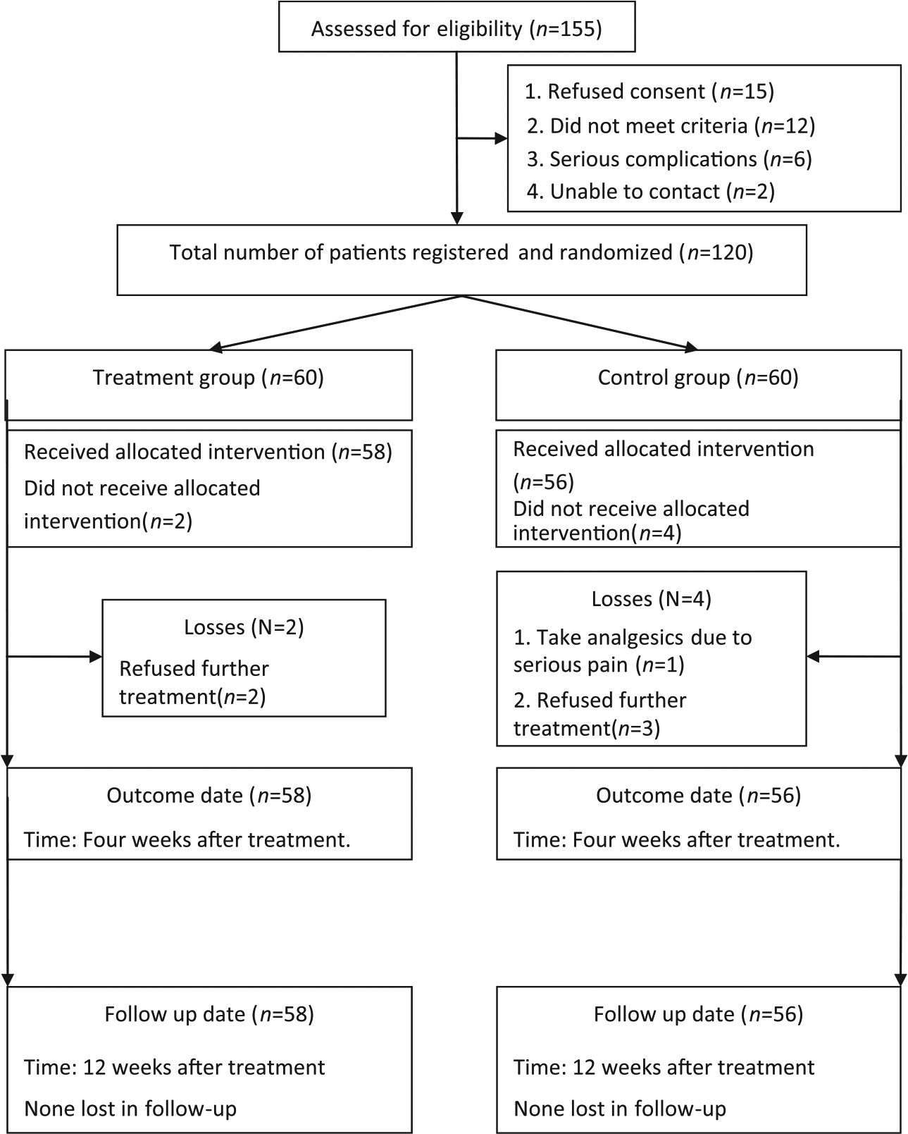
Of the 155 stroke patients admitted to the study, 120 patients met the inclusion criteria. They were invited to sign the informed consent before randomization (shown in Figure 1). Following that, a statistician who was not involved in the screening, treatment or assessment procedures generated random allocation sequence using the SPSS version 17.0 (SPSS Inc., Chicago, IL, USA). The range of random digit was 120. Participants with random number less than 60 were included in the treatment group and the others were placed in the control group. The random number and allocation message were placed in an envelope to ensure that the randomization was performed fairly by the physicians. In addition, the randomization information could only be opened when the participant had been registered and the physician, statistician and assessor were blinded to group assignment and intervention method of the modified wheelchair arm-support.
Overview of the study design.
The inclusion criteria were as follows:
Specific diagnosis of stroke. 16 Strokes including infarcts and haemorrhages were diagnosed by computed tomography or magnetic resonance imaging;
Duration of disease ranges from two weeks to six months;
Age ranges from 20 to 85 years;
Brunnstrom 17 scale ranges from I to II. Patients with shoulder pain caused by spasticity were excluded;
Healthy mental status and could be able to answer questions.
The exclusion criteria were as follows:
Patients with brain trauma, thalamic lesions or peripheral neuropathy;
Patients with a history of cervical spondylosis, periarthritis, fracture or trauma at the shoulder, osteoporosis or myocardial infarction which could cause shoulder pain;
Patients who developed cerebral oedema or coma following stroke as it was difficult to measurement or provide intervention for these patients;
Patients with serious infection or disorders of the heart, liver or kidney were excluded.
Intervention
The interventions included routine services and experimental treatments, which were administered by several physical therapists. They were trained for consistent physiotherapy before intervention and did not participate in the outcome measurements. Participants in the control group received all the routine services, which include the following: (1) maintaining normal limb position, in which the patient lies in supine position flat on the bed, and a pillow was placed under the shoulder to keep the arm stretched forward; followed by positioning the patient on the healthy side, a pillow was placed in front of the chest and the paralyzed arm was placed over the pillow with the assistance of the physiotherapist; furthermore, the patient lies on the disabled side, and the paralyzed arm was stretched in front of the body; the patient was instructed to sit on the bed, and a mini table was placed in front of the body and the paralyzed arm was placed on it; finally, the patient was instructed to stand or walk, and the paralyzed arm was supported by a shoulder strap. (2) Passive rehabilitation practices tailored to the individual’s needs. (3) Offering medical treatment according to the guidelines of Western and Chinese medicine.16,18 (4) Offering ordinary wheelchair with ordinary armrest for assistance. The rehabilitation training was offered six times per week during the four-week treatment period.
In addition to the routine services, the participants in the treatment group additionally received experimental equipment, which included the modified wheelchair arm-support, after they signed the informed consent. As shown in Supplementary Figure 2(a) and (b), the board was designed to support the arm, with a thick handle equipped at the end of the board which could be held by hand. Supplementary Figure 2(c) and (d) shows, when patient is sitting in the wheelchair, a physiotherapist placed the paralyzed upper limb on the modified wheelchair arm-support and assisted the patient to hold the thick handle. This position made the shoulder joint stretch forward, assisting the extension of the elbow joint, while maintaining the forearm in mid-position and fingers half extended. In addition, the arm-support could support the arm and prevent the arm from falling, which may protect the upper limb, as well as prevent shoulder dislocation. All the positions above may decrease the reflex stiffness of muscles and prevent shoulder pain following stroke. Participants may receive other treatment which should not affect their shoulder pain extending from one week before the start of the study to the follow-up periods.
Patients in the treatment and control groups separately received interventions at least 60 minutes a day, for six days a week in the period of four weeks. The patients were nursed, trained and evaluated at the hospital during the four weeks of the intervention process. Generally, the hospitalization period in the rehabilitation department was four weeks, after which the patients will be discharged. The modified wheelchair arm-support was rented to the patients for eight weeks supporting home rehabilitation. During the eight-week follow-up period, the patients received intervention for at least 30 minutes a day, three days a week, for eight weeks. The intervention, observation and evaluation period was 12 weeks. Furthermore, the patients submit ¥200 (approximately US$30) for renting the wheelchair arm-support, which would be returned to the patients when the wheelchair compensator was given back. The nursing staff, relatives or care providers received appropriate training at the hospital or at home to avoid shoulder trauma during transfer. When patients were discharged from the hospital, they still received basic rehabilitation by relatives or care assistants at home, including normal limb positioning, basic Western and Chinese medicine and simple limb movements, recommended by the physician. ‘The modified wheelchair arm-support’ use compliance was recorded by an assessor.
Outcome measures
The primary outcome was Visual Analogue Pain Scale or Numeric Pain Rating Scale, which is commonly used to measure subjective pain intensity level. The scale indicates from 0 (no pain) to 10 (the most intense pain level).19,20 The Visual Analogue Pain Scale was used for pain measurement if the patient was unable to communicate the pain experience. The Numeric Pain Rating Scale was used for pain measurement if the patient could communicate easily or when interviewed via telephone at follow-up. The secondary outcome measures include Upper Extremity Fugl-Meyer Assessment scale, Modified Barthel Index and Quality of Life Index. The Upper Extremity Fugl-Meyer Assessment scale (range = 0–66; 0 = no function, 66 = normal function) is a well-established stroke motor measurement, iteratively determining active movement at each joint of the upper extremity.21–23 The Upper Extremity Fugl-Meyer Assessment scale items are organized into scales that discern isolated movements at increasingly distal upper extremity regions. The Barthel Index score is a 10-item measure of activities of daily life. It has been proven that the Barthel Index has excellent inter-rater reliability for standard administration, following stroke. 24 The Modified Barthel Index (range = 0–100; 0 = no independence, 100 = normal independence), modified from Barthel Index by Shah et al., 25 is more sensitive to small changes in functional assessment than Barthel Index. The Quality of Life Index (range = 0–5; 0 = no quality, 5 = high quality) is a generic measure for the evaluation of quality of life that was initially used in patients with chronic diseases or pain. It has been reported that Quality of Life Index has good reliability and validity in measuring pain disorders. 26
All measurements were conducted by an independent assessor blinded to the participant’s group allocation. The Visual Analogue Pain Scale or Numeric Pain Rating Scale was measured based on patient interviews, while the Upper Extremity Fugl-Meyer Assessment scale, Modified Barthel Index and Quality of Life Index were measured using a questionnaire. All participants were assessed three times, the first was baseline measurement before intervention, and then at 4 and 12 weeks following intervention. The baseline and four-week measurements, including Visual Analogue Pain Scale or Numeric Pain Rating Scale, Upper Extremity Fugl-Meyer Assessment scale, Modified Barthel Index and Quality of Life Index, were evaluated at the hospital by the assessor. After four weeks, patients were discharged from hospital. The 12-week measurements, including Visual Analogue Pain Scale or Numeric Pain Rating Scale, Modified Barthel Index and Quality of Life Index, were conducted by the same assessor. At a certain time point, the assessor will perform telephone interview to collect physical examination results’ information and conduct assessments based on the description. The physical examination results’ information was obtained from the patient’s regular check-up at the local hospital. The Upper Extremity Fugl-Meyer Assessment scale data had been excluded from the final follow-up results due to potential assessment bias that may result from the Upper Extremity Fugl-Meyer Assessment scale measurement.
Statistical analysis
The analysis was conducted by an independent scientist and an independent statistician. SPSS version 17.0 (SPSS, Inc.) was applied for statistical analysis, and a P-value of 0.05 was considered to be statistically significant.
Descriptive statistics were calculated for gender, side of lesion, age (years) and months post stroke. Independent t-test was carried out on the comparison between the two groups if the measurement data conformed to a normal distribution. The Wilcoxon rank sum test was used if the above is not possible. The chi-square test or rank test was used for comparison of the count data between the two groups. The data conformed to normal distribution were expressed as mean ± standard deviation. If not, the data were expressed as median, interquartile range. All the patients’ information including data was protected by the statistician unless the information is required for scientific research.
The primary outcome Visual Analogue Pain Scale or Numeric Pain Rating Scale was used to calculate the sample size. A change of 2.5 was deemed clinically relevant. A sample size of 120 was sufficient to have a significance level of more than 95% and power of 80%, allowing for 15% attrition based on previous stroke rehabilitation studies. 11
Since there were six patients not completing the study, intention-to-treat analysis was performed to evaluate the significance of the experimental data.
Results
Figure 1 shows the flow diagram of eligible patients in the study. A total of 155 participants were admitted to the study in the beginning and 35 (22.6%) were excluded for several reasons (details are shown in Figure 1). Of these, 120 (77.4%) patients met the inclusion criteria and consented to receive intervention and follow-up assessment. Six were lost to follow-up for several reasons (details are shown in Figure 1). Eventually, 114 (95%) patients in the treatment group (n = 58) and the control group (n = 56) completed the study.
The comparisons and statistical analyses between groups were made at baseline, 4 weeks and 12 weeks. The baseline characteristics are shown in Table 1. The average age of the participants was 62.41 ± 12.26 years, and the average duration of stroke of all patients was 1.9 ± 1.3 months. There were no significant differences (P < 0.05) between the two groups in baseline characteristics, including gender, age, duration of stroke, hemiparetic side and baseline measurements.
Baseline characteristics of the participants.
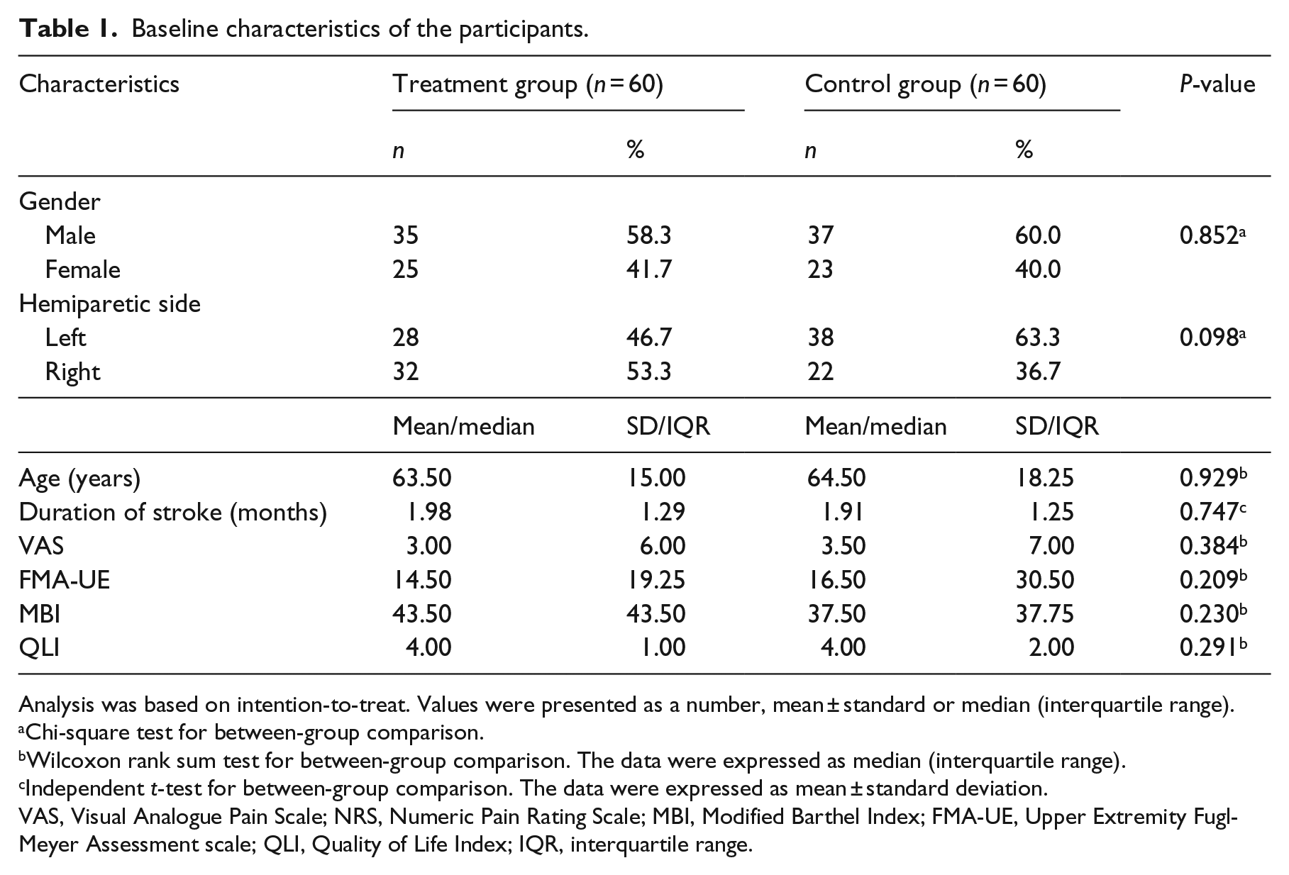
Analysis was based on intention-to-treat. Values were presented as a number, mean ± standard or median (interquartile range).
Chi-square test for between-group comparison.
Wilcoxon rank sum test for between-group comparison. The data were expressed as median (interquartile range).
Independent t-test for between-group comparison. The data were expressed as mean ± standard deviation.
VAS, Visual Analogue Pain Scale; NRS, Numeric Pain Rating Scale; MBI, Modified Barthel Index; FMA-UE, Upper Extremity Fugl-Meyer Assessment scale; QLI, Quality of Life Index; IQR, interquartile range.
The comparisons within groups for all measurements are shown in Table 2. It is noted that both groups showed improvements in shoulder pain, upper extremity movements, independence of daily living and quality of life during the four weeks of intervention and at the 12 weeks of follow-up, and the differences within groups were significant (P < 0.001).
Comparison of VAS/NR, FMA-UE, MBI and QLI within groups at 4 weeks and 12 weeks.
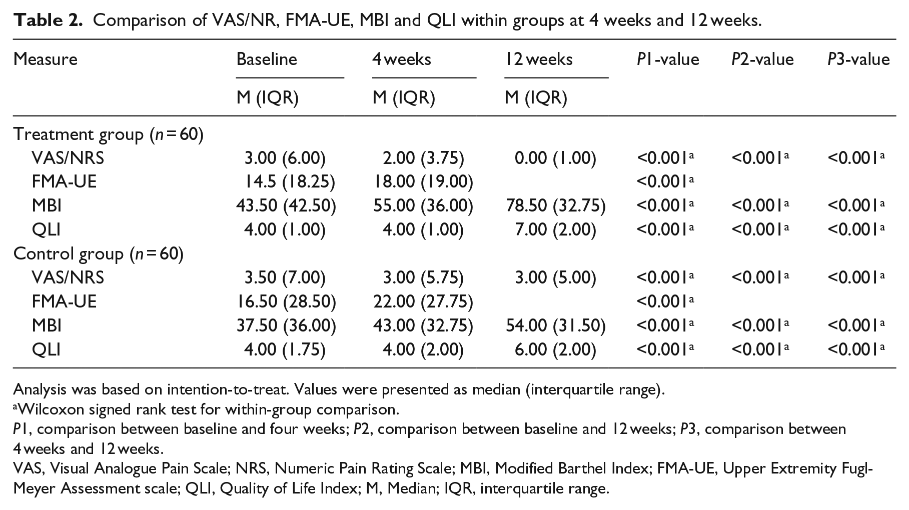
Analysis was based on intention-to-treat. Values were presented as median (interquartile range).
Wilcoxon signed rank test for within-group comparison.
P1, comparison between baseline and four weeks; P2, comparison between baseline and 12 weeks; P3, comparison between 4 weeks and 12 weeks.
VAS, Visual Analogue Pain Scale; NRS, Numeric Pain Rating Scale; MBI, Modified Barthel Index; FMA-UE, Upper Extremity Fugl-Meyer Assessment scale; QLI, Quality of Life Index; M, Median; IQR, interquartile range.
The comparisons of all measurements between the two groups are shown in Table 3. There were no significant differences in Visual Analogue Pain Scale or Numeric Pain Rating Scale between the two groups at baseline and four weeks (P > 0.05). However, the difference reached statistical significance at 12 weeks (P < 0.001). All subjects demonstrated an increase in Fugl-Meyer Assessment scale after four weeks of intervention, but the differences were not significant between the groups. There were no follow-up comparisons on the scale at 12 weeks because of assessment limits. The median of Modified Barthel Index reached significant difference between the two groups at 4-week and 12-week follow-up (P < 0.05). Similarly, the median of Quality of Life Index was significantly different between the groups at 4 weeks and 12 weeks (P < 0.05).
Comparison of VAS/NRS, FMA-UE, MBI and QLI between groups at 4 weeks and 12 weeks.
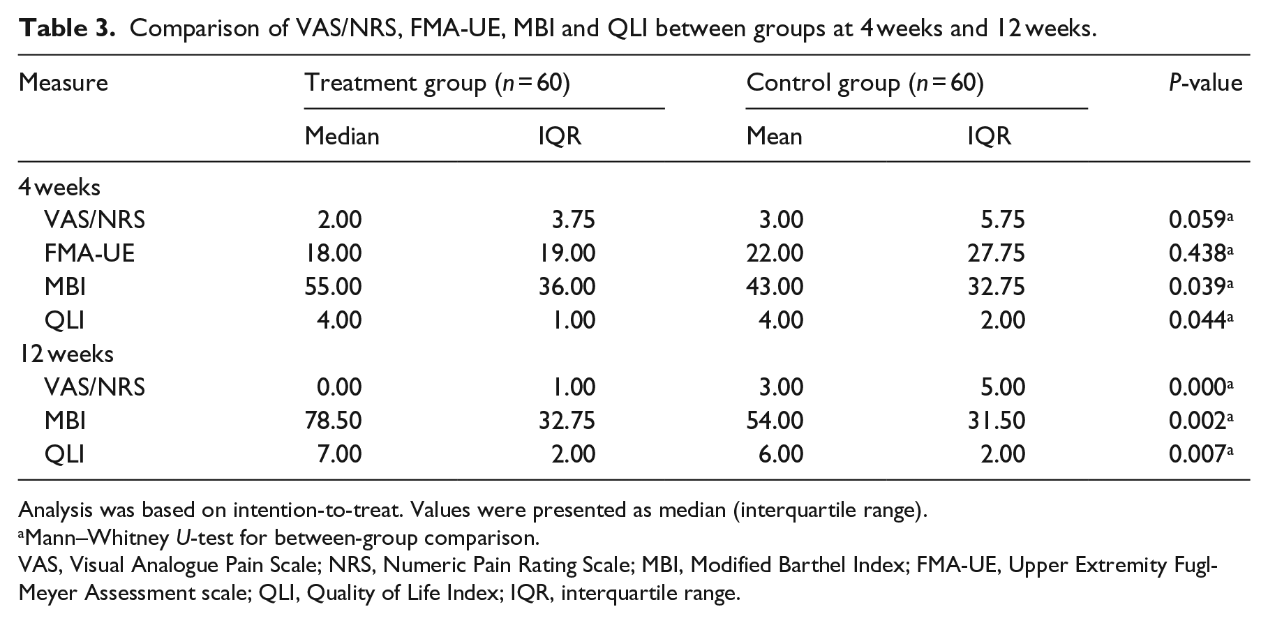
Analysis was based on intention-to-treat. Values were presented as median (interquartile range).
Mann–Whitney U-test for between-group comparison.
VAS, Visual Analogue Pain Scale; NRS, Numeric Pain Rating Scale; MBI, Modified Barthel Index; FMA-UE, Upper Extremity Fugl-Meyer Assessment scale; QLI, Quality of Life Index; IQR, interquartile range.
Table 4 demonstrates the comparisons of the changes in the measurements during the intervention period. The changes at four weeks were measured from baseline to four weeks, and the changes at 12 weeks were measured from baseline to 12 weeks. The results showed that after four weeks of intervention, there were no significant differences (P > 0.05) in the change of Visual Analogue Pain Scale or Numeric Pain Rating Scale and Quality of Life Index between the two groups. However, the changes of Upper Extremity Fugl-Meyer Assessment scale and Modified Barthel Index were all significantly different between the groups (P < 0.05). Interestingly, after 12 weeks of intervention, there were significant differences in the change of the Visual Analogue Pain Scale or Numeric Pain Rating Scale, Modified Barthel Index and Quality of Life Index between the two groups (P < 0.05).
Comparison of the VAS/NRS, FMA-UE, MBI and QLI improvement between groups at 4 weeks and 12 weeks.
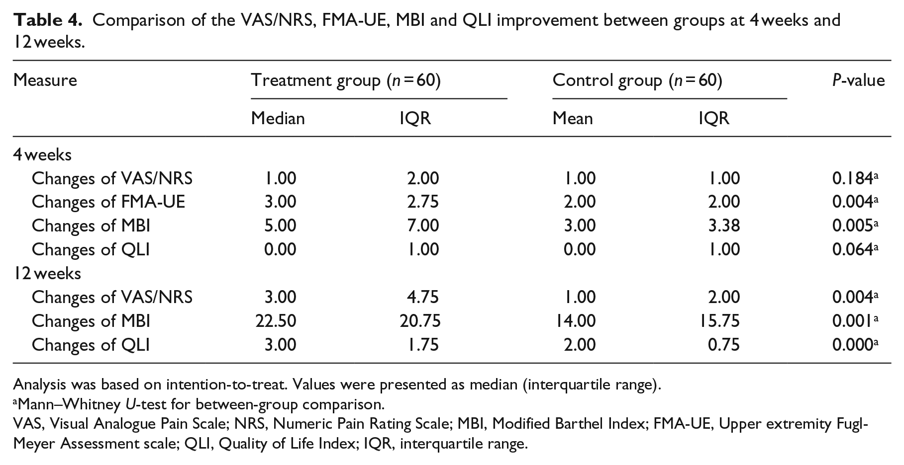
Analysis was based on intention-to-treat. Values were presented as median (interquartile range).
Mann–Whitney U-test for between-group comparison.
VAS, Visual Analogue Pain Scale; NRS, Numeric Pain Rating Scale; MBI, Modified Barthel Index; FMA-UE, Upper extremity Fugl-Meyer Assessment scale; QLI, Quality of Life Index; IQR, interquartile range.
Table 5 demonstrates that, before the treatment, 25 patients in the treatment group and 20 patients in the control group do not suffer shoulder pain. After 12 weeks, only one patient suffers from shoulder pain in the treatment group. However, in the control group, six patients appeared to suffer from shoulder pain. This shows a significant difference in the incidence of shoulder pain between the two groups (P = 0.034).
Incidence of hemiplegic shoulder pain between groups.
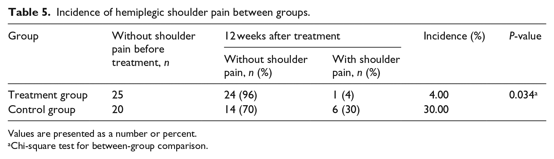
Values are presented as a number or percent.
Chi-square test for between-group comparison.
Discussion
The results showed a significant difference in the measurements of Visual Analogue Pain Scale or Numeric Pain Rating Scale and Quality of Life Index between groups appeared at 12 weeks rather than at 4 weeks, which indicates that the effectivity of the modified wheelchair arm-support increased upon longer period of use. The morbidity of hemiplegic shoulder pain in the treatment group was much lower than average, 1 which suggests that the application of modified wheelchair arm-support could prevent the occurrence of shoulder pain, following stroke. The reason may be that the modified wheelchair arm-support could prevent the upper limb from falling, which may contribute to soft tissue injury or spasticity and lead to shoulder pain.
Nadler and Pauls 27 concluded that majority of patients wearing orthosis showed modest improvement in shoulder pain, similar to the results of this study. Therefore, it could be inferred that an antigravity position of hemiplegic upper limb is effective to mitigate and prevent shoulder pain. However, the result failed to reveal the appropriate time of intervention. We have not conducted a stratification analysis of the time period as to whether earlier intervention is more effective and this needs further studies.
The goal of rehabilitation therapy for hemiplegic patients is to restore independence in limb movement and everyday activities. We selected the Upper Extremity Fugl-Meyer Assessment scale score to reflect improvements in upper limb activity and the Modified Barthel Index score to measure the independence to perform everyday activities. Both scores reflect effective upper limb rehabilitation using the modified wheelchair arm-support.
However, there is an incertitude regarding how the support device could improve mobility. There could be several factors that may influence this. One reason is that the device could keep the paralyzed upper limb in reflex inhibition pattern, which could prevent the development of inefficient movement and ensure that normal position is maintained in the paralyzed limb. 28 The normal position of paralyzed upper limb could contribute to functional recovery. Alternatively, the application of the modified wheelchair arm-support could prevent and relieve shoulder pain, which may encourage the patients to exercise proactively.
The rehabilitation in this study combined physical exercise with position correction. Therefore, we cannot conclude that the modified wheelchair arm-support could improve the limb and body activity function by itself, but it could be beneficial when combined with physical exercise in the recovery and rehabilitation progress.
We select Quality of Life Index to assess the life quality, which is the goal of rehabilitation. Sudlow and Warlow 29 concluded that hemiplegic shoulder pain is an independent determinant of subsequent quality of life. For every stroke patient, the aim of rehabilitation is not only to regain motor function of the body but also to help them return to the society. In this study, the decrease in shoulder pain improves upper limb function resulting in better daily performance using the modified wheelchair arm-support. In other words, using the modified wheelchair arm-support may contribute to quality-of-life improvements.
Previous studies rarely focused on the effect of wheelchair in preventing or reducing hemiplegic shoulder pain. The common method is to attach the forearm trough or placing a board to the wheelchair to prevent shoulder subluxation. 28 These methods have some limitations in maintaining a reflex inhibiting pattern, which could prevent the development of inefficient movement patterns and muscle spasm of the upper limb. 30
In our study, most functional limb posture could be obtained using the modified wheelchair arm-support, including arm in external rotation, wrist in neutral position, finger half extended and thumb abduction. Veneman et al. 31 designed a novel wheelchair posture support device to assure correct sitting posture for post-acute stroke patients and found it could reduce shoulder pain and the number of repositioning manoeuvres. The device was designed with trunk support and a table to support the hemiplegic upper limb, which has similar therapeutic effect as our compensator device. Both studies demonstrate that upper limb support proves to be beneficial for hemiplegic patients with shoulder pain.
We include patients with Brunnstrom scale range from I to II in our study, which means that we excluded hemiplegic patients with shoulder pain caused by spasticity. Therefore, the effects of the modified wheelchair arm-support on hemiplegic shoulder pain were suitable for patients with flaccid hemiplegia. The effects of the equipment on spastic hemiplegia need further exploration.
There are some limitations in this study. First, since the device was too large to conceal, the study has to be designed to be a single-blind trial, which could lead to some observational bias. Second, the rehabilitation practices and medical treatments were kept consistent as far as possible, but there are still some individual differences based on the patients’ needs, which may produce some interference to the results. Third, the follow-up assessment was conducted by an independent assessor via telephone to collect physical examination results’ information rather than face-to-face interview, which may result in measurement errors. Fourth, no stratified analysis of the time and complications was performed, so we cannot estimate the best intervention time point and which type of complications would benefit the most using the modified wheelchair arm-support. Fifth, shoulder pain was measured by two types of scales, which could lead to a measurement bias. Therefore, future studies should focus on testing the effect of modified wheelchair arm-support on different kinds of shoulder complications or different time periods after stroke. A multicentre, multilevel, long-term study is needed to confirm these results.
It is concluded that using the modified wheelchair arm-support could mitigate hemiplegic shoulder pain, as well as reduce the incidence of pain in stroke patients. It may also improve upper limb motor function, daily activities’ performance and quality of life compared to using an ordinary wheelchair. This could be an effective therapy for all patients with hemiplegic shoulder pain.
Clinical Messages
The application of modified wheelchair arm-support can mitigate hemiplegic shoulder pain as well as reduce the incidence of shoulder pain better than ordinary wheelchair for stroke patients.
The application of modified wheelchair arm-support could also improve the upper limb motor function, daily activities and quality of life for stroke patients.
Footnotes
Acknowledgements
The authors would like to thank all the professors and coworkers for all the help they have given. They would also like to thank Mr Hjalte Holm Andersen from Center for Sensory-Motor Interaction, Aalborg University, for his valuable comments on this article. Thanks also to Dong guan Kean Medical Instrument Co., Ltd for producing and providing the modified wheelchair arm-support used in this study. Ruihuan Pan and Mingchao Zhou contributed equally to this work.
Declaration of Conflicting Interests
The author(s) declared no potential conflicts of interest with respect to the research, authorship and/or publication of this article.
Funding
The author(s) disclosed receipt of the following financial support for the research, authorship and/or publication of this article: This study was supported by the Guangdong Science and Technology Department. (2012B091100487).
References
Supplementary Material
Please find the following supplemental material available below.
For Open Access articles published under a Creative Commons License, all supplemental material carries the same license as the article it is associated with.
For non-Open Access articles published, all supplemental material carries a non-exclusive license, and permission requests for re-use of supplemental material or any part of supplemental material shall be sent directly to the copyright owner as specified in the copyright notice associated with the article.