
Abstract
Non-steroidal anti-inflammatory drugs (NSAIDs) can cause histopathological changes in the kidney and liver of fish. Still, it is unclear whether exposure to treated municipal wastewater that contains NSAID residues causes similar effects. We therefore conducted a comprehensive, critical review on claimed histopathological changes in fish exposed either to NSAIDs or to treated municipal wastewater in the laboratory or downstream from treatment plants. A detailed scrutinization questioned the basis for several findings. Hepatocellular necrosis, hepatocellular vacuolation, and an increase of developing nephrons/basophilic clusters (DNs/BCs) were overlapping findings, but the lowest observed effect concentrations (LOEC) for the hepatic endpoints were well above concentrations frequently encountered in treated effluents. An increase of DNs/BCs were reported at lower NSAID concentrations, but with some concerns regarding reliability. Hence, there is no clear documentation that histopathological effects caused by NSAIDs are present in fish exposed to municipal effluents. Study design, including the species studied, exposure regimes, endpoints analyzed, and applied methodology varied widely between studies, all of which could make overlapping effects difficult to detect. In addition, limitations in both experimental design and reporting standards in fish histopathology studies prevent any firm conclusions. More comparable study designs in future studies would facilitate comparisons.
Introduction
Over the past decades, there has been a growing concern about pharmaceuticals in the environment. The most remarkable example is the near extinction of several vulture species in India and Pakistan caused by exposure to the non-steroidal anti-inflammatory drug (NSAID) diclofenac. When vultures were scavenging on carcasses of livestock previously treated with diclofenac, their kidney function rapidly deteriorated, leading to severe visceral gout, and ultimately death.28,30 The resulting lack of vultures also led to vast amounts of animal carcasses not being eaten, thereby becoming a public health risk as vectors for infectious diseases. 31 To protect the vultures, diclofenac was banned for veterinary use in India and Pakistan in 2006, however, diclofenac could still be detected in carcasses of ungulates years after the ban, which points to an illegal use. 9
As of today, diclofenac is still a very commonly used drug in humans to treat inflammation, pain, and fever. A well-recognized side effect from NSAIDs, including diclofenac, is altered blood flow to the kidneys, which in certain patients can lead to renal failure. 33 This indicates that kidney impairment due to diclofenac may be found in a wide range of vertebrates. Indeed, laboratory studies in fish exposed to diclofenac reports histopathological effects, including various changes in the kidneys, at low µg/L.5,27 A recent mesocosm study also report increased mortality in three-spined sticklebacks at 3.82 µg/L. 18 In Europe, levels of diclofenac around or exceeding 1 µg/L can often be found in effluents from municipal wastewater treatment plants (WWTPs),6,13,24,38 raising concern about possible effects in the aquatic environment as well. Such observations led to diclofenac being added to the watch list of priority substances within the Water Framework Directive in the European Union 11 with the objective of improving knowledge on environmental exposure levels. In 2020, an expert group was assigned by the department Directorate-General Environment (DG ENV) at the European Commission (EC) to propose a draft for an Environmental Quality Standard for diclofenac derived from the available literature. During this exercise, the reliability of some of the diclofenac studies was questioned. 10 The draft was then evaluated by the Scientific Committee on Health, Environmental, and Emerging Risks (SCHEER) on behalf of DG ENV. An Annual Average Quality Standard (AA-QS) of 40 ng/L in fresh water and 4 ng/L in sea water was suggested by the expert group and supported by SCHEER, but no decision has yet been made by the European Council or the European Parliament. 34
Studies reporting effects in fish at low or even sub µg/L of NSAIDs together with observed or predicted concentrations in waterways suggest that effects of diclofenac are possible, and even plausible. Still, there seems to be a lack of studies directly addressing if effects typically caused by diclofenac exposure, can be found in fish exposed to treated municipal wastewater. Such municipal wastewater contains mixtures of chemicals, including several NSAIDs. Given the similar mode of action of different NSAIDs, one can expect that they would act additively in fish, although direct empirical evidence appears to be scarce. There are also data suggesting that bioconcentration of NSAIDs may be higher in fish exposed to complex wastewaters than single compounds, 8 potentially leading to underestimation of risks if only effect data from single-compound exposures are used.
Sublethal effects in fish, such as histopathological alterations, can add evidence of causality but the specificity and sensitivity of such effects are important to consider. Here we aimed to conclude if specific histopathological alterations in fish identified after exposure to NSAIDs also are reported in fish exposed to treated municipal wastewaters. This was addressed by a comprehensive review where histopathological effects in fish after NSAID or wastewater exposure were compared and evaluated based on both relevance and reliability. The chosen approach is most similar to a critical review, as defined by Grant and Booth, 15 but has some of the characteristics of a systematic review, such as a clearly defined research question, a standardized and fully transparent search and clear inclusion/exclusion criteria. As histopathology and especially fish histopathology is a complex scientific field where misdiagnoses and misinterpretations are common, 40 a secondary aim was to evaluate the technical (section preparation and imaging) and diagnostic quality for each reported histopathological effect.
Materials and Methods
Rationale for Scope
The current review focuses on kidney and liver only, two commonly analyzed organs where histopathological effects of NSAIDs at low concentrations have been reported.17,27 Gills are also frequently analyzed with reported effects after NSAID exposure.5,23,35 However, from our own experience and also stressed by Wolf et al, 40 out of the commonly investigated organs in fish histopathology, gills are the most technically challenging during preparation and they are also prone to artifacts to a higher degree than liver and kidney. For this reason, we did not evaluate reported effects on gills here and limit our conclusions to liver and kidney.
There is a vast literature on effects of gonad histology linked to exposure to treated municipal wastewater. Although gonads in general are relatively easy to prepare for histological analyses and are more resilient to artifacts than gills, some of the observed effects on gonads are linked to endocrine disrupting chemicals in the wastewater.2,19 Also, we are not aware of any extensive support for specific histopathological effects on fish gonads by NSAIDs at low exposure concentrations. This motivated our choice to not include gonad histology in the present review.
Database Search on Treated Municipal Wastewater Exposure
The search was performed by JN using the database Web of Science™. 7 A research question was formulated: What histopathological findings are reported in the kidney or liver of fish exposed to treated municipal wastewaters? The search settings were “Database: Web of Science Core Collection” and “Editions: All” and the publication date was set to January 1, 1970, to March 31, 2025. Full search strings can be found in the supplementary material (Table S1). To ensure an accurate search, previously downloaded articles regarding treated municipal wastewater exposure and histology in fish were sought for among the final hits, and 11/12 were found which was considered acceptable. The remaining article not found, was added to the final search hits (supplementary material, Table S3).
Database Search on NSAID Exposure
The search for NSAID articles was also performed by JN using the database Web of Science™, 7 with the corresponding research question: What histopathological findings are reported in the kidney or liver of fish exposed to NSAIDs? The search strings were similar to the wastewater search but replacing the first string with appropriate terms for NSAIDs as well as approximately fifty different NSAIDs (supplementary material, Table S2, Figure S1). This string was divided into three sub-strings due to space limitations in the search engine and for clarification. The same settings as for the wastewater search were used. Previously downloaded relevant articles were sought for among the final hits and 12/13 were found. The missing article was added to the final search hits (supplementary material, Table S4).
Inclusion Criteria and Screening Workflow
Only original articles in English were included in the screening. Prior to the screening of the search hits, a set of inclusion criteria was formulated. Only articles quantitatively or semiquantitatively analyzing liver or kidney histopathology in fish exposed to municipal WWTP effluent or exposed to a single NSAID or a mixture of NSAIDs and compared with a suitable control were included in the review (Table 1).
Article inclusion criteria.
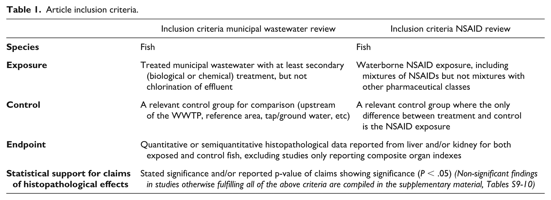
Effluents from non-municipal sources were considered irrelevant since they would not normally contain NSAIDs. Untreated or only primary treated wastewaters could certainly contain elevated NSAID levels but were also considered out of scope here. Chlorinated effluents were excluded due to the special nature of such effluents, including risks for generating toxic disinfection byproducts and/or residual chlorine. Furthermore, NSAID exposure had to be waterborne to facilitate a comparison between the exposure levels with levels found in wastewaters or surface waters. Only articles reporting statistically significant differences in histological effects between exposed fish and fish used as controls were included in the detailed evaluation. This was mainly because of the many additional challenges in interpreting reported negative results in non-standardized tests. These also include unspecific claims, such as “no histological alterations,” leaving the reader unaware of which alterations that actually were scored. For completeness, non-significant findings in studies otherwise fulfilling all of the above criteria were compiled in the supplementary material, Tables S9 and S10. Another cause for exclusion was when the histological diagnoses were added together, generating an organ index without reporting individual effects. This is problematic for several reasons, including that it does now allow exact identification of what was affected, which makes comparisons between studies practically impossible. While detailed information on both methods used for obtaining measurements and on approaches to minimizing bias (eg, randomized sampling and blinded evaluation), are important for judging validity of quantitative or semiquantitative data, it was not used as formal inclusion criteria.
The screening was performed by JN and started with the article title and if it was clear that the article was out of scope, it was excluded with no further evaluation. Otherwise, the abstract was evaluated and articles not providing evidence for exclusion continued to full-text evaluation. After full-text evaluation, if an article was to be excluded, at least one of the reasons for exclusion was recorded. DGJL performed an additional evaluation in case of any ambiguities with regards to inclusion/exclusion. Articles that remained after the full-text evaluation were included in the review. See supplementary material (Tables S3-S8) for full details, including exclusion reasons after the full-text evaluation.
Review of Included Articles
General information regarding study design and histological analysis was compiled for each article in tables. A general comment was also added when relevant, which illustrated identified ambiguities within each paper. All claimed histological diagnoses in liver and kidney were also compiled in tables.
A comparison of histological findings in fish exposed to wastewaters or NSAIDs was made to identify, and highlight overlaps and potential lack thereof. Findings with representative photomicrographs included, were evaluated by a fish pathologist (JN) based on criteria modified from Wolf and Maack, 41 created for scoring credibility of histopathology data. “Figure image quality” was defined as an adequate technical quality including image resolution and magnification of the provided figure, while “histologic specimen quality” was defined as adequate preservation and processing of the tissue. Both criteria were adopted but instead of a numeric combined score as in Wolf and Maack, 41 only yes/no or N/A (= not assessable) was used. If the image quality of the histological figure is not adequate, the quality of the histological specimen is often impossible to assess and thus the need for N/A. Diagnostic accuracy was discussed on a case per case basis as we found it too complex to present in a tabular way.
We then assigned a “weight of evidence” scored as low, moderate, or high, to each claim (each histological endpoint in each article). This was based on an overall assessment of experimental design including clarity of methodology, relevance, and number of control groups, coherence in results between multiple control groups, potential confounders between groups (eg, sex, maturation, and size differences), observed clear dose-responses, the level of statistical replication, in particular with regards to aquaria replication, appropriate statistical handling of data, the level of statistical significance, and if the diagnosis was supported or not in provided histological images. Although there will inevitably remain some level of subjectivity in the determination of “weight of evidence,” to achieve the score “high,” the article must provide histological figures of adequate technical and histological quality. Given the low number of articles in the review with even fewer including photomicrographs, and the evaluator’s (JN) familiarity with the articles in the NSAID review, blinding the photomicrographs before the evaluation would not have increased the objectivity and hence, the assessment was performed with the evaluator aware of the origin of the histological images. Finally, each overlapping diagnosis was then evaluated, including a discussion of relevance and reliability.
Results and Discussion
Database Search on Treated Municipal Wastewater and NSAID Exposure
A total of 866 articles were generated in the Web of Science search for histological effects from municipal effluent exposure. Adding the previously downloaded article not found in the search resulted in 867 articles. Since the search was wide to retrieve as many relevant articles as possible, many were clearly out of scope. After the title and abstract screening, only 68 articles continued to full-text evaluation where only seven articles passed the criteria to be included in the review. The majority of the 68 articles were excluded due to factors, such as effluent arising from non-municipal sources (eg, metallurgical effluent) or the wastewater was untreated, only primary treated or chlorinated. There were also papers where the control was not suitable, that is, other pollution sources than treated municipal wastewater differed between exposure and control sites, or in some cases, controls were missing and hence, made it impossible to assign any histological outcomes to the wastewater exposure. For additional information, see supplementary material (Tables S5 and S7). No study passed all inclusion criteria while simultaneously claiming no statistical significance for any of the histological effect investigated.
As for the NSAID exposure articles, 177 articles were generated in the Web of Science search producing a total of 178 when the previously downloaded article not found in the search was added. Thirty-six articles were still included after the title and abstract screening but only seven of them remained after full-text evaluation. Reasons for exclusion were similar to the evaluation on municipal effluent, where many of the search hits were addressing a completely different subject. Regarding the articles that did expose fish to NSAIDs, the majority of them lacked a quantitative or semiquantitative histological analysis, which is necessary to statistically evaluate the results. Some articles were also excluded because only organ indexes were reported. For additional information, see supplementary material (Tables S6 and S8). One article fulfilled all criteria except claiming any statistical significance for histological endpoints evaluated in liver and kidney. 23
The applied inclusion criteria largely represent a set of minimum requirements to facilitate an evaluation of claimed findings. The low number of studies fulfilling these criteria could reflect either the lack of studies in this area, or a sign of suboptimal experimental design in existing studies. The latter is problematic in many ways, ranging from the unnecessary use of research animals to the risk of misdirecting policies and decisions based on non-robust data.
Review of Treated Municipal Wastewater and NSAID Studies
Seven articles were included in the municipal wastewater exposure review and seven in the NSAID exposure review. The experimental setup on wastewaters differed widely between studies. None of them studied the same fish species and the studies were conducted on three different continents. Most studies investigated free-living wild fish in rivers downstream from WWTPs, but fish were also held in cages in rivers downstream from WWTPs or exposed to treated municipal wastewater in aquaria. The controls consisted mainly of river water upstream from the WWTPs, but also from nearby lakes or ponds or tap water. The exposure duration lasted from three weeks to up to close to a year. Exposure duration was obviously undetermined for all studies that analyzed free-living wild fish. Choosing to study free-living wild fish when it comes to evaluating effects of WWTP effluent exposure can come with some limitations as many uncontrolled variables may affect the outcome. Water temperature, food availability, fish density, or other properties may differ between sites, while not being attributed to the wastewater exposure as such. Hence, although field studies in some sense reflect actual exposure scenario better than laboratory studies, it can be difficult to link effects observed in fish to the wastewater exposure specifically. It is also impossible to know the exact level or timing of exposure since the fish in most cases can migrate closer to or further from where the effluent enters the study area. The characteristics of the control conditions are equally important. Comparing caged fish in a river downstream from a WWTP with fish in a tank receiving tap water can have its disadvantages since tap water and river water (without the wastewater) may also differ in their effects on fish. Exposing control fish in cages upstream from the WWTP would be more suitable. More details on the respective study design can be found in Table 2.
Summary of study design in the wastewater articles.
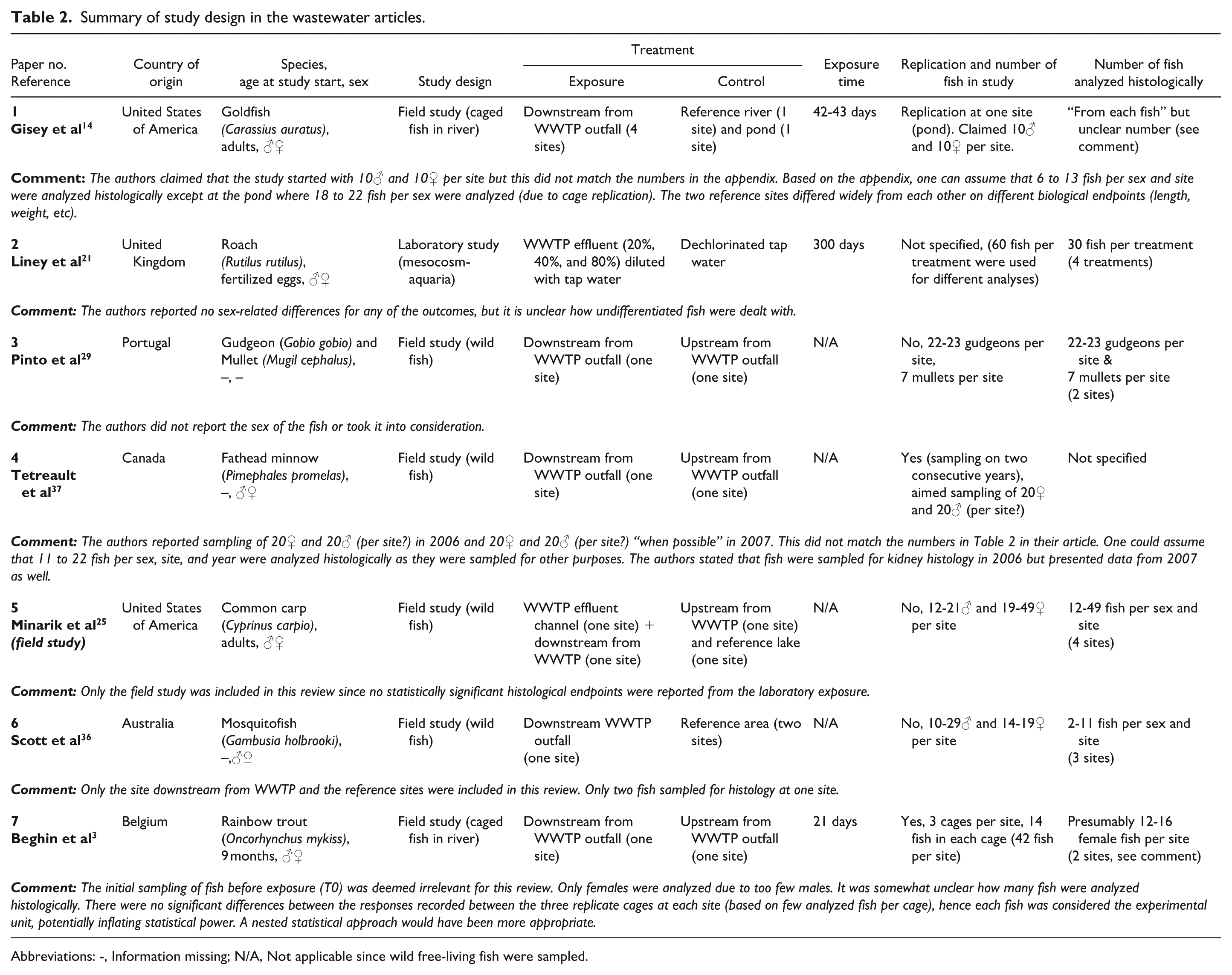
Abbreviations: -, Information missing; N/A, Not applicable since wild free-living fish were sampled.
All studies included in the NSAID review were conducted in Europe. The most investigated drug was diclofenac (5/7 studies). All included studies measured the actual exposure levels, which increases their overall reliability, and many argue this should be a requirement in these types of studies. 16 The exposure concentrations were in the µg or sub-µg/L range, except in one study where only mg/L-concentrations were evaluated. The latter is not considered relevant for any exposure scenario, with the possible exception of direct discharges from manufacturing. 20 Salmonid species were used in three out of seven studies and all studies lasted for 21 to 28 days. Studies on pharmacokinetics of NSAIDs in fish are sparse but suggest this is probably sufficient to allow reasonably steady states of internal exposure concentrations to be achieved. 6 On the contrary, one cannot exclude that certain histological changes could take longer to manifest, particularly at lower concentrations. Additional details on the respective study designs are presented in Table 3.
Summary of study design in the NSAID articles.
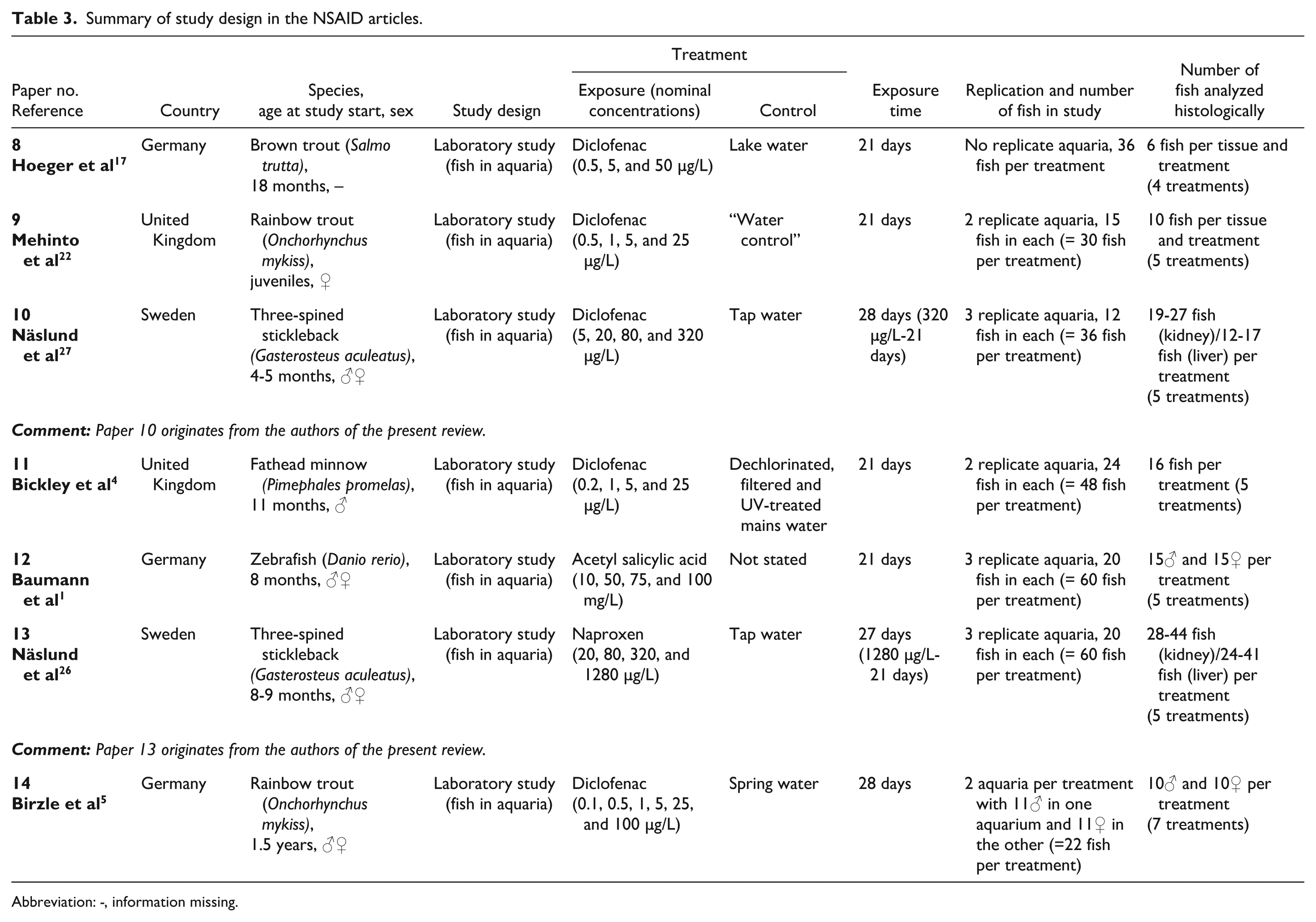
Abbreviation: -, information missing.
Regarding the histological analysis in both reviews, most of the articles (11/14) did not report if the scoring was done with the evaluator unaware of the treatment (Tables 4 and 5). Due to the subjective nature of histological scoring where a semiquantitative approach is common, it is important to minimize observer bias by masking the slides before the final analysis. Blinded scoring was, however, not used as an inclusion criterion since it was suspected to exclude too many papers, which unfortunately could be confirmed afterwards with only 3 of 14 claiming a blinded evaluation. Some of the articles reported which histological diagnoses they would analyze, and this varied between one diagnosis to more than ten different diagnoses. Others applied a more general but unspecific approach and stated they would analyze “histological alterations.” This could be problematic since lesion severity, and the experience level of the examiner will affect the likelihood of a finding to be detected. In addition, it is not always the case of identifying an entity with increased magnitude but also detecting what is missing. Therefore, a histological investigation with “no findings” is not always equal to no effect. As clearly demonstrated in the review by Wolf, 39 regarding morphologic findings involving fish exposures to diclofenac, out of 12 papers with a total of 62 claimed histological findings (electron microscopy analysis excluded), less than 15% were judged as credible or highly credible. Approximately, 65% of the findings were judged as having no or dubious credibility. Although that study was funded by a company that markets products with diclofenac and by a single author, we judge the reliability of the credibility assessment as high. This is not only due to Wolf’s recognized experience in fish histopathology, but also from our own previous study independently raising very similar critique about the histological assessment in some of the reviewed papers. 27 We strongly recommend describing which diagnoses have been evaluated (eg, necrosis, vacuolation, and inflammation) and not only “histological alterations.” This will make the reader aware if a specific finding has been evaluated or not. Such standardization will also decrease subjectivity since the same diagnoses are evaluated in all slides.
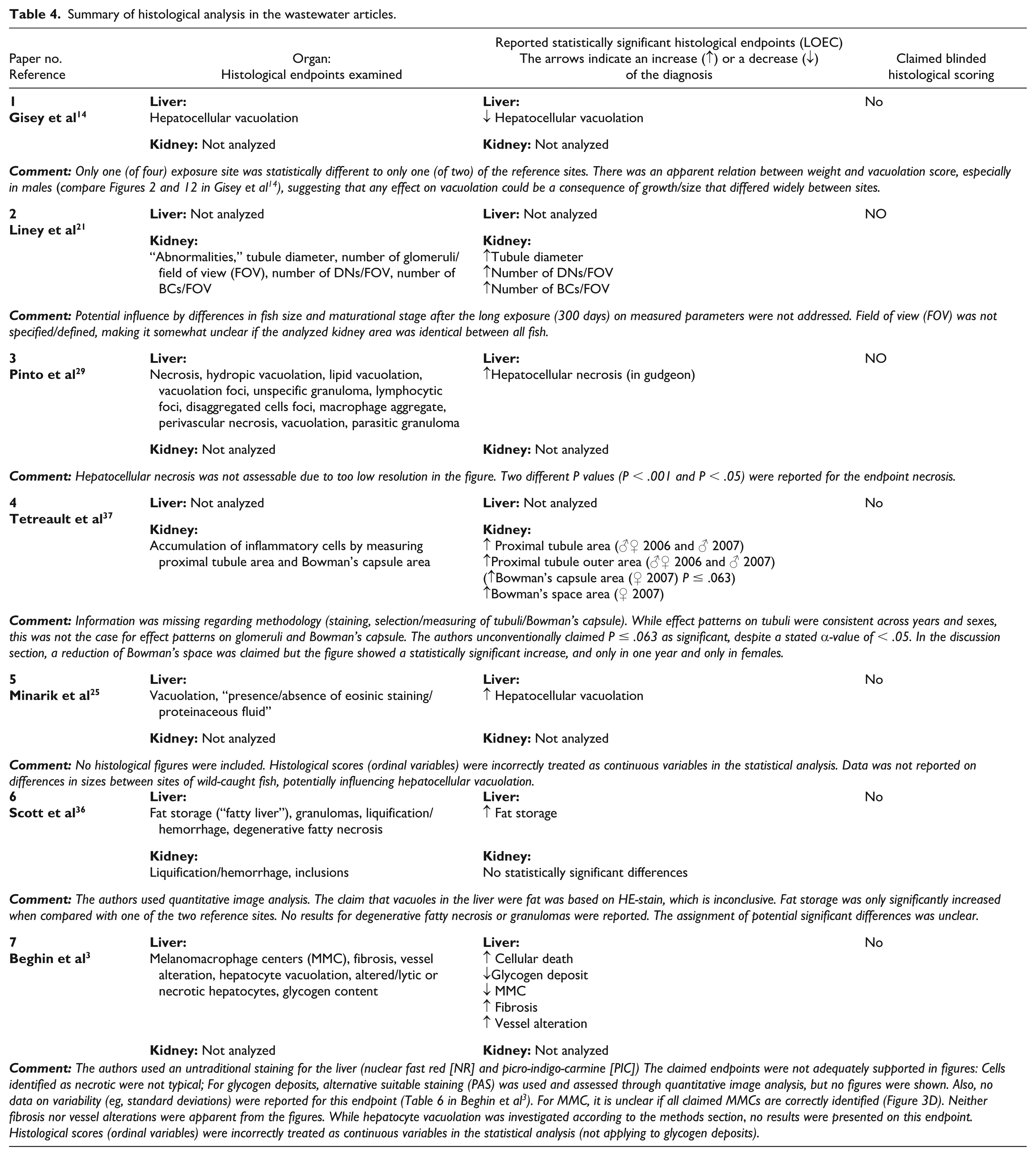
Summary of histological analysis in the wastewater articles.
Summary of the histological analysis in the NSAID articles.
Some of the histological findings reported from the articles were consistent within and between the exposure types but contradictory diagnoses were also identified. In 2/14 articles, no histological figures were provided. Even though it is not mandatory to include histological figures in papers analyzing histology, we strongly encourage it. This will facilitate the possibility for the reader to not only evaluate the histological quality of the samples but also allows for an external evaluation of the diagnosis. While single images are not sufficient to reliably confirm a diagnosis, they play an important role in supporting (or contradicting) claimed diagnoses. Since article space is limited, there is always the possibility to include large amounts of data including histological figures as supplementary material. The reported histopathological diagnoses will be reviewed further in the next section.
Evaluation of Reported Histological Changes
All diagnoses from Tables 4 and 5 are divided by organ and exposure type, with similar diagnoses grouped together (Table 6). The majority of the included histological images were of both adequate image and histological specimen quality. However, the overall weight of evidence for respective histological claim was considered low in 15/33 cases (Table 6). Three histological diagnoses showed some overlap between wastewater and NSAID-exposed fish but there were also diagnoses with contradictory results. Hepatocellular necrosis, hepatocellular vacuolation and developing nephrons/basophilic clusters (DNs/BCs) were reported in both exposure types. The overlaps, however, should be viewed considering the limitations pointed out in Tables 4 and 5 where both reliability and relevance are questionable in several of the studies. This is further evaluated below the respective diagnosis. To illustrate overlapping histological findings as well as renal hematopoietic hyperplasia, we include photomicrographs from our own collections of slides from three-spined stickleback. Reports of no significant effects are listed in Tables S9 and S10.
Overview of the claimed significant histological changes from studies of fish exposed to treated municipal wastewater or NSAIDs alone.
Related diagnoses are grouped. Text in combined boldface and italics further indicate similar effects reported both in fish exposed to wastewater and to NSAIDs.
This study originates from the authors of the present review.
Abbreviations: ASA, acetyl salicylic acid; DCF, diclofenac; NPX, naproxen; N/A, not assessable due to inadequate quality of histological figure; -, no histological image representing endpoint included in article.
Hepatocellular Necrosis
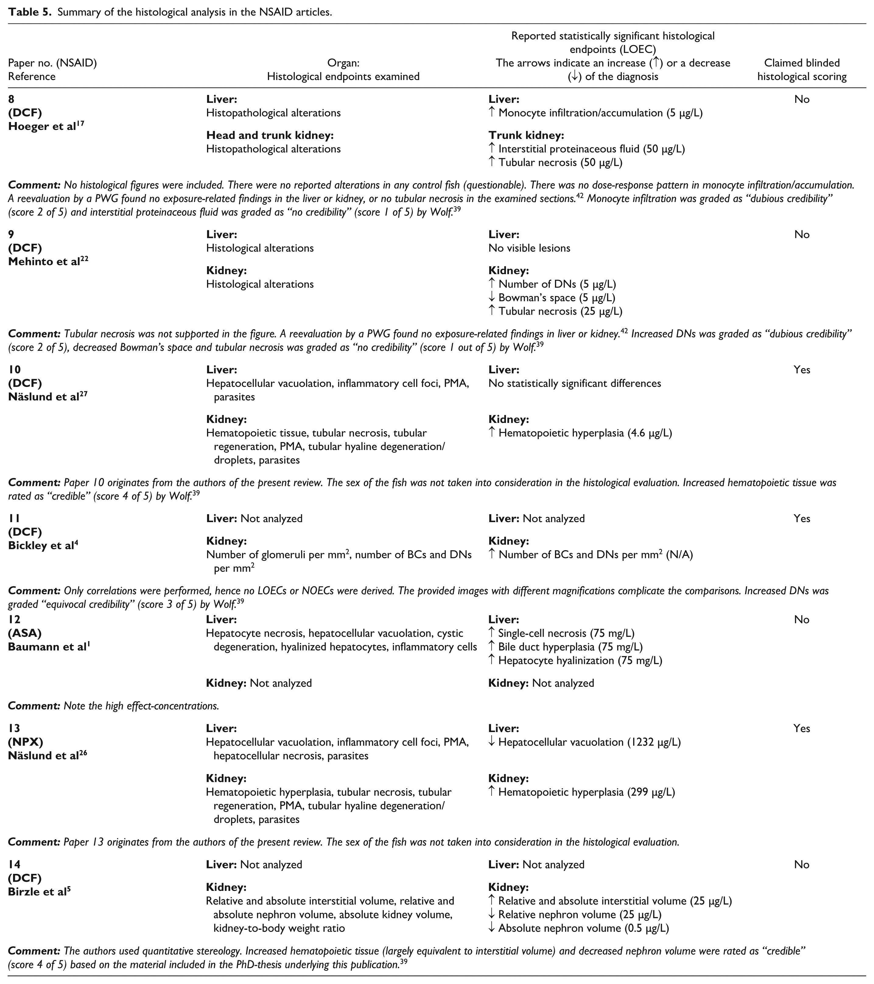
Hepatocellular necrosis (Figure 1A and B) in fish can be caused by a wide range of etiologies, both infectious and non-infectious but it is also frequently overdiagnosed in the scientific literature. 40 Pinto et al 29 and Beghin et al 3 both reported hepatocellular necrosis to be increased after exposure to treated municipal wastewater and Baumann et al 1 after NSAID exposure (Table 6). None of the two wastewater studies provide histological images that conclusively support their claims. In the study of Beghin et al, 3 the liver is stained with an untraditional stain, nuclear fast red and picro-indigo-carmine, why we prefer to be a bit cautious in our interpretation. There are some signs that could point to cellular alterations, such as focal loss of cell nuclei and occasional more dark-stained hepatocytes. However, in our view, they are not typical necrotic cells and artifacts cannot be excluded. The image intended to illustrate necrosis in the study by Pinto et al 29 was unfortunately not of sufficient quality to support the proposed finding. In addition, Pinto et al 29 did not take the sex of the wild-caught fish into consideration. The hepatosomatic index was also significantly larger in downstream fish, possibly related to different sex ratios and/or exposure to estrogenic substances. This bears relevance as hepatocellular necrosis of reproductively active female fish has been described previously by Wolf and Wheeler. 43 This is only one of the reasons why sex of the fish should be accounted for in histopathological investigations. Beghin et al 3 analyzed females only but applied a statistical approach with exaggerated power. Baumann et al 1 provided adequate images of high quality supporting the diagnosis and supported even further by an evident dose-response. However, the LOEC/NOEC (lowest observed effect concentration/no observed effect concentration) were 75,000/50,000 µg/L (acetyl salicylic acid), which is well above concentrations found in treated municipal wastewaters. In addition, in our own studies, only one case of liver necrosis was detected in close to 250 sticklebacks exposed to two different NSAIDs. The sole case of necrosis detected was most likely caused by a previous parasite migration.26,27 Taken together, the support that treated municipal wastewater can cause hepatocellular necrosis and that this finding is caused by NSAIDs is low.
Hepatocellular necrosis in the three-spined stickleback (Gasterosteus aculeatus). (A) Centrally, there is a large necrotic area (Ne, dashed line) with disintegrated cells with pale eosinophilic cytoplasm, karyolysis, and nuclear pyknosis. There is also a generalized hepatocellular vacuolation of lipid type (clear round cytoplasmatic vacuoles) across the entire section. A previous parasite migration was the suspected etiology of the necrosis. H&E, original objective 20X. (B) Higher magnification of the necrotic area (Ne) from the liver section in A. H&E, original objective 40X.
Hepatocellular Vacuolation
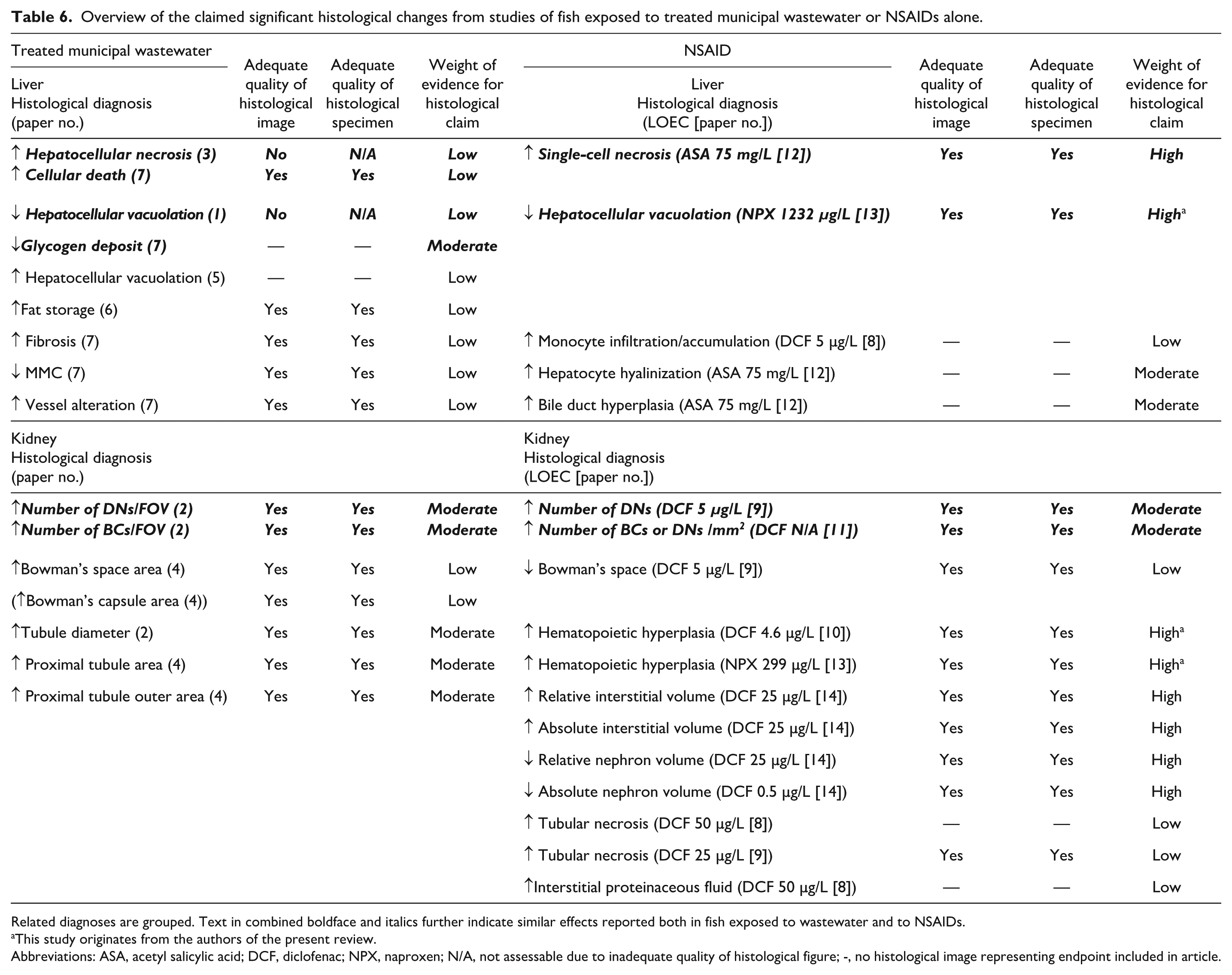
Hepatocellular vacuolation is a commonly scored endpoint in fish histology but evaluation demands caution due to high variability of the “normal” level. Differences in the level of vacuolation are, for example, due to species, sex, and nutritional and reproductive status. The appearance of the vacuoles can provide a hint on their origin (glycogen or lipid), but special stains are required for correct identification. Figure 2A to D demonstrates different levels and types of hepatocellular vacuolation in the three-spined stickleback. Two wastewater studies and one NSAID study identified a decrease in hepatocellular vacuolation or a related diagnosis (glycogen storage) (Table 6). Giesy et al 14 studied caged fish upstream and downstream from WWTPs and reported a statistically significant decrease only at one out of four exposure sites and only when compared with one of the two reference sites. However, the observed difference was similar for both females and males, but there was an apparent relation between weight and vacuolation score, which suggest that vacuolation could be a consequence of growth/size which differed widely between sites. In addition, the images provided were not of sufficient quality to support the diagnosis. Beghin et al 3 studied caged fish upstream and downstream from one WWTP. They claimed they evaluated and scored hepatocyte vacuolization, but no data or conclusions were presented. They reported decreased glycogen, but no supportive images were included. However, they used periodic acid Schiff (PAS) staining to identify glycogen and quantitative image analysis to evaluate the amount in the liver, which is considered a good approach. While claiming a significant difference between the two sites, results were presented without providing any measure of variance.
Liver from three-spined stickleback (Gasterosteus aculeatus) with different degrees of vacuolation. (A) Reproductively active female with minimal vacuolation, and hypertrophied, basophilic hepatocytes. (B) Reproductively active male with a higher degree of hepatocellular vacuolation and a more eosinophilic cytoplasm compared to the female in A. (C) Reproductively inactive male with extensive hepatocellular vacuolation with clear round vacuoles in the cytoplasm suggestive of a lipid origin. The nucleus is commonly located more peripherally, but this is not always easily discerned. (D) Reproductively inactive male with extensive hepatocellular vacuolation with more irregular shapes of the vacuoles and occasionally a more centrally located nucleus suggestive of glycogen type. H&E, original objective 40X (A-D).
Näslund et al 26 (own study) identified a decrease in hepatocellular vacuolation after exposure to naproxen. The study design included three replicate aquaria with both females and males, but the sex of the fish was not accounted for in the statistical comparisons. However, this has been investigated in retrospect, and including sex in the statistical analyses did not affect the previously reported conclusions (unpublished data). Critically, the LOEC of naproxen was 1232 µg/L, which is much higher than levels reported in treated municipal wastewater.
In addition, even though Memmert et al 23 did not report any significant histological changes in trout liver after chronic exposure to diclofenac and hence, are not included in this review, a pathology working group (PWG) reanalyzed the histological slides and identified decreased glycogen as a novel finding. 42 No histological images were included in the original article, or in the PWG article. The LOEC was 1000 µg/L, which is in line with the findings for naproxen by Näslund et al. 26 In conclusion, there is some evidence that wastewater exposure leads to decreased hepatocellular vacuolization/glycogen storage, noting that both Minarik et al 25 and Scott et al 36 report the opposite. The latter studies investigated wild fish which complicates comparisons. Decreased hepatocellular vacuolation after NSAID exposure is a reliable finding, but it has only been demonstrated at around 1 mg/L or higher. Hence, the relative insensitivity makes it implausible that NSAIDs in treated municipal wastewaters could cause such effects. As mentioned earlier, several factors such as species, sex, and reproductive and nutritional status can all affect hepatocellular vacuolation. A preferred strategy is therefore to account for such factors either experimentally or in the statistical analysis.
Developing Nephrons/Basophilic Clusters
Renal tubuli can regenerate after injury but the fish kidney has also the ability to develop new nephrons. DNs arise from BCs and the presence of both BCs and DNs in histological sections (Figure 3A and B), can be a normal finding, especially in growing animals. 12 However, Reimschuessel et al 32 demonstrated an increase of DNs/BCs after renal injury. Increasing numbers of DNs/BCs were reported in roach exposed for 300 days to treated wastewater 21 and in rainbow trout 2 and fathead minnow 4 exposed to diclofenac. The image quality and the quality of the histological specimens were considered adequate for all three articles and DNs/BCs could be identified in the provided figures.
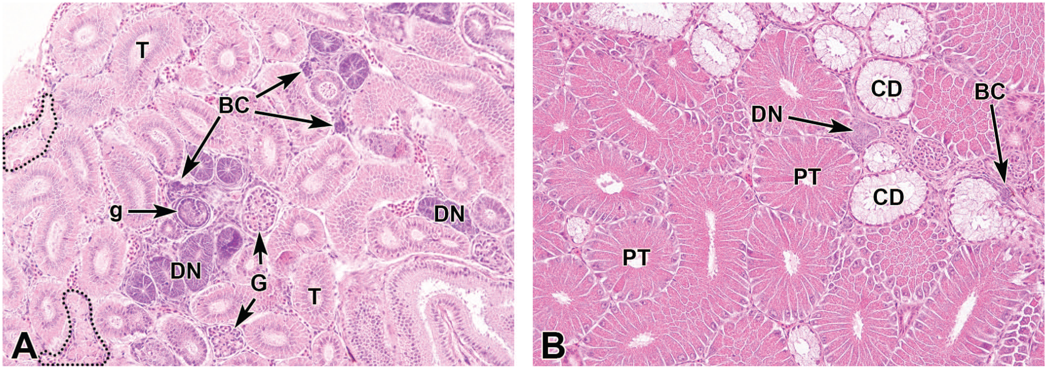
Kidney with nephron neogenesis in the three-spined stickleback (Gasterosteus aculeatus). The renal tubules may appear very different in reproductively active male sticklebacks compared to females or to reproductively inactive male sticklebacks. (A) Kidney from a reproductively active female stickleback, basophilic clusters (BCs) and developing nephrons (DNs) are easily recognized in H&E. A newly formed glomerulus (g) is also present and can be compared with older glomeruli (G). Numerous tubuli (T) and two pigmented macrophage aggregates (dashed line) are also visible. (B) Kidney from a reproductively active male stickleback. The proximal tubuli (PTs) are hypertrophied and more eosinophilic due to spiggin accumulation. Basophilic clusters (BCs) and developing nephrons (DNs) are also visible and are of comparable sizes as in A, hence demonstrating the massive increase in tubuli size. The collecting ducts (CDs) are clear as they contain large amounts of acidic mucus. H&E, original objective 20X (A-B).
In Figure 4 in the article by Liney et al 21 , the kidney from exposed fish is approximately twice as thick compared to the depicted control. Accordingly, the author mentioned significant differences in fish size between treatments. Hence, differential growth over the 300 days exposure could very well have led to secondary effects, including differences in the maturational stage of the kidneys. Furthermore, the field of view (FOV) was not defined, which raises the question whether the absolute number (eg, per cross-section, which in turn is inherently related to fish size) or the relative number (eg, per area unit) were reported.
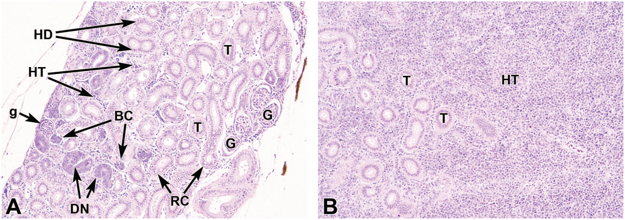
Kidney from three-spined stickleback (Gasterosteus aculeatus) with different degrees of hematopoietic hyperplasia. Hyperplasia of hematopoietic tissue can occur as a general response to different stimuli, both infectious and non-infectious. (A) Renal hematopoietic hyperplasia scored as minimal (normal level in the three-spined stickleback) with barely visible hematopoietic tissue (HT). Tubuli (T), glomeruli (G), developing nephrons (with several mitoses) (DNs), basophilic clusters (BCs), a newly formed glomerulus (g), hyaline droplets (HDs), and rodlet cells (RCs) are also seen. (B) Renal hematopoietic hyperplasia scored as severe. There is a marked proliferation of hematopoietic tissue (HT) and very few tubuli (T) can be found. Due to the hyperplasia, the kidney is much larger than in the fish illustrated in A and therefore extends beyond the entire field of view despite the same magnification. Parasagittal section of kidney in situ, H&E, original objective 20X (A-B).
Mehinto et al 22 reported a clear concentration-response, with significant increased number of DNs at 5 and 25 µg/L, but not at lower concentrations. Histological slides from a subset of fish (5) from the control group and the groups exposed to 0.5 and 1 µg/L were reevaluated blindly by the PWG mentioned earlier. 42 No significant differences were observed, which is in agreement with the original study at these concentrations. Histological sections from the 5 and 25 µg/L groups were additionally reevaluated by a single reviewing pathologist who reported no increase of DNs at any concentration but without providing a fully comparable quantitative comparison across all groups. 42 Notably, it was pointed out that slides from 40% of the fish in the study did not cover any or enough urinary kidney tissue for diagnostic purposes. This raises the question of what part of the kidney that was studied in the original study.
Bickley et al 4 reported the relative amount of DNs and BCs to the cross-sectional area. However, they sectioned the kidney transversely, and at several levels along the longitudinal axis, but it is unclear which levels they have compared.
In conclusion, an increase of DNs and BCs were reported at considerably lower NSAID concentrations than the histopathological changes in the liver, but the reliability of the finding in both Mehinto et al 22 and Bickley et al 4 is not without doubt. In addition, if the increase of DNs or BCs is due to toxic insult to the kidney, one might expect other histological signs of this, such as vacuolation or necrosis of tubuli cells. Mehinto et al 22 do indeed claim increased tubular necrosis, but this finding is neither supported by the histological images provided in the paper, nor was it detected by the PWG reanalysis mentioned earlier. The reliability of the findings by Liney et al 21 is also uncertain. Although not completely dismissed, the evidence for NSAID residues causing an increased number of DNs or BCs in fish exposed to treated municipal wastewater is weak.
Other Diagnoses
Among all histopathological diagnoses identified in this review, one diagnosis particularly worth mentioning is renal hematopoietic hyperplasia (Figure 4A and B). As stated by Wolf et al 40 some diagnoses, including renal hematopoietic hyperplasia, have not been traditionally evaluated in the past and hence there is a risk of them being underreported. We have earlier demonstrated an increase in renal hematopoietic hyperplasia, both after exposure to diclofenac and naproxen with an LOEC for diclofenac at 4.6 µg/L and for naproxen at 299 µg/L.26,27 The paper by Birzle et al 5 also supports this where both the absolute and relative volume of interstitial tissue, which mainly consists of hematopoietic tissue, were increased and the corresponding finding of an absolute and relative volume of nephron tissue was decreased. The LOEC for the most sensitive endpoint was 0.5 µg/L of diclofenac. This was analyzed by quantitative image analysis, a method that may be highly sensitive and have potential to overcome some sources of subjective errors. As stated by Wolf et al, 42 40% of the kidney sections examined in Mehinto et al 22 consisted mainly or solely of hematopoietic kidney where few or no urinary elements were found. If this was due to differences in sampling levels along the longitudinal axis of the kidney, or an actual increase in hematopoietic tissue with concurrent decrease of urinary tissue remains unknown. Unfortunately, none of the papers in the municipal wastewater review reported an evaluation of this specific lesion and there is therefore a risk that this might be overlooked.
Conclusion
This study investigated whether there is support that fish exposed to treated municipal wastewater develop similar histological alterations in liver and kidney as those identified after NSAID exposure. Comprehensive literature reviews of relevant articles fulfilling basic criteria including relevant controls and statistical comparisons only resulted in seven studies for each exposure type. Most of the included studies still had significant limitations, such as unclear methodology, lack of histological figures, histological figures with insufficient quality or not supporting the diagnoses, and use of only unrealistically high exposure concentrations, to name a few. Based on this critical review, we find no clear evidence in the literature that histological alterations in fish exposed to treated municipal wastewaters are caused by residues of NSAIDs. However, limitations in both experimental design and reporting standards of fish histopathology studies prevent any firm conclusions. Well-designed studies evaluating effects from wastewater or NSAID exposure, in which as many factors as possible, with regards to experimental design and analysis, have been kept constant, would certainly facilitate interpretations. In addition, given the largely unlimited possibilities to provide supplementary data with today’s publications, including histological images of adequate quality, representing both treated and control animals, with both high and low magnification, and with clear notations should not be an issue and are strongly encouraged. The current lack of evidence for effects of NSAID residues on liver and kidney histology in fish does not rule out histopathological effects in other organs, including gills. It also does not preclude other types of effects in fish or in other taxa.
Supplemental Material
sj-docx-1-tpx-10.1177_01926233261423895 – Supplemental material for No Clear Evidence of Histopathological Effects Linked to NSAIDs in the Kidney or Liver of Fish Exposed to Treated Municipal Wastewaters
Supplemental material, sj-docx-1-tpx-10.1177_01926233261423895 for No Clear Evidence of Histopathological Effects Linked to NSAIDs in the Kidney or Liver of Fish Exposed to Treated Municipal Wastewaters by Johanna Näslund, Leif Norrgren and D. G. Joakim Larsson in Toxicologic Pathology
Supplemental Material
sj-xlsx-2-tpx-10.1177_01926233261423895 – Supplemental material for No Clear Evidence of Histopathological Effects Linked to NSAIDs in the Kidney or Liver of Fish Exposed to Treated Municipal Wastewaters
Supplemental material, sj-xlsx-2-tpx-10.1177_01926233261423895 for No Clear Evidence of Histopathological Effects Linked to NSAIDs in the Kidney or Liver of Fish Exposed to Treated Municipal Wastewaters by Johanna Näslund, Leif Norrgren and D. G. Joakim Larsson in Toxicologic Pathology
Footnotes
Author Contributions
Declaration of Conflicting Interests
The authors declared no potential conflicts of interest with respect to the research, authorship, and/or publication of this article.
Funding
The authors received no financial support for the research, authorship, and/or publication of this article.
Ethical Considerations
This article does not contain any studies with human or animal participants.
Consent to Participate
Not applicable.
Consent for Publication
Not applicable.
Supplemental Material
Supplemental material for this article is available online.
References
Supplementary Material
Please find the following supplemental material available below.
For Open Access articles published under a Creative Commons License, all supplemental material carries the same license as the article it is associated with.
For non-Open Access articles published, all supplemental material carries a non-exclusive license, and permission requests for re-use of supplemental material or any part of supplemental material shall be sent directly to the copyright owner as specified in the copyright notice associated with the article.