
Abstract
A retrospective analysis in C57BL6/J mice used in dietary carcinogenicity studies was performed to determine the survival rate, causes of death and incidences of spontaneous non-tumoral and tumoral findings. Data were collected from 1600 mice from control dose groups of sixteen 18-month carcinogenicity assays performed between 2003 and 2021 at the same test facility with similar environmental conditions and experimental procedures. The survival rate was high in both sexes (81%-85%) and the causes of humane euthanasia or death were mainly non-tumoral (chronic ulcerative dermatitis, atrial thrombosis). Benign tumors were more frequent than malignant tumors and females were more affected than males. Pituitary gland adenoma in females, lymphoma, bronchioloalveolar adenoma, and harderian gland adenoma in both sexes were the most common tumors. Systemic amyloidosis, the most frequent non-tumoral lesion, was observed variably across studies without sex predilection. The analysis by cohort (3 time periods of 6 years) showed a tendency toward higher incidences of lymphoma and pituitary gland adenoma and lower incidences of amyloidosis over time. The results presented here provide for the first time a robust set of control historical data in untreated C57BL/6J mice kept for 18 months contributing to build in depth knowledge of this animal model.
Introduction
The information provided by carcinogenicity assays in 2 rodent species remains a cornerstone for the market authorization of agrochemicals worldwide. Nowadays, their outcomes are used to confirm via negative results (absence of treatment-related tumors or absence of human relevance) the safety of a new active ingredient which has been selected through a long process based on in silico, in vitro (high and medium throughput) assays, and animal toxicity studies. Despite some limitations, the 2-year carcinogenicity studies in rodents remain the gold standard regulatory wise that ensures the safe use of agrochemicals. 18 The assay is first designed to identify genotoxic and non-genotoxic carcinogens, but also gives information on non-tumoral pathology, either induced by the treatment or exacerbated by the treatment (eg, spontaneous lesions of aging animals). In these studies, 23 the test article is given at 3 different doses (low, medium, and high) in a large cohort of rodents (at least fifty animals of each sex). The route of administration is oral by diet, which is the relevant route of exposure for regulatory toxicity studies of agrochemicals, as it mimics the potential human intake of the agrochemical via treated crops. The No-Observed-Adverse-Effect Level (NOAEL), often derived from non-tumoral adverse changes, helps to define the average daily intake (ADI) for consumers. 5 The current effort to get similar or better information in the context of Next Generation Risk Assessment of chemicals without animal models, promoted by the European Food Safety Authority (EFSA) and the US Environmental Protection Agency (US EPA), will certainly lead to New Approach Methods (NAMs) in toxicology.13,14,29 Until now, these alternative methods do not cover the full range of possible modes of action and target organs identified in the regulatory studies, which remain mandatory. 18
The carcinogenicity assay must be run in 2 different rodent species for an agrochemical. 23 The study duration is 2 years in the rat and 18 months to 2 years in the mouse, depending on the strain. 22 Two main outbred strains of rats (Sprague Dawley and Wistar) are commonly used. The outbred CD-1 strain is the standard model for the mouse species and the study duration is also 2 years. 6 In parallel, the inbred C57BL/6 strain has been used more frequently as a relevant model in toxicology and especially in the plant protection products (PPPs) industry. 6 This model exhibits a very low spontaneous incidence of liver foci and tumors 10 and has a lower sensitivity to liver carcinogens compared with the rat. 25
The outcomes of the assay are compared with concurrent control groups but also with Historical Control Data (HCD) generated at the test facility where the study is conducted. 16 This knowledge becomes even more critical in the case of rare tumors or non-proliferative changes for revealing true positive or safe compounds. To have robust HCD, the 3 pillars of the animal model (the source of animal itself, the food, and the environment) must be fixed and controlled.
In this article, the authors wish to share their knowledge on the histopathological lesions observed in aging C57BL6/J mice. The data were collected from C57BL6/J control mice among sixteen successive 18-month carcinogenicity studies, performed in the same environment and management at Bayer Crop Science Research Center (France) over the past 18 years. This article illustrates the most observed tumors but also hyperplastic and non-proliferative changes in control C57BL6/J mice. To identify a possible drift over time, the most frequent tumors and selected non-tumoral lesions were analyzed by cohort. When possible, a comparison was made with the data already published in the untreated aged C57BL/6 mouse.
Materials and Methods
In Vivo Phase
One thousand six hundred C57BL/6J mice (800 males and 800 females), obtained from Charles River Laboratories (Saint-Germain-sur-l’Arbresle, France), were maintained as control animals in sixteen 18-month carcinogenicity studies via oral diet administration performed from 2003 to 2021 at Bayer Crop Science Research Center (France) (Table 1). After at least 15 days of acclimatization, animals were randomly selected and housed individually in suspended, stainless and wire mesh cages, except for the two most recent studies (studies O and P) in which two females were housed per cage. They were approximately 6-week-old at the beginning of each study and were identified by a micro-identification implant from Biomedic Data Systems (The Netherlands) or Biolog-id (Bernay, France). Animals were maintained at a room temperature of 20 to 24°C and at 40% to 70% relative humidity with a 12-hour light/dark cycle and 15 filtered air changes per hour, with constant monitoring. All mice had free access to filtered and softened tap water from an automatic watering system. They were fed ad libitum for 78 weeks with a certified powdered and irradiated diet A04AC-10 P1 from S.A.F.E. (Scientific Animal Food and Engineering, Epinay-sur-Orge, France). Drinking water and diet were routinely subjected to chemical analysis to monitor for nutritional quality and for contaminants. All studies were conducted in accordance with contemporary guidelines for animal welfare (Guide for the Care and Use of Laboratory Animals, Public Health Service, National Institute of Health, NIH, publication No. 86-23, revised 1985; Directive 2010/63/EU of the European Parliament and the Council of September 22, 2010 on the protection of animals used for scientific purposes, by the Official Journal of the European Union, L276/33-79, 2010; Decree No. 2013-118 relating to the protection of animals used in scientific experiments described in the Journal Officiel de la République Française on February 1, 2013).
List of carcinogenicity studies.
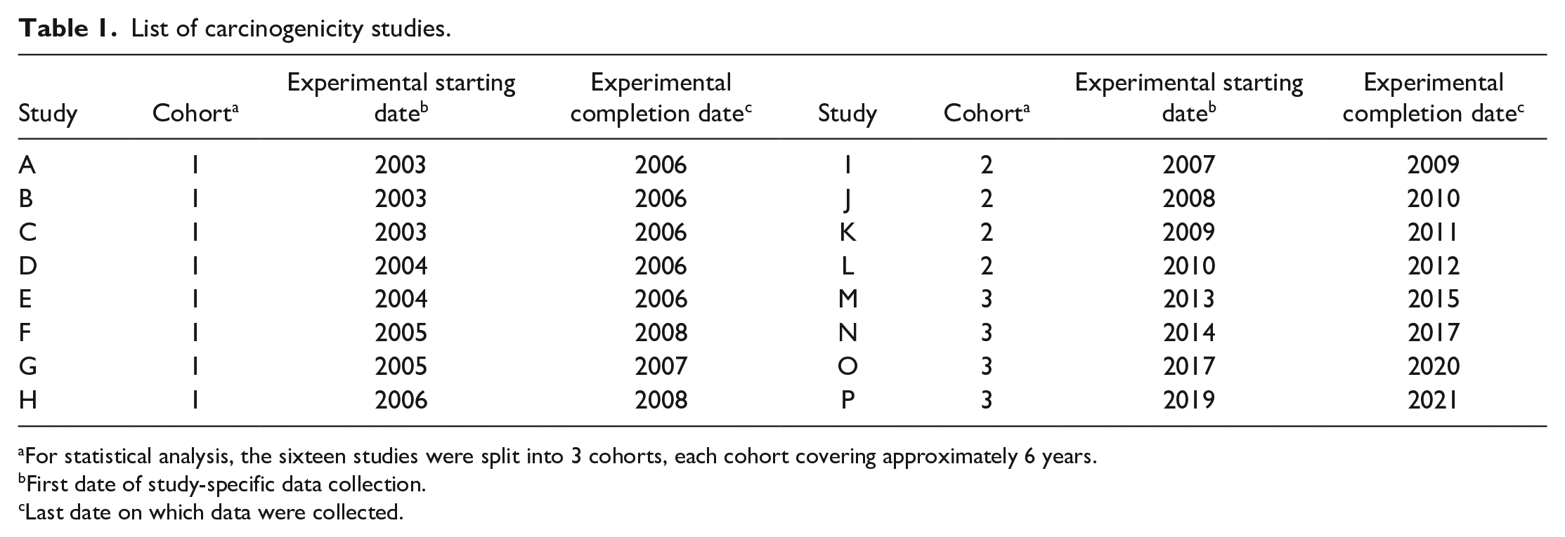
For statistical analysis, the sixteen studies were split into 3 cohorts, each cohort covering approximately 6 years.
First date of study-specific data collection.
Last date on which data were collected.
Mortality and clinical signs were checked twice daily, except on weekends and public holidays, during which it was done once a day. Body weight and food consumption measurements (weekly), and blood sampling for hematology at approximately 12 and 18 months were performed (data not shown). Detailed physical examinations including palpation for masses were performed weekly. The onset, location, size, appearance, progression, and duration of each mass were recorded. Any animal suffering from severe distress, in a moribund condition or considered unlikely to survive was humanely euthanized before the end of the study and submitted to a detailed necropsy.
Terminal Procedures and Histopathological Evaluation
Mice from terminal necropsies and animals humanely euthanized were anesthetized by intraperitoneal injection of pentobarbital (studies A and B) or inhalation of isoflurane (studies C to P) and then exsanguinated under deep anesthesia before necropsy. Any animal found dead during the study was necropsied as soon as possible within the 24 hours of the time of discovery. The terminal body weight (TBW) of all animals at scheduled sacrifice was recorded after overnight dietary fasting in all studies, except the two most recent studies (studies O and P), in which the mice were not diet fasted (animal welfare refinement 8 ). A complete macroscopic examination was performed, including external surfaces, all orifices and body cavities, and representative organs or tissues from all major systems were collected (Table 2). Macroscopic findings including masses and their regional lymph nodes were also sampled. All tissues were fixed in 10% neutral buffered formalin, except testes, epididymides, eyes, optic nerves and harderian glands which were preserved in Davidson’s fixative. Tissues were embedded in paraffin wax, sectioned at approximately 5 µm and histological slides were stained with hematoxylin and eosin.
Collected tissues and organs.
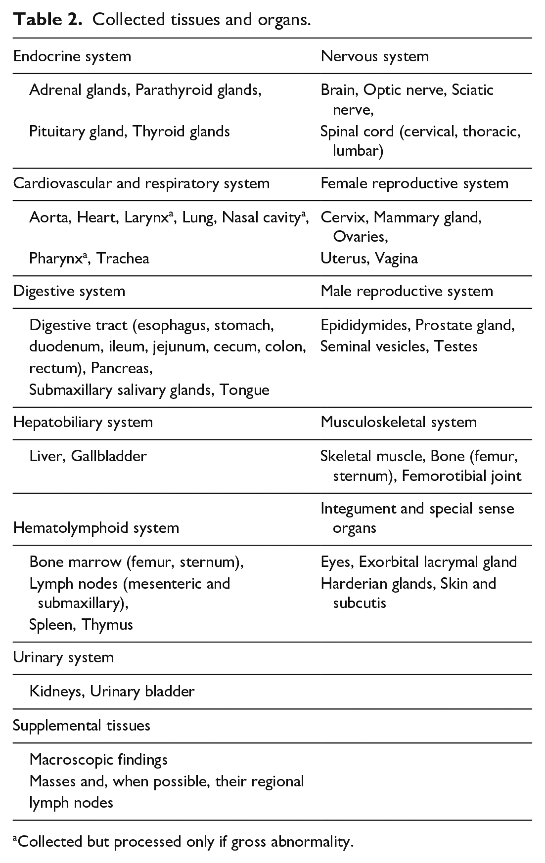
Collected but processed only if gross abnormality.
All the slides were examined by four experienced and board-certified study pathologists for diagnosis of non-tumoral and tumoral findings. Each study was evaluated by one study pathologist. Generally accepted terms were used for the diagnosis of proliferative and non-proliferative lesions. Tumor identification was based on recognized international nomenclature (International Agency for Research on Cancer, RITA-Registry of Industrial Toxicology Animal data, NTP-National Toxicology Program, and International Harmonization of Nomenclature and Diagnostic Criteria for Lesions in Rats and Mice [INHAND] initiative since 2005). A 5-stage severity grading system, from minimal to severe, was used for non-tumoral lesions but is not presented in this article. For each animal found dead or humanely euthanized before the end the study, the study pathologist established a cause of death, when possible. Following the initial evaluation, a pathology peer review of representative slides, including all tumors and hyperplastic lesions and all slides from 10% of animals in control groups, was performed. The same MRCPath board-certified pathologist was in charge of the peer review for all studies (except the most recent one, study P), guaranteeing the homogeneity of the nomenclature used and the consistency of the thresholds applied.
All animal data, including in vivo and post-mortem data, were recorded online using a secured dedicated computer system (Xybion Path/Tox System version 4.2.2. for studies A to J and Xybion Pristima version 7.0.0 build22 for studies K to P).
Statistical Methods
Statistical analyses were conducted to compare the incidence of lesions between males and females. A Pearson’s chi-squared test was performed using R (version 4.1.1.) on incidences of tumoral and non-tumoral findings (Tables 3-5). To identify a possible drift over time (data collected for 18 years), statistical analyses were also conducted by cohort on selected lesions (Table 6). The sixteen studies were split into 3 cohorts, each cohort covering approximately 6 years. Cohort 1 included studies A to H (800 mice, 2003-2008); Cohort 2 included studies I to L (400 mice, 2007-2012); Cohort 3 included studies M to P (400 mice, 2013-2021). Cohort 1 was the largest since 8 studies were conducted between 2003 and 2008. For all tests, a P-value ≤ 0.05 was considered statistically significant.
Incidence summary of spontaneous tumors in control C57BL/6 mice.
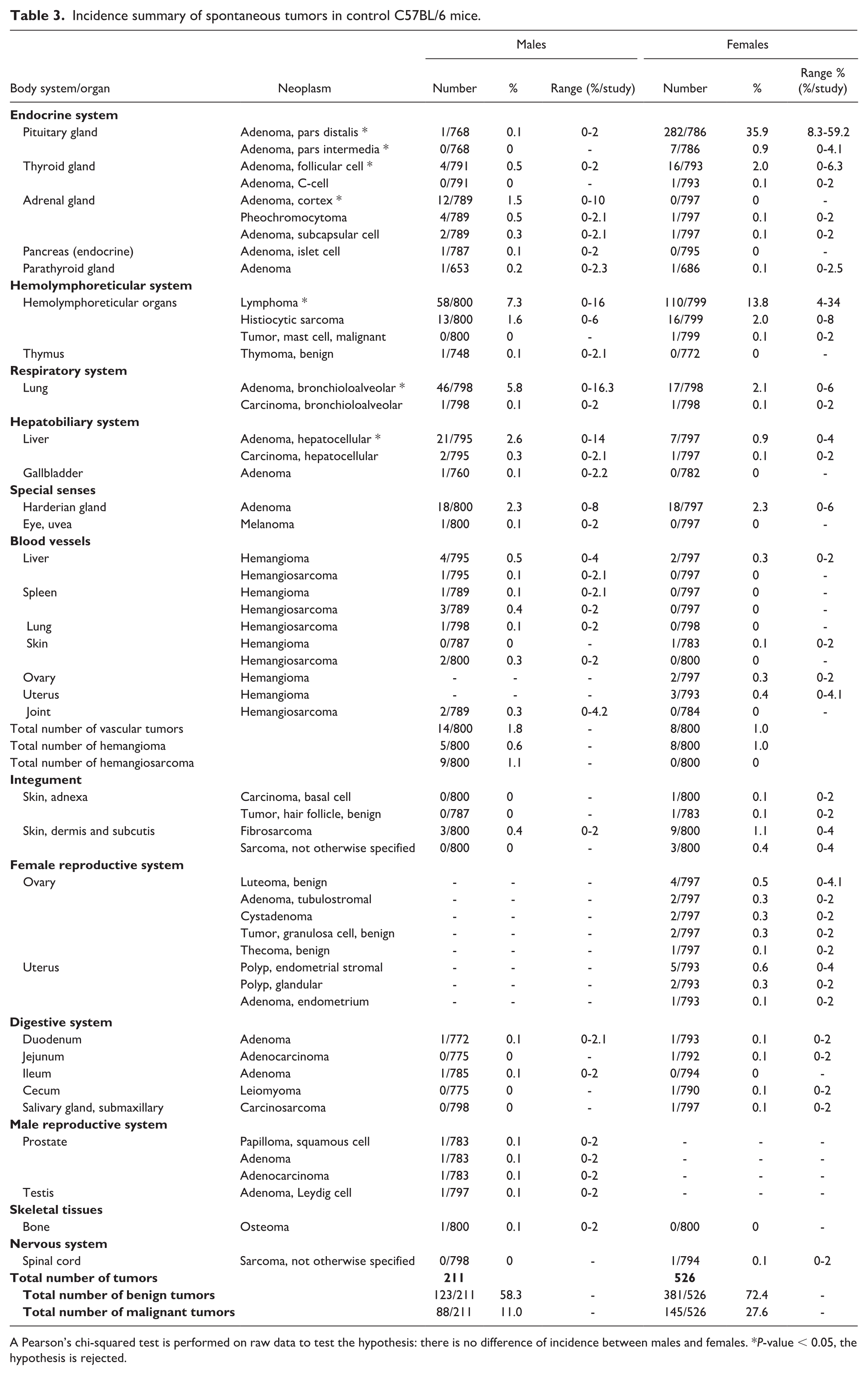
A Pearson’s chi-squared test is performed on raw data to test the hypothesis: there is no difference of incidence between males and females. *P-value < 0.05, the hypothesis is rejected.
Incidence summary of selected non-tumoral lesions in control C57BL/6 mice.
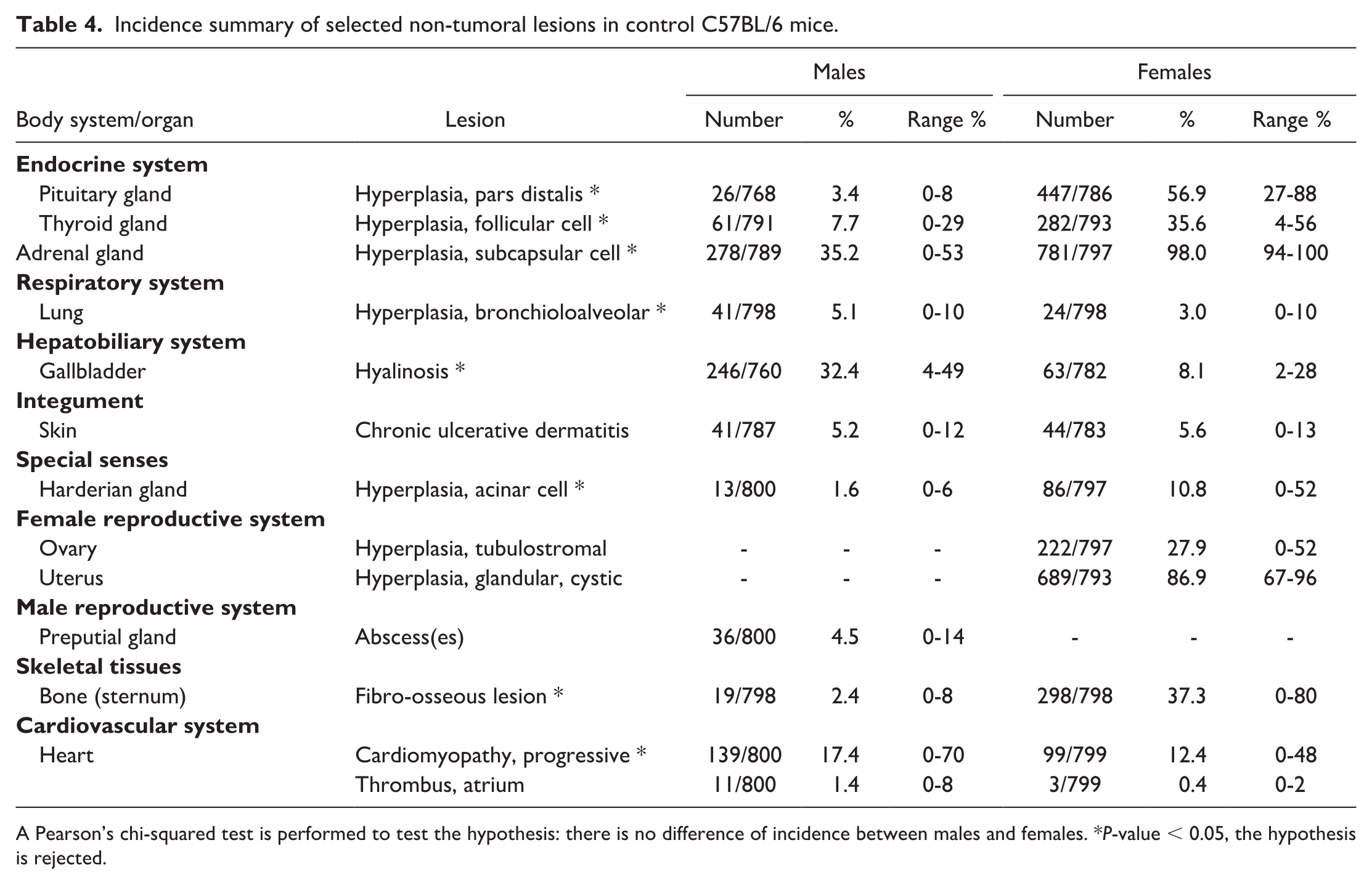
A Pearson’s chi-squared test is performed to test the hypothesis: there is no difference of incidence between males and females. *P-value < 0.05, the hypothesis is rejected.
Incidence summary of amyloid deposition in control C57BL/6 mice.
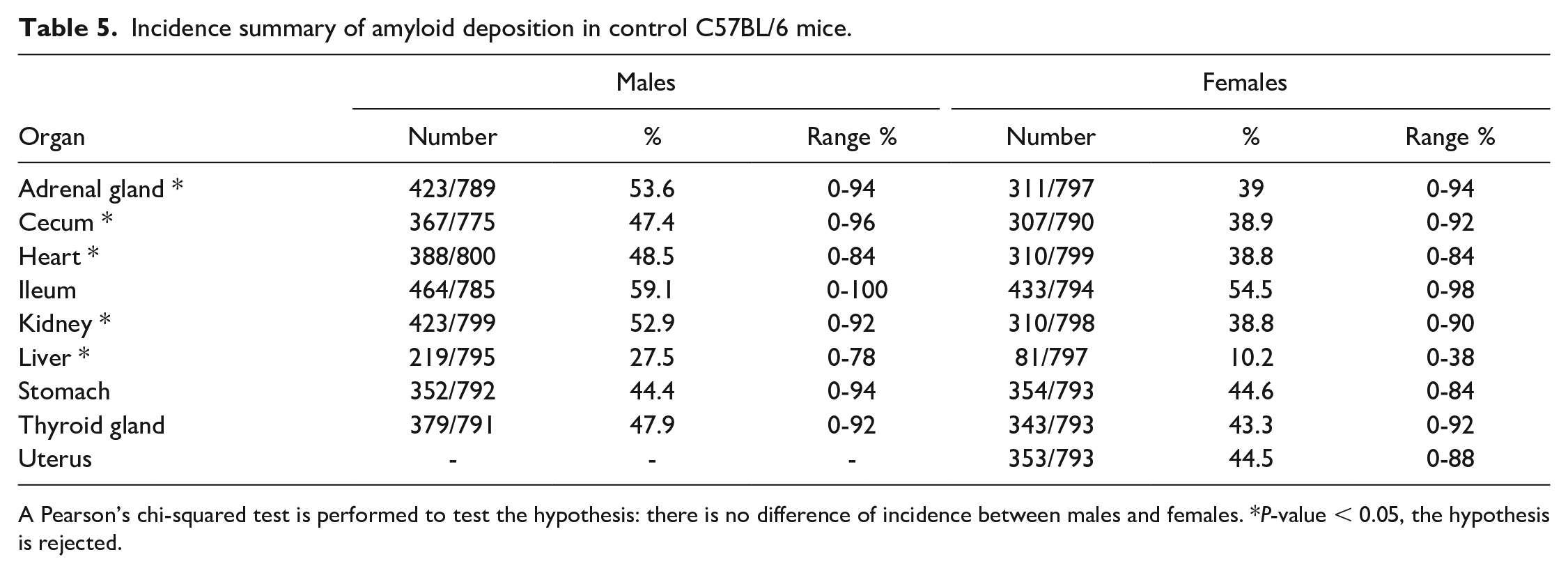
A Pearson’s chi-squared test is performed to test the hypothesis: there is no difference of incidence between males and females. *P-value < 0.05, the hypothesis is rejected.
Incidence summary of selected lesions by cohort.
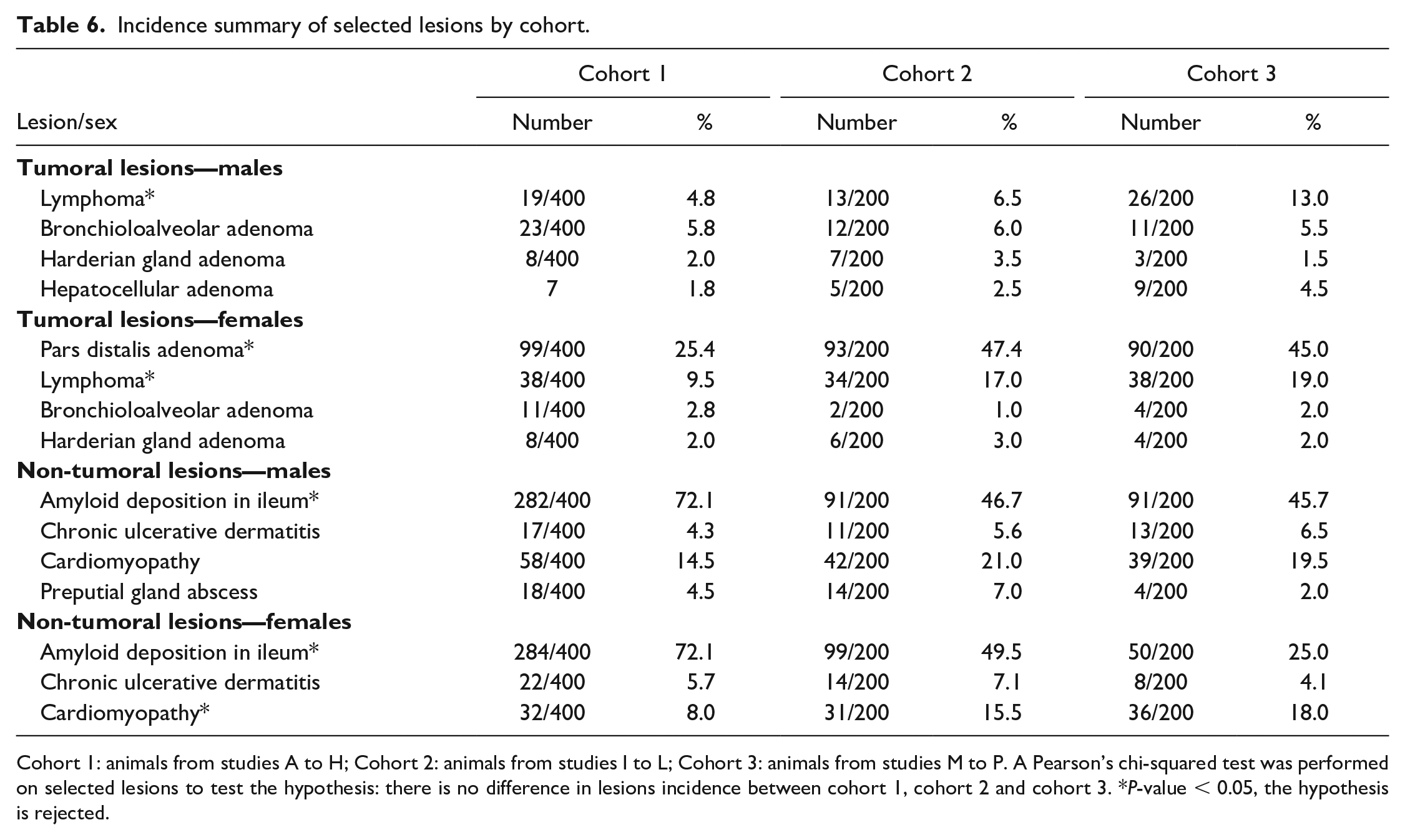
Cohort 1: animals from studies A to H; Cohort 2: animals from studies I to L; Cohort 3: animals from studies M to P. A Pearson’s chi-squared test was performed on selected lesions to test the hypothesis: there is no difference in lesions incidence between cohort 1, cohort 2 and cohort 3. *P-value < 0.05, the hypothesis is rejected.
Results
Terminal Body Weight
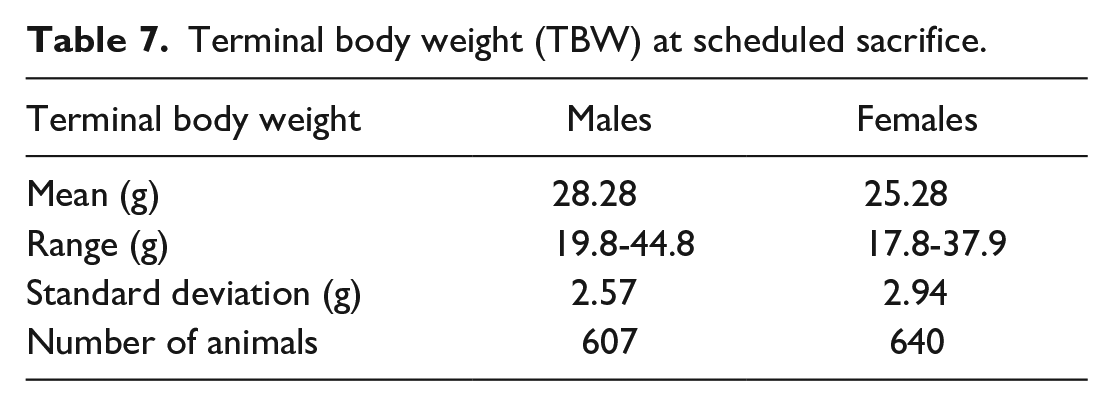
Terminal body weight (TBW) data registered at scheduled euthanasia (78 weeks) are presented in Table 7 and Figure 1. The mean TBW was 28.28 g in males and 25.28 g in females. The data showed general consistency across the studies, with a small shift in the two most recent ones (studies O and P), in which slightly higher TBW were observed, especially in males.
Terminal body weight (TBW) at scheduled sacrifice.
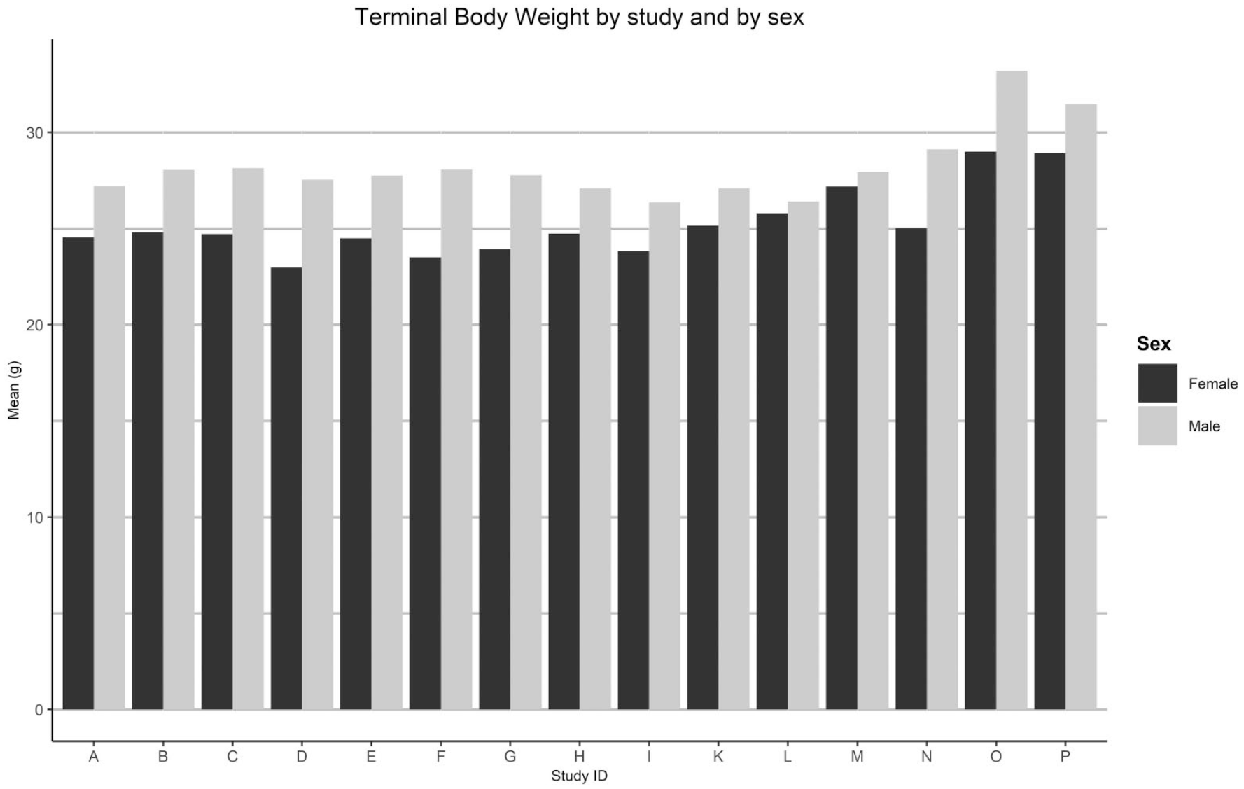
Terminal body weight by study and by sex.
Survival Data and Causes of Death
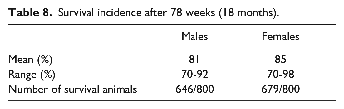
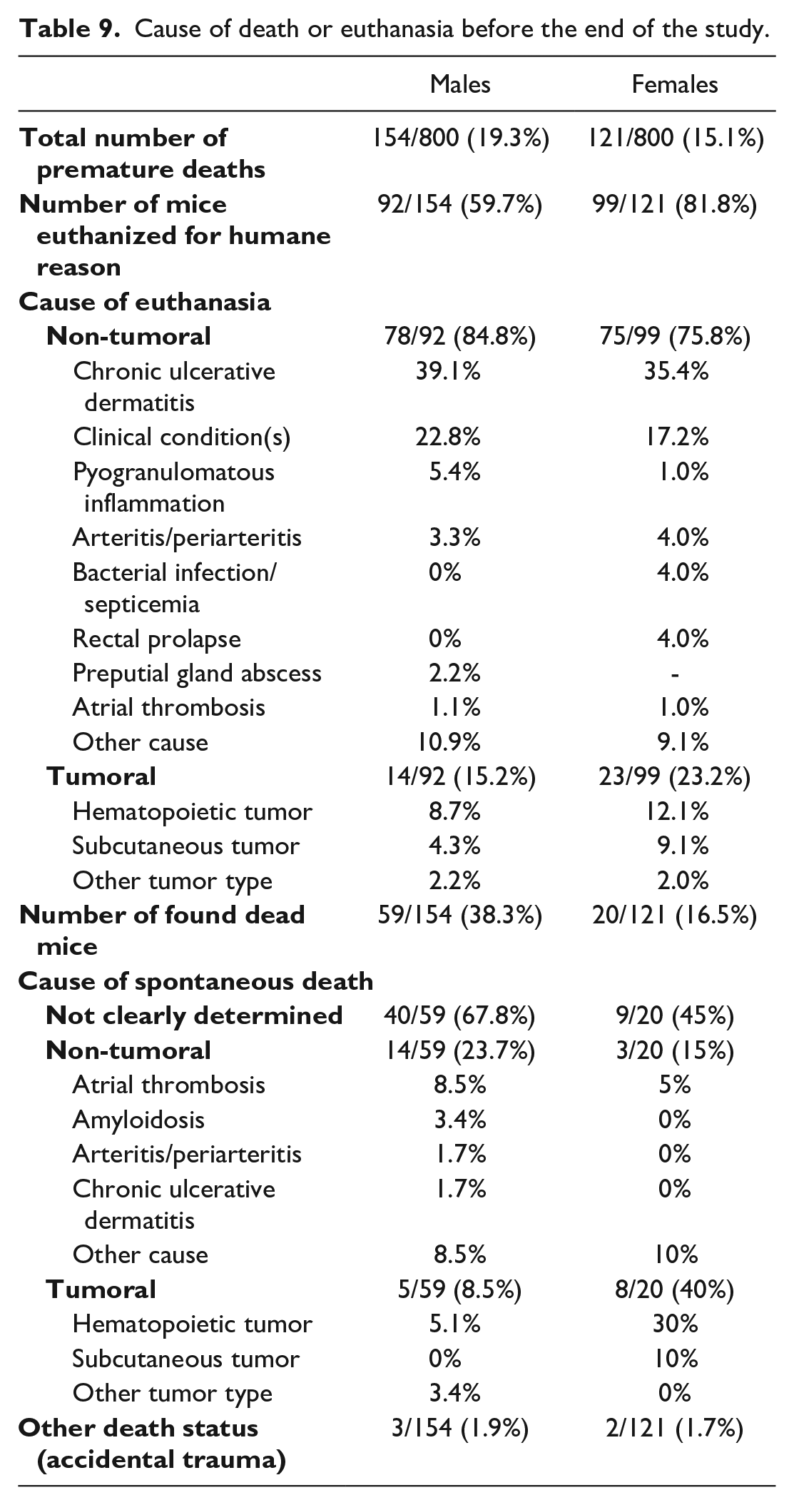
Survival data until scheduled euthanasia (78 weeks) are shown in Table 8 and Figure 2. The survival rate was high and comparable between males and females (81% and 85%, respectively). Among the sixteen studies, the range of variation was quite important in both sexes (from 70% to 92% in males and from 70% to 98% in females), the highest survival rate being observed in the most recent study (study P). The death status and the main causes of death or humane euthanasia in the sixteen studies are summarized in Table 9 and Figure 3. For humane, a decreased body weight was indicated when no pathological lesions could account for the moribund status of the mouse. For found dead mice, “not clearly determined” was indicated when the cause of death could not be established at the pathological examination. The death status was mainly euthanasia for humane reasons, followed by spontaneous death (found dead mice) and significant differences were noted between males and females. Of the 154 (out of 800) males who died prematurely, 92 (59.7%) were humanely euthanized and (62) 38.3% were found dead. Among the 121 (out of 800) premature decedents in females, a large proportion of mice were humanely euthanized (81.8%) and only 16.5% of animals were found dead. Other deaths were related to accidental trauma (less than 2% in both sexes). In found dead mice, no clear cause of death could be established at the pathological examination in 67.8% of males and 45% of females. The causes of spontaneous death were mainly non-tumoral in origin in males (23.7%) and comprised atrial thrombosis, amyloidosis, systemic arteritis/periarteritis and chronic ulcerative dermatitis. Tumors accounted for 40% of deaths in females whereas only for 8.5% in males. The most frequent tumoral causes of death were hematopoietic tumors (mainly lymphoma) and subcutaneous tumors (mainly fibrosarcoma). Regarding the mice humanely euthanized, chronic ulcerative dermatitis was the most common cause of euthanasia (39.1% in males and 35.4% in females), followed by poor clinical condition (clinical signs and/or loss of body weight without relevant histopathological finding), pyogranulomatous inflammation (in skin and other tissues), and arteritis/periarteritis. The same tumor types as in found dead mice accounted for only 15.2% (males) and 23.2% (females) of euthanasia. Considering the overall mortality, a tumor was the cause of death in 12% of males and 26% of females.
Survival incidence after 78 weeks (18 months).
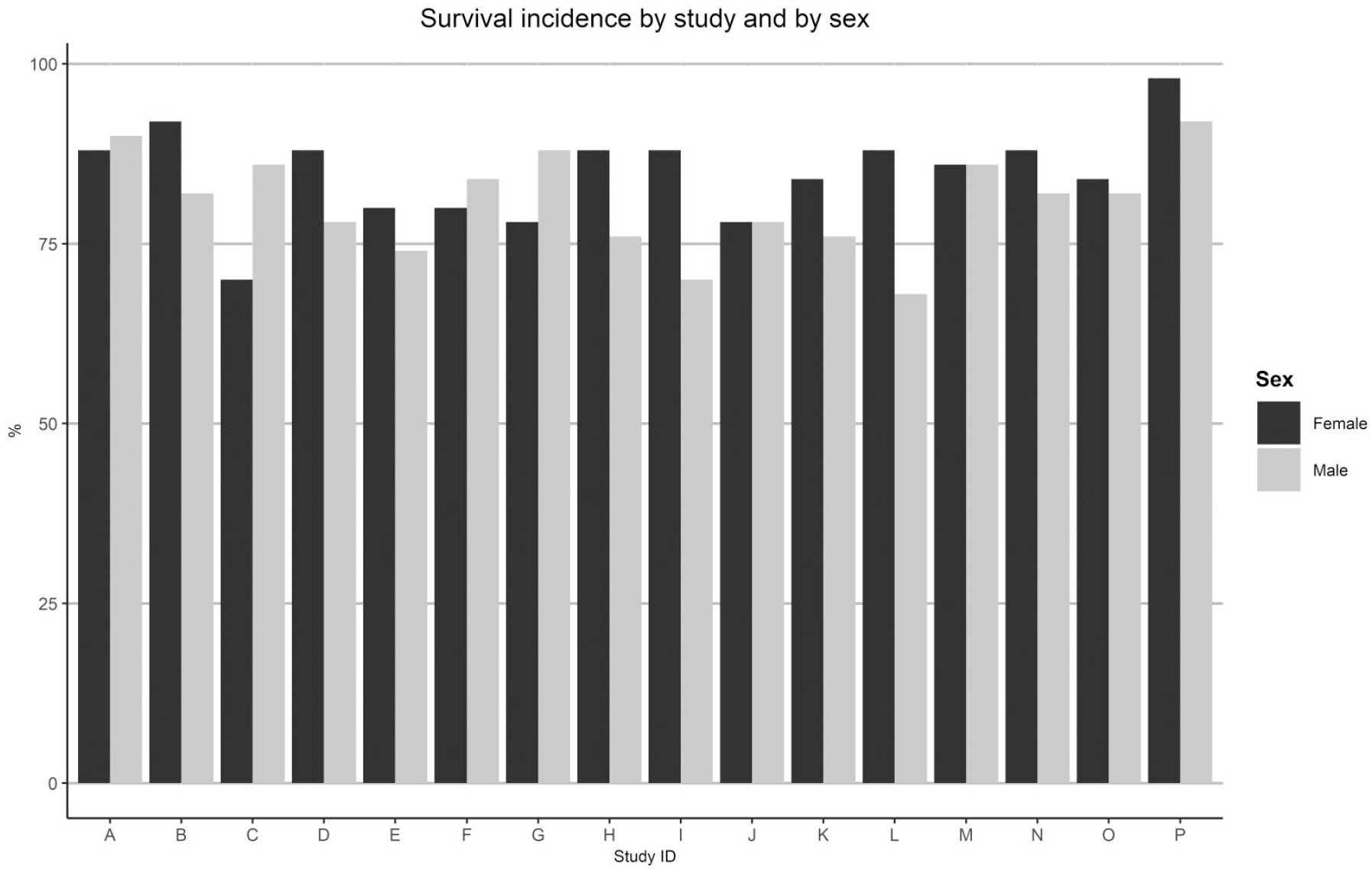
Survival incidence by study and by sex.
Cause of death or euthanasia before the end of the study.
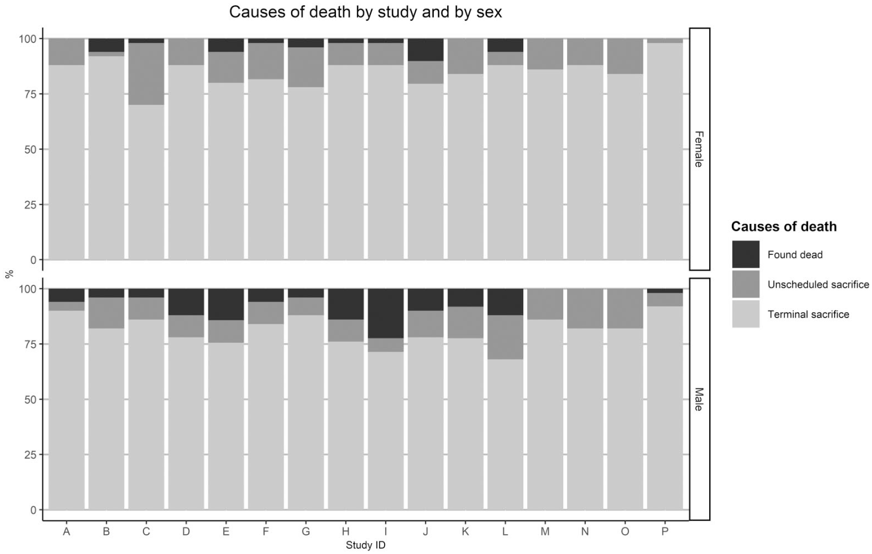
Causes of death by study and by sex.
Tumoral Lesions
The overall incidence of spontaneous tumors observed in 1600 untreated C57BL/6J mice during the sixteen carcinogenicity studies is summarized in Table 3. The tumors are presented by organ system and by order of frequency, the most frequently affected system first (endocrine system). For each organ, the percentages of total number of tumors out of total number of organs examined (800 or lower number, for each sex) were calculated. The very slight variation in the number of organs examined was explained by occasional missing tissues (lost at necropsy or during processing) or autolysis precluding examination. The range (% per study) is also presented for each tumor type.
The total number of observed tumors (526) was much higher in females compared with males (211). In both sexes, the proportion of benign tumors was higher (58.3% in males and 72.4% in females) compared with malignant tumors.
The endocrine system (in females only) and the hemolymphoid system (in both sexes) were the most affected organ systems. In males, the most observed tumors were lymphoma (7.3%) (Figure 4a-c), bronchioloalveolar adenoma (5.8%) (Figure 5a), hepatocellular adenoma (2.6%), harderian gland adenoma (2.3%) (Figure 5b), vascular tumors (1.8%), histiocytic sarcoma (1.6%), and cortical adenoma in the adrenal gland (1.5%). In females, the most frequent tumors were pars distalis adenoma (35.9%) (Figure 5c), lymphoma (13.8%), harderian gland adenoma (2.3%), bronchioloalveolar adenoma (2.1%), follicular cell adenoma (2.0%), histiocytic sarcoma (2.0%) (Figures 6a-c), subcutaneous fibrosarcoma (1.1%), and hemangioma (1.0%). Other tumors were observed at a very low incidence, which means less than 8 cases out of 800 males or females.
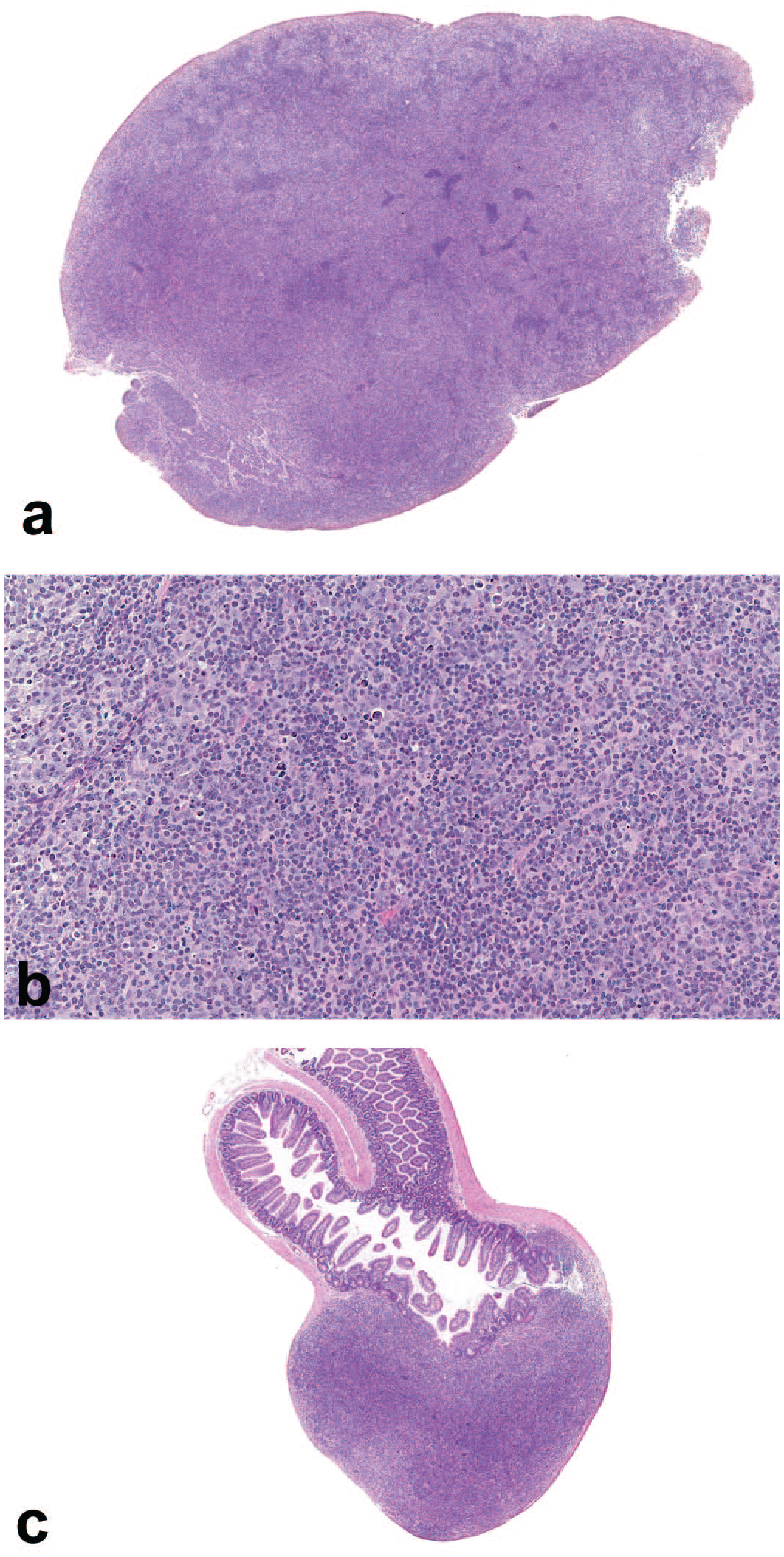
Malignant lymphoma in C57BL/6 mice. (a) Bronchial lymph node—Malignant lymphoma. HE. Original scan 1×. (b) Bronchial lymph node—Malignant lymphoma. HE. Original scan 20×. (c) Ileum, Peyer’s patch—Malignant lymphoma. HE. Original scan 1.8×.
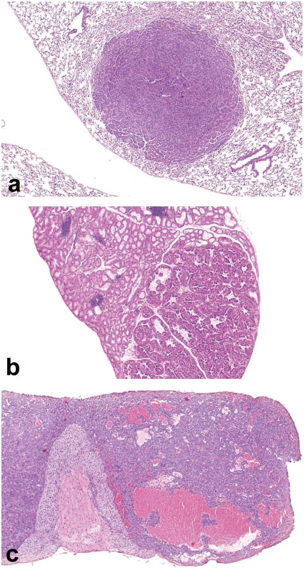
Benign tumors in C57BL/6 mice. (a) Lung—Bronchioloalveolar adenoma. HE. Original scan 4×. (b) Harderian gland—Adenoma. HE. Original scan 4×. (c) Pituitary gland—pars distalis adenoma. HE. Original scan 6.5×.
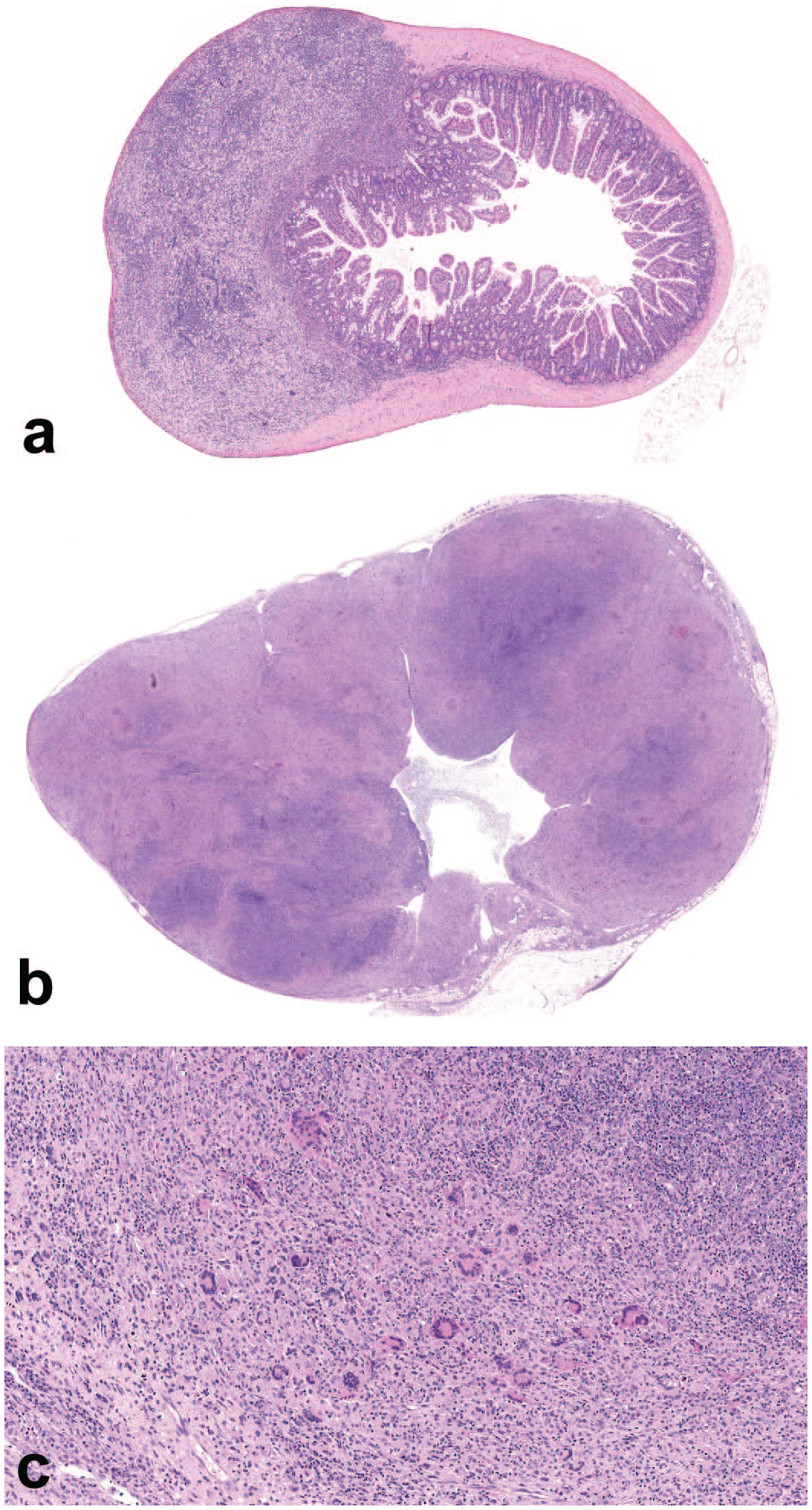
Histiocytic sarcoma in C57BL/6 mice. (a) Ileum, Peyer’s patch—Histiocytic sarcoma. HE. Original scan 2×. (b) Renal lymph node—Histiocytic sarcoma. HE. Original scan 2×. (c) Renal lymph node—Histiocytic sarcoma. HE. Original scan 20×.
Except pituitary gland adenoma in pars distalis, which was by far the most frequent tumor in females (282 occurrences), lymphoma (168 occurrences) and bronchioloalveolar adenoma of the lung (63 occurrences) were the most common tumors in both sexes.
The only tumors observed in all carcinogenicity studies were pituitary gland adenoma (from 8.3% to 59.2%, depending on the study) and lymphoma (from 4% to 34%), both in females. Regarding other tumors, the variability according to the study was also quite significant (eg, a range from 0% to 16% for bronchioloalveolar adenoma and lymphoma in males). Lymphoma was recorded in many organs, including mainly lymph nodes, spleen, Peyer’s patches, liver, and thymus. The mesenteric lymph node was by far the most common anatomic location, either alone or with the largest tumor mass, suggesting that it was probably the most frequent primary site of lymphoma. In the liver, the spontaneous incidence of tumors was low, especially in females (0.9%) and mostly comprised hepatocellular adenoma, malignant hepatic tumor being rare in both sexes (0.3% in males and 0.1% in females).
Rare tumors, which were observed at a single occurrence (1 out of 1600 mice), were: C-cell adenoma of the thyroid gland, pancreatic islet cell adenoma, benign thymoma, malignant mast cell tumor, gallbladder adenoma, uveal melanoma in the eye, basal cell carcinoma and benign hair follicle tumor in the skin, adenocarcinoma and leiomyoma in the digestive tract, salivary gland carcinosarcoma, and bone osteoma.
Noteworthy, no tumors were observed in the brain, mammary gland, kidney, urinary bladder, exocrine pancreas, skeletal muscle and seminal vesicles in males in the 1600 examined mice.
Non-Tumoral Lesions
The incidences of selected non-tumoral lesions are presented in Table 4. Only hyperplastic lesions, lesions related to early mortality or lesions specific to the C57BL6/J mouse are presented in this table.
Focal hyperplasia was mainly noted in the endocrine system, lung, and harderian gland. In the endocrine system, high incidences of focal hyperplasia of the pars distalis in the pituitary gland (56.9%) and follicular cell hyperplasia in the thyroid gland (35.6%) were noted in females. In the harderian gland, the incidence of focal hyperplasia was higher in females (10.8%) compared with males (1.6%). Focal bronchioloalveolar hyperplasia was observed in the lung of 5.1% of males and 3% of females. In the adrenal gland, focal subcapsular cell hyperplasia was noted in almost all females (98%) and in 35.2% of males. In the female reproductive system, diffuse glandular cystic hyperplasia in the uterus was noted in most females (86.9%) and tubulostromal hyperplasia in the ovary was observed in 27.9% of females.
Other non-tumoral lesions presented in Table 4 were related to mortality, either spontaneous or due to humane euthanasia. They included chronic ulcerative dermatitis (around 5% in each sex) (Figure 7a), preputial gland abscess in males (4.5%) and atrial thrombosis in the heart (from 0.4 to 1.4%). Hyalinosis in the gallbladder was noted with a higher incidence in males (32.4%) compared with females (8.1%). Fibro-osseous bone lesion was observed with a higher incidence in females (37.3%) compared with males (2.4%). Chronic progressive cardiomyopathy was observed in both sexes (17.4% in males and 12.4% in females).
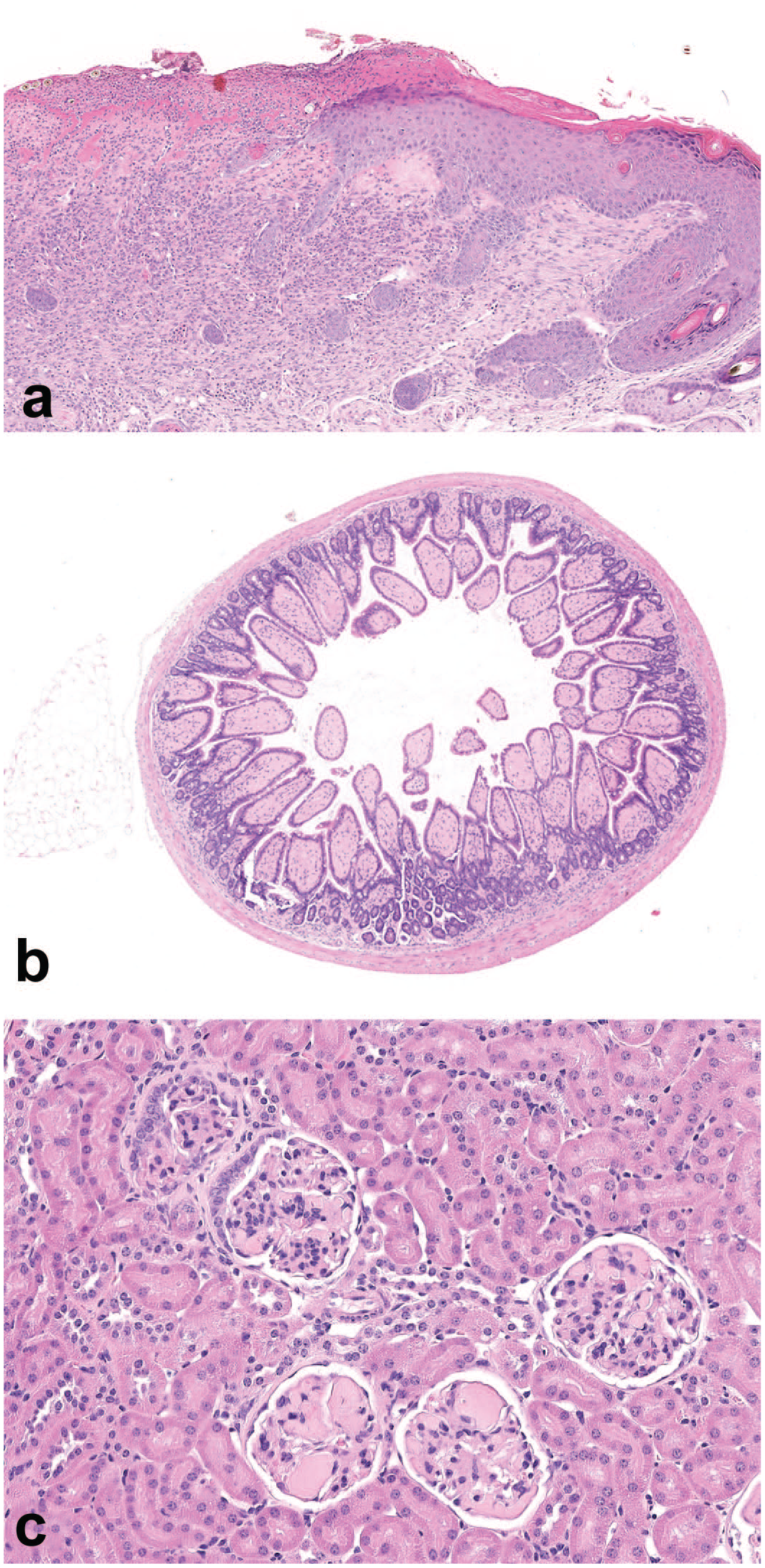
Non-tumoral findings in C57BL/6 mice. (a) Skin—Chronic ulcerative dermatitis. HE. Original scan 8×. (b) Ileum-Amyloid deposition. HE. Original scan 20× (c) Kidney, glomeruli—Amyloid deposition. HE. Original scan 20×.
Amyloid deposition was one of the most common non-tumoral lesions and was observed at different anatomic locations, as presented in Table 5. The ileum (Figure 7b) was the most common anatomic location in both sexes (59.1 and 54.5% in males and females, respectively). Other commonly affected organs (from 38.8% to 53.6%) were the adrenal gland, cecum, heart, kidney (Figure 7c), stomach, thyroid gland, and uterus. Among the most common locations, the liver presented the lowest incidences (10.2% in females and 27.5% in males). A statistically higher overall incidence of amyloid deposition was noted in males compared with females, in the following organs: adrenal gland, cecum, heart, kidney, and liver. There was a large inter-study variability, as shown in Figure 8 (eg, a range from 0% to 100% in the ileum in males).
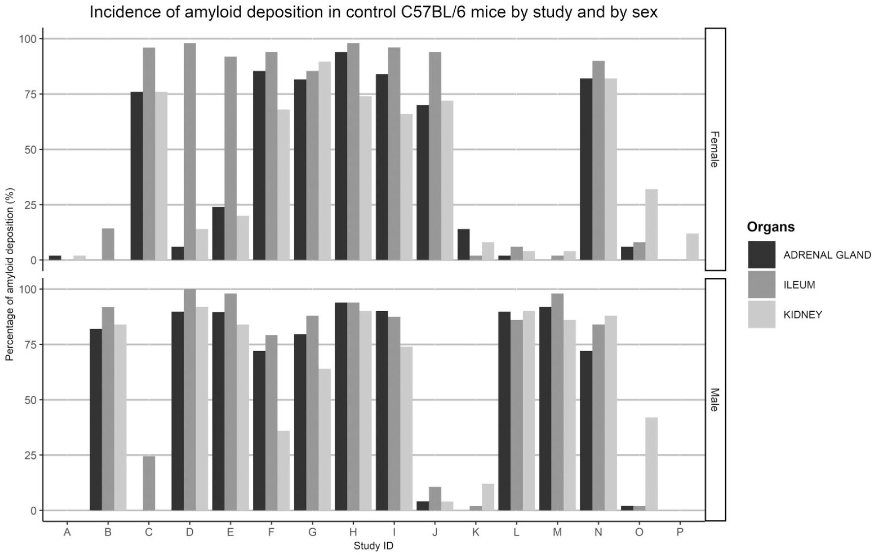
Incidence summary of amyloid deposition in control C57BL/6 mice by study and by sex.
Other common spontaneous findings, not presented in Table 4, were observed with a high incidence (superior to 70%) and included pigment deposition (in both sexes) and atrophy of the cortex (males) in the adrenal gland; dilatation of seminal vesicles in males; focal/multifocal degeneration/atrophy of seminiferous tubules in the testis; atrophy of the ovaries; basophilic tubules in the kidney; degeneration of lens in the eye; mineralization in the brain (thalamus).
Data Analysis by Cohort
The most frequent tumors (lymphoma, pars distalis adenoma, bronchioloalveolar adenoma, harderian gland adenoma, and hepatocellular adenoma) and selected non-tumoral lesions (amyloid deposition in the ileum, chronic ulcerative dermatitis, cardiomyopathy, and preputial gland abscess) were analyzed by time cohort (2003-2008, 2007-2012, and 2013-2021) to identify a possible drift over time (Table 6). The incidences of tumors were comparable, except for the incidences of lymphoma in both sexes and the pars distalis adenoma in females, which were statistically different between the 3 cohorts. A tendency toward higher incidence of lymphoma was observed in males in the cohort 3 (13%) compared with cohorts 1 and 2 (4.8 and 6.5%, respectively). In females, higher incidences of lymphoma were observed in the cohorts 2 and 3 (17 and 19%, respectively) compared with cohort 1 (9.5%). Higher incidences of pars distalis adenoma in females were observed in the cohorts 2 and 3 (47.4 and 45%, respectively) compared with cohort 1 (25.4%).
For non-tumoral lesions, lower incidences of amyloid deposition in ileum were observed in the cohorts 2 (46.7% and 49.5%, in males and females, respectively) and 3 (45.7% and 25% in males and females, respectively) compared with cohort 1 (72.1% in both sexes). A tendency toward higher incidence of cardiomyopathy was observed in males in the cohorts 2 and 3 (15.5% and 18%, respectively) compared with cohort 1 (8%).
Discussion
Terminal body weight data, survival data, causes of death, and pathology data generating from 1600 control C57BL/6J mice maintained in sixteen 18-month carcinogenicity studies over a period of 18 years were detailed above. These data, generated in a single laboratory with the same source of animals, same environment, and same food, constitute a robust database of HCD. In the following sections, the results are analyzed and compared with the data already published in the untreated aged C57BL/6 mouse.1,9,17,27
Terminal Body Weight
Although consistency was observed across the 16 studies, a slightly higher mean TBW was observed in both sexes in the two most recent studies (O and P) compared with the previous ones. This was considered related to the animal welfare refinement and the suppression of overnight diet fasting before scheduled euthanasia.
The mean TBW data were noticeably different from previous data published with C57BL/6 strains. Mean body weights of C57BL/6 mice (NCTR Specific Pathogen Free breeding colony) were higher at 18 months in ad libitum-fed males (slightly above 40 g) and females (approximately 30 g) according to Blackwell et al 1 and Turturro et al. 27 In both reports, the diet was a cereal-based diet in pellets (NIH-31). The origin of the strain and the composition of the diet could explain these differences.
Survival Data and Cause of Death
A high survival rate comparable in males and females (81% and 85%, respectively) was observed in our data. In the data published by Blackwell et al, 1 obtained from 911 ad libitum-fed and diet-restricted C57BL/6 mice maintained up to 42 months, the survival rate was slightly higher: approximately 90% of survival animals in both sexes at 18 months. In addition, Blackwell et al presented the age at 50% survival which was around 27 months in mice fed ad libitum, and up to 33.5 months in mice with diet restriction. As in our data, there was no significant difference between males and females.
In our study, the causes of premature death were mainly non-tumoral in origin. This result is in contradiction with Blackwell et al 1 who found a tumoral cause of death in approximately 70% of C57BL/6 mice. This difference could be attributed to the evolution of animal welfare considerations and refinement of humane endpoints between 1986 4 and 2010. 3 Nevertheless, the lymphoma was the most frequent tumoral cause of death in both data sets. The urogenital system was involved in 4.5% of C57BL/6J males with the preputial gland abscess as a common cause of humane euthanasia. Despite the high incidence of obstructive uropathy in male aging mice in the literature,7,19 related lesions such as distended urinary bladder, hydroureters, or renal pelvic dilatation were not commonly seen and were not considered as factors related to death in our studies. Systemic amyloidosis, even though frequently observed in our data, was a cause of death in 3.4% of C57BL/6 male mice only.
The main non-tumoral cause of euthanasia in our data was the chronic ulcerative dermatitis in both sexes (39.1% in males and 35.4% in females). This skin disease is a well-known idiopathic, spontaneous and debilitating syndrome of black mice with C57BL/6 background.11,26 Both sexes were similarly affected, which was not the case in the data set presented by Blackwell et al, 1 in which the females were more affected. The hybrid B6C3F1 is also known to develop cutaneous ulceration, 12 due to its B6 parental origin. From animal welfare point of view, it is recommended to euthanize the animal if more than 10% of the total body surface area is affected. 15 This explains the high incidence of chronic ulcerative dermatitis as a cause of humane euthanasia in our data set. In conclusion, slight to important differences in the longevity and in the cause of death can be observed within the same strain. This is directly linked to environmental conditions, especially diet. 16 It is key to maintain the standardized conditions of the animal model to reproduce the same data over time, as illustrated Figure 2, with a constant survival rate.
Tumors
Our data showed that C57BL/6 female mice had twice more tumors than males and that benign tumors were more frequent than the malignant ones. Pars distalis adenoma, as the most common tumor in females, and lymphoma, as a common tumor in both sexes, were described similarly by Blackwell et al 1 and Frith et al. 9 In our studies, malignant lymphoma was diagnosed without further classification 30 (e., subtype modifiers as lymphoblastic, pleomorphic or immunoblastic, or T- cell or B-cell type modifiers). Indeed, the focus of the rodent carcinogenicity studies is on risk assessment (rather than on translational research for clinical outcome and treatment) and the overall incidence of lymphomas as an initial assessment of treatment-related effects is acceptable. If an effect is observed, a further characterization of subtypes of lymphoma (with immunohistochemistry) would be necessary, but it has not been the case in our studies so far. Nevertheless, the majority of lymphomas affecting mostly the mesenteric lymph node, spleen and Peyer’s patches and the morphological appearance (mixed, with small and large lymphocytes) were consistent with the pleomorphic subtype, which is described as the most common lymphoma subtype in aged mice in most strains. 30 Considering all the hematolymphoid tumors observed, an important differential diagnostic would be myeloid leukemia, described as common in the spleen and occasional in the bone marrow in mice in general. 28 Leukemia is not described as a common tumor in C57BL/6 mice,1,9,17,27 and it is confirmed in our data with no cases observed in control mice. Of note, sporadic cases were observed in treated groups of some studies (data not shown).
Interestingly for these 2 most frequent tumors, the analysis by cohort showed higher incidences of lymphoma (both sexes) and pars distalis adenoma (females) in the cohorts 2 (2007-2012) and 3 (2013-2021) compared with cohort 1 (2003-2008). The similar genetic background of this C57BL/6J model (inbred) and the tight control of environmental parameters should have prevented this drift over time. However, recent research has revealed new insights into the genetic fragility and relative instability of inbred mouse strains. 2 Indeed, inbred mice are far from being completely isogenic and sources of genetic polymorphisms (single-gene mutations or polygenic mutational variability) are continuously uploading into inbred populations, despite the efforts of breeders to maintain genetic stability.
In contrast, histiocytic sarcoma was the most common tumor in C57BL/6 males (42.5%) according to Blackwell et al 1 whereas it represented only 1.6% of tumors in males from our data. Hemangiosarcoma was the most common tumor according to Frith et al, 9 whereas it accounted for only 1.8% tumors in males from our data. Although it could be surprising to observe such differences with an inbred strain, the variability could be explained by a difference in the substrain. 20 Due to its popularity, several substrains of C57BL/6 have been established and significant genetic and phenotypic variations exist between them. The most common C57BL/6 substrains include C57BL/6J (maintained at The Jackson Laboratory, which is the origin of our mice) and C57BL/6N (established at National Institute of Health).
The C57BL/6J was selected for its low propensity to develop spontaneous hepatocellular tumors and foci. 10 Our data confirm this important condition which has driven the selection of this strain as one possible model for agrochemicals.
Non-tumoral lesions
To the authors’ knowledge, this is the first report of the incidence of focal hyperplasia in aged C57BL6/J mice. Noteworthy in the pituitary gland of females, there was a good correlation between the high incidences of focal hyperplasia in the pars distalis (56.9%) and adenoma (35.9%).
Amyloid deposition was one of the most common non-tumoral lesions and its occurrence was variable according to the study, without clear sex difference. It was not associated with mortality, in contrast to the CD-1 mouse, 19 in which systemic amyloidosis was a frequent cause of death. Of note, it was observed with strong variations from one study to another and between sexes and the analysis by cohort showed a tendency toward lower incidences of amyloid deposition in ileum in the cohorts 2 (2007-2012) and 3 (2013-2021) compared with cohort 1 (2003-2008). The inter-study differences observed for this pathological condition confirm the importance of building knowledge on HCD. Without this knowledge, it would have been easy to attribute an increase in amyloidosis to the treatment.
The C57BL/6J mouse is not the most used strain in toxicology as a carcinogenicity model23,24 despite high interest for this strain in medical research. The authors wanted to illustrate the importance of establishing a solid pathological understanding of the animal model itself, in the interest of improving confidence in the overall interpretation of the study. Concurrent control groups and most importantly, HCD, provided insights generated in the test facility where the study is conducted, 16 and the basis for a strong trial conclusion. In the lack of those internal data or as complementary information, it is also possible to refer to the RITA 21 database. However, internal HCD, in addition to the concurrent control group, remains the most relevant tool when interpreting the data, especially when produced under similar experimental conditions. Nevertheless, many sources of bias can still influence the results, including genetic drift, standards of animal welfare, and other environmental conditions (housing, presence of endogenous or exogenous viruses). In addition to the development of NAMs, the publication of HCD showing the good knowledge for one specific model remains an activity of high scientific value. The data presented may provide a valuable source of information to support pathologists and toxicologists involved in the interpretation of pathology data from toxicology studies in C57BL/6J mice.
Footnotes
Declaration of Conflicting Interests
The author(s) declared no potential conflicts of interest with respect to the research, authorship, and/or publication of this article.
Funding
The author(s) received no financial support for the research, authorship, and/or publication of this article.
Institutional Review Board Approval
This paper has been validated by Bayer institutional board via an approval process for scientific publications.