
Abstract
A retrospective study was performed to establish the causes of mortality and incidence patterns of tumors in young (<50 weeks) control CD-1® mice from Charles River Laboratories. Tumor incidences (fatal and nonfatal) and nonneoplastic causes of death observed during the first 50 weeks of the study were collected from 48 thirteen-week toxicity studies conducted between 2009 and 2018 and from 43 carcinogenicity studies conducted between 2005 and 2018. Thirteen-week studies had a mortality rate of 8/620 (1.3%) in males and 4/620 (0.65%) in females. The major factors contributing to death were integument lesions in males (3/8) and experimental procedure–related injuries in females (3/4). All tumors recorded were nonfatal. Bronchiolo-alveolar adenoma was the commonest tumor with the same incidence in both males and females (4/620, 0.65%); a single lymphoma (0.16%) and uterine leiomyosarcoma (1/620 0.16%) were reported in females. The mortality rates of males and females that died or were euthanized during the first 50 weeks in carcinogenicity studies were 192/2830 (6.8%) and 198/2830 (7%), respectively. The most common fatal tumor in this age group was lymphoma in both sexes, with an incidence of 18/192 (9.3%) and 41/198 (20.7%) in males and females, respectively. In males tumors were responsible for fewer deaths than in females (17% vs. 32.3%). The major nonneoplastic causes of death or moribundity were cutaneous lesions (44/192, 22.9%), and obstructive uropathy (39/192, 20.3%) in males, and chronic progressive nephropathy (40/198, 20.2%) in females. Only minor differences were evident compared to a similar study performed 15 years ago; these might reflect changes in terminology and diagnostic criteria, and stricter animal welfare endpoints.
Keywords
Introduction
Rodent carcinogenicity studies are required by regulatory agencies for pharmaceuticals intended for long-term clinical use and for chemicals that humans will have long-term exposure to. The mouse is one of the accepted species for these evaluations. Historically, CD-1 mice are the strain of mouse most commonly used for chronic studies in Europe. 1 Knowledge of the profile of spontaneously occurring neoplasms of rodents is essential for differentiating spontaneous and test item–related tumor patterns. The profile of common background tumors in aged CD-1 mice in conventional 2-year rodent studies is well-documented and has been described by several authors.1-6 The factors influencing spontaneous tumor profiles have also been described.7-9 However, the literature is largely based on lifetime data from carcinogenicity studies. Despite their common usage, specific information on the incidence of spontaneous neoplastic and nonneoplastic findings in younger CD-1 mice is not readily available, and most publications on young mice are based on B6C3F1, C57BL6, BALB/c, or transgenic mice. Therefore, in short-term studies, it is sometimes difficult to assess the significance of unusual nonneoplastic findings and isolated tumors that occur in animals given the test item, given the limited information available.10,11 To help put these occurrences into perspective, and assess their relationship to mortality, data on neoplastic and nonneoplastic causes of death or euthanasia during the first year of life were collated and tabulated. This report is intended to establish a general profile of tumor occurrence and nonneoplastic causes of death in control CD-1 mice which were on study for <13 or ≤50 weeks based on data at our laboratories. In addition, to identify any changes in mortality or tumor incidence patterns over the years, these data were compared with a similar survey by Son and Gopinath 11 published in 2004, performed on mouse studies run at Labcorp Early Development Laboratories Limited, Huntingdon (then Huntingdon Life Sciences) between 1990 and 2002.
Materials and Methods
Animals
Male and female CD-1® mice, obtained from Charles River Laboratories Limited (UK), were assigned to 13-week toxicity studies or 78- to 104-week carcinogenicity studies in control groups, as per individual study plans. Studies were conducted either at the Huntingdon or Eye facilities of Labcorp Early Development Laboratories Limited. Different studies were housed in different buildings depending on the type of protocol. However, housing conditions, husbandry, animal management, study protocols, diet, and scheduling are identical across sites and therefore the use of separate facilities was not considered to influence the results.
At the age of 5 to 6 weeks, 1 to 4 randomly selected mice were placed in solid-bottom polycarbonate cages with a stainless-steel mesh lid. Sterilized wood flakes and Nestlets were used as bedding.
In forty-eight studies of 13 weeks’ duration housing conditions included two or three animals of the same sex per cage in 40 studies; males individually caged, and females were housed three of same sex in four studies; animals were individually housed in four studies.
In the forty-three studies of long-term duration, animals were housed as two or three of one sex per cage in 37 studies; males individually caged and females housed three of the same sex in three studies, and animals individually housed in three studies.
Animal room temperature and relative humidity controls were maintained between ranges of 20°C and 24°C and 38% to 68%, respectively, with a 12-hour light/dark cycle and 15 filtered air changes per hour. The mice had free access to tap water in bottles with sipper tubes and to Mouse No. 1 modified maintenance diet (SDS Special Diets, Witham). Drinking water and diet were routinely analyzed for contaminants that might influence the studies. Food hoppers and water bottles were changed at daily to twice-weekly intervals. Studies commenced after an acclimatization period of 14 days, during which veterinary officers monitored the animals’ health. Animals were euthanized when showing signs of pain or distress below the severity welfare limits as specified in the guidelines included in the study protocol. All studies were Good Laboratory Practice compliant. All study protocols were approved by the Labcorp animal care and use committee in accordance with the UK Animals (Scientific Procedures) Act 1986, which conforms to the European Convention for the Protection of Vertebrate Animals Used for Experimental and Other Scientific Purposes (Strasbourg, Council of Europe).
Histopathology
Animals were humanely euthanized by a rising concentration of carbon dioxide and exsanguinated via femoral veins, except in inhalation studies where they were injected intraperitoneally with pentobarbital. Comprehensive necropsies were performed on all decedents. Standardized lists of tissues were fixed in 10% neutral buffered formalin (except eyes, which were fixed in Davidson fluid, and testes and epididymides, which were initially fixed in Bouin solution and then transferred to 70% industrial methylated spirit). Samples of all macroscopically abnormal tissues (including tissue masses) were also routinely fixed, processed, and examined. All tissues were embedded in paraffin wax, sections were cut at 4 to 6 µm and stained with hematoxylin and eosin. A full list of tissues was microscopically examined for all decedent animals. After initial examination by a study pathologist, results were peer reviewed by a second pathologist. Tumor identification was based on in-house nomenclature, which followed the current guidelines for proliferative lesions of each organ system published by the Society of Toxicologic Pathology.
Study Design
13-week toxicity studies
The survey is based on data from forty-eight studies of 13 weeks’ duration, run between January 2009 and June 2018. Only control animals were included in this data compilation. These comprised 48 groups, consisting of 620 males and 620 females in total. Group size varied ranging from 8 to 20 animals of each sex in each study. Mortality occurring before the scheduled end of the study was reviewed. The nonneoplastic causes of death and tumor incidences were tabulated for 4-weekly intervals.
Carcinogenicity studies
The survey is based on data from forty-three studies of 78 to 104 weeks’ duration and comprises 2830 males and 2830 females in total. Group size varied, but at least 50 males and 50 females were present in each study. Mortality occurring within the first 50 weeks of study was reviewed, and the tumor incidences (fatal and nonfatal) tabulated for 4-weekly intervals. In addition, nonneoplastic causes of death were also collated. These data were compared with the publication of Son and Gopinath, which analyzed the mortality and tumor incidences from decedent animals in studies run at Labcorp Early Development Laboratories Limited, Huntingdon (then Huntingdon Life Sciences) between 1990 and 2002.
Results
Thirteen-Week Toxicity Studies
Mortality
In 48 studies of 13 weeks’ duration, there were decedents in only 9 studies. Eight males and 4 females were found dead or euthanized for welfare reasons during the course of these studies, giving a mortality rate of 8/620 (1.3%) in males and 4/620 (0.65%) in females.
Nonneoplastic cause of death
Among the decedent males, integument lesions comprising skin ulceration were the cause of death or early euthanasia in three males. A urogenital lesion (preputial gland abscess) was the cause of unscheduled euthanasia in one male and the cause of death/poor clinical condition was not identified in the remaining male. The other two males died due to experimental procedure–related trauma (Table 1).
Nonneoplastic causes of death of decedents from the control groups expressed by time of death in CD-1 mice enrolled in 13-week studies—Males.
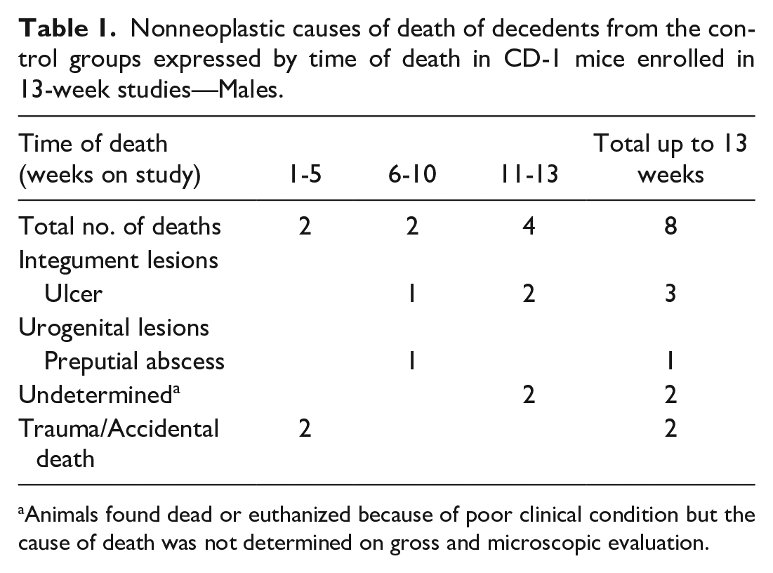
Animals found dead or euthanized because of poor clinical condition but the cause of death was not determined on gross and microscopic evaluation.
Of the four females, the cause of poor clinical condition was undetermined in one. The remaining three animals died or were euthanized due to experimental gavage procedure–related trauma (Table 2).
Nonneoplastic causes of death of decedents from the control groups expressed by time of death in CD-1 mice enrolled in 13-week studies—Females.
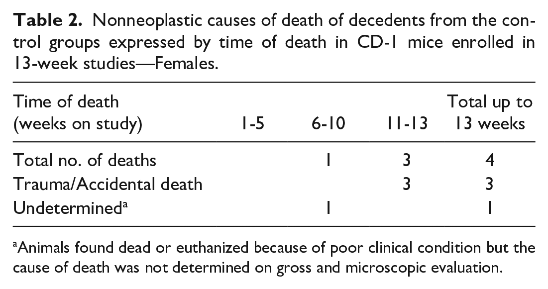
Animals found dead or euthanized because of poor clinical condition but the cause of death was not determined on gross and microscopic evaluation.
Tumor occurrence pattern
All reported tumors occurred in animals euthanized at scheduled timepoint as nonfatal findings. The tumor incidences in males and females are presented in Tables 3 and 4.
Tumor incidence profiles expressed by time of death in CD-1 mice enrolled in 13-week studies—Males.
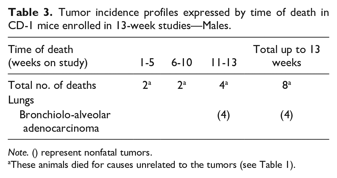
Note. () represent nonfatal tumors.
These animals died for causes unrelated to the tumors (see Table 1).
Tumor incidence profiles expressed by time of death in CD-1 mice enrolled in 13-week studies—Females.
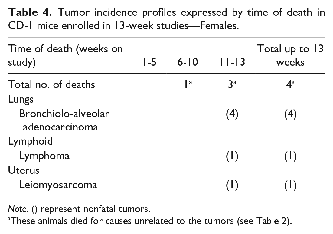
Note. () represent nonfatal tumors.
These animals died for causes unrelated to the tumors (see Table 2).
The only tumor seen in males was bronchiolo-alveolar adenoma of the lungs, four occurrences (4/620, 0.65%) from four separate studies were reported. Two of the four tumors were observed macroscopically as masses.
The most common tumor in females was also bronchiolo-alveolar adenoma of the lungs. Four occurrences (4/620, 0.6%) of this tumor were reported in scheduled euthanasia animals from four separate studies. Three tumors were noted macroscopically and sampled as abnormalities.
A lymphoma, confined to the mandibular lymph nodes, was reported in a female euthanized at the end of the 13-week treatment period. There were no macroscopic findings associated with this tumor. A leiomyosarcoma of the uterus was also reported in a single female, where it correlated with a firm pale mass seen at necropsy.
Carcinogenicity Studies
Mortality
These data are based on forty-three studies of 78 to 104 weeks’ duration, comprising 2830 males and 2830 females in total. Decedents within the first 50 weeks of study comprised 192 males and 198 females, giving a mortality rate of 192/2830 (6.8% range [0-18.3]) in males and 198/2830 ([7%] range [0-16.6]) in females. A pathological cause of death or a major factor contributing to euthanasia of the animal was established in 154/192 (76.1%) males and 164/198 (74.7%) females of animals found dead or euthanized during the first 50 weeks of the study. The remaining 53/192 (27.6%) of males and 46/198 (23.2%) of females comprised trauma/accidental deaths and deaths where no diagnosis could be made (undetermined) for various reasons, including autolysis and cannibalism.
Nonneoplastic causes of death
In males and females, nonneoplastic lesions were diagnosed as the cause of death or unscheduled euthanasia in 106/192 (55.2%) and 88/198 (44.4%) of decedents, respectively. The incidences of these lesions per organ system are shown in Tables 5 and 6. When death was attributed to poor clinical condition, with no specific organ pathology identified as a major contributory factor, the cause of death was categorized as “undetermined.” In females, fourteen deaths were classified as accidental, generally due to trauma, misdosing, or death in the tube during the inhalation procedure.
Nonneoplastic causes of death of decedents from the control groups expressed by time of death in CD-1 mice enrolled in carcinogenicity studies—Males.
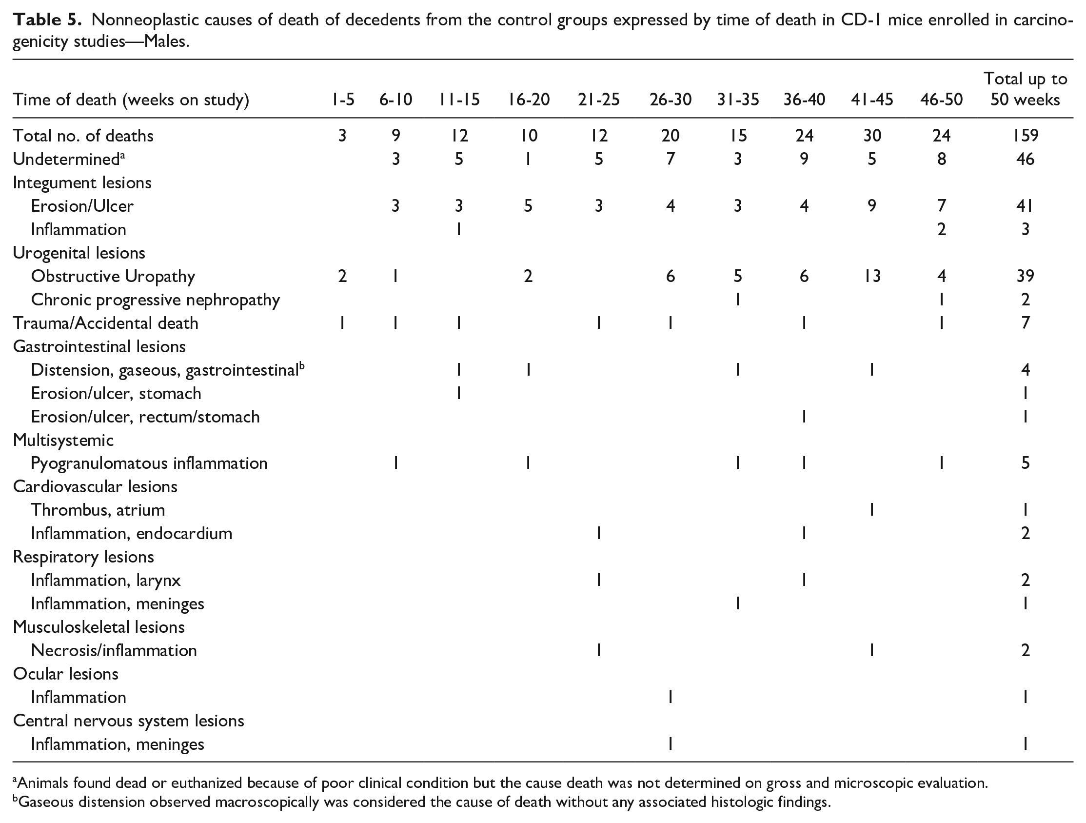
Animals found dead or euthanized because of poor clinical condition but the cause death was not determined on gross and microscopic evaluation.
Gaseous distension observed macroscopically was considered the cause of death without any associated histologic findings.
Nonneoplastic causes of death of decedents from the control groups expressed by time of death in CD-1 mice enrolled in carcinogenicity studies—Females.
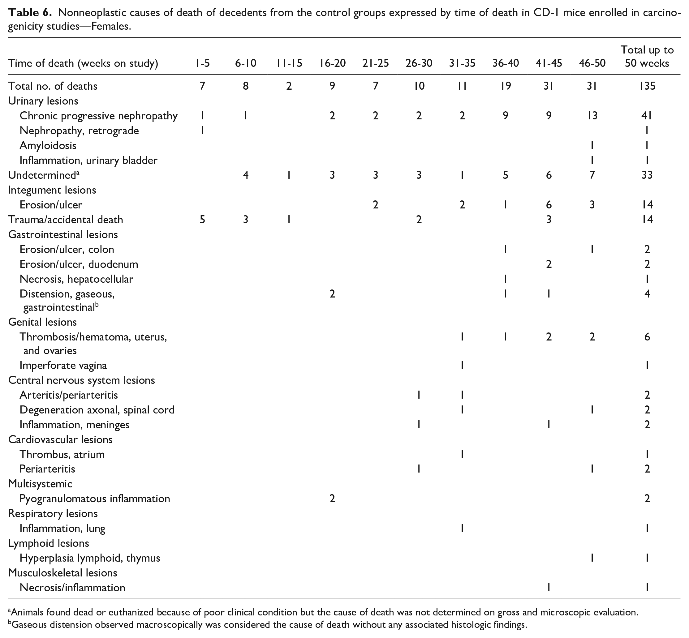
Animals found dead or euthanized because of poor clinical condition but the cause of death was not determined on gross and microscopic evaluation.
Gaseous distension observed macroscopically was considered the cause of death without any associated histologic findings.
In males, integument lesions were the commonest cause of death or unscheduled euthanasia (44/192, 22.9%) and comprised a range of histopathologic findings including multifocal epidermal ulceration with secondary scab formation, neutrophilic and pyogranulomatous inflammation (abscesses), and bacterial aggregates in the dermis and subcutis (Figure 1).
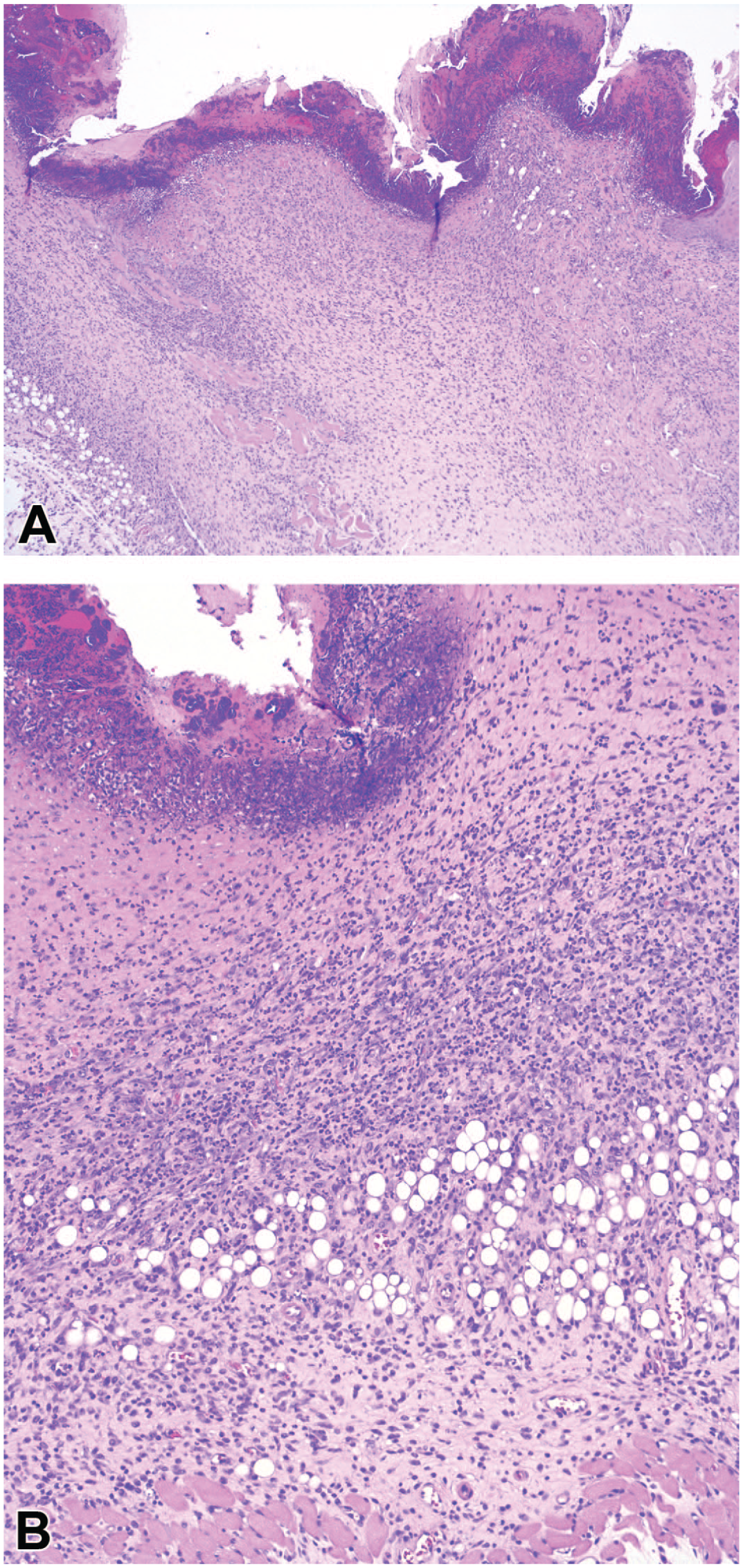
Skin, control male CD-1 mouse enrolled in a 104-week carcinogenicity study, euthanized for welfare reasons on study day 293 (week 42) (hematoxylin and eosin). (A) The epidermis is diffusely ulcerated, and diffuse inflammation disrupts the dermis and subcutis. Original objective 5X. (B) Higher magnification of skin lesion in Figure 1A. The ulcerated epidermis is covered by a serocellular crust comprising multifocal bacterial aggregates; moderate diffuse neutrophilic inflammation expands the dermis and subcutis. Original objective 10X. The integument lesions were considered the major factor contributing to death.
Urogenital lesions were the second commonest cause of death or unscheduled euthanasia and comprised a spectrum of changes, predominantly obstructive uropathy (39/192, 20.3%). The latter was generally associated with changes in the urinary bladder (Figure 2), with pyelonephritis and cystitis, with proteinaceous urethral plugs sometimes evident, and correlated with penile protrusion.
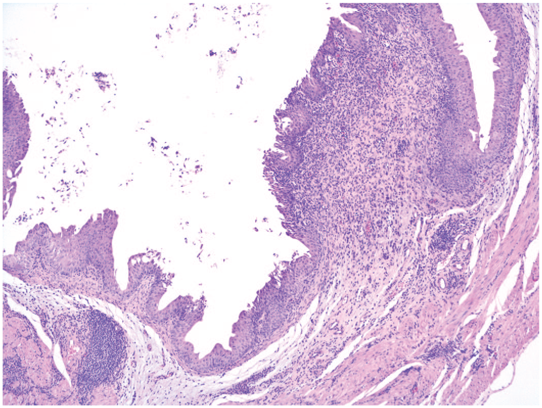
Urinary bladder, control male CD-1 mouse enrolled in a 104-week carcinogenicity study, found dead on study day 217 (week 31) (hematoxylin and eosin). Moderate urothelial hyperplasia associated with multifocal moderate lymphoplasmacytic inflammatory infiltration in the mucosa and submucosa possibly resulting from obstructive uropathy or ascending infections. Original objective 5X.
Other organ system pathologies were seen in relatively small numbers of males. Gastrointestinal lesions, namely, ulceration of the glandular stomach and rectum and gaseous distension of the gastrointestinal tract, were observed in 6/192 (3.1%) males. Multisystemic pyogranulomatous inflammation was recorded in 5/192 (2.6%) males.
Cardiovascular lesions occurred in a few animals and encompassed atrial thrombosis and endocardial inflammation. Respiratory lesions were characterized by inflammation. Ocular lesions were described as inflammatory.
Skeletal lesions comprised bone necrosis and pyogranulomatous inflammation of the femuro-tibial joint and osteomyelitis. Inflammation of the central nervous system accounted for a single animal.
Chronic progressive nephropathy (CPN) (Figure 3), associated with amyloidosis in one case, was the most common cause of early mortality in females (41/198, 20.7%), followed by ulcerative lesions of the integument (14/198, 7.1%).
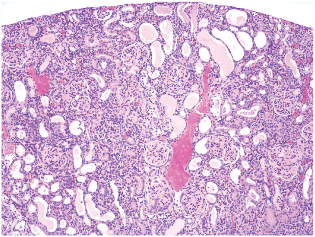
Kidney, control female CD-1 mouse enrolled in a 104-week carcinogenicity study, euthanized for welfare reasons on study day 344 (week 50) (hematoxylin and eosin). Marked chronic progressive nephropathy comprising numerous clusters of basophilic tubules, mild glomerulosclerosis with thickened basement membranes, tubular dilatation, and eosinophilic casts. Original objective 10X. Chronic progressive nephropathy was considered the major factor contributing to death.
Genital lesions were the cause of death in 7 females and included thrombosis/hematoma of the uterus and ovaries in 6 cases. Imperforate vaginal septa associated with inflammation of the genital tract was the cause of death in one female.
Gastrointestinal lesions affected 9/198 (4.5%) females and comprised ulceration of the duodenum and colon, hepatocellular necrosis, and gaseous distension of the gastrointestinal tract. Central nervous system pathologies were noted in 6/198 animals (3%) and comprised arteritis/periarteritis of the meninges, inflammation, and axonal degeneration of the spinal cord with inflammation. All other organ system pathologies were fatal in small numbers of decedents only (1-2 animals). The thymic lesion was characterized by lymphoid hyperplasia.
Tumor Occurrence Pattern
In males, tumors accounted for 33/192 (17.2%) of all unscheduled deaths. The earliest identified tumor was a fatal lymphoma (Figure 4A and 4B) seen in a male at week 14. The second earliest occurring tumor, seen in week 18, was a fatal astrocytoma of the brain.
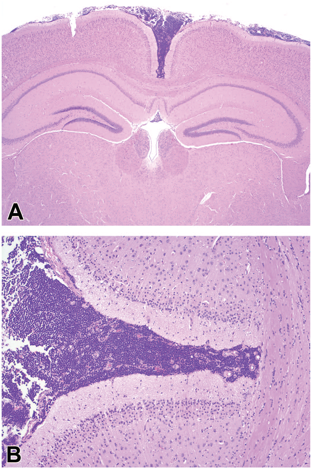
Brain, control male CD-1 mouse enrolled in a 104-week carcinogenicity study, euthanized for welfare reasons on study day 261 (week 38). Coronal at the hippocampus level (hematoxylin and eosin). (A) Lymphoma is expanding the meningeal subarachnoid space. Original objective 2.5X. (B) Higher magnification of Lymphoma in Figure 4A. Original objective 10X. The neoplasm was considered the major factor contributing to death.
The commonest tumor type was lymphoma (54.5% of all tumors); with eighteen cases recorded in total (Table 7). All lymphomas were fatal. Five bronchiolo-alveolar carcinomas of the lung, all fatal, were the second most common tumor (Figure 5A and 5B). In addition, two nonfatal bronchiolo-alveolar adenomas were noted in animals dying of other causes. Tumors noted as single occurrences, including sarcomas of various origins, colonic adenocarcinoma, hemangioma, and osteosarcoma, among others, were ascribed as causes of death (Table 7). Two Harderian gland adenomas, a chondroma, a hepatic adenoma, a subcutaneous lipoma, an adrenal subcapsular adenoma, a testicular hemangioma, and a nasal odontoma, were noted as nonfatal findings (Table 7).
Tumor incidence profiles expressed by time of death in CD-1 mice enrolled in carcinogenicity studies—Males.
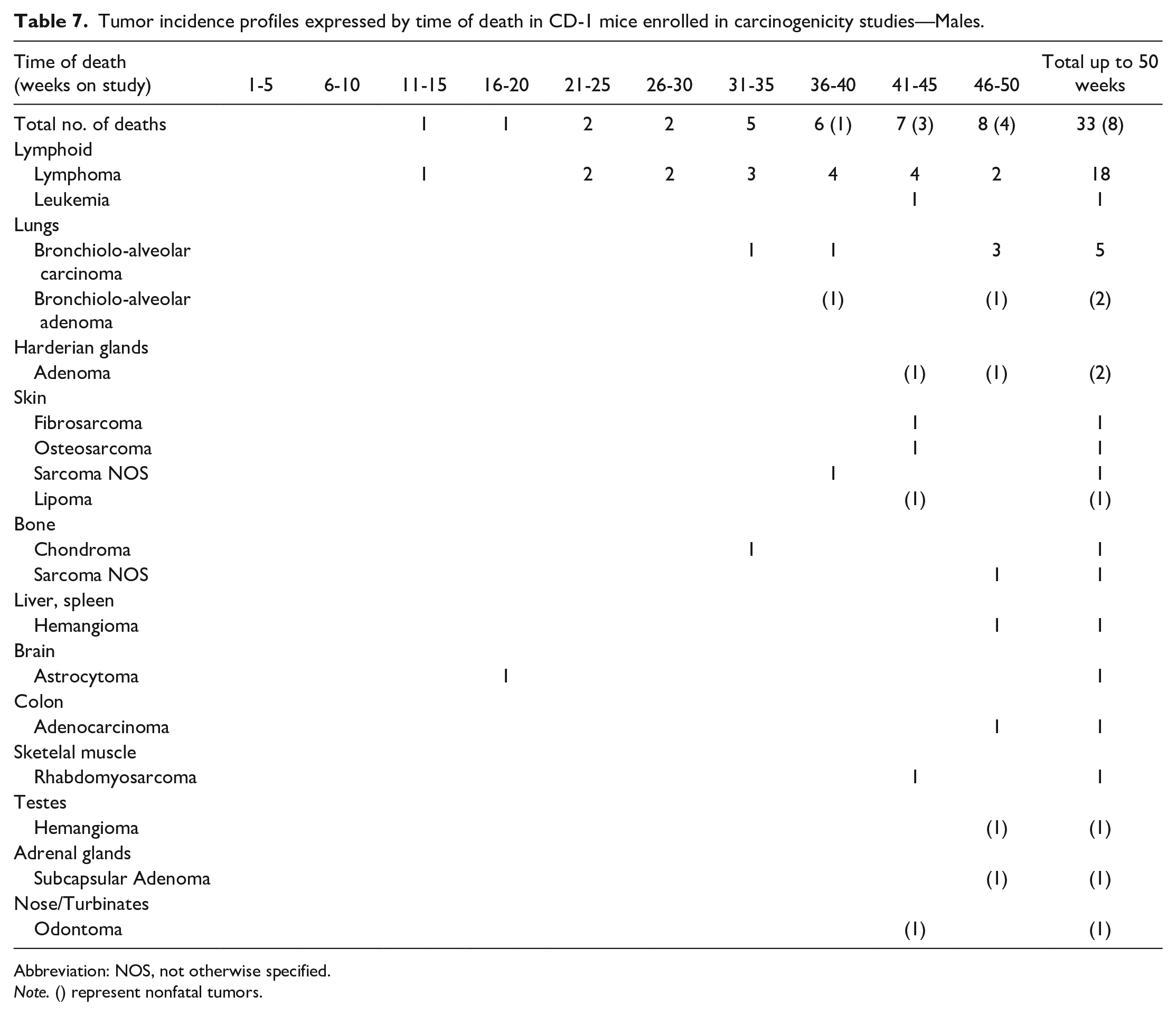
Abbreviation: NOS, not otherwise specified.
Note. () represent nonfatal tumors.
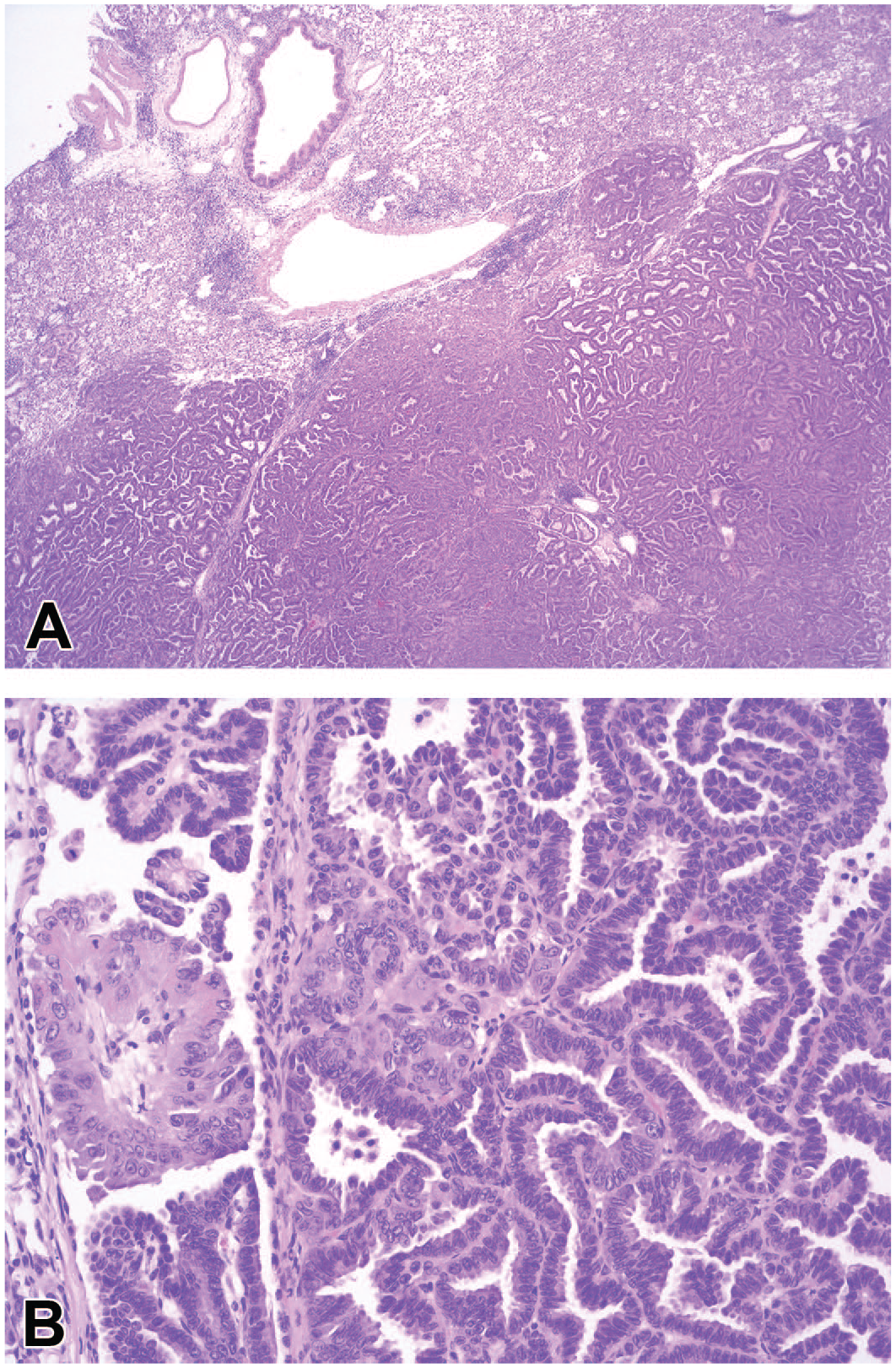
Lung, control male CD-1 mouse enrolled in a 104-week carcinogenicity study, found dead on study day 221 (week 32) (hematoxylin and eosin). (A) The pulmonary parenchyma is expanded and disrupted by a multilobulated bronchio-alveolar carcinoma. Adjacent alveolar spaces contain many macrophages and peribronchial/perivascular infiltrates of lymphocytic and mononuclear cells are present. Original objective 2.5X. (B) Higher magnification of bronchio-alveolar carcinomas in Figure 5A. The neoplasm is composed by branching papillae lined by 1 to 2 layers of polygonal cells, supported by a fine axis of fibrovascular stroma. Moderate anisocytosis and anisokaryosis present. Original objective 20X. The neoplasm was considered the major factor contributing to death.
In females, tumors accounted for 63/198 (31.8%) of all unscheduled deaths. As in males, lymphoma was the commonest tumor type (41/64, 64.1%) and was fatal in all affected females. Lymphoma was also the earliest tumor to occur, with the first one noted in week 20 (Table 8). Bronchiolo-alveolar adenomas were the second most frequently recorded tumors, noted as nonfatal findings in 4 females (6.3%). The uterus was a common site for tumors, which included endometrial adenocarcinoma, hemangiosarcoma, hemangioma, endometrial stromal sarcoma, polyps, granular cell tumor, and histiocytic sarcoma (Table 8). All other tumors were seen at low incidences, including bronchiolo-alveolar carcinomas, mammary adenocarcinomas and adenoacanthoma, basal cell tumor, fibrosarcoma of the skin, and skeletal osteosarcomas (Table 8).
Tumor incidence profiles expressed by time of death in CD-1 mice enrolled in carcinogenicity studies—Females.
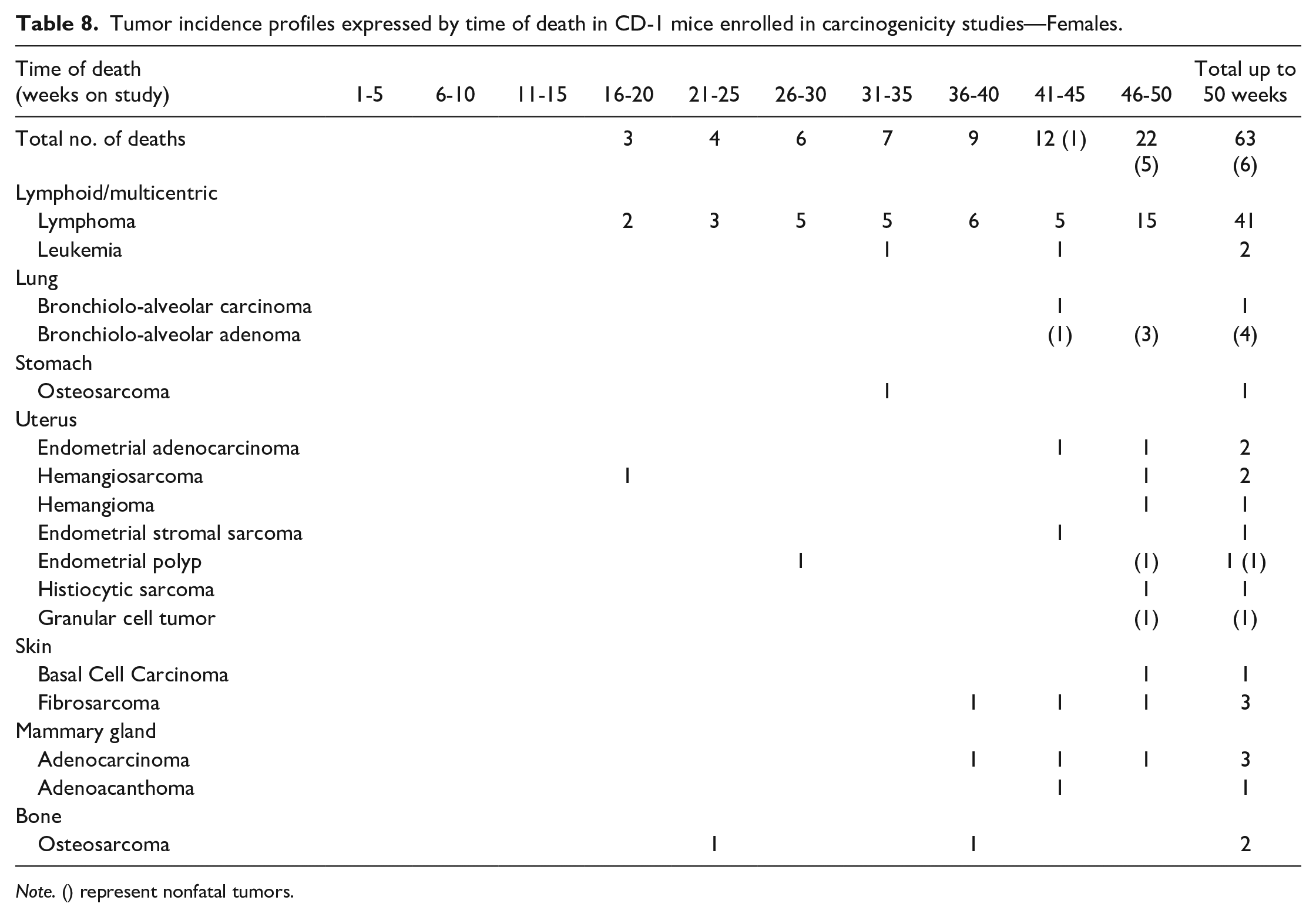
Note. () represent nonfatal tumors.
Discussion
Mortality patterns were compared with a similar study published by Son and Gopinath. 11 In that study, the author reported a 2.06% mortality of males and 0.69% mortality of females up to week 20 of study. 11 Mortality in males was lower (1.3%) in our data, whereas similar results were found for the females, in our 13-week studies. This variation could be related to the longer endpoint of 20 weeks in Son and Gopinath’s survey, compared with 13 weeks in our study. The same author reported a male mortality of 6.95% in animals up to week 50 of study, which is similar to the males in our study (6.8%). Mortality within the first 50 weeks of study was recorded by Son and Gopinath as 4.75% for females, which is lower than the 7% mortality seen in females in the first 50 weeks of our survey. 11 The reasons for this are unclear. A shift to stricter criteria for euthanasia of females due to clinical signs, particularly masses, may be a contributory factor.
In males, skin lesions were the most common findings among nonneoplastic causes of deaths in both 13-week studies and long-term studies. Housing conditions in both short- and long-term studies mostly comprised gang-housed animals (2-3 animals of the same sex in the same cage in 77/91 studies). Similar incidences of skin wounds were reported by Son and Gopinath. 11 Fighting among cage mates has been pointed out as a cause of traumatic skin lesions (abrasions and ulcers) that may lead to death in male mice.12-14 Mouse obstructive uropathy has also been associated with cutaneous lesions in the urogenital area, caused by aggression between males or self-mutilation, 15 and accounted for the second most common nonneoplastic causes of death or unscheduled euthanasia in the first 50 weeks of study in this report. Staphylococcal abscesses or Corynebacterium infection may lead to pyogranulomatous inflammation, which is considered to be a sporadic secondary infection. 16 CD-1 mice have been enumerated among the strains with a high propensity to aggression. 14 In a recent study, an increase in the prevalence of aggression was found when male mice were housed in cages of three; on the contrary, previous indications concluded that a more stable dominance hierarchy is achieved with smaller groups (i.e., three animals per cage). 17 Other recommendations to mitigate aggressive behavior are, among others, selecting animals into a cage by litter rather than randomly. 14 Skin lesions resulting in premature euthanasia of the animal were also observed in studies comprising individually housed males. As abrasions were found in males only, it is possible that the lesions were a consequence of them being individually housed, therefore less well-groomed or possibly exhibiting repetitive behaviors. 18
In this study, CPN was the most common cause of early mortality in females (41/198, 20.7%) with a higher incidence when compared with the previous study (6/69, 8.7%). 11 Amyloidosis was very rarely a feature of CPN in these studies. The CPN in mice comprises a wide spectrum of chronic degenerative renal lesions, including glomerulosclerosis, glomerular dilation, regressive changes and atrophy of tubules, increased connective tissue, and interstitial mononuclear cell infiltrates, the latter being especially common in mice. 19 In animals with advanced lesions including pelvic dilatation, the disease may constitute the major cause of death. 3 A doubled to tripled incidence of CPN has been reported in female mice versus males.3,10 The spectrum of changes has similarities to those seen in chronic rat CPN, although these changes tend to occur with a lower incidence and severity in mice than in rats. 15 In rats, CPN is more common than in mice and can occur at a relatively young age, with some studies reporting a 100% incidence of the histological lesions at 4 to 5 months of age in males. 20 In long-term studies where rats were fed with conventional high-protein diets, CPN represented the major cause of death, due to chronic renal failure. 21 In addition, in rats this spontaneous disease is more severe in males than in females, and sex predisposition is linked to the presence of androgen.21,22 Mechanistic pathways that have recently been involved in the pathogenesis of progressive age-related decline in rodent kidney function include intrarenal renin-angiotensin system (RAS) alterations that may be critical to CPN development and progression. 19
The profile of tumors seen in young animals was similar to that noted in the literature for older animals. Lymphoma, as noted by other authors, was the commonest tumor type in both sexes.1,3,11,23 A few differences between the earlier data survey from this laboratory, 11 and the data presented here were evident, and these are discussed below.
In the current survey, the earliest occurring tumor in males was a lymphoma, noted between weeks 6 and 10, which is very similar to the previous data. 11 The incidence of lymphoma in male decedents in the current survey (18/192 [9.3%]) was lower than previously reported (23/101 [22.8%]) at this laboratory. Hepatocellular adenomas were not identified in our survey, whereas Son and Gopinath noted it in three males that died between 36 and 45 weeks of study; with two being nonfatal findings and one being the cause of death. Histiocytic sarcoma and myeloid leukemia, each seen in 2/101 decedent males in the previous survey, were not recorded in males in the current survey. Conversely, several tumor types noted in the current data were absent in Son and Gopinath’s review. These include adenomas of the Harderian gland, astrocytoma of the central nervous system, hemangiomas in several organs, chondroma of the bone, osteosarcoma of the stomach, and an adenocarcinoma of the colon.
In females, the earliest identified lymphoma was in the 19th week of study. This occurrence was slightly earlier than noted by Son and Gopinath (25 weeks). 11 The incidence of lymphoma in female decedents in the current survey (41/198 [20.7%]) was lower than previously reported (19/69 [27.5%]) at this laboratory. 11 In a previous study from another institution, lymphoma in females occurred with a 20% incidence in 2-year carcinogenicity studies. 3 Also, histiocytic sarcoma and myeloid leukemia, each seen in 2/68 decedent females by Son and Gopinath, were not recorded in this survey. Mammary and lung tumors occurred with similar profiles in both surveys. One uterine polyp and one uterine adenocarcinoma were noted in our survey but not in the previous survey. An extraskeletal osteosarcoma in the stomach, not recorded in the earlier survey, was also present in the current data.
In summary, only slight differences in tumor occurrence patterns were noted when our data were compared with the older publication from this laboratory, by Son and Gopinath, 11 suggesting that there is no significant genetic drift affecting the tumor occurrence over a twenty-year period.
Conclusion
Mortality was similar in both sexes up to week 50 of study. In both sexes, the majority of deaths were due to nonneoplastic causes, but in males, tumors were responsible for far fewer deaths than in females (17% vs 32.3%). In mice from 13-week studies, bronchiolo-alveolar adenomas were the commonest tumor in both sexes, all occurring as nonfatal findings at the end of study phase in all cases. In decedents of <50 weeks from carcinogenicity studies, lymphomas were the earliest occurring tumor in both males and females and were also the commonest tumor in both sexes. This pattern is very similar to the results from a previous survey done at this laboratory and the literature from other laboratories.
Lesions of the integument in males and CPN in females were the most commonly identified nonneoplastic causes of death or early euthanasia.
Only minor differences in incidence and nature of tumors were evident between a similar survey performed approximately 15 years ago and the present survey, and these at least partially reflect changes in terminology and diagnostic criteria.
Footnotes
Declaration of Conflicting Interests
The author(s) declared no potential conflicts of interest with respect to the research, authorship, and/or publication of this article.
Funding
The author(s) received no financial support for the research, authorship, and/or publication of this article.
References
Supplementary Material
Please find the following supplemental material available below.
For Open Access articles published under a Creative Commons License, all supplemental material carries the same license as the article it is associated with.
For non-Open Access articles published, all supplemental material carries a non-exclusive license, and permission requests for re-use of supplemental material or any part of supplemental material shall be sent directly to the copyright owner as specified in the copyright notice associated with the article.