
Abstract
Mink (Mustela vison) exposed to 2,3,7,8-tetrachlorodibenzo-p-dioxin (TCDD)-like chemicals have been reported to develop mandibular and maxillary squamous cell proliferation that results in the destruction of alveolar bone and eventual tooth loss. This jaw lesion has been reported in wild mink collected from areas contaminated with TCDD-like compounds and is a potential biomarker for exposure to these chemicals. The blue iris strain of domestic mink is prone to develop severe periodontal disease, which results in destruction of bone and tooth loss that is grossly similar to the lesion induced by exposure to TCDD-like chemicals. A histological assessment of jaws from blue iris mink and natural dark mink exposed to 3,3′,4,4′,5-pentachlorobiphenyl (PCB 126) was done to determine whether the oral lesions are similar. The jaw tissue from the blue iris mink had lesions indicative of lymphoplasmacytic gingivitis and osteomyelitis, caused by inflammation entering the dental sulcus, while the jaw tissue from the mink exposed to PCB 126 exhibited squamous epithelial proliferation. Therefore, it was determined that the tooth loss and bone destruction seen in these mink are of different origin despite the similarity of the gross clinical signs.
Introduction
One particular effect of 2,3,7,8-tetrachlorodibenzo-p-dioxin (TCDD) and 3,3′,4,4′,5-pentachlorobiphenyl (PCB 126) reported in mink (Mustela vison) is a jaw lesion characterized as mandibular and maxillary squamous cell proliferation that was first described by Render et al. (2000a, 2000b, 2001) as proliferation of squamous cells arising out of the gingival tissue adjacent to the teeth. These proliferations coalesce to form squamous cysts (SC) that invade the alveolar bone (AB) to cause osteoporosis, eventually causing loose, displaced teeth.
The occurrence of mandibular and maxillary squamous cell proliferation recently has been associated with environmental contamination by TCDD-like chemicals at a number of locations, and therefore has the potential to be further developed as a biomarker for exposure to these chemicals. The research on this topic has included two feeding studies in which mink were fed diets containing fish collected from the Saginaw River in Michigan (Bursian et al. 2006a) and the Housatonic River in Massachusetts (Bursian et al. 2006b, 2006c). The lesion was induced in mink at TCDD toxic equivalent (TEQ) concentrations as low as 47 and 9.2 ng TEQs/kg feed in the Saginaw River (Bursian et al. 2006a) and Housatonic River (Bursian et al. 2006c) studies, respectively. The incidence of the lesion in the Saginaw River study was 10 of the 15 mink in the treatment groups displaying the lesion and 9 of the 18 mink in the treatment groups having the lesion in the Housatonic River study. In addition to laboratory studies, 4 of the 9 wild mink collected along a polychlorinated biphenyl (PCB)-contaminated stretch of the Kalamazoo River in Michigan exhibited histological signs of the lesion (Beckett et al. 2005) as did 1 of the 3 mink collected from a PCB-contaminated site on the south shore of Lake Ontario (Haynes et al. 2009).
Many species of wildlife have been reported to have gross clinical signs indicative of a jaw lesion similar to those reported for mink exposed to TCDD-like chemicals, although the cause of these clinical signs has not been established. Mortenson et al. (1992) described “skull-bone lesions” in harbor seals (Phoca vitulina) in the Baltic region, which included considerable loss of AB around teeth and other parts of the jaw, as well as multiple loss of teeth. Beland et al. (1993) and De Guise et al. (1995) both reported tooth loss and periodontitis in beluga whales (Delphinapterus leucas) found in the St. Lawrence Estuary, and Sonne et al. (2007) reported similar effects in polar bears (Ursus maritimus) in Greenland. The cause of these jaw bone effects has not been established, but coplanar PCBs and other TCDD-like chemicals are known to affect high trophic level predators such as seals, whales, and polar bears. The presence of these chemicals has been documented in the environments mentioned above. Therefore, it is possible that a lesion induced by TCDD and PCB 126, such as has been described in mink (Render et al. 2000a, 2000b, 2001; Beckett et al. 2005; Bursian et al. 2006a, 2006c; Haynes et al. 2009), could be similar to the condition described in other wildlife species.
In a recent report, Hammer et al. (2005) described an oral lesion in a color strain of ranch mink that appears grossly to be very similar to the condition reported in mink exposed to TCDD-like chemicals. Blue iris mink can develop severe destructive periodontal disease, which results in the loss of teeth. These mink are susceptible to an autosomal disorder known as Chediak-Higashi syndrome (CHS), which is associated with a deficiency of the immune system. This makes blue iris mink, unlike natural dark mink, more susceptible to various infections including skin abscesses (Hammer et al. 2005). The authors of this report studied the association between destructive periodontal disease (measured by radiographic images of mink jaws) and counts of abnormal polymorphonuclear leukocytes (which indicates CHS). It was concluded that tooth loss was related to destructive periodontal disease, although a significant relationship to the number of CHS leukocytes was not shown.
Several blue iris mink were obtained by the Michigan State University (MSU) Experimental Fur Farm from a commercial fur rancher for an Aleutian Disease trial. Upon receipt of the mink, gross clinical signs of loose teeth and bleeding gums were observed in some of these animals (Figure 1a), which appeared to be similar to gross clinical signs in natural dark mink exposed to PCB 126 (Figure 1b). It was of interest to compare the histology of the jaw tissue from the blue iris mink to jaw tissue from natural dark mink exposed to a TCDD-like chemical to determine whether or not the two conditions were the same. Because exposure to TCDD-like chemicals can suppress the immune system (Vos, De Heer, and Van Loveren 1997), it is possible that the jaw lesion reported in mink exposed to TCDD-like chemicals is the result of periodontal disease arising as a result of a suppressed immune system.
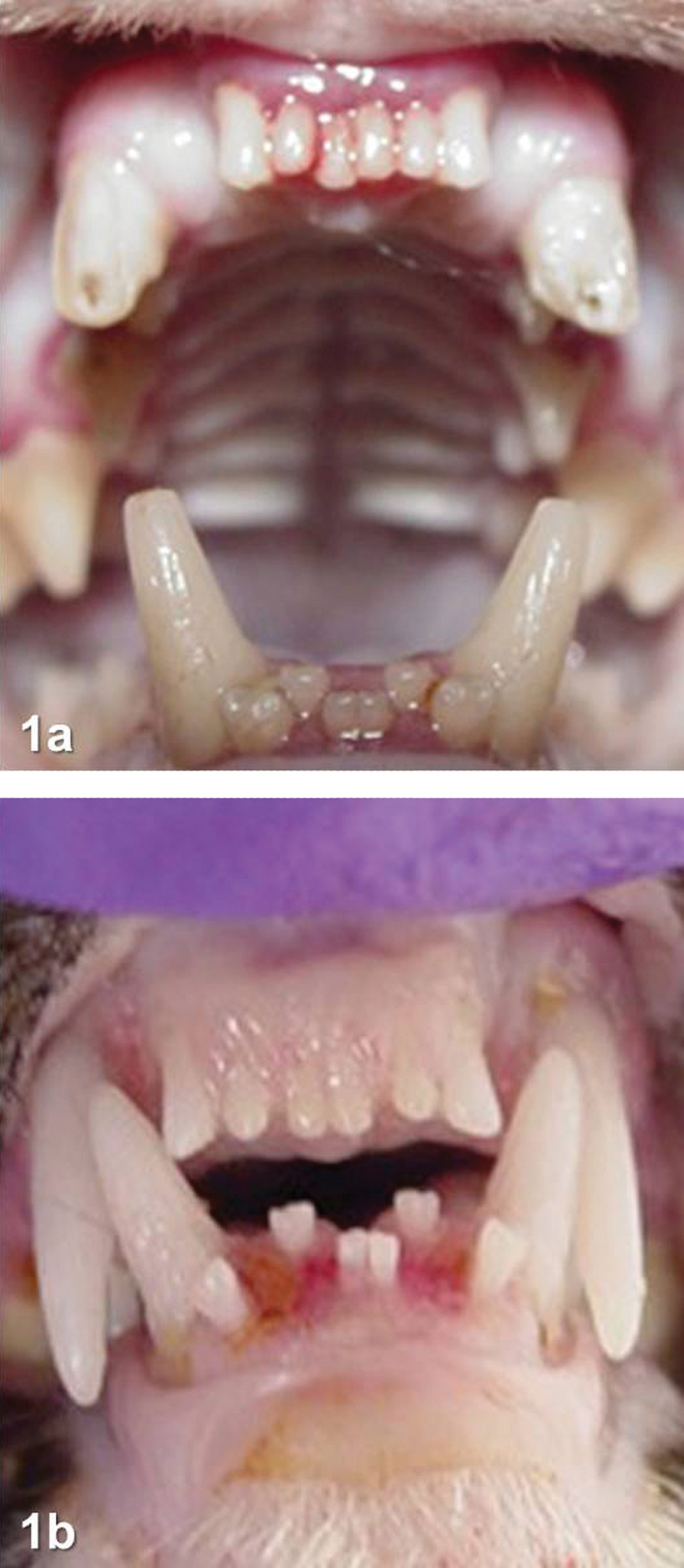
Gross frontal view of the mouth of a blue iris mink (a) and a 3,3′,4,4′,5-pentachlorobiphenyl (PCB 126)-exposed, natural dark mink (b). Notice in both cases the swollen gingiva and loose and misaligned incisors.
Method
Twenty-four 18-month-old, female, blue iris mink were obtained from a commercial mink ranch for a trial related to Aleutian Disease, to which the blue iris mink are particularly susceptible. Upon delivery, six of the animals were identified as having bloody gums and loose teeth, a condition that the rancher reported to be not uncommon in this strain of mink. The blue iris mink were maintained in cages in an open-side pole barn at the MSU Experimental Fur Farm for 4 months prior to being transferred to an environmentally controlled room in MSU’s University Research Containment Facility, where the Aleutian Disease trial was conducted. After 7 weeks, when approximately half of the mink had developed rhinorrhea, the Aleutian Disease trial was terminated and animals were necropsied. Mink were euthanized with carbon dioxide and jaws were collected from the six animals that had been initially identified as having swollen gums and loose teeth. Jaws were removed, stripped of adhering skin and tissue, and placed in 10% neutral buffered formalin (VWR International, West Chester, PA) for subsequent processing. Jaws were then placed in Rapid Decal (Leica Biosystems Peterborough, Peterborough, UK) for 36 to 48 hr for decalcification. After decalcification, jaws were trimmed, embedded in paraffin, sectioned (5 µm), and stained with H&S. A board-certified pathologist examined the slides under light microscope for distinguishing histological signs.
In a separate trial, twenty-three healthy, 5-month-old, natural dark mink from the MSU Experimental Fur Farm were dosed with 30 µg PCB 126/kg body weight via a single intraperitoneal injection. The mink were housed separately in cages in an open-sided pole barn and were provided the standard ranch diet and drinking water ad libitum throughout the trial. The mink were euthanized with carbon dioxide 9 weeks after exposure to PCB 126, and their jaws were collected and processed as described above. All of the exposed mink had gross clinical signs indicative of the jaw lesion associated with exposure to TCDD-like compounds. There was no apparent sex-related difference in development of the jaw lesion, and the lesions were equally severe on the maxilla and mandibles of the animals.
Both sets of animals were covered under protocols approved by the MSU Institutional Animal Care and Use Committee.
Results and Discussion
The jaw tissue from the blue iris mink had lesions indicative of lymphoplasmacytic gingivitis and osteomyelitis, caused by immunosuppression and inflammation entering the dental sulcus (Figure 2a and b), originally described by Hammer et al. (2005). The jaw tissue from the mink exposed to PCB 126 developed severe SC surrounding the teeth, which invade the AB, but not the teeth themselves (Figure 2c and d). Despite the similarity in gross clinical signs between the blue iris mink and mink exposed to PCB 126 (Figure 1a and b), histological examination of jaw tissue indicated that the inflammatory lesions in the blue iris mink were unrelated to the SC that are indicative of a proliferative/neoplastic condition (Render et al. 2000a, 2000b, 2001). While the destruction of the AB appears similar in the blue iris mink and mink dosed with PCB 126, the underlying cause is different. One additional difference is that the inflammatory destruction also affects the teeth, whereas the proliferative lesions destroy only the surrounding AB, leaving the teeth intact. Ranch mink exposed to environmentally relevant concentrations of TCDD-like compounds in fish collected from contaminated rivers (Bursian et al. 2006a, 2006c), as well as wild mink collected from areas contaminated with TCDD-like compounds (Beckett et al. 2005; Haynes et al. 2009) were reported to have histological effects like those reported for ranch mink fed diets containing PCB 126 or TCDD (Render et al. 2000a, 2000b, 2001).
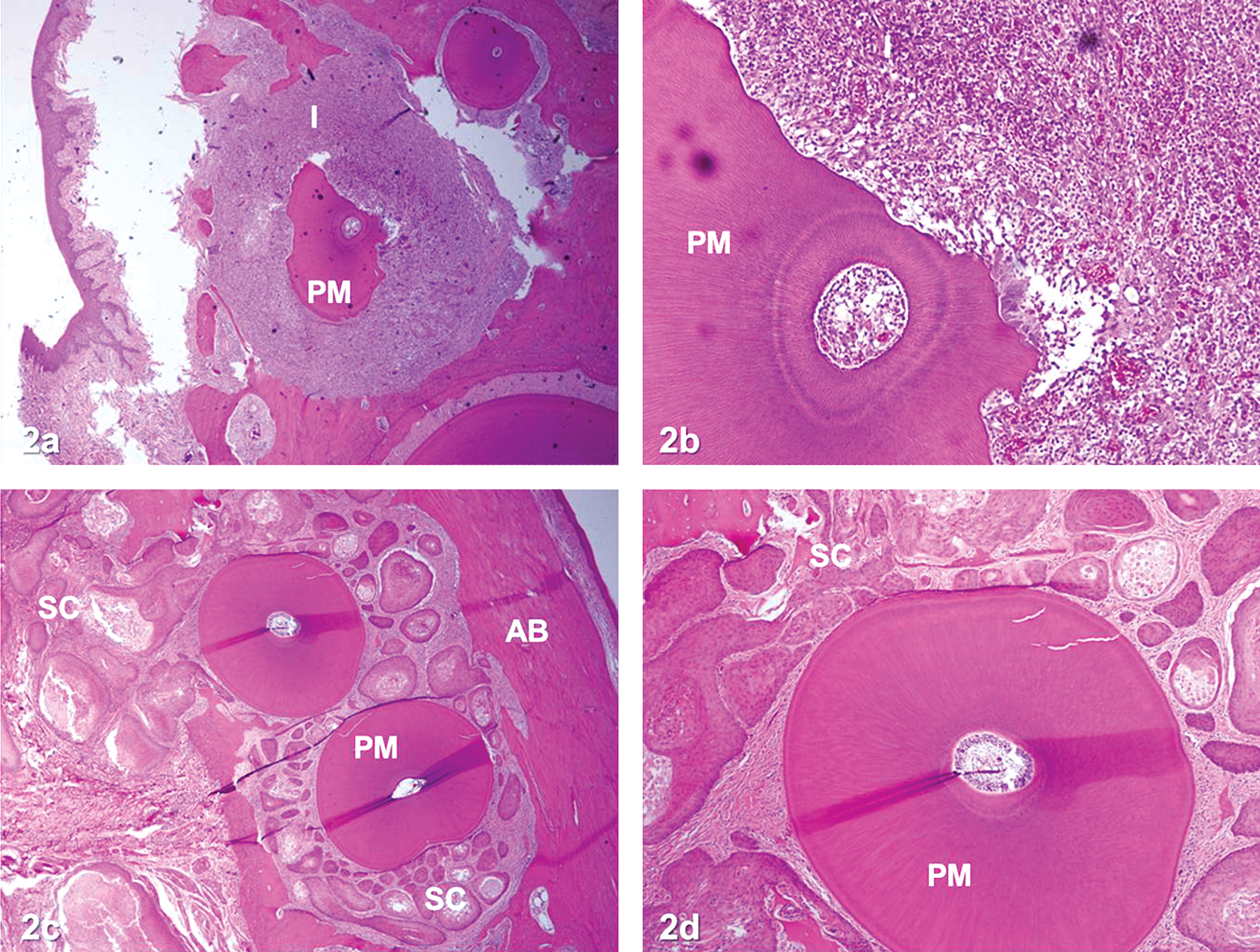
Photomicrograph (H&S) of the mandible showing premolars (PM) from a blue iris mink at 4× (a) and 10× (b) and a natural dark mink exposed to PCB 126 at 4× (c) and 10× (d). In the blue iris mink (a,b), notice the destructive lymphocytic inflammation (I) that destroys both the alveolar bone (AB) and the PM tooth, giving the tooth an irregular outer surface. In the 3,3′,4,4′,5-pentachlorobiphenyl (PCB 126)-exposed mink (c,d), notice the squamous cysts (SC) surrounding the tooth, which destroys the surrounding AB, but not the PM teeth.
Gross oral lesions seen in seals, whales, and polar bears (Mortensen et al. 1992; Beland et al. 1993; De Guise et al. 1995; Sonne et al. 2007; Bergman, Olsson, and Reiland 1992) could be indicative of either exposure to TCDD-like compounds or an inflammatory response. The results of this study suggest that histological examination of jaw tissue collected opportunistically from wildlife species will therefore be necessary to differentiate between the two conditions, or perhaps indicate a different etiology.
Footnotes
Abbreviations
Acknowledgments
This research was assisted by Angelo Napolitano and the Michigan State University Experimental Fur Farm.
The authors declared no potential conflicts of interest with respect to the research, authorship, and/or publication of this article.
The authors received no financial support for the research, authorship, and/or publication of this article.